Re activity o f anti-thyro id antibo die s
to thyro glo bulin tryptic fragm e nts:
co m pariso n o f auto im m une and
no n-auto im m une thyro id dise ase s
Laboratório de Imunologia Clínica, Faculdade de Ciências Médicas, Universidade Estadual de Campinas, Campinas, SP, Brasil
L.H.B. Boechat and R.L. Zollner
Abstract
Studies concerning the antigenicity of thyroglobulin fragments allow the characterization of the epitopes but do not consider the role of heavier antigenic fragments that could result in vivo from the action of endoproteases. Here we assess the relative importance of the frag-ments obtained from thyroglobulin by limited proteolysis with trypsin and compare by immunoblotting their reactivity to serum from pa-tients with autoimmune (Graves disease and Hashimotos thyroiditis) and non-autoimmune (subacute thyroiditis) disease. The results showed no difference in frequency of recognition of any peptide by sera from patients with autoimmune thyroiditis. In contrast, sera from patients with subacute thyroiditis reacted more frequently with a peptide of 80 kDa. These results suggest the presence of antibody subpopulations directed at fragments produced in vivo by enzymatic cleavage of thyroglobulin. This fragment and antibodies to it may represent mark-ers for subacute thyroiditis.
Co rre spo nde nce R.L. Zollner
Laboratório de Imunologia Clínica Departamento de Clínica Médica FCM, UNICAMP
Caixa Postal 6111 13081-970 Campinas, SP Brasil
E-mail: zollner@ obelix.unicamp.br
Research supported by CNPq (No. 132405/94-6). L.H.B. Boechat was the recipient of a CNPq fellowship. Publication supported by FAPESP.
Received April 30, 1998 Accepted February 12, 1999
Ke y wo rds
·Autoantibodies
·Autoimmunity
·Thyroglobulin
·Trypsin
·Thyroiditis
·Proteases
Intro ductio n
Thyroglobulin is a protein of 660 kDa and the main product of thyroid follicular cells. Hormone liberation from this heavy precursor depends on its proteolysis at pre-determined sites in an organized fashion (1). Endopeptidases such as cathepsin D and cys-teine-proteases such as cathepsin B, H and L have been implicated in this process and their synergistic action seems to be neces-sary for hormone secretion (2,3).
Studies trying to determine the sites of proteolytic action and correlate them with
the tertiary structure of thyroglobulin have demonstrated the presence of areas sensitive to proteolysis located at the C-terminal end and especially in peptide sequences inserted among blocks of internal homology. Gentile et al. (4) have proposed that human thyroglo-bulin could be spontaneously broken be-tween residues 503 (leucine) and 505 (serine), located between the fourth and fifth blocks of type 1 homology. These regions are also susceptible to trypsin, thermolysin and ca-thepsins B, D and L (1-3).
ac-tivity at low enzyme/substrate ratios is analo-gous to that of thyroid endoproteases, lead-ing to the production of peptides similar to those derived from lysosomal processing of this prohormone. Although many investiga-tors have tried to determine the relationship between thyroglobulin structure and the im-mune system, reports concerning the reac-tivity of peptides obtained from the action of endoproteases in vivo are not frequent in the literature. Thus, analysis of the immuno-genic characteristics and protein sequence of these thyroglobulin fragments can help understand the origin and role of antithyro-globulin antibodies in autoimmune and non-autoimmune thyroid diseases. Moreover, the study of thyroglobulin antigenicity can pro-vide new data for the understanding of the involvement of thyroglobulin as an autoanti-gen in autoimmune and non-autoimmune thyroid diseases.
The aim of the present study was to evalu-ate the reactivity of the peptide panel ob-tained by limited trypsin hydrolysis of thyro-globulin. Sera from patients with autoim-mune thyroid disease and subacute thyroidi-tis were reacted with the fragments, in order to identify possible differences between these disorders.
Mate rial and Me tho ds
Patie nts and se ra
Sera from patients with Graves disease (5), Hashimotos thyroiditis (5) and sub-acute thyroiditis (6) were studied. The diag-nosis of Graves disease was made on the basis of clinical findings of thyrotoxicosis, diffuse goiter and ophthalmopathy and con-firmed by detection of high free thyroxin and reduced levels of thyrotropin (TSH) (Baxter Diagnostics Inc., Deerfield, IL, USA). Hashimotos thyroiditis was diagnosed by clinical findings of hypothyroidism associ-ated with high levels of TSH and low free thyroxin. For both diseases, an autoimmune
origin was suggested by the elevation of antithyroglobulin or antithyroperoxidase an-tibodies. Diagnosis of subacute thyroiditis was based on findings of cervical pain irradi-ating to ear and jaw, accompanied by asthe-nia and fever. The presence of a high eryth-rocyte sedimentation rate, low radioiodine uptake during scintigraphy, and giant multi-nucleate cells in thyroid aspirates was also considered for the diagnosis. All blood samples were obtained during the first visit to the hospital when none of the patients was receiving anti-thyroid drugs. Patients pre-senting other autoimmune or infectious dis-eases were excluded from the study. The mean levels of anti-thyroglobulin antibodies were 872 ± 344.3 IU/ml in patients with Hashimotos thyroiditis and 1235 ± 369.9 IU/ml (P = 0.478) in patients with Graves disease when tested by radioimmunoassay (Serono Diagnostics, Norwell, MA, USA). Sera from patients with subacute thyroiditis did not present detectable antibodies by this method. Sera from five healthy individuals were used as negative controls.
Thyro glo bulin pre paratio n
human thyroglobulin. This was confirmed by SDS-PAGE, immunoblotting and radio-immunoassay (Diagnostic Products Co., Los Angeles, CA, USA).
Limite d trypsinizatio n o f thyro glo bulin
Human thyroglobulin was dialyzed against 50 mM Tris-HCl and 130 mM NaCl, pH 8.4, and its concentration was adjusted to 2 mg/ml by dilution in the same buffer. Thyroglobulin was then submitted to trypsin hydrolysis (Trypsin type III, Sigma Chemi-cal Co., St. Louis, MO, USA) for 60 min at 30o
C at an enzyme/substrate ratio (w/w) of 1/1000. Tryptic digestion was stopped by dilution in SDS-PAGE sample buffer (62.5 mM Tris-HCl, 10% glycerol and 0.001% bromophenol blue) and boiling for 90 s in the presence of 10% (v/v) 2-mercaptoetha-nol (Sigma Chemical Co.).
SD S-PAGE
Electrophoresis was performed on a ver-tical slab by the method of Laemmli (7) at room temperature. When appropriate, the disulfide bridges of the samples were re-duced by adding 10% (v/v) 2-mercaptoetha-nol followed by boiling for 2 min. The gels were stained with 0.2% Coomassie brilliant blue R 250 by the usual methods.
Immuno blo tting
Thyroglobulin or its fragments were trans-ferred electrophoretically to a nitrocellulose membrane (Trans-Blot Transfer Medium, 0.45 micron; Bio-Rad, Hercules, CA, USA) at 4oC as described by Towbin (8). Nonspe-cific reactions were blocked with 3% fetal calf serum, 0.3% gelatin, and 0.05% Tween 20 for 1 h. Nitrocellulose was washed in PBS and then incubated with patient sera diluted 1:1000 for 3 h. After a second cycle of washing, the membrane was incubated with peroxidase-conjugated goat anti-human IgG
for 1 h in the dark. Membranes were stained with 3,3diaminobenzidine (DAB, Sigma Chemical Co.) in H2O2 as described by De Blas and Cherwinski (9).
Statistics
The frequency of positive reaction of each tryptic fragment with sera from the groups studied was compared by the c2
test with Yates correction or, when indicated, by the Fisher exact test, with the level of signif-icance set at P<0.05.
Re sults
Thyro glo bulin pre paratio n and limite d
trypsin hydro lysis
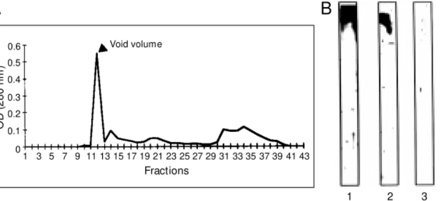
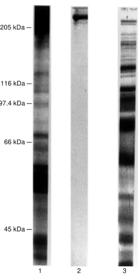
The crude thyroid extract contained 4.4 mg/ml protein and showed bands ranging from about 300 kDa to 15 kDa when ana-lyzed by SDS-PAGE. After filtration through a Sephadex G-200, the initial peak corre-sponding to the void volume of the column was identified as thyroglobulin by SDS-PAGE on the basis of immunoblotting and radioimmunoassay (Figure 1). After limited proteolysis, thyroglobulin was degraded to peptides with molecular masses ranging from 205 to fragments smaller than 29 kDa, as shown in Figure 2.
O
D
(
2
8
0
n
m
)
0.6
0.5
0.4
0.3
0.2
0.1
0
1 3 5 7 9 11 13 15 17 19 21 23 25 27 29 31 33 35 37 39 41 43 Fractions
1 2 3
A
B
Void volume
standing of its behavior as an autoantigen became possible. Its high molecular mass (2748 amino acids) impairs the production of recombinant thyroglobulin, and therefore studies on the whole monomer are performed with protein material obtained from human or animal glands. Many experimental meth-ods have been used, including acid precipi-tation (6,11) and chromatographic fraction-ation (12). In the present study, we decided to use the latter approach, since it excludes potential risks of structural modifications produced by exposing the protein to pH lev-els outside the physiological range (13). It was possible to obtain human thyroglobulin with a high level of purity, as confirmed by SDS-PAGE, immunoblotting and radioim-munoassay analysis.
Thyroid hormone synthesis includes the capture of iodinated thyroglobulin from the follicular lumen and its processing by endoproteases, finally leading to T4 and T3 synthesis in late endosomes (14). This seems to be a controlled process and cathepsins B, D, H and L are the best known enzymes whose proteolytic action could produce frag-ments with conserved antigenic characteris-tics. Gentile and Salvatore (1) used the method of limited proteolysis, with low en-zyme/substrate ratios and variable times of reaction to determine the preferential sites of action of trypsin and thermolysin. Compar-ing their results with those of Dunn et al. (2,3), it can be seen that these enzymes cleave thyroglobulin at sites of action similar to those of cathepsins.
In the present study, we performed lim-ited thyroglobulin proteolysis with an en-zyme/substrate ratio of 1/1000, since these conditions permitted us to obtain peptides ranging from <29 to >205 kDa. Our results are comparable to those of Gentile and Salvatore (1) who described peptides of hu-man thyroglobulin of 265, 230, 145, 105, 80, 56 and 29 kDa. However, we have also observed other fragments with molecular masses close to these. This may have been
Figure 2 - SDS-PAGE of thyro-globulin and fragments obtained by limited trypsin hydrolysis. The samples contained 10 µg protein w hich w as heat denatured in the presence of 2-mercaptoethanol. Protein w as detected w ith Coo-massie blue staining. Lane 1, Crude extract of thyroid tissue; lane 2, thyroglobulin purified by gel filtration (Figure 1A); lane 3, limited trypsin hydrolysate of thyroglobulin.
205 kDa
-97.4 kDa
-66 kDa
-45 kDa -116 kDa
-1 2 3
Re co gnitio n o f tryptic fragme nts o f
thyro glo bulin by auto antibo die s pre se nt
in patie nt se ra
Only peptides with molecular masses above 45 kDa were recognized (Figure 3). There was no significant difference in the recognition of any peptide by sera from pa-tients with Graves disease or Hashimotos thyroiditis. Thus, these data were grouped for comparison with patients presenting sub-acute thyroiditis. Peptides with molecular masses of 80, 185 and >205 kDa reacted more frequently with sera from patients with subacute thyroiditis than with those present-ing autoimmune thyroid disease (Table 1).
D iscussio n
under-due to differences in the trypsin preparations used, since in our case some residual chymo-trypsin action was probably present.
Many investigators have published data concerning the antigenicity of thyroglobulin fragments. However, these studies generally focus on small synthetic peptides or on those obtained by proteolysis with higher enzyme/ substrate ratios and for longer periods of time (15). These approaches allow the char-acterization of epitopes but do not consider the role of heavier antigenic fragments that could result, in vivo, from the action of endoproteases. Our findings suggest that antithyroglobulin antibodies recognize many antigenic sites in thyroglobulin, since immu-noblotting reactions were positive for all peptides above 45 kDa. The inability to rec-ognize smaller fragments could be related to spatial changes and loss of conformational epitopes.
There was no difference in frequency of recognition of any peptide by sera from pa-tients with Graves disease and Hashimotos thyroiditis and therefore these data were pooled into a single group concerning au-toimmune thyroid disease. In contrast, sera from patients with subacute thyroiditis re-acted more frequently with peptides of 80, 185 and >205 kDa.
The action of trypsin on residue 522 of thyroglobulin produced fragments of 80 and 265 kDa. This hydrolysis site, located be-tween the fourth and fifth area of internal homology type 1, is also susceptible to hy-drolysis by thermolysin and cathepsin B and L, and also is an area of spontaneous cleav-age of thyroglobulin (1,2,4). On this basis, the 80-kDa fragment may correspond to the first 521 amino acids of the amino-terminus containing the hormonogenic site at tyrosine 5 and four of the ten areas of internal homol-ogy type 1. These characteristics suggest a fragment with a rigid structure and probably able to preserve conformational epitopes.
Epitope mapping of thyroglobulin indi-cates the existence of at least 11 antigenic
205 kDa
-97.4 kDa
-66 kDa
-45 kDa -116 kDa
-1 2 3
29 kDa
--80 kDa
4
Figure 3 - Representative im-munoblots of thyroglobulin peptide fragments obtained by limited trypsin hydrolysis. The sera used w ere: lane 1, normal serum; lane 2, serum from a patient w ith Base-dow -Graves’ disease; lane 3, serum from a patient w ith Hashimoto’s thyroiditis; lane 4, serum from a patient w ith subacute thyroiditis.
Table 1 - Absolute and relative frequencies of reactivity of anti-thyroglobulin antibody w ith tryptic fragments of human thyroglobulin in patients w ith autoimmune thyroid disease and subacute thyroiditis.
aP = 0.0400; bP = 0.0230; cP = 0.0233 all compared to subacute thyroiditis (c2 test).
Fragments (kDa) Autoimmune thyroid disease Subacute thyroiditis
>205 17/36 (47.2% )a 9/10 (90% )
185 12/36 (33.3% )b 8/10 (80% )
170 4/36 (11.1% ) 0/10 (0% )
160 4/36 (11.1% ) 4/10 (40% )
145 18/36 (50.0% ) 6/10 (60% )
130 11/36 (30.5% ) 2/10 (20% )
115 12/36 (33.3% ) 3/10 (30% )
110 16/36 (44.4% ) 7/10 (70% )
100 9/36 (25.0% ) 6/10 (60% )
90 17/36 (47.2% ) 5/10 (50% )
80 9/36 (25.0% )c 7/10 (70% )
75 8/36 (22.2% ) 5/10 (50% )
70 10/36 (27.7% ) 0/10 (0% )
66 10/36 (27.7% ) 1/10 (10% )
60 13/36 (36.1% ) 4/10 (40% )
56 11/36 (30.5% ) 1/10 (10% )
50 2/36 (5.5% ) 1/10 (10% )
areas mainly located in the central region, but also comprising the C- and N-terminal ends (16). Henry et al. (17,18) proposed that antithyroglobulin autoantibodies from pa-tients with autoimmune thyroid diseases are related to epitopes in the central and C-terminal areas. Bouanani et al. (5) published data indicating differences in the pattern of antigen recognition between healthy sub-jects and patients with autoimmune diseases. Similarly, Ruf et al. (19) proposed that the repertoire of thyroglobulin autoantibodies is the same in healthy individuals and in pa-tients with autoimmune diseases or thyroid carcinoma, with differences only in the fre-quency of epitope commitment. Based on these facts, they suggested that different mechanisms could be responsible for the elevation of antithyroglobulin antibody ti-ters in these situations, namely disorders of immunoregulation in thyroid autoimmune diseases and antigen liberation associated with polyclonal activation in carcinomas.
In subacute thyroiditis, follicles are dis-rupted and large amounts of thyroglobulin and thyroid hormones are released into the blood (20). This is probably associated with viral infection, where many defense mecha-nisms are activated. Polyclonal activation occurs in response to some viral infections, mainly through the action of superantigens (21). Thus, circulating thyroglobulin or its fragments may result in the amplification of antithyroglobulin antibodies, in this case di-rected at epitopes exposed on the surface and not necessarily related to immunodomi-nant regions. The reactivity of serum from subacute thyroiditis patients to fragments of thyroglobulin and not to the whole molecule (present in commercial kits) suggests hidden antigenic epitopes and should be further
in-vestigated to determine whether this event reflects a physiological or an autoim-mune disturbance.
Data obtained from models of experi-mental autoimmune thyroiditis show that thyroglobulin peptides are able to induce the amplification of antithyroglobulin antibod-ies, especially when administered in combi-nation with polyclonal activators such as Freunds adjuvant. These peptides, obtained by proteolysis or synthesized in the labora-tory, are normally localized in central or C-terminal areas of thyroglobulin (22-24). In-formation about experimental thyroiditis in-duced by N-terminal sequences is not com-mon in the literature. The more frequent reaction to the peptide of 80 kDa in patients with subacute thyroiditis does not indicate a pathogenic role but rather seems to represent an epiphenomenon, since follicular destruc-tion and cell death probably release not only thyroglobulin, but also endoproteases and peptides produced by their action. Many authors have reported the development of Hashimotos thyroiditis after subacute thy-roiditis (25-28). It would be interesting to determine the repertoire of antithyroglobulin antibodies in these patients in order to eluci-date some of the immune mechanisms in-volved or to identify a subtype of antithyro-globulin antibody.
Ackno wle dgm e nts
Re fe re nce s
1. Gentile F & Salvatore G (1993). Preferen-tial sites of proteolytic cleavage of bovine, human and rat thyroglobulin. The use of limited proteolysis to detect solvent-ex-posed regions of the primary structure. European Journal of Biochemistry,218: 603-621.
2. Dunn AD, Crutchfield HE & Dunn J (1991). Proteolytic processing of thyroglobulin by extracts of thyroid lysosomes. Endocri-nology, 128: 3073-3080.
3. Dunn AD, Crutchfield HE & Dunn J (1991). Thyroglobulin processing by thyroid pro-teases. M ajor sites of cleavage by cathep-sins B, D and L. Journal of Biological Chemistry,266:20198-20204.
4. Gentile F, Palum bo G & Salvatore G (1992). The origin of the electrophoretic doublet of thyroglobulin. Biochemical and Biophysical Research Communications, 186: 1185-1191.
5. Bouanani M , Hanin V, Bastide M & Pau B (1992). New antigenic clusters on human thyroglobulin defined by an expanded panel of monoclonal antibodies. Immu-nology Letters,32: 259-264.
6. Lemansky P & Herzog V (1992). Endocy-tosis of thyroglobulin is not mediated by mannose-6-phosphate receptors in thyro-cytes. European Journal of Biochemistry, 209: 111-119.
7. Laemmli UK (1970). Cleavage of struc-tural proteins during the assembly of the head of bacteriophage T4. Nature, 227: 680-685.
8. Tow bin H, Staehelin T & Gordon J (1979). Electrophoretic transfer of proteins from polyacrylam ide gels t o nit rocellulose sheets and some applications. Proceed-ings of the National Academy of Sciences, USA,76: 4350-4354.
9. De Blas AL & Cherw inski HM (1983). De-tection of autoantigens on nitrocellulose paper immunoblots w ith monoclonal anti-bodies. Analytical Biochemistry, 133: 214-219.
10. M althièry Y & Lissitzky S (1987). Primary
structure of thyroglobulin deduced from the sequence of its 8448-base comple-mentary DNA. European Journal of Bio-chemistry,165: 491-498.
11. Gruffat D, Venot N, M arriq C & Chabaud O (1992). Thyroid hormone synthesis in thyroglobulin secreted by porcine thyroid cells cultured on porous bottom cham-bers: effect of iodide. Endocrinology,131: 2921-2927.
12. Chan CTJ, Byfield RL, Himsw orth RL & Shepherd P (1987). Human autoantibod-ies to thyroglobulin are directed tow ards a restricted number of specific epitopes. Clinical and Experimental Immunology, 69: 516-523.
13. Edelhoch H (1960). The properties of thy-roglobulin: the effects of alkali. Journal of Biological Chemistry,235: 1326-1334. 14. Rousset B (1991). Traffic intracellulaire et
clivage protéolytique de la thyroglobuline, la prohorm one t hyroïdienne. Annales d’Endocrinologie,52: 355-360.
15. Salamero J, Remy JJ, M ichel-Bechet M M & Charreire J (1987). Experimental au-toimmune thyroiditis induced by a 5-10 kDa tryptic fragment from porcine thyro-globulin. European Journal of Immunol-ogy,17: 843-848.
16. M althièry Y, Henry M & Zanelli E (1991). Epitope mapping of the human thyroglo-bulin reveals a central immunodominant region. FEBS Letters,279: 190-192. 17. Henry M , Zanelli E, Piechaczyk M , Pau B
& M althièry Y (1992). A major human thy-roglobulin epitope defined w ith mono-clonal antibodies is mainly recognized by human autoantibodies. European Journal of Immunology,22: 315-319.
18. Henry M , M althièry Y, Zanelli E & Charvet B (1990). Epitope mapping of human thy-roglobulin: heterogeneous recognition by thyroid pathologic sera. Journal of Immu-nology,145: 3692-3698.
19. Ruf J, Carayon P & Lissitzky S (1995). Various expressions of a unique anti-hu-man thyroglobulin antibody repertoire in
normal state and autoimmune disease. European Journal of Immunology, 15: 268-272.
20. Volpé R (1986). Autoimmune thyroiditis. In:Ingbar SH & Braverman LE (Editors), The Thyroid. A Fundamental and Clinical Text.JB Lippincott Co., Philadelphia. 21. M iethke T, Wahl C, Heeg KK & Wagner H
(1995). Superantigens: the paradox T-cell activation versus inactivation. Interna-tional Archives of Allergy and Immunol-ogy,106: 3-7.
22. Chronopoulou E & Carayanniotis G (1992). Identification of a thyroiditogenic se-quence w ithin the thyroglobulin molecule. Journal of Immunology,149: 1039-1044. 23. Hutchings PR, Cooke A, Daw e K, Cham-pion BR, Geysen M , Valerio R & Roitt IM (1992). A thyroxin-containing peptide can induce murine experimental autoimmune thyroiditis. Journal of Experimental M edi-cine,175: 869-872.
24. Texier B, Bèdin C, Tang H, Camoin L, Laurent-Winter C & Charreire J (1992). Characterization and sequencing of a 40-amino acid peptide from human thyroglo-bulin inducing experimental autoimmune thyroiditis. Journal of Immunology,148: 3405-3411.
25. Ivy HK (1961). Permanent myxedema: an unusual complication of granulomatous thyroiditis. Journal of Clinical Endocrinol-ogy and M etabolism,21: 1384-1389. 26. Tikkanen M J & Lamberg BA (1982).
Hy-pothyroidism follow ing subacute thyroidi-tis. Acta Endocrinologyca,101: 348-353. 27. M arinoni U & M arinoni P (1984).
Evoluzi-one, a lunga distanza, della tiroide acuta e subacuta. Giornale di Clinica M edica,65: 433-439.