Cop
yright
© ABE&M t
odos os dir
eit
os r
eser
vados
.
Undetectable pre-ablation
thyroglobulin levels in patients
with differentiated thyroid cancer:
it is not always what it seems
Níveis indetectáveis de tireoglobulina pré-ablação em pacientes com câncer de tireoide: nem sempre é o que parece
Fabián Pitoia1, Maria F. Bueno1, Erika Abelleira1, Maria E. Salvai1, Liliana Bergoglio2, Markus Luster3, Hugo Niepomniszcze1†
ABSTRACT
Objective: To establish the frequency of U Tg (undetectable pre-ablation thyroglobulin) in TgAb- negative patients and to evaluate the outcome in the follow-up. Subjects and methods: We re-trospectively reviewed 335 patients’ records. Twenty eight patients (9%) had U Tg. Mean follow--up was 42 ± 38 months. All subjects had undergone total thyroidectomy, and lymph nodes were positive in 13 (46%) patients. Tg and TgAb levels were measured 4 weeks after surgery by IMA technology in hypothyroid state. No evidence of disease (NED) status was deined as undetectable (< 1 ng/mL) stimulated Tg and negative Tg-Ab and/or negative WBS, together with normal imaging studies. Results: Seventeen patients (61%) were considered with NED. Four pa-tients (14%) had persistent disease (mediastinum, n = 1, lung n = 2, unknown n = 1), and 7 (25%) had detectable TgAb by other method during their follow-up. Conclusions: U Tg levels usually is associated to a complete surgery. However, in a low percentage of patients, this may be related to false negative Tg or TgAb measurement. Arq Bras Endocrinol Metab. 2013;57(4):300-6
Keywords
Undetectable thyroglobulin levels; thyroglobulin; ablation; thyroid; cancer
RESUMO
Objetivo: Estabelecer a frequência de U Tg (tireoglobulina indetectável pré-ablação) em pa-cientes com TgAb negativo e avaliar o prognóstico no seguimento. Sujeitos e métodos: Foram analisados retrospectivamente 335 registros de pacientes. Vinte e oito pacientes (9%) tiveram U Tg. O acompanhamento médio foi de 42 ± 38 meses. Todos os participantes receberam uma tireoidectomia total, e os linfonodos foram positivos em 13 (46%) pacientes. Tg e TgAb foram medidos quatro semanas após a cirurgia pelo método IMA em estado de hipotireoidismo. A não evidência de doença (NED) foi deinida como níveis indetectáveis (<1 ng/mL) de Tg estimu-lada com anticorpos anti-Tg negativos e/ou PCI negativo, com estudos de imagem normais.
Resultados: Dezessete pacientes (61%) foram considerados com NED. Quatro pacientes (14%) tiveram doença persistente (mediastino, n = 1, pulmão n = 2, n = desconhecido 1), e 7 (25%) apresentavam anticorpos anti-Tg detectáveis por outro método durante acompanhamento.
Conclusões: U Tg geralmente indica uma cirurgia completa. No entanto, em uma pequena por-centagem de pacientes, pode estar relacionada com uma medida de Tg ou de anticorpos anti--Tg falsamente negativos. Arq Bras Endocrinol Metab. 2013;57(4):300-6
Descritores
Níveis de tiroglobulina indetectáveis; tireoglobulina; câncer; tiroide
1 Division of Endocrinology, Hospital de Clinicas, University of Buenos Aires, Buenos Aires, Argentina 2 Endocrine Laboratory, Hospital de Clinicas, Córdoba, Argentina 3 Department of Nuclear Medicine, University of Marburg, Germany † In memoriam
Correspondence to: Fabián Pitoia
Division of Endocrinology, Hospital de Clinicas, University of Buenos Aires Av. Córdoba, 2351, 5º piso, 1120 – Buenos Aires, Argentina [email protected]
Cop
yright
© ABE&M t
odos os dir
eit
os r
eser
vados
.
INTRODUCTION
S
erum thyroglobulin (Tg) is usually used as a post--surgical marker to deine disease status in patients with differentiated thyroid cancer (1,2). Up to 20% of patients may present Tg-Antibodies (TgAb), which mi-ght obscure the evidence of detectable Tg (3,4). TgAb are also considered as surrogate markers for the persis-tence of disease (1,2,6-8).Variability in the detection of TgAb according to the methodology employed (immunometric, IMA vs. radioimmunometric, RIA) is a well-known situation with many specimens with interfering TgAb being mis-classiied as TgAb-negative when manufacturer-recom-mended cutoff values are considered (9). This situation is even more complex when patients are followed up with measurements by different methods, as it is com-mon practice in Argentina, mainly when patients are evaluated in private practices.
On the other hand, undetectable pre-ablation stim-ulated Tg (U Tg) is a condition that might indicate complete previous surgery, secretion of non-immuno-reactive Tg, or the presence of non-measurable serum TgAb (9-12).
The objective of this study was to establish the fre-quency of U Tg in patients with negative TgAb mea-sured by IMA technology (considering the analytical sensitivity for each methodology), and to evaluate the outcome of these patients in the follow-up.
SUBJECTS AND METHODS
Approval from the review board of our institution was obtained for the study. We retrospectively reviewed the charts of 335 patients with diagnosis of differentiated thyroid carcinoma between January 2000 and May 2010: papillary thyroid cancer, n = 325 (97%), and follicular thyroid cancer, n = 10 (3%), who had under-gone total thyroidectomy (with or without neck dissec-tion), and were radioiodine-ablated after surgery in hy-pothyroid state (TSH levels > 30 mU/L). All included subjects had to have pre-ablation U-Tg associated with undetectable TgAb considering the analytical sensitivi-ty as a cutoff value for each assay (IMA methodology), with no suspicion of metastatic systemic disease at diag-nosis. Patients had to be followed up for at least 12 months after ablation.
Twenty eight patients met these criteria, 27 women and 1 man (Table 1). All of them were diagnosed with papillary thyroid cancer. The prevalence of U Tg in this
series was 9%. Mean age of included patients was 48 ± 14 years, TNM Stages (American Joint Committee of Cancer, 6th Edition)were: Stage I: n = 17, Stage II: n
= 2, Stage III: n = 5, Stage IV: n = 4. Risk of Recur-rence according to the Latin American Thyroid Society (LATS) classiication was: very low, n = 3; low n = 12; high n = 13 (2); and according to American Thyroid Association (ATA) was: low, n = 15, intermediate, n = 5, high, n = 8 (1).
Diffuse Hashimoto’s thyroiditis was observed in the pathology of 11/28 (39%) of the patients. Mean follow-up for the full cohort of patients was 42 ± 38 months (range 13-153 months).
Methods
All included subjects received total thyroidectomy in a specialized center and lymph node dissection was per-formed in 18/28 (64%) patients.
Lymph nodes were dissected when intrasurgical anatomopathological analysis proved the presence of metastasis (frozen section). This was true for 8/18 (44%) patients (central and lateral neck dissection). In the remaining subjects, 10/18 (66%), lymph node dis-section in the central neck compartment (level VI) was mostly indicated after conirmation of tumor size (T3) and/or when suspicious lymph nodes were noted dur-ing the surgical procedure. Five out of these 10 patients inally had metastatic microscopic central lymph nodes. In these series, the prevalence of lymph node metastasis was 46% (13/28 patients), as shown in table 1.
Cop
yright
© ABE&M t
odos os dir
eit
os r
eser
vados
.
Table 1. Characteristics of the 28 patients with papillary thyroid cancer with undetectable Tg levels after total thyroidectomy and radioiodine ablation
n = 28 patients
Sex
M/F 27 (96.5%)/1(3.5%)
Mean age (range) 48 ± 14 (27-73)
Papillary thyroid cancer 28 (100%)
Variant
Classic 23 (82.5%)
Follicular 1 (3.5%)
Oncocytic 1 (3.5%)
Tall Cell 2 (7%)
Diffuse sclerosing 1 (3.5%)
TNM stage
I 17 (61%)
II 2 (7%)
III 5 (18%)
IV 4 (14%)
ATA risk of recurrence
Low 15 (54%)
Intermediate 5 (18%)
High 8 (28%)
Bilateral tumor 2 (7%)
Multifocal tumor 6 (21%)
LN metastasis
Present 18 (64%)
Central 10 (71%)
Central and lateral 8 (29%)
No neck dissection 10 (36%)
Mean radioiodine dose for RA (mCi) 123 ± 40
Follow-up in months (range) 42 ± 38 (13-153)
Measurement of Tg and TgAb in different centers is a common practice in our country, as health care insur-ance companies reimburse the cost of laboratory tests according to their own agreement with speciic institu-tions.
Follow-up
After thyroid remnant ablation, all patients were started on thyroid hormone therapy. Thyroid ultrasound scan-ning (US) was performed in all patients by using an 11 MHz linear array transducer every 6 months. Twelve to eighteen months after ablation, TSH-stimulated Tg was measured (in 18 patients after two 0.9 mg rhTSH administration and in 10 after thyroid hormone
wi-thdrawal). In 16/28 patients whole body scan (WBS) was also performed, in 6 after administration of 4 mCi of 131-I, and in the remaining 10 patients, after a new radioiodine dose (mean activity of 96 ± 40 mCi 131-I). No new radioiodine doses or diagnostic WBS were performed after 24 months of follow-up at the moment of the analysis.
In patients with measurable Tg in the follow-up (un-der L-T4 treatment or thyroid hormone withdrawal), and/or abnormal neck ultrasound, and/or persistence of TgAb after a 24-36 months of follow-up, morpho-logical evaluations, including computed tomography (n = 5), and 18luorodesoxyglucose positron emission
tomography (n = 3), were performed.
The no evidence of disease (NED) status was deined after the measurement of undetectable stimulated Tg in association with negative TgAb and/or negative diag-nostic or post-dose WBS, together with ultrasonogra-phy and/or CT/MRI/PET-CT showing no evidence of disease.
Statistical analysis
Quantitative data were expressed as means and SD, and qualitative data were expressed in percentages.
RESULTS
The clinical characteristics of the 28 patients with U Tg are shown in table 1,and the outcome of these patients is presented in igure 1.
n = 17 (61%) NED Follow-up: 40 ± 37 months
Lung metastasis (n = 2)
Mediastinal lymph node (n = 1)
Detectable stimulated Tg level (2.5 ng/ml) NED (n = 1)
n = 3 TgAb (-) (22 ± 5 m) Stimulated undetectable
Tg: NED
n = 4 NED; follow-up 60 ± 27 m Detectable TgA band NED n = 28
n = 4 (14%) Persistence (All of them with detectable
Tg in follow-up)
n = 7 TgAb (+) (25%) Follow-up: 61± 36 months (100% Hashimoto’s thyroiditis in
the pathology)
NED: no evidence of clinical disease; Tg: thyroglobulin; TgAb: anti-thyroglobulin antibodies; (+): positive; (-): negative.
Cop
yright
© ABE&M t
odos os dir
eit
os r
eser
vados
.
Post-ablation WBS results showed only thyroid bed uptake in the neck in all 28 included subjects, except for one who had mediastinal uptake.
Seventeen patients (61%) were considered with NED. These patients were followed up for 40 ± 37 months after thyroid remnant ablation, and did not show any evidence of recurrent disease. In this group of patients, TNM stages were Stage I: n = 14 (82%); Stage II: n = 2 (12%) and Stage III: n = 1 (6%), and risk of re-currence (LATS) was: very low and low for n = 13 (76%) and high for n = 4 (24%), whereas ATA recurrence risk was: low n = 13 (76%) and intermediate n = 4 (24%).
Four patients (14%) had persistent/recurrent dis-ease (mediastinum, n = 1, lung n = 2, unknown n = 1). The only patient with mediastinal persistence was diagnosed after abnormal uptake in the WBS after the irst radioiodine activity of 100 mCi 131-I. Another radioiodine dose of 150 mCi 131-I was administered 12 months later, showing persistent mediastinal uptake, together with abnormal uptake in the same site with the 18FDG in the PET/CT, and stimulated detectable
Tg of 5.4 ng/mL. Mediastinal lymph node dissection was then indicated. One of 7 lymph nodes was positive for metastatic papillary thyroid carcinoma. A new stim-ulated Tg level, performed 12 months after surgery, showed persistent detectable Tg level of 2.4 ng/mL.
Both patients diagnosed with lung metastasis had a post ablation WBS showing only thyroid bed uptake (100 mCi 131-I). The appearance of detectable Tg level under thyroid hormone suppressive therapy (2.5 and 1.9 ng/mL, 12 and 24 months after remnant abla-tion, respectively) without any other suspicious inding (normal US and chest CT), led us to administrate a second radioiodine dose of 100 mCi 131-I. Post-dose WBS showed diffuse radioiodine uptake in the lungs associated with stimulated Tg level of 69 and 144 ng/ mL, respectively. The fourth patient had biochemical persistence with a stimulated Tg level of 2.5 ng/mL measured 72 hours after two recombinant human TSH injections, without any other sign of disease localiza-tion. The TNM stages for three of these patients were IVa (two classic and one tall cell PTC variant), and one patient was staged as IVb with diffuse sclerosing variant of PTC. The risk of recurrence was high for all of them, according to both LATS and ATA classiications.
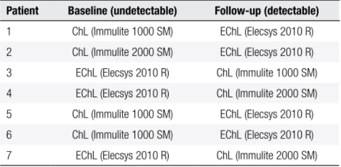
Seven patients (25%) presented TgAb detection by other IMA methodology in their follow-up (Table 2). All of these patients had diffuse Hashimoto’s thyroiditis in the pathological analysis.
Table 2. Laboratory IMA used in the irst negative TgAb assessment and in the second detectable TgAb measurement in 7 patients with papillary thyroid cancer
Patient Baseline (undetectable) Follow-up (detectable)
1 ChL (Immulite 1000 SM) EChL (Elecsys 2010 R)
2 ChL (Immulite 2000 SM) EChL (Elecsys 2010 R)
3 EChL (Elecsys 2010 R) ChL (Immulite 1000 SM)
4 EChL (Elecsys 2010 R) ChL (Immulite 2000 SM)
5 ChL (Immulite 1000 SM) EChL (Elecsys 2010 R)
6 ChL (Immulite 1000 SM) EChL (Elecsys 2010 R)
7 EChL (Elecsys 2010 R) ChL (Immulite 2000 SM)
ChL: chemiluminescence; EChL: electrochemiluminescence.
Three of these 7 patients were inally considered with NED (undetectable Tg and TgAb in the follow-up). This situation ocurred 22 ± 5 months after rem-nant ablation. All were TNM stage I, but with high (LATS) or intermediate (ATA) risk of recurrence.
The remaining 4 patients still persisted with detect-able TgAb, with no evidence of disease after a mean follow-up of 60 ± 27 months.
DISCUSSION
Thyroglobulin is a speciic tumor marker when total thyroidectomy is performed and remnant ablation indi-cated. Most differentiated thyroid cancer cells synthesi-ze Tg, although there may be differences in the mole-cular conformation of this tumor-derived Tg (13,14). The presence of detectable Tg after total thyroidec-tomy and remnant ablation usually indicates persistence or recurrence of disease (1). Pre-ablation stimulated Tg levels < 10 ng/mL under hypothyroid conditions has been proven to have a high negative predictive value in the deinition of free of disease patients (15,16).
However, serum Tg measurement remains techni-cally challenging even after 30 years of experience with different assays (17). Despite the introduction of a Tg reference preparation (CRM 457) more than 10 years ago (18), current methods to measure Tg still have an unacceptable intermethod variability (14,19), and this issue would need to be addressed before considering Tg as a marker of the presence of thyroid tissue. On top of that, false negative Tg levels from 4 up to 35% of DTC patients with evidence of local or metastatic disease have been reported in literature (20-24).
Cop
yright
© ABE&M t
odos os dir
eit
os r
eser
vados
.
risk patients (11). In the study of Phan and cols., who speciically analyzed low risk patients with undetectable Tg at ablation, only 3.4% of these patients had metas-tasis on postoperative US or WBS, suggesting that the measurement of a stimulated Tg at 12-18 months in order to deine the disease status was unnecessary (10). We agree that this is true for most patients with U Tg, but we observed 14% recurrence (including distant metastatic disease not detected by the measurement of the irst stimulated Tg). We believe that, in these cases, the method that had been used to measure the irst stimulated Tg level did not detect it accurately, yielding a false negative value. Otherwise, Tg would not have been detectable on thyroid hormone suppressive ther-apy 12 and 24 months after thyroid remnant ablation.
Furthermore, 25% of our patients who had unde-tectable TgAb turned out to be positive in successive measurements with other laboratory kits (Table 2). It is probable that if we continued measuring TgAb with the same IMA methodology, we could have missed these positive TgAb cases.
Given the high prevalence of TgAb, it is critical to ensure that specimens do not contain TgAb before au-thenticating a serum Tg result, because even low levels of TgAb can interfere with Tg measurement (1,2,9,17). Interference of serum antibodies remain the most se-rious problem, limiting the clinical utility of Tg testing. Tg-Ab is usually detected in around 20% of patients with DTC, compared with the 10% generally seen in the general population (20). The presence of TgAb is characterized by discordance in Tg values between RIA and IMA methodologies (detectable Tg using RIA and low or undetectable Tg using IMA) (9,14).
Table 2 shows the different kits used for TgAb measurement in our series. Because of TgAb method variability, the same method needs to be employed for long-term monitoring. This requirement is problem-atic for laboratories because manufacturers sometimes withdraw or change their methods without any notii-cation. Furthermore, it is not unusual for patients to change physicians and/or insurance plans that offer dif-ferent laboratories to continue their follow-up.
Although undetectable pre-ablation Tg levels usu-ally indicate complete previous surgery, our data rein-force earlier observations that, in a low percentage of patients, this situation may be related to non-detectable Tg or to the presence of TgAb (12-30,31). We believe that, when concurrent chronic thyroiditis is found by the pathologist, or when initial TNM stages are dis-with DTC, compared dis-with the 10% generally seen in
the general population (20). The presence of TgAb is characterized by discordance in Tg values between RIA and IMA methodologies (detectable Tg using RIA and low or undetectable Tg using IMA) (14).
When TgAb are initially detectable, their serum lev-el may decrease over subsequent months and years after adequate therapy, as thyroid tissue mass and Tg antigen levels decline (25,26). Patients may not achieve nega-tive TgAb status during the irst postoperanega-tive year, and may even exhibit a rise (or de novo appearance of TgAb during the 6 months after a radioiodine treatment, when there is release of Tg antigen secondary to lytic cell damage of thyroid tissue) (27,28). Clearly, it is the long-term trend in TgAb concentrations that is more valuable than any single TgAb level per se (29). Kim and cols. found that less than 1% of patients who became TgAb-negative or displayed more than 50% decrease in TgAb levels over the 6 to 12 months period after radioiodine treatment had recurrence detected during follow-up. In contrast, 19% of patients in whom TgAb decreased less than 50%, as well as the 37% in whom TgAb concentrations rose, were diagnosed with recur-rence (29).
On the other hand, undetectable Tg levels at the time of ablation may relect variable situations, accord-ing to different publications (12,16,30,31). Almost 30% of the reported patients in these series have an un-detectable Tg levels after surgery. In our experience, inding undetectable pre-ablation stimulated Tg le-vels is not frequent, as it was shown in this study. In a retrospective investigation performed by the Thyroid Department (of the Argentinian Society of Endocrinol-ogy and Metabolism), more than 90% of patients had detectable Tg levels at the moment of ablation (16). Similarly, we had 9% of patients of our database with an undetectable pre ablation Tg level, and only 61% of them were considered free of disease. Whether this is a condition associated with the surgery technique usually employed in our country or to other circumstances, it remains unclear.
Cop
yright
© ABE&M t
odos os dir
eit
os r
eser
vados
.
cordant with pre-ablation Tg levels, it is imperative to measure TgAb with different laboratory kits in order to avoid misdiagnosing a patient with undetectable Tg level as free of disease.
Disclosure: no potential conlict of interest relevant to this article was reported.
REFERENCES
1. Cooper DS, Doherty GM, Haugen BR, Kloos RT, Lee SL, Mandel SJ, et al. Revised American Thyroid Association management guidelines for patients with thyroid nodules and differentiated thyroid cancer. Thyroid. 2009;19(11):1167-214.
2. Pitoia F, Ward L, Wohllk N, Friguglietti C, Tomimori E, Gauna A, et al. Recommendations of the Latin American Thyroid Society on diagnosis and management of differentiated thyroid cancer. Arq Bras Endocrinol Metabol. 2009;53(7):884-7.
3. Kumar A, Shah DH, Shrihari U, Dandekar SR, Vijayan U, Sharma SM. Signiicance of antithyroglobulin autoantibodies in differen-tiated thyroid carcinoma. Thyroid. 1994;4(2):199-202.
4. Pacini F, Mariotti S, Formica N, Elisei R, Anelli S, Capotorti E, et al. Thyroid autoantibodies in thyroid cancer: incidence and rela-tionship with tumor outcome. Acta Endocrinol. 1988;119(3):373-80.
5. Spencer CA, Takeuchi M, Kazarosyan M, Wang CC, Guttler RB, Singer PA, et al. Serum hyroglobulin autoantibodies: prevalence, inluence on serum thyroglobulin measurement and prognostic signiicance in patients with differentiated thyroid carcinoma. J Clin Endocrinol Metab. 1998;83(4):1121-7.
6. Görges R, Maniecki M, Jentzen W, Sheu SN, Mann K, Bockisch A, et al. Development and clinical impact of thyroglobulin antibodies in patients with differentiated thyroid carcinoma during the irst 3 years after thyroidectomy. Eur J Endocrinol. 2005;153(1):49-55. 7. Kim WG, Yoon JH, Kim WB, Kim TY, Kim EY, Kim JM, et al. Chan-ge of serum antithyroglobulin antibody levels is useful for pre-diction of clinical recurrence in thyroglobulin-negative patients with differentiated thyroid carcinoma. J Clin Endocrinol Metab. 2008;93(12):4683-9.
8. Seo JH, Lee SW, Ahn BC, Lee J. Recurrence detection in differen-tiated thyroid cancer patients with elevated serum level of anti-thyroglobulin antibody: special emphasis on using 18F-FDG PET/ CT. Clin Endocrinol (Oxf). 2010;72(4):558-63.
9. Spencer C, Petrovic I, Fatemi S. Current thyroglobulin autoanti-body (TgAb) assays often fail to detect interfering TgAb that can result in the reporting of falsely low/undetectable serum Tg IMA values for patients with differentiated thyroid cancer. J Clin Endo-crinol Metab. 2011;96(5):1283-91.
10. Phan HT, Jager PL, van der Wal JE, Sluiter WJ, Plukker JT, Dierckx RA, et al. The follow-up of patients with differentiated thyroid can-cer and undetectable thyroglobulin (Tg) and Tg antibodies during ablation. Eur J Endocrinol. 2008;158(1):77-83.
11. Park EK, Chung JK, Lim IH, Park do J, Lee DS, Lee MC, et al. Re-current/metastatic thyroid carcinomas false negative for serum thyroglobulin but positive by posttherapy I-131 whole body scans. Eur J Nucl Med Mol Imaging. 2009;36(2):172-9.
12. Rosario PW, Xavier AC, Calsolari MR. Value of postoperative thyroglobulin and ultrasonography for the indication of ablation and ¹³¹I activity in patients with thyroid cancer and low risk of recurrence. Thyroid. 2011;21:49-53.
13. Schulz R, Bethäuser H, Stempka L, Heilig B, Moll A, Hüfner M. Evidence for immunological differences between circulating and
thyroid tissue-derived thyroglobulin in men. Eur J Clin Invest. 1989;19(5):459-63.
14. Spencer CA, Bergoglio LM, Kazarosyan M, Fatemi S, LoPresti JS. Clinical impact of thyroglobulin (Tg) and Tg autoantibody method differences on the management of patients with differentiated thyroid carcinomas. J Clin Endocrinol Metab. 2005;90(10):5566-75.
15. Polachek A, Hirsch D, Tzvetov G, Grozinsky-Glasberg S, Slutski I, Singer J, et al. Prognostic value of post-thyroidectomy thyroglo-bulin levels in patients with differentiated thyroid cancer. J Endo-crinol Invest. 2011;34(11):855-60.
16. Cabezón C, Löwenstein A, Orlandi A, Sartorio G, Sobrado P y Miembros del Departamento de Tiroides de la Sociedad Argen-tina de Endocrinología y Metabolismo (SAEM). Usefulness of preablation serum thyroglobulin as a predictor of the evolution of patients with differentiated thyroid carcinoma. Rev Arg Endo-crinol Metab. 2011;48(4):25-33.
17. Spencer CA, Lopresti JS. Measuring thyroglobulin and thyroglo-bulin autoantibody in patients with differentiated thyroid cancer. Nat Clin Pract Endocrinol Metab. 2008;4(4):223-33.
18. Feldt-Rasmussen U, Proilis C, Colinet E, Black E, Bornet H, Bour-doux P, et al. Human thyroglobulin reference material (CRM 457). 2nd Part: Physicochemical characterization and certiication. Ann Biol Clin (Paris). 1996;54(10-11):343-8.
19. Spencer CA, Takeuchi M, Kazarosyan M. Current status and per-formance goals for serum thyroglobulin assays. Clin Chem. 1996;42(1):164-73.
20. Spencer CA, Takeuchi M, Kazarosyan M, Wang CC, Guttler RB, Singer PA, et al. Serum thyroglobulin autoantibodies: prevalence, inluence on serum thyroglobulin measurement, and prognostic signiicance in patients with differentiated thyroid carcinoma. J Clin Endocrinol Metab. 1998;83(4):1121-7.
21. Grant S, Luttrell B, Reeve T, Wiseman J, Wilmshurst E, Stiel J, et al. Thyroglobulin may be undetectable in the serum of patients with metastatic disease secondary to differentiated thyroid car-cinoma. Follow-up of differentiated thyroid carcar-cinoma. Cancer. 1984;54(8):1625-8.
22. Westbury C, Vini L, Fischer C, Harmer C. Recurrent differentiated thyroid cancer without elevation of serum thyroglobulin. Thyroid. 2000;10(2):171-5.
23. Mertens IJ, De Klerk JM, Zelissen PM, Thijssen JH, Sie-Go DM,Han SH, et al. Undetectable serum thyroglobulin in a patient with me-tastatic follicular thyroid cancer. Clin Nucl Med 1999;24(5):346–9. 24. Brendel AJ, Lambert B, Guyot M, Jeandot R, Dubourg H, Roger
P, et al. Low levels of serum thyroglobulin after withdrawal of thyroid suppression therapy in the follow-up of differentiated thyroid carcinoma. Eur J Nucl Med. 1990;16(1):35–8.
25. Görges R, Maniecki M, Jentzen W, Sheu SN, Mann K, Bockisch A, et al. Development and clinical impact of thyroglobulin antibodies in patients with differentiated thyroid carcinoma during the irst 3 years after thyroidectomy. Eur J Endocrinol. 2005;153(1):49-55.
26. Chiovato L, Latrofa F, Braverman LE, Pacini F, Capezzone M, Mas-serini L, et al. Disappearance of humoral thyroid autoimmunity after complete removal of thyroid antigens. Ann Intern Med. 2003;139(5):346-51.
27. Rubio IG, Silva MN, Knobel M, Romao R, Possato R, Gebrin EM, et al. Peripheral blood levels of thyroglobulin mRNA and serum thyroglobulin concentrations after radioiodine ablation of multi-nodular goiter with or without pre-treatment with recombinant human thyrotropin. J Endocrinol Invest. 2007;30(7):535-40. 28. Benvenga S, Bartolone L, Squadrito S, Trimarchi F. Thyroid
Cop
yright
© ABE&M t
odos os dir
eit
os r
eser
vados
.
29. Kim WG, Yoon JH, Kim WB, Kim TY, Kim EY, Kim JM, et al. Chan-ge of serum antithyroglobulin antibody levels is useful for pre-diction of clinical recurrence in thyroglobulin-negative patients with differentiated thyroid carcinoma. J Clin Endocrinol Metab. 2008;93(12):4683-9.
30. Tuttle RM, Tala H, Shah J, Leboeuf R, Ghossein R, Gonen M, et al. Estimating risk of recurrence in differentiated thyroid cancer after total thyroidectomy and radioactive iodine remnant abla-tion: using response to therapy variables to modify the initial risk
estimates predicted by the new American Thyroid Association staging system. Thyroid. 2010;20(12):1341-9.