Key words:
Tumor Markers, Biological; CA-19-9 Antigen; Odds Ratio
Int Braz J Urol. 2013; 39: 631-8
__________________ Submitted for publication: January 16, 2013
__________________ Accepted after revision: August 07, 2013 Objectives: Although the glycoprotein group tumor marker CA 19-9 has been detected
in both serum and urine of bladder cancer patients, information about their comparative role in screening of low grade transitional cell carcinoma (LGTCC) and high grade tran-sitional cell carcinoma (HGTCC) is rare.
Materials and Methods: In this study we measured both the urinary and serum levels of CA 19-9 in 35 LGTCC and 20 HGTCC patients by ELISA and determined the cut off value of both urinary and serum CA 19-9 levels by receiver operator characteristic curve (ROC) for both patient groups. Odds ratio (OR) for CA 19-9 was analyzed with its range at 95% confidence interval to analyze the role of this tumor marker as a screening parameter for both of these cancer types.
Results: For urinary CA 19-9 the OR was 20.16 with an interval of 4.91-82.71 whereas for the serum CA 19-9 it was 7.5 with an interval of 2.28-24.62.
Conclusions: From these data we suggest that urinary CA 19-9 is a better screening pa-rameter with optimum sensitivity and specificity than its serum counterpart for diagnosis of low grade and early stages of transitional cell carcinoma of urinary bladder. Further-more, it can be suggested that urinary CA 19-9 can be used as better prognostic marker for LGTCC than its serum counterpart.
INTRODUCTION
Tumor markers are biochemical substan-ces elaborated by tumor cells either due to the cause or effect of malignant process. These ma-rkers can be normal endogenous products that are produced at a greater rate in cancer cells or the products of newly switched on genes that re-mained quiescent in the normal cells. They may be present as intracellular substances in tissues or may be released into the circulation and appe-ar in serum. An ideal tumor mappe-arker should be highly sensitive and specific, and should be able to detect neoplastic growth from non-neoplastic growth. Its level should rise before the tumor
proceeds, so that it can be used as an early detec-tion marker in any cancer (1,2).
Bladder cancer is a common urologic can-cer. Bladder cancer is the fourth most common cancer in men in the United States, after pros-tate, lung, and colorectal cancer. Bladder cancer is the 10th most common cancer in women (3). The most common type of bladder cancer in the United States is urothelial carcinoma, former-ly known as transitional cell carcinoma (TCC) (4) which happens to be the 7th most common cancer in men and the 17th most common can-cer in women worldwide (5). The urothelium in the entire urinary tract may be involved, inclu-ding the renal pelvis, ureter, bladder, and urethra.
Comparison of Urinary and Serum CA 19-9 as Markers
of Early Stage Urothelial Carcinoma
_______________________________________________
Suparna Roy, Anindya Dasgupta, Kaushik Kar
Dept. of Biochemistry, Calcutta National Medical College, Kolkata-14, India
ABSTRACT
ARTICLE
INFO
The clinical course of bladder cancer carries a broad spectrum of aggressiveness and risk. The detection of bladder cancer mostly depends on urinary cytology and cystoscopy. Cystoscopy has been proven quite successful in surveillance and follow-up of patients with treated bladder cancer. The drawback of cystoscopy is that it is quite expensive, invasive and uncomfortable (6). Urine cytology has been the gold standard for bladder cancer screening and surveillance in the past (7) but it is subjective and requires adequa-te number of exfoliaadequa-ted cells in the urine, and cellular alterations are likely due to change in collection conditions and therapeutic interven-tions. Urine cytology is performed at the same time as cystoscopy, although its routine use for screening is controversial (8). Urine cytology is associated with a significant false-negative rate, especially for low-grade carcinoma (10-50% ac-curacy rate). Studies have evaluated the clinical significance of urinary CA19-9 levels in bladder cancer patients classified according to various combinations of Lewis (Le) and Secretor (Se) ge-notypes (9). The CA 19-9 concentration correla-ted well with the clinical response to treatment. CA 19-9 increases very early during recurrence in patients with a mean lead-time of 4-6 mon-ths before the clinical diagnosis. The marker is 210 kDa tumor- associated glycoprotein antigen present as carbohydrate determinant on glycoli-pid and glycoprotein. Carbohydrate antigen 19-9 was established from a colon cancer cell line (10) and it has been clinically applied as a useful tu-mor marker of pancreatic and gastrointestinal carcinoma (11-13). Many immunohistochemical studies have shown carbohydrate antigen 19-9 expression in various normal tissues, including the pancreas, gall bladder, stomach, colon, bron-chial tree, endometrium, salivary glands, kidney and prostate (14-17). Furthermore, it is well kno-wn that the serum level is elevated in some non-malignant diseases. In 99.6% of healthy adults, serum CA 19-9 levels are lower than 37µ/mL. Va-lue less than 100µ/mL is considered as grey zone values in which malignant and benign diseases may overlap. Pal et al. reported urinary CA 19-9 to be a sensitive marker for an early diagnosis of transitional cell carcinoma but did not evaluate
its comparative role with its serum counterpart (18). Although studies have evaluated the clinical significance of urinary CA19-9 levels in bladder cancer patients classified according to various combinations of Lewis (Le) and Secretor (Se) ge-notypes, reports regarding its sensitivity as an early marker in low and high grade transitional cell carcinomas are scarcely available, particular-ly in comparison to its serum counterpart. Hence, we hypothesized that the urinary and serum le-vel of this marker may not exhibit same degree of sensitivity and specificity for early detection of urothelial carcinoma. Accordingly, we made an effort to evaluate the usefulness of urinary CA19-9 in comparison to the serum CA 19-9 for an early diagnosis of urothelial carcinoma in the present study. The aim of this study was to detect urinary level of CA 19-9 in different stages of bladder cancer and its role in early diagnosis of cancer, and also to establish it as a good nonin-vasive diagnostic tool in conjunction with urine cytology and cystoscopy.
MATERIALS AND METHODS
Study design
The study was conducted in the Institute of Postgraduate Medical Education & Research, West Bengal, India as a cross-sectional observa-tional study conducted over a period of 2 years, from September 2008 to September 2010. The study followed the rules and regulations of the modified Helsinki Declaration and was approved by the properly constituted institutional ethical committee for studies involving human subjects.
Selection of cases and controls
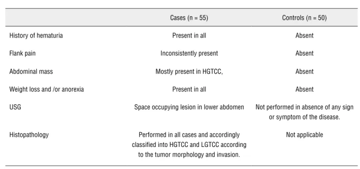
patients diagnosed as suffering from transitional cell carcinoma in the urology department of our hospital were included. The total study period was from September 2008 to September 2010. Exclusion criteria for selection of subjects in the case group were patients suffering from i) any other malignant disorders as evident from careful history, clinical examination and routine clinical investigation, ii) any endocrinological disorders, any pathological le-sion in prostate, kidney and gastrointestinal tracts. Control subjects were selected from those having no previous history of any urological disorder accor-ding to criteria: i) history of hematuria, ii) history of flank pain, iii) abdominal mass, iv) anorexia, v) weight loss, vi) dysuria, vii) H/O prostatism eg. noc-turia, difficulty in starting and stopping the urine stream, overflow dribbling, poor urine stream, and other obstructive symptoms were excluded from the present study. All control subjects were scree-ned for any inflammatory or malignant disorder by obtaining detailed history and clinical examination. Overall criteria for choosing cases and controls are presented in Table-1.
Informed consent obtained from each patient before participation and the study was approved by institutional ethical committee. Following the above mentioned criteria 55 and 50 persons were finally se-lected in the case and control group respectively.
Collection of tissue samples
Tissue samples from the case group were collected during their surgery (exploration or re-moval of the tumor). When partial or complete removal was performed tissue samples were isola-ted from several area of the affecisola-ted organ to ma-ximize the histopathological diagnosis. For ino-perable masses an appropriate amount of tissue was isolated. Although the process was limited by invasion of the tumor mass into the surrounding area, multiple tissue samples were removed as far as practicable without disturbing the surrounding area. Urine and blood samples were collected from the patients just before their surgery.
Study Methods:
a) Concentration of urine sample: Uri-ne sample was concentrated 20 times by Biogel-p. Dried gels having pore size 90-180µm were added to a measured volume of urine as weighed granules. Water and small molecules were attracted into the gel by osmosis. The exclusion limit of the biogel p is 6000 dalton. Those molecular weights > 6000 dalton were excluded by pore size. It is almost always necessary to concentrate the urine before test. The urine samples were taken and concentrated by
Table 1 - Criteria for selection of cases and controls.
Cases (n = 55) Controls (n = 50)
History of hematuria Present in all Absent
Flank pain Inconsistently present Absent
Abdominal mass Mostly present in HGTCC, Absent
Weight loss and /or anorexia Present in all Absent
USG Space occupying lesion in lower abdomen Not performed in absence of any sign
or symptom of the disease.
Histopathology Performed in all cases and accordingly
classified into HGTCC and LGTCC according to the tumor morphology and invasion.
passing them through biogel in 1:20 ratio. 50µg of biogel was taken in an aliquot and to it 1000µl of urine added and kept in 2-4 ºC for 5 hours. The CA 19-9 is a tumor ma-rker of molecular weight of 210 kD. So the supernatant urine will contain the CA19-9 molecules and the low molecular wei-ght particles will be adsorbed by the gel. The supernatant of the urine sample were collected and tested for CA19-9 level by ELISA method.
b) Measurement of urinary and serum CA19-9 levels: measurement of urinary and serum CA19-9 levels of both patients and controls was done by enzyme immunoassay from Monobind- AcuBind ELISA kit.
c) Detection of malignant cell in urine was done by urinary cytology with Papanicolaou (PAP) staining for malignant cell detection. d) Histological grading of tumor was done by histopathology of malignant tissues de-rived from the urinary bladder. The LGTCC were diagnosed mainly by the features of minimal nuclear atypia, infrequent mitotic figures predominantly towards the base, and mid variation on nuclear size and sha-pe. The HGTCC were diagnosed mainly by the features of frank anaplasia, frequent mitotic figures including atypical ones, and a much higher incidence of invasion into the muscular layer.
Data analysis
Data obtained for the selected parameters from the case and control groups were analyzed for
significance of differences between urinary and se-rum parameters in case and control groups. Receiver operator characteristic curve (ROC) was used to find out cut off value for the tumor markers at a definite level of true sensitivity against false positivity. All statistical analysis was performed using SPSS sof-tware version 16.0 for Windows.
RESULTS
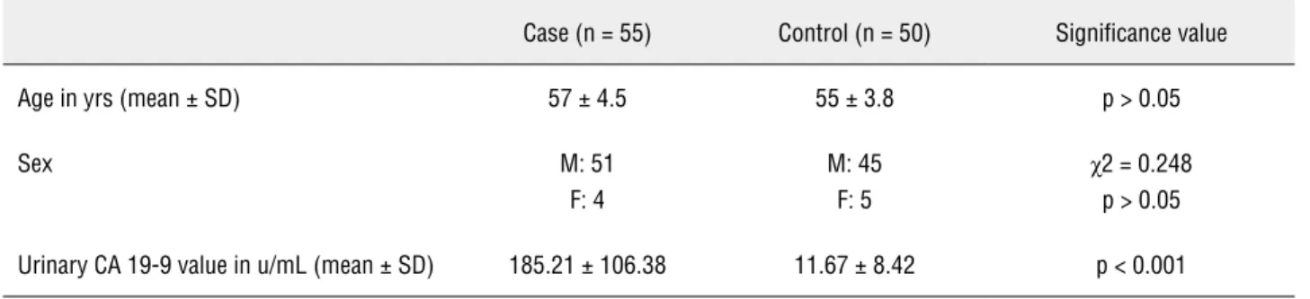
From the comparative values shown in the Table-2 it is evident that there is no statistically significant difference between the age and sex ra-tio of case and control groups. Furthermore, it is seen that urinary CA 19-9 values are significantly higher in the case group (Figure-1).
A sensitivity of 0.943 and false positivity of 0.450 was chosen at the left uppermost corner of the curve (marked) with the corresponding cut off value of urinary CA19-9 of 114.5 IU/l.
A sensitivity of 0.714 and false positivity of 0.250 was chosen at the left uppermost corner of the curve (marked) with the corresponding cut off value of serum CA19-9 of 17.90 IU/l.
Table-3 shows that Odd’s ratio (OR) is 20.16 and its range for 95% CI = 4.91-82.71. The range of OR is above 1 that suggests that urinary level of CA19-9 in low grade TCC patients is highly signifi-cant than observed in high grade cases.
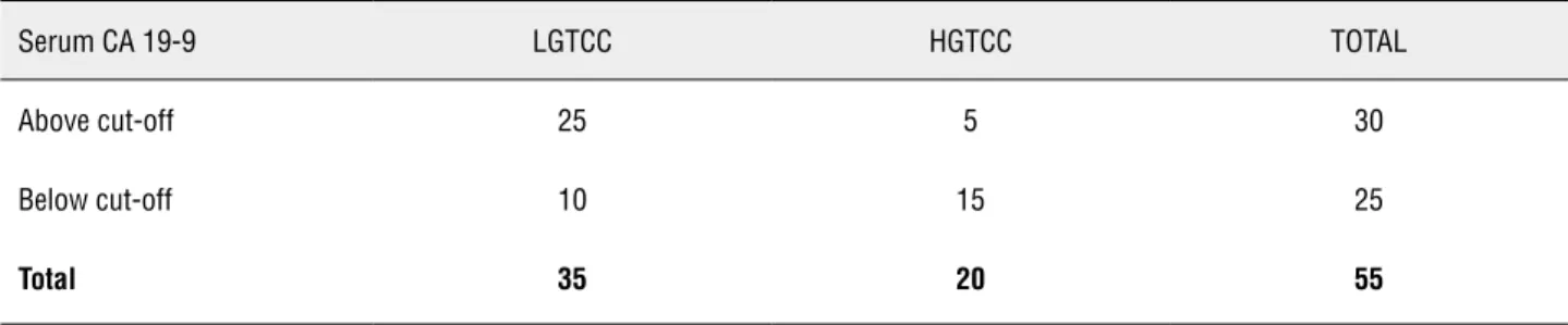
Table-4 shows that Odd’s ratio is 7.5 and its range for 95% CI = 2.28-24.62. The range of OR is above 1 so it suggests that serum level of CA19-9 in low grade TCC is significant but its sig-nificance is lower than urinary CA19-9 level.
Urinary cytology was performed only in the patient group. The sensitivity for it in our study
Table 2 - Comparative data analysis of age, sex and urinary CA 19-9 between case and control groups.
Case (n = 55) Control (n = 50) Significance value
Age in yrs (mean ± SD) 57 ± 4.5 55 ± 3.8 p > 0.05
Sex M: 51
F: 4
M: 45 F: 5
χ2 = 0.248 p > 0.05
population was 20 and 40 percent for the LGTCC and HGTCC categories respectively. In contrast to these values the sensitivity detected were 94.2 and 45 percent for the urinary CA 19-9 in the LGTCC and HGTCC groups respectively as obtained from our ROC curve (Table-3 and Figure-2).
DISCUSSION
In this study an attempt was made to as-sess the role of CA19-9 as a tumor marker in urine
and serum of patients of histopathologically con-firmed bladder cancer. Bladder cancer is amenable to biomarker development because many tumor--associated molecules are secreted in urine. Tumor cells are shed in urine, and, therefore, tests that detect tumor cell-surface markers have also been developed to diagnose bladder cancer and monitor its recurrence. Several bladder tumor markers show higher sensitivity than cytology, but most have lo-wer specificity. In isolated papers it has been ob-served that the serum CA19-9 level is higher in
Figure 1 - Cut off value serum CA 19-9 as deduced from ROC Curve.
Diagonal segments are produced by ties 1 - Specificity
ROC Curve 1.0
0.8
0.6
0.4
0.2
0.0
0.0 0.2 0.4 0.6 0.8 1.0
Sensitivity
Table 3 - Comparison of urinary level of CA19-9 between low grade and high grade transitional cell carcinoma with cut-off value of urinary CA19-9 of 114.5 IU/l.
Urinary CA19-9 LGTCC HGTCC TOTAL
Above cut-off 33 9 42
Below cut-off 2 11 13
Total 35 20 55
HGTCC = High grade transitional cell carcinoma. LGTCC = Low grade transitional cell carcinoma x2= 17.12
Figure 2 - Cut off value of urinary CA 19-9 as deduced from ROC Curve.
Diagonal segments are produced by ties 1 - Specificity
0.0 0.2 0.4 0.6 0.8 1.0 ROC Curve
1.0
0.8
0.6
0.4
0.2
0.0
Sensitivity
Table 4 - Comparison of serum level of CA19-9 between Low grade and High grade transitional cell carcinoma with a cut-off of value for serum CA-19-9 of 17.9 IU/l.
Serum CA 19-9 LGTCC HGTCC TOTAL
Above cut-off 25 5 30
Below cut-off 10 15 25
Total 35 20 55
HGTCC = High grade transitional cell carcinoma. LGTCC = Low grade transitional cell carcinoma
x2= 11.06
Odds ratio = 7.5 (95% CI = 2.28-24.62)
few benign urological disease but more often it is associated with transitional cell carcinoma of blad-der (19). It is also reported that serum CA19-9 is a diagnostic marker in case of urachal carcinoma as the embryological origin of urachus and colon are the same (20). Although cytology accurately detects high grade lesions, it lacks the sensitivity to detect the low grade tumors that characterize most blad-der cancer cases (21,22). Urine cytology is associa-ted with a significant false-negative rate, especially
The false-positive rate is 1-12%, but it has a 95% accuracy rate for diagnosing high-grade carcinoma and CIS. The sensitivity of urine cytology can be in-creased by obtaining a bladder barbotage cytology (70%) as opposed to a voided cytology (30%).
pre-and so, an easily available tumor marker is needed, that can be easily measured with lesser time, shows significantly high sensitivity with minimum false positivity and is easily reproducible.
In the present study we used ROC curves to find out the cut off value of the tumor marker CA 19-9 from its corresponding values in 35 LGTCC and 20 HGTCC case subjects. The cut off value at the left uppermost corner of the ROC curve was selected as it signified maximum sensitivity with minimum false positivity. Tables 3 and 4 show the distribu-tion of CA 19-9 in these two groups based on this cut off value in urine and serum respectively. For the urinary CA 19-9, we found an Odds Ratio to be 20.16 with a range of 4.91 to 82.71 at 95% CI that suggested a significant elevation in the urinary level of this tumor marker in all cases. When we analyzed the Odds Ratio for the same tumor marker in serum a value of 7.5 with its range from 2.28 to 24.62 at 95% CI was obtained. Although it signified a significantly raised level of this tumor marker in the serum of the case group, its predictive value was much lower in comparison to that of its urinary le-vel (7.5 vs. 20.16). Our data analysis thus indicated that increases in the urinary level of CA19-9 was more closely and significantly associated with the low grade TCC patients than their high grade coun-terparts. The importance of the urinary level of CA 19-9 in diagnosis and prognosis of bladder cancer has been reiterated in some other studies. Urinary CA19-9 and DU-PAN-2 levels were measured as units per mg creatinine (U/mg Cr) in 121 patients with bladder cancer and in 31 patients with other urologic diseases The cut-off value determined using receiver operating characteristics analyses was 37.6 U/mg Cr. Approximately 70% of bladder cancer patients with both Le and Se alleles de-monstrated urinary CA19-9 levels above the cut--off value. In contrast, only 16% of patients with other urologic diseases were above the cut-off value which suggested that the urinary CA19-9 level could be a new effective diagnostic tool in bladder cancer patients with both Le and Se alle-les (9). Keeping in track with these observations our study furthermore suggests that urinary level is not only a sensitive marker for early diagnosis but it is also can be utilized as a better predictor for early stages of urothelial carcinoma than its
corresponding serum level. In bladder cancers, as urinary excretion of this tumor marker occurs directly from its source tissue, its urinary appea-rance is more probable at an earlier phase than its serum counterpart. However, in advanced stages of tumor progression the serum level is raised to a significant amount due to increased production and possible spread to other organs that leads to almost similar type of appearance of this tumor marker in both urine and serum. Hence and from the findings of our study we suggest that urinary CA 19-9 level merits particular importance for diagnosis of low grade transitional cell carcinoma which is of much importance if the treatment and prognosis of the disease are concerned. However, these observations have to be correlated to and integrated with the re-sults obtained for other tumor marker for bladder carcinoma in future studies involving larger selec-tion of people from different areas of world. Our results should be interpreted with these limitations.
ABBREVIATIONS
PAP = Papanicolaou stain
HGTCC = High grade transitional cell carcinoma LGTCC = Low grade transitional cell carcinoma TCC = Transitional cell carcinoma
ROC = Receiver operator characteristic curve OR = Odds ratio
ELISA = Enzyme-linked immunosorbent assay
CONFLICT OF INTEREST
None declared.
REFERENCES
1. Vaidyanathan K, Vasudevan DM: Organ Specific Tumor Mark-ers: What’s New? Indian J Clin Biochem. 2012; 27: 110-20. 2. Taguchi A, Hanash SM: Unleashing the power of proteomics
to develop blood-based cancer markers. Clin Chem. 2013; 59: 119-26.
3. Jemal A, Siegel R, Ward E, Hao Y, Xu J, Thun MJ: Cancer sta-tistics, 2009. CA Cancer J Clin. 2009; 59: 225-49.
4. Greenlee RT, Murray T, Bolden S, Wingo PA: Cancer statistics, 2000. CA Cancer J Clin. 2000; 50: 7-33.
6. Rodríguez-Rubio F, Sanz G, Garrido S, Sánchez C, Estudillo F: Patient tolerance during outpatient flexible cystoscopy--a prospective, randomized, double-blind studycomparing plain lubrication and lidocaine gel. Scand J Urol Nephrol. 2004; 38: 477-80.
7. Papanicolaou GN, Marshall VF: Urine sediment smears as a diagnostic procedure in cancers of the urinary tract. Science. 1945; 101: 519-20.
8. Grossfeld GD, Litwin MS, Wolf JS Jr, Hricak H, Shuler CL, Agerter DC, et al.: Evaluation of asymptomatic microscopic hematuria in adults: the American Urological Association best practice policy--part II: patient evaluation, cytology, voided markers, imaging, cystoscopy, nephrologyevaluation, and follow-up. Urology. 2001; 57: 604-10.
9. Nagao K, Itoh Y, Fujita K, Fujime M: Evaluation of urinary CA19-9 levels in bladder cancer patients classified according to the combinations of Lewis and Secretor blood group geno-types. Int J Urol. 2007; 14: 795-9.
10. Del Villano BC, Brennan S, Brock P, Bucher C, Liu V, McClure M, et al.: Radioimmunometric assay for a monoclonal antibody-defined tumor marker, CA 19-9. Clin Chem. 1983; 29: 549-52. 11. Atkinson BF, Ernst CS, Herlyn M, Steplewski Z, Sears HF,
Ko-prowski H: Gastrointestinal cancer-associated antigen in im-munoperoxidase assay. Cancer Res. 1982; 42: 4820-3. 12. Fong ZV, Winter JM: Biomarkers in pancreatic cancer:
diag-nostic, progdiag-nostic, and predictive. Cancer J. 2012; 18: 530-8. 13. Bluvshteĭ’n GA, Lysenko VG, Zakharova NB, Kitaev IV: Tu-mor markers p53, sFAS, FASL, CEA and CA 19-9 in eval-uating the effectiveness of surgical and pharmaco nutri-tionaltreatment of patients with gastric cancer. Eksp Klin Gastroenterol. 2012; 2: 41-9.
14. Ohshio G, Ogawa K, Kudo H, Yamabe H, Higuchi K, Nakashima Y, et al.: Immunohistochemical distribution of CA19-9 in nor-mal and tumor tissues of the kidney. Urol Int. 1990; 45: 1-3.
15. Terracciano D, Mariano A, Macchia V, Di Carlo A: Analysis of glycoproteins in human colon cancers, normal tissues and in human colon carcinoma cells reactive withmonoclonal anti-body NCL-19-9. Oncol Rep. 2005; 14: 719-22.
16. Aybek H, Aybek Z, Sinik Z, Demir S, Sancak B, Tuncay L: Eleva-tion of serum and urinary carbohydrate antigen 19-9 in benign hydronephrosis. Int J Urol. 2006; 13: 1380-4.
17. Parra JL, Kaplan S, Barkin JS: Elevated CA 19-9 caused by Hashimoto’s thyroiditis: review of the benign causes of in-creased CA 19-9 level. Dig Dis Sci. 2005; 50: 694-5. 18. Pal K, Roy S, Mondal SA, Chatterjee U, Tiwari P, Bera M:
Uri-nary level of CA19-9 as a tumor marker in urothelial carci-noma of the bladder. Urol J. 2011; 8: 203-8.
19. Barone D, Onetto M, Conio M, Paganuzzi M, Saccomanno S, Aste H, et al.: CA 19-9 assay in patients with extrahepatic cho-lestatic jaundice. Int J Biol Markers. 1988; 3: 95-100. 20. Whelan P, Britton JP, Dowell AC: Three-year follow-up of
blad-der tumours found on screening. Br J Urol. 1993; 72: 893-6. 21. Sheldon CA, Clayman RV, Gonzalez R, Williams RD, Fraley EE:
Malignant urachal lesions. J Urol. 1984; 131: 1-8.
22. Schwalb DM, Herr HW, Fair WR: The management of clini-cally unconfirmed positive urinary cytology. J Urol. 1993; 150: 1751-6.
_____________________
Correspondence address: