Induction of G
2
/M arrest, caspase activation and apoptosis by
a
-santonin
derivatives in HL-60 cells
José Roberto Oliveira Ferreira
a, Bruno Coêlho Cavalcanti
a, Patricia Marçal da Costa
a,
Francisco Frederico Perlinson de Arantes
b, Elson Santiago de Alvarenga
b, Célia Regina Alvares Maltha
b,
Luiz Cláudio de Almeida Barbosa
c, Gardenia Carmen Gadelha Militão
d, Claúdia Pessoa
a, Paulo Michel
Pinheiro Ferreira
e,f,⇑aDepartment of Physiology and Pharmacology, Federal University of Ceará, Fortaleza, Brazil bDepartment of Chemistry, Federal University of Viçosa, Viçosa, Brazil
cDepartment of Chemistry, Federal University of Minas Gerais, Belo Horizonte, Brazil dDepartment of Physiology and Pharmacology, Federal University of Pernambuco, Recife, Brazil
eDepartment of Biological Sciences, Campus Senador Helvídio Nunes de Barros, Federal University of Piauí, Picos, Brazil fPostgraduate Program in Pharmaceutical Sciences, Federal University of Piauí, Teresina, Brazil
a r t i c l e
i n f o
Article history:
Received 28 November 2012 Accepted 16 March 2013 Available online 27 March 2013
Keywords:
Sesquiterpene lactones Santonin
Cytotoxicity Apoptosis
a b s t r a c t
Sesquiterpene lactones (SLs) are natural products with a variety of biological activities. Previously, we demonstrated the cytotoxic effects of three newa-santonin derivatives on different tumor cell lines with low toxic effects upon peripheral human leukocytes. Here, we evaluated the mechanism of action trig-gered by these derivatives. HL-60 cell cycle determined after 24 h treatment revealed a significant inhi-bition on cell-cycle progression and leading to an increasing of cells in G2/M [7.6% and 9.0% for compound 3% and 9.0% and 8.6% for compound 4 (1 and 2lM, respectively)]. However, after 48 h exposure, all com-pounds caused G2/M reduction and a significant DNA fragmentation. Compounds 2, 3 and 4 were able to induce apoptosis on leukemia cells, which was corroborated by phosphatidyserine externalization and activation of caspases-3 and -7 after 24 h exposure. None of the derivatives analyzed caused depolariza-tion of mitochondrial membrane within 24 h of incubadepolariza-tion, suggesting the involvement of the extrinsic apoptotic pathway in the death process. The antiproliferative action of these compounds is related to the DNA synthesis inhibition and cell cycle arrest, which probably lead to apoptosis activation. Therefore, these santonin derivatives are promising lead candidates for development of new cytotoxic agents.
Ó2013 Elsevier Ltd. All rights reserved.
1. Introduction
Cancer is a disease caused by disorderly growth of cells that of-ten invade tissues and organs. Considerable insight has been gained into the mechanisms by which some chemicals affect cellu-lar growth and this knowledge has been used to design new more selective chemotherapeutic drugs towards cancer cells than to nor-mal cells and reduce side effects (Benz and Yau, 2008).
The development of antineoplasic agents is important to dimin-ish the mortality caused by cancer. Since cell homeostasis depends on the balance between proliferation and cell death, effective com-pounds that increase apoptosis without concomitant increases cel-lular proliferation appear to be a relevant strategy to suppress
tumor growth (Kaufmann and Earnshaw, 2000; Ferreira et al.,
2010).
Despite the interest in molecular modeling and combinatorial chemistry, the search for novel anticancer drugs from natural and non-natural sources has continued through the collaboration of scientists worldwide in looking for new bioactive compounds
(Kiran et al., 2008; Cragg et al., 2009; Ferreira et al., 2011). Among
the large sources of potential compounds natural products offer opportunities to evaluate not only totally new chemical classes of anticancer agents, but also novel and potentially relevant mech-anisms of action. The majority of anticancer drugs are natural products or their derivatives and more than 200 drugs derived from natural products are in preclinical or clinical development and evaluation (Ghantous et al., 2010; Newman and Cragg, 2012). Sesquiterpene lactones (SLs) are a class of naturally occurring plant terpenoids of the Asteraceae family, known for their various biological activities such as anti-inflammatory, phytotoxic, antimi-crobial, antiprotozoal, and cytotoxic against different tumor cell lines (Hehner et al., 1998; Mazor et al., 2000; Schmidt et al.,
0887-2333/$ - see front matterÓ2013 Elsevier Ltd. All rights reserved.
http://dx.doi.org/10.1016/j.tiv.2013.03.010
⇑Corresponding author. Address: Campus Senador Helvídio Nunes de Barros
(UFPI), Cícero Duarte 905, ZIP 64607-670, Picos, Piauí, Brazil. Tel.: +55 89 34221008; fax: +55 89 34221024.
E-mail addresses:[email protected],[email protected](P.M.P. Ferreira).
Contents lists available atSciVerse ScienceDirect
Toxicology in Vitro
2002; Zhang et al., 2005).
a
-Santonin, a sesquiterpene lactone iso-lated fromArtemisia santonica presents antipyretic, anti-parasitic and anti-inflammatory properties (Ivasenko et al., 2006). Somea
-santonin derivatives also act as inhibitors of phospholipase A2enzymes from Bothrops jararacussu (De Alvarenga et al., 2011). Additionally, we have reported the activity of synthetic
a
-santonin derivatives against several human cancer cell lines (HL-60, leuke-mia; SF-295; glioblastoma; HCT-8, colon; MDA/MB-435, mela-noma) with low antiproliferative effects upon normal human leukocytes (Arantes et al., 2009,Arantes et al., 2010). Therefore, these results indicate that SLs and related compounds may repre-sent a promising class of biological agents. In this work, we de-scribed, for the first time, the mechanism of induction of cell death on human promyelocytic leukemia HL-60 cell line triggered by threea
-santonin derivatives.2. Methods
2.1. Chemicals
Fetal calf serum was purchased from Cultilab (Campinas, SP), RPMI 1640 medium, trypsin–EDTA, penicillin and streptomycin were purchased from GIBCOÒ
(Invitrogen, Carlsbad, CA, USA). Pro-pidium iodide (PI), acridine orange (AO), ethidium bromide (EB) and Rhodamine 123 (Rho-123) were purchased from Sigma–Al-drich Co. (St. Louis, MO, USA). Doxorubicin (DoxolemÒ
) was pur-chased from Zodiac Produtos Farmacêuticos S/A, Brazil. All other chemicals and reagents used were of analytical grade.
a
-Santonin (compound 1) (97%) was procured from Sigma–Aldrich Co. (Mil-waukee, WI, USA) and was utilized without further purification. The transformation ofa
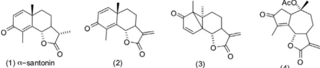
-santonin (compound 1) into lactone (com-pound 2), and its further transformation into (com(com-pound 3) and (compound 4) were carried out as previously described (Arantes et al., 2010) (Fig. 1).2.2. Study of the mechanisms involved in the cytotoxic activity on HL-60 cells
Since HL-60 cell line was the most sensitive cell line to
a
-santo-nin derivatives (Arantes et al., 2010), it was selected to evaluate the mechanism underlying to their cytotoxic effects after 24 h expo-sure. The compounds dissolved in DMSO (0.1%) were added to cell cultures of HL-60 cells (3105cells/mL) to obtain finalconcentra-tions of 1 and 2
l
M. Doxorubicin (0.6l
M) was used as a positive control (Dox). All flow cytometry analyses were performed in a GuavaÒEasyCyte Mine using Guava Express Plus CytoSoft 4.1 soft-ware (Guava Technologies Inc., Industrial Blvd., Hayward, CA, USA). Five thousand events were evaluated per experiment and cell deb-ris was omitted from the analysis.
2.2.1. Trypan blue exclusion test
Cell viability was determined by the trypan blue dye exclusion test (Kepp et al., 2011). The cell samples were diluted in trypan blue dye of an acid azo exclusion medium by preparing a 1:1
dilution of the cell suspension using a 0.4% trypan blue solution. Non-viable cells were labeled in blue and are visible with bright-field optics and viable cells were unstained, since viable cells main-tain the capacity to extrude this vital dye. The count was performed under the microscope in four 11 mm squares of a
Neubauer chamber. Number of cells (104cells/mL) was stated
and viable and non-viable cells were expressed as a percentage of total cells.
2.2.2. Inhibition of DNA synthesis detected by immunocytochemistry
Cells were plated in 24-well tissue culture plates (2 mL/well) and treated with the compounds. After 21 h exposure, 20
l
L of 5-bromo-20-deoxyuridine (BrdU, 10 mM) was added and incubatedfor 3 h at 37°C. To determine the amount of BrdU incorporated
into DNA (Pera et al., 1977), cells were harvested, transferred to cytospin slides, and allowed to dry for 2 h at room temperature (25°C). Cells were labeled using direct peroxidase
immunocyto-chemistry by the chromogen diaminobenzidine (DAB) staining those cells that incorporated Brd. Slides were counterstained with hematoxylin and coverslipped. The determination of BrdU positiv-ity was performed by light microscopy (Olympus, Tokyo, Japan). Two hundred cells were counted per sample to determine the per-centage of BrdU-positive cells (Costa et al., 2008).
2.2.3. Morphological analysis with fluorescence microscopy
To determine whether the growth inhibition activity of com-pounds 2–4 was related to the induction of apoptosis or necrosis, morphological analysis of treated cells was investigated by fluores-cent microscopy using acridine orange/ethidium bromide (AO/EB) staining. After 24 h incubation, cells were pelleted and each sample was mixed with 1 L of aqueous AO/EB solution (100 g/mL of AO in PBS; 100 g/mL EB in PBS) just prior to fluorescence microscopy and quantification (Olympus, Tokyo, Japan). Three hundred cells were counted per sample and scored as follows: viable cells, apoptotic cells and necrotic cells (Cury-Boaventura et al., 2004; Tamatani
et al., 2012). The percentage of apoptotic and necrotic cells was
then calculated.
2.2.4. Cell cycle distribution and internucleosomal DNA fragmentation analysis
Cell cycle distribution and DNA fragmentation analysis were evaluated by the incorporation of propidium iodide (50
l
g/mL). Briefly, 24 h-treated and untreated cells (3105cells/mL) wereincubated at 37°C for 30 min in the dark, in a lysis solution
con-taining 0.1% citrate, 0.1% Triton X-100 and 50
l
g/mL propidium io-dide and fluorescence was measured afterwards.2.2.5. Cell membrane integrity
Cell membrane integrity was evaluated by the exclusion of pro-pidium iodide. Briefly, 100
l
L of treated and untreated cells were incubated with propidium iodide (50l
g/mL). The cells were then incubated for 5 min at 37°C. Fluorescence was measured and cellmorphology, granularity and membrane integrity were determined
(Darzynkiewicz et al., 1992).
2.2.6. Phosphatidylserine (PS) externalization
PS externalization was analyzed by flow cytometry (Annexin V) according toVermes and co-works (1995)using Guava Nexin As-say Kit. Briefly, cells (3105cells/mL) were washed twice with
cold PBS and then resuspended in 135
l
L of PBS with 5l
L of 7-aminoactinomycin D (7-AAD) and 10l
L of Annexin V-PE. Cells were gently vortexed and incubated for 20 min at room tempera-ture (22 ± 2°C) in the dark. Afterwards, cells were analyzed by flowcytometry (EasyCyte from GuavaÒ
Technologies). Annexin V is a phospholipid-binding protein that has a high affinity for PS. 7-AAD, a cell impermeant dye, is used as an indicator of membrane structural integrity. Fluorescence of Annexin V-PE was measured in yellow 583 nm and 7-AAD in red fluorescence-680 nm. The percentage of early and late apoptotic cells and necro-tic cells was then calculated.
2.2.7. Detection of caspase 3 and 7
Active catalytically caspases-3/7 were analyzed by flow cytom-etry using GuavaÒ
EasyCyte Caspase Kit after 24 h of incubation. HL-60 cells (3105cells/mL) were incubated with Fluorescent
La-beled Inhibitor of Caspases (FLICAs) and maintained for 1 h at 37°C
and 5% CO2. After incubation, 80
l
L of washing buffer were added and cells were centrifuged at 2000 rpm for 5 min. The resulting pellet was resuspended in 200l
L of washing buffer and centri-fuged again. Then, cells were resuspended in the working solution(propidium iodide 1:200 in 1 washing buffer) and analyzed
immediately by flow cytometry.
2.2.8. Measurement of mitochondrial transmembrane potential (D
w
m)Mitochondrial transmembrane potential was determined by rhodamine 123 dye retention using flow cytometry. Rhodamine 123 is a cell-permeable, cationic, fluorescent dye that is readily sequestered by lively mitochondria without inducing cytotoxic ef-fects. Cells (3105cells/mL) were washed with PBS, incubated
with rhodamine 123 at 37°C for 15 min in the dark. Cells were
incubated again in PBS at 37°C for 30 min in the dark, and
fluores-cence was measured (Militão et al., 2006).
2.3. In vitro alkaline comet assay
Heparinized blood was collected from healthy, non-smoker do-nors who had not taken any medication for at least 15 days prior to sampling and with no history of recent exposure to potentially genotoxic substances (i.e., pesticides, drugs, alcohol, tobacco or ionizing radiation, such as X-rays). All studies were performed in accordance with Brazilian (Law 196/96, National Council of Health) and international (Declaration of Helsinki) guidelines.
Human peripheral blood mononuclear cells (PBMCs) were iso-lated by the standard method of density-gradient centrifugation over Histopaque-1077. Cells were washed and resuspended in RPMI 1640 medium supplemented with 20% fetal bovine serum, 2 mM glutamine, 100 U/mL penicillin and 100
l
g/mL streptomy-cin, at 37°C under 5% CO2. Phytohemagglutinin (4%) was addedat the beginning of culture. After 24 h of culture, PBMC were trea-ted with the test substances.
The alkaline comet assay was performed as described bySingh
et al. (1988)with minor modifications (Hartmann and Speit, 1997),
and following the recommendations of the International Workshop on Genotoxicity Test Procedures (Tice et al., 2000). At the end of the treatment, cells were washed with ice-cold PBS, detached with 100
l
L trypsin (0.15%) and resuspended in complete RPMI med-ium. Next, 20l
L of cell suspension (106cells/mL) were mixedwith 100
l
L of 0.75% low melting point agarose and immediately spread onto a glass microscope slide precoated with a layer of 1% normal melting point agarose. Agarose was allowed to set at 4°Cfor 5 min. The slides were incubated in ice-cold lysis solution (2.5 M NaCl, 10 mM Tris, 100 mM EDTA, 1% Triton X-100 and 10% DMSO, pH 10.0) at 4°C for a minimum of 1 h to remove cellular
proteins, leaving the DNA as ‘‘nucleoids’’. After the lysis procedure, the slides were placed on a horizontal electrophoresis unit. The unit was filled with fresh buffer (300 mM NaOH and 1 mM EDTA, pH > 13.0) to cover the slides for 20 min at 4°C to allow DNA
unwinding and expression of alkali-labile sites. Electrophoresis was carried out for 20 min at 25 V and 300 mA (0.86 V/cm). After electrophoresis, the slides were neutralized (0.4 M Tris, pH 7.5), stained with ethidium bromide (20
l
g/mL) and analyzed using a fluorescence microscope. All the above steps were conducted un-der yellow light or in the dark to prevent additional DNA damage. Images of 100 randomly selected cells (50 cells from each of two replicate slides) were analyzed for each concentration of test sub-stance. Cells were scored visually and classified in 5 grades accord-ing to the tail size (from undamaged-0 to maximally damaged-4), and a damage index value was calculated for each sample of cells. Damage index thus ranged from 0 (completely undamaged: 100 cells0) to 400 (with maximum damage: 100 cells4) (Collins, 2004). The frequency of tailed cells, a DNA damage frequency indi-cator, was also calculated based on the number of cells with or without tails.2.4. Statistical analysis
In order to determine differences among treatments, data were compared by one-way analysis of variance (ANOVA) followed by the Newman–Keuls test (p <0.05) using the Graphpad program (Intuitive Software for Science, San Diego, CA). All studies were car-ried out in triplicate represented by independent biological evaluations.
3. Results
3.1. Antiproliferative effects of santonin derivatives
The indirect inhibitory growth effects of
a
-santonin derivatives (2–4) on HL-60 cells were determined by MTT assay in a previous study (Arantes et al., 2010, 2009). The derivatives showed high activity, possessing IC50values in the range of 1.1–2.3l
M on HL-60 cells. Regarding to normal cells (PBMC), IC50values ranged from3.2 to 13.4
l
M and were less pronounced than those found in can-cer cells.As shown inFig. 2A, compounds 2, 3 and 4 caused reduction in HL-60 cell number in the concentration of 2
l
M after 24 h treat-ment and evaluation by trypan blue exclusion test (46.7 ± 2.0, 43.0 ± 2.1 and 48.5 ± 3.3104cells/mL, respectively) whencom-pared to control cells (65 ± 5.5104cells/mL) (p <0.05), while
no differences between the compounds were noticed (p >0.05). The positive control Dox also caused a significant reduction on via-ble cell population (42.2 ± 1.0104cells/mL, p <0.05).
revealed that all compounds were able to reduce the BrdU incorpo-ration, presenting the compound 4 the highest potential to dimin-ish BrdU-positive cells in both dose tested (1
l
M and 2l
M, 28.5 ± 2.2% and 28 ± 1.9%, respectively) in comparison to negative control (51.4 ± 3.15%).3.2. Santonin derivatives induced cell cycle arrest and DNA fragmentation
To define the mechanism responsible for the action of santonin derivatives involved on HL-60 cell death, cell-cycle distribution was assessed after 24 h and 48 h of treatment (Fig. 3A and B). A sig-nificant inhibition on HL-60 cell-cycle progression was observed within 24 h, where Dox (37 ± 3.4%), compound 3 (7.6 ± 0.5% and 9.0 ± 0.9%) and 4 (9.0 ± 0.9% and 8.6 ± 9.6%) (1 and 2
l
M, respec-tively) caused an increasing of cells in G2/M phase when comparedto untreated cells (3.4 ± 0.5%). On the other hand, 48 h exposure provoked G2/M reduction [(2.6 ± 0.7% and 1.5 ± 1.0%), (1.7 ± 0.3%
and 1.5 ±.0.5%) and (0.6 ± 0.2% and 1.0 ± 0.8%), for compounds 2, 3 and 4, respectively] when compared to negative control (5 ± 0.8%) (Fig. 3C,p <0.05), findings indicating time and concen-tration dependent activity of the molecules. Interestingly, only compound 2 at highest concentration was able to increase sub-G0/G1DNA content after 24 h (34 ± 4.8%, indicated in pink part)
in comparison with control (13 ± 1.3%) (Fig. 3D). However, after
48 h exposure,
a
-santonin derivatives 3 and 4 also caused increas-ing on DNA fragmentation [(45.3 ± 1.2% and 91.0 ± 2.0%) and (64.4 ± 1.8% and 83.8 ± 4.8%), respectively] in both concentrations tested (1l
M a 2l
M, p< 0.05), though the compound 2 has in-creased DNA fragmentation only at 2l
M (75 ± 5.6%, p <0.05,Fig. 3D).
3.3. Santonin derivatives induces cell death by apoptosis
To corroborate the suggestion the mechanism of action, we ex-plored some hallmarks of apoptosis during a 24 h HL-60 cell expo-sure to the
a
-santonin derivatives (2, 3 and 4).3.3.1. Acridine orange/ethidium bromide staining
For this purpose, HL-60 cells treated with the lactones 2, 3, and 4 were stained with AO/EB in order to discriminate cells undergo-ing necrosis or apoptosis. The compounds 2, 3 and 4 were able to reduce the number of viable cells at higher concentrations [2
l
M (77.3 ± 1.5%, 70.7 ± 0.1% and 70.1 ± 2.1%)] and to expand the apop-tosis level (20.5 ± 1.6%, 26.6 ± 0.4% and 26.4 ± 1.5%), respectively (p <0.05). On the other hand, compound 4 was the single concen-tration capable to decrease the number of viable cells at 1l
M (84.1 ± 1.5%) when compared to negative control (92.5 ± 0.5%) (p <0.05, respectively). At lowest concentrations, compounds 2, 3 and 4 also induced apoptosis (14.0 ± 1.1% and 11.8 ± 0.6% and 13.6 ± 1.6%, respectively) (Fig. 4,p <0.05), though in lower levels.The positive control (Dox, 0.6
l
M) reduced viable cells(60.0 ± 7.3%) and increased apoptosis (36.2 ± 4.8%).
When examined under light microscopy, control cells exhibited a typical non-adherent and round morphology, while derivatives-treated cells displayed chromatin condensation, nuclear fragmen-tation and shrinking in all concentrations tested (Fig. 4). Dox also induced cell reduction and nuclear disintegration.
3.3.2. Santonin derivatives induces phosphatidylserine externalization
Phosphatidylserine externalization was determined using An-nexin V test as a marker of apoptosis. AnAn-nexin V, a 35 kDa Ca2+
phospholipid-binding protein, binds to the phosphatidylserine on the outer layer of the plasma membrane with a high affinity due to loss of polarity whereas propridium iodide (PI) bind to cells that lost membrane integrity (Krysko et al., 2008). After 24 h exposure, compounds 2, 3 and 4 at 2
l
M were able to reduce cell viability (90.2 ± 1.5%, 89.5 ± 1.6% and 86.7 ± 2.7%), to induce early(7.5 ± 0.8%, 7.6 ± 1.0% and 8.7 ± 0.7%) and late apoptosis
(0.8 ± 0.1%, 0.6 ± 0.1% and 0.7 0.2%) and necrosis (1.6 ± 0.3%, 1.4 ± 0.1% and 1.6 ± 0.4%) on leukemia cells in comparison with control (92.5 ± 0.6%, 5.9 ± 1.0%, 0.2 ± 0.1% and 0.4 ± 0.1%, respec-tively) (Fig. 5A,p <0.05). Meanwhile, Dox-treated tumor cells also revelaed cell viability decreasing (50.5 ± 0.2%), high levels of early apoptosis (47.5 ± 0.3%) and necrosis (1.6 ± 0.1%) following 24 h of treatment (p <0.05).
3.3.3. Caspase cell activation
The main characteristic of cell undergoing apoptosis is the acti-vation of caspases. The caspases can be categorized into initiator (8, 9 and 10) and executing caspases (3, 6 and 7) (Hanahan and
Weinberg, 2011). At highest concentration, the compounds 2, 3
and 4 reduced cell viability (83.2 ± 5.2%, 83.4 ± 6.6% and 76.3 ± 8.5%) and increased the number of early (7.3 ± 2%, 5.8 ± 2.5% and 9.1 ± 4.1%) and late apoptosis cells (4.5 ± 0.8%, 5.0 ± 0.7% and 4.8 ± 0.5%, respectively) in comparison with nega-tive control (94.3 ± 1.5%, viable cells; 1.7 ± 0.9%, early apoptosis and 1.5 ± 0.2%, late apoptosis) (p <0.05) (Fig. 5B). Similarly, Dox also caused a significant cell viability decreasing (16.1 ± 0.1%) and early apoptosis rising (83.2 ± 0.1%).
C Dox 1 2 1 2 1 2
0 20 40 60 80
* * *
Number of cells (x10
4 cells
/mL
)
2 3 4
*
(µM)
A
C Dox 1 2 1 2 1 2
0 20 40 60 80 100
Viable cells Non-viable cells
2
(µM)
3 4
B
Remaining cells (%)
C Dox 1 2 1 2 1 2
0 20 40 60 80
100 *
2 3 4
(µM)
C
Membrane integrity (%)
Fig. 2.Effects of thea-Santonin derivatives 2, 3 and 4 (1 and 2lM) on HL-60 leukemia cell number (A), remaining cell viability (B) and determined by trypan blue exclusion test and membrane integrity analyzed using flow cytometry after 24 h exposure (C). Negative control was incubated with the vehicle used to dilute
the molecules (DMSO 0.1%). Doxorubicin (0.6lM) was used as positive control
(Dox). Results are expressed as mean ± standard error of measurement (SEM) from
three independent experiments. p <0.05 compared by one-way analysis of
3.3.4. Mitochondrial membrane potential (D
w
m)Another early marker of the apoptotic process is the depletion of mitochondria membrane potential. In this work, none of the compounds evaluated in 24 h of treatment significantly alter the mitochondrial membrane potential (p> 0.05), suggesting that only the extrinsic pathway was activated within 24 h. However, in 48 h exposure, compound 4 (2
l
M) caused depolarization of mitochon-drial membrane potential (37.3 ± 4.6%,Fig. 5C) when compared to negative control (4.7 ± 0.6%,p <0.05). Dox, positive control, causeintense membrane depolarization after 24 h (44.0 ± 2.3%) and 48 h (46.9 ± 5.4%) of incubation.
3.4. Comet assay
The DNA damage induced by the
a
-santonin derivatives was evaluated in human mononuclear cells. DNA damages were not de-tected with the concentrations tested (p >0.05, data not shown).C Dox 1 2 1 2 1 2
0 20 40 60 80 100
G1
S
G2/M
2 3 4
*
*
*
*
A
(µM)
Cell number after 24h (%)
C Dox 1 2 1 2 1 2
0 20 40 60 80 100
G1
*
*
*
*
B
S
G2/M
*
*
*
2 3 4
(µM)
Cells number after 48h (%)
C Dox 1 2 1 2 1 2
0 20 40 60 80 100
24h 48h
2 3 4
(µM)
* *
*
* *
* *
*
DNA Fragmentation (%)
Control Dox 0.6µM Compound 2 -
1µM
Compound 2 - 2µM
Compound 3 - 1µM
Compound 3 - 2µM
Compound 4 -
1µM Compound 4 - 2µM
D
C
Fig. 3.Effects of thea-santonin derivatives 2, 3 and 4 (1 and 2lM) on HL-60 cell cycle evaluated by flow cytometry using propidium iodide, Triton X-100 and citrate. General
view of the cell cycle (A and B), percentage of cell number in G2/M phase (C) and DNA fragmentation (D) after 24 h and 48 h of treatment. Negative control (C) was treated
with the vehicle used for diluting the tested substance. Dox (0.6lM) was used as positive control (Dox). Results are expressed as mean ± standard error of measurement
4. Discussion
Sesquiterpene lactones (SLs) are plant-derived compounds of-ten used in traditional medicine against several human diseases such as inflammation and cancer (Ghantous et al., 2010). Previous researches showed no cytotoxic activity of the
a
-santonin mole-cule, even at high concentrations (100l
M) (Kim et al., 2002;Konaklieva and Plotkin, 2005). Then, we designed three cytotoxic
sesquiterpene lactones based on
a
-santonin (Arantes et al., 2009; 2010) with activity on different cancer cell lines and low toxicity on PBMC. In this work, we propose the mechanism responsible for this cytotoxicity using the HL-60 cell line as experimentalmodel and the compounds tested (1 and 2
l
M) after 24 h oftreatment.
Initially, we showed that the antiproliferative potential of the
a
-santonin derivatives is not related to direct membrane damages, since the trypan and propidium iodide exclusion techniques did re-veal membrane permeability of remaining cells. In fact, it is possi-ble that apoptosis or other process might have already compromised cell proliferation, but membrane integrity is still maintained (Kepp et al., 2011). We previously reported that these derivatives did not produce cell membrane disruption of mouse erythrocytes (Arantes et al., 2010).Some studies have been pointed that SLs inhibit tumor growth by selective alkylation of growth-regulatory biological macromole-cules, such as DNA and key enzymes, which control cell division, thereby inhibiting a variety of cellular functions, which leads cells into apoptotic death (Fernandes et al., 2008; Rozenblat et al., 2008). Herein, all molecules reduced BrdU incorporation by HL-60 treated cells, suggesting inhibition of DNA synthesis. Other SLs caused inhibition of DNA synthesis by BrdU test such as
enhydin, uvedalin and sonchifolin (Siriwan et al., 2011). The apop-totic effect of these SLs is associated with caspase 3/7 activation.
All compounds (2, 3 and 4) increased cell death with morpho-logical characteristics of apoptosis and reduced the number viable cells at 2
l
M, whose concentration decreased plasma membrane integrity as seen by trypan blue test. Furthermore, AO/BE staining analysis after 24 h of incubation revealed treated cells displaying typical apoptotic and necrotic features, including reduction in cell volume, intense karyorrhexis, pyknotic nuclei typical of necrotic processes and signs of plasma membrane destabilization, which indicates quick activation of apoptosis pathways that culminate in secondary necrosis activation (de Bruin and Medema, 2008). Dose-dependent regulation of cellular processes is one of the most important characteristics of signaling molecules naturally occur-ring in cells. Therefore, depending on the concentration used, many different processes may be influenced and/or altered. Indeed, trea-ted cells displayed apoptotic features at concentrations as low as 1l
M with an increase of necrotic cells at 2l
M, probably as a result of a later apoptosis stage.To elucidate the probable mechanism by the antiproliferative effects of
a
-santonin derivatives (B–D), we first examined whether inhibition of cell viability by the SLs was associated with changes in cell cycle progression. Compounds 3 and 4 produced cell cycle arrest at G2/M transition. The cell cycle arrest reflects arequire-ment to repair cell damages; if not repaired, apoptotic mechanisms are often activated (Rozenblat et al., 2008). Other SLs are known to arrest cell cycle. Thus, the molecules 6-O-angeloylenolin and dehydrocostuslactone induced cell-cycle arrest and apoptosis in human nasopharyngeal and ovarian cancer cells, respectively (Su
et al., 2011). Tomentosin (36 and 54
l
M) and Inuviscolide (36and 72
l
M) caused cell cycle arrest at G2/M, phosphatidylserine Fig. 4.Cell death pattern determined by acridine orange/ethidium bromide (AO/EB) after 24 h exposure. Control (C) was treated with the vehicle used for diluting the testedsubstance. Doxorubicin (0.6lM) was used as positive control (Dox). Results are expressed as mean ± standard error of measurement (SEM) from three independent
exposition and caspase-3 activation in SK-28 cells (human melanoma).
G0/G1 subpopulation represented DNA fragmentation on flow
cytometry cell cycle assay (Krysko et al., 2008). In this event, only the compound 2 at highest concentration was able to cause DNA fragmentation following 24 h exposure. On the other hand, after 48 h all compounds induced DNA fragmentation. Internucleosomal DNA fragmentation is a nuclear feature of apoptosis and double-stranded DNA disintegration is attributed to caspases (Huerta
et al., 2007), cysteine aspartate-specific proteases synthesized as
zymogens that cleave different proteins (Krysko et al., 2008). These enzymes are involved in two different apoptotic pathways: the intrinsic and extrinsic pathways, each possessing your specific ini-tiator enzymes (caspase-9 and -8, respectively). Both pathways can activate executor caspases (caspase-3, -6 and -7), being caspase-3 the major effector caspase that predominantly triggers laminin and nuclear mitotic apparatus collapse (Hanahan and Weinberg,
2000, 2011; Widlak and Garrard, 2009). While necrotic cells
pres-ent random DNA fragmpres-entation, the caspases produce a pattern
damaged DNA with intervals of 180–200 base pairs (Krysko
et al., 2008). Herein, all molecules assessed induced caspases
acti-vation, with intensification in early and late apoptotic processes. The phosphatidylserine externalization induced at 2
l
M is an additional finding that corroborates activation of pathwayssuggestive of apoptosis. Loss of membrane polarity, caused by translocation of phosphatidylserine from the inner to outer plasma membrane, thereby exposing phosphatidylserine for external envi-ronment, stimulates recognition and engulfment of dying cells by macrophages and other antigen presenting cells (Kroemer et al.,
1997).
In order to improve comprehension about cell signaling, it was performed a mitochondrial depolarization analyses. Apoptosis stimulation by the mitochondrial via starts with a pore formation in the external membrane mitochondrial and release of cyto-chrome c, with permeability changes and mitochondrial mem-brane potential collapse. This failure can be measured by cationic lipophilic fluorochromes, such as rhodamine 123 (de Thonel and
Eriksson, 2005). Within 24 h of treatment,
a
-santonin derivativesdid not induce modifications in mitochondrial potential. However, following 48 h, the compound 4 induced mitochondrial depolariza-tion, an indicative of apoptosis intrinsic pathway activation com-monly caused in reaction to DNA damage, absence of survivor factors and several types of cellular stress. Whereas caspase-8 was activated, it is likely that it had been occurred convergence into apoptosis intrinsic pathway downstream from the extrinsic route. Costunolide, a sesquiterpene lactone structure close to
a
-santonin, induces G2/M cell cycle arrest and cause apoptosis onseveral cell tumor lines through extrinsic pathway before intrinsic
C Dox 1 2 1 2 1 2
0 20 40 60 80 100
Viable cells Early apoptotic cells Later apoptotic cells Necrotic cells
* *
*
*
* *
*
* *
* * *
2 3 4
(µM) Phosphatidylserine externalization (%)
A
*
* *
C Dox 1 2 1 2 1 2
0 20 40 60 80 100
Viable cells Early apoptotic cells Later apoptotic cells Necrotic cells
2 3 4
(µM)
*
* *
** * * **
* *
Caspases-3 and -7
activation (%)
B
C Dox 1 2 1 2 1 2
0 5 10 15 20
24h
48h
2 3 4
(µM)
*
* *
C
Mitochondrial potential (%)
Fig. 5.Flow cytometry analysis of the death pattern in HL-60 cells treated witha-santonin derivatives 2, 3 and 4 (1 and 2lM) performed with Annexin V-PE and 7-amino-actinomycin-D (phosphatidylserine externalization) (A) and FLICA™ solution (activation of caspases-3 and -7) after 24 h (B) and rhodamine 123 (mitochondrial
transmembrane potential) at 24 h and 48 h exposure (C). Negative control (C) was treated with the vehicle used for diluting the tested substance. Doxorubicin (0.6lM) was
used as positive control (Dox). Results are expressed as mean ± standard error of measurement (SEM) from three independent experiments.p< 0.05 compared by one-way
activation, leading to the caspase-8 Bid stimulation, a pro-apopto-tic protein belonging to BCL-2 family that also activates mitochon-drial pathways (Liu et al., 2011). This indirectly mitochondrial involvement could explain a longer extensive interval required (48 h) by
a
-santonin derivatives 2 and 4 to instigate cell damage, which culminates with mitochondrial depolarization and DNA destabilization (de Thonel and Eriksson, 2005; Liu et al., 2011; Choiet al., 2012; Guzman and Jordan, 2005).
Sesquiterpene lactones have a privileged selectivity for tumor cells (Ghantous et al., 2010). Previously, compounds 2, 3 and 4 were tested against normal cells (PBMC) and showed low toxicity (Arantes et al., 2010). Here, the same ones were tested in alkaline comet assay to analyze their ability to cause DNA alkylation on PBMC. None of compounds were able to DNA disruptions in both concentrations tested (data not shown). Similarly, it was displayed that Parthenolide, another sesquiterpene lactone, selectively kill primitive leukemia cells without affecting normal stem and pro-genitor hematopoietic cells (Guzman and Jordan, 2005).
5. Conclusions
We propose the extrinsic pathway of apoptosis as the central via involved in cell death induced by these
a
-santonin derivatives. The compounds showed low toxicity effects on normal and higher cytotoxic on tumor cells, a very desired advantage in new lead anticancer chemicals to overwhelmed adverse effects due to ther-apeutic narrow window, pharmacological multiple resistance and morphological and physiological similarities between transformed and normal cells.Conflict of interest statement
The authors have declared that there is no conflict of interest.
Acknowledgements
We are grateful to the Brazilian agencies Conselho Nacional de Desenvolvimento Científico e Tecnológico (CNPq), Fundação de Amparo à Pesquisa de Minas Gerais (FAPEMIG) and Fundação de Amparo à Pesquisa do Estado do Piauí (FAPEPI) for financial support.
References
Arantes, F.F., Barbosa, L.C., Alvarenga, E.S., Demuner, A.J., Bezerra, D.P., Ferreira, J.R., Costa-Lotufo, L.V., Pessoa, C., Moraes, M.O., 2009. Synthesis and cytotoxic activity ofa-santonin derivatives. Eur. J. Med. Chem. 44, 3739–3745.
Arantes, F.F.P., Barbosa, L.C.A., Maltha, C.R.A., Demuner, A.J., Costa, P.M., Ferreira, J.R.O., Costa-Lotufo, L.V., Moraes, M.O., Pessoa, C., 2010. Synthesis of novela -santonin derivatives as potential cytotoxic agents. Eur. J. Med. Chem. 45, 6045– 6051.
Benz, C.C., Yau, C., 2008. Ageing, oxidative stress and cancer: paradigms in parallax. Nat. Rev. Cancer 11, 875–879.
Choi, Y.K., Seo, H.S., Choi, H.S., Choi, H.S., Kim, S.R., Shin, Y.C., Ko, S.G., 2012. Induction of Fas-mediated extrinsic apoptosis, p21WAF1-related G2/M cell
cycle arrest and ROS generation by costunolide in estrogen receptor-negative breast cancer cells, MDA-MB-231. Mol. Cell Biochem. 363, 119–128.
Collins, A.R., 2004. The comet assay for DNA damage and repair: principles, applications, and limitations. Mol. Biotechnol. 26, 249–261.
Costa, P.M., Ferreira, P.M.P., Bolzani, V.S., Furlan, M., Santos, V.A.F.F.M., Corsino, J., Moraes, M.O., Costa-Lotufo, L.V., Montenegro, R.C., Pessoa, C., 2008. Antiproliferative activity of pristimerin isolated from Maytenus ilicifolia (Celastraceae) in human HL-60 cells. Toxicol In Vitro 22, 854–863.
Cragg, G.M., Grothaus, P.G., Newman, D.J., 2009. Impact of natural products on developing new anti-cancer agents. Chem. Rev. 109, 3012–3043.
Cury-Boaventura, M.F., Pompéia, C., Curi, R., 2004. Comparative toxicity of oleic acid and linoleic acid on Jurkat cells. Clin. Nutr. 23, 721–732.
Darzynkiewicz, Z., Bruno, S., Del Bino, G., Gorczyca, W., Hotz, M.A., Lassota, P., Traganos, F., 1992. Features of apoptotic cells measured by flow cytometry. Cytometry 13, 795–808.
De Alvarenga, E.S., Silva, S.A., Barbosa, L.C., Demuner, A.J., Parreira, A.G., Ribeiro, R.I., Marcussi, S., Ferreira, J.M., Resende, R.R., Granjeiro, P.A., Silva, J.A., Soares, A.M.,
Marangoni, Da Silva, S.L., 2011. Synthesis and evaluation of sesquiterpene lactone inhibitors of phospholipase A2fromBothrops jararacussu. Toxicon 57,
100–108.
de Bruin, E.C., Medema, J.P., 2008. Apoptosis and non-apoptotic deaths in cancer development and treatment response. Cancer Treat. Rev. 34, 737–749.
de Thonel, A., Eriksson, J.E., 2005. Regulation of death receptors-relevance in cancer therapies. Toxicol. Appl. Pharm. 207, 123–132.
Fernandes, M.B., Scotti, M.T., Ferreira, M.J., Emerenciano, V.P., 2008. Use of self-organizing maps and molecular descriptors to predict the cytotoxic activity of sesquiterpene lactones. Eur. J. Med. Chem. 43, 2197–2205.
Ferreira, P.M.P., Santos, A.G., Tininis, A.G., Costa, P.M., Cavalheiro, A.J., Bolzani, V.S., Moraes, M.O., Costa-Lotufo, L.V., Montenegro, R.C., Pessoa, C., 2010. Casearin X exhibits cytotoxic effects in leukemia cells triggered by apoptosis. Chem. Biol. Interact. 188, 497–504.
Ferreira, P.M.P., Costa-Lotufo, L.V., Moraes, M.O., Barros, F.W., Martins, A.M., Cavalheiro, A.J., Bolzani, V.S., Santos, A.G., Pessoa, C., 2011. Folk uses and pharmacological properties ofCasearia sylvestris: a medicinal review. An. Acad. Brás. Cienc. 83, 1373–1384.
Ghantous, A., Gali-Muhtasib, H., Vuorela, H., Saliba, N.A., Darwiche, N., 2010. What made sesquiterpene lactones reach cancer clinical trials? Drug Discov. Today 15, 668–678.
Guzman, M.L., Jordan, C.T., 2005. Feverfew: weeding out the root of leukaemia. Expert Opin. Biol. Ther. 5, 1147–1152.
Hanahan, D., Weinberg, R.A., 2000. The hallmarks of cancer. Cell 100, 57–70.
Hanahan, D., Weinberg, R.A., 2011. Hallmarks of cancer: the next generation. Cell 144, 646–674.
Hartmann, A., Speit, G., 1997. The contribution of cytotoxicity to DNA-effects in the single cell gel test (comet assay). Toxicol. Lett. 90, 183–188.
Hehner, S.P., Heinrich, M., Bork, P.M., Vogt, M., Ratter, F., Lehmann, V., Schulze-Osthoff, K., Dröge, W., Schmitz, M.L., 1998. Sesquiterpene lactones specifically inhibit activation of NF-kappa B by preventing the degradation of I kappa B-a
and I kappa B-beta. J. Biol. Chem. 273, 1288–1297.
Huerta, S., Goulet, E.J., Huerta-Yepez, S., Livingston, E.H., 2007. Screening and detection of apoptosis. J. Surg. Res. 139, 143–156.
Ivasenko, S.A., Edil’baeva, T.T., Kulyyasov, A.T., Atazhanov, G.A., Drab, A.I., Turdybekov, K.M., Raldugin, V.A., Adekenov, S.M., 2006. Structure and biological activity of a-santonin chloro-derivatives. Chem. Nat. Compd. 42, 36–40.
Kaufmann, S.H., Earnshaw, W.C., 2000. Induction of apoptosis by cancer chemotherapy. Exp. Cell Res. 1, 42–49.
Kepp, O., Galluzzi, L., Lipinski, M., Yuan, J., Kroemer, G., 2011. Cell death assays for drug discovery. Nat. Rev. Drug Discov. 10, 221–237.
Kim, R., Tanabe, K., Uchida, Y., Emi, M., Inoue, H., Toge, T., 2002. Current status of the molecular mechanisms of anticancer drug-induced apoptosis. The contribution of molecular-level analysis to cancer chemotherapy. Cancer Chemoth. Pharm. 50, 343–352.
Kiran, Y.B., Devendranath, R.C., Gunasekar, D., Suresh, R.C., Leon, A., Barbosa, L.C., 2008. Synthesis and anticancer activity of new class of bisphosphonates/ phosphanamidates. Eur. J. Med. Chem. 43, 885–892.
Konaklieva, M.I., Plotkin, B.J., 2005. Lactones: generic inhibitors of enzymes? Mini. Rev. Med. Chem. 5, 73–95.
Kroemer, G., Zamzami, N., Susin, S.A., 1997. Mitochondrial control of apoptosis. Immunol. Today 18, 44–51.
Krysko, D.V., Vanden, B.T., D’Herde, K., Vandenabeele, P., 2008. Apoptosis and necrosis: detection, discrimination and phagocytosis. Methods 44, 205–221.
Liu, C.Y., Chang, H.S., Chen, I.S., Chen, C.J., Hsu, M.L., Fu, S.L., Chen, Y.J., 2011. Costunolide causes mitotic arrest and enhances radiosensitivity in human hepatocellular carcinoma cells. Radiat. Oncol. 6, 56.
Mazor, R.L., Menendez, I.Y., Ryan, M.A., Fiedler, M.A., Wong, H.R., 2000. Sesquiterpene lactones are potent inhibitors of interleukin 8 gene expression in cultured human respiratory epithelium. Cytokine 12, 239–245.
Militão, G.C., Dantas, I.N., Pessoa, C., Falcão, M.J., Silveira, E.R., Lima, M.A., Curi, R., Lima, T., Moraes, M.O., Costa-Lotufo, L.V., 2006. Induction of apoptosis by pterocarpans fromPlatymiscium floribundumin HL-60 human leukemia cells. Life Sci. 78, 2409–2417.
Newman, D.J., Cragg, G.M., 2012. Natural products as sources of new drugs over the 30 years from 1981 to 2010. J. Nat. Prod. 75, 311–335.
Pera, F., Mattias, P., Detzer, K., 1977. Methods for determining the proliferation kinetics of cells by means of 5-bromodeoxyuridine. Cell Tissue Kinet. 10, 255– 264.
Rozenblat, S., Grossman, S., Bergman, M., Gottlieb, H., Cohen, Y., Dovrat, S., 2008. Induction of G2/M arrest and apoptosis by sesquiterpene lactones in human
melanoma cell lines. Biochem. Pharmacol. 75, 369–382.
Schmidt, T.J., Brun, R., Willuhn, G., Khalid, S.A., 2002. Anti-trypanosomal activity of helenalin and some structurally related sesquiterpene lactones. Planta Med. 68, 750–751.
Singh, N.P., McCoy, M.T., Tice, R.R., Schneider, E.L., 1988. A simple technique for quantitation of low levels of DNA damage in individual cells. Exp. Cell Res. 175, 184–191.
Siriwan, D., Naruse, T., Tamura, H., 2011. Effect of epoxides anda-methylene-c -lactone skeleton of sesquiterpenes from yacon (Smallanthus sonchifolius) leaves on caspase-dependent apoptosis and NF-jB inhibition in human cercival cancer cells. Fitoterapia 82, 1093–1101.
Tamatani, T., Ferdous, T., Takamaru, N., Hara, K., Kinouchi, M., Kuribayashi, N., Ohe, G., Uchida, D., Nagai, H., Fujisawa, K., Miyamoto, Y., 2012. Antitumor efficacy of sequential treatment with docetaxel and 5-fluorouracil against human oral cancer cells. Int. J. Oncol. 41, 1148–1156.
Tice, R.R., Agurell, E., Anderson, D., Burlinson, B., Hartmann, A., Kobayashi, H., Miyamae, Y., Rojas, E., Ryu, J.C., Sasaki, Y.F., 2000. Single cell gel/comet assay: guidelines for in vitro and in vivo genetic toxicology testing. Environ. Mol. Mutagen. 35, 206–221.
Vermes, I., Haanen, C., Steffens-Nakken, H., Reutelingsperger, C., 1995. A novel assay for apoptosis. Flow cytometric detection of phosphatidylserine expression on
early apoptotic cells using fluorescein labelled Annexin V. J. Immunol. Methods 184, 39–51.
Widlak, P., Garrard, W.T., 2009. Roles of the major apoptotic nuclease-DNA fragmentation factor-in biology and disease. Cell Mol. Life Sci. 66, 263–274.