Organ-Specific Splice Variants of Aquaporin Water
Channel AgAQP1 in the Malaria Vector
Anopheles
gambiae
Hitoshi Tsujimoto
1, Kun Liu
2, Paul J. Linser
3, Peter Agre
2, Jason L. Rasgon
1*1 Department of Entomology, Center for Infectious Disease Dynamics and the Huck Institutes of the Life Sciences, Pennsylvania State University, University Park, Pennsylvania, United States of America, 2 Malaria Research Institute and Department of Molecular Microbiology and Immunology, Bloomberg School of Public Health, Johns Hopkins University, Baltimore, Maryland, United States of America, 3 University of Florida, Whitney Laboratory, Gainesville, Florida, United States of America
Abstract
Background: Aquaporin (AQP) water channels are important for water homeostasis in all organisms. Malaria transmission is dependent on Anopheles mosquitoes. Water balance is a major factor influencing mosquito survival, which may indirectly affect pathogen transmission.
Methodology/Principal Findings: We obtained full-length mRNA sequences for Anopheles gambiae aquaporin 1 (AgAQP1) and identified two splice variants for the gene. In vitro expression analysis showed that both variants transported water and were inhibited by Hg2+. One splice variant (AgAQP1A) was exclusively expressed in adult
female ovaries indicating a function in mosquito reproduction. The other splice variant (AgAQP1B) was expressed in the midgut, malpighian tubules and the head in adult mosquitoes. Immunolabeling showed that in malpighian tubules, AgAQP1 is expressed in principal cells in the proximal portion and in stellate cells in the distal portion. Moreover, AgAQP1 is expressed in Johnston’s organ (the “ear”), which is important for courtship behavior.
Conclusions And Significance: These results suggest that AgAQP1 may play roles associated with mating (courtship) and reproduction in addition to water homeostasis in this important African malaria vector.
Citation: Tsujimoto H, Liu K, Linser PJ, Agre P, Rasgon JL (2013) Organ-Specific Splice Variants of Aquaporin Water Channel AgAQP1 in the Malaria Vector Anopheles gambiae. PLoS ONE 8(9): e75888. doi:10.1371/journal.pone.0075888
Editor: Kristin Michel, Kansas State University, United States of America
Received April 12, 2013; Accepted August 20, 2013; Published September 16, 2013
Copyright: © 2013 Tsujimoto et al. This is an open-access article distributed under the terms of the Creative Commons Attribution License, which permits unrestricted use, distribution, and reproduction in any medium, provided the original author and source are credited.
Funding: This work was supported by a Johns Hopkins Malaria Research Institute Pilot grant and start-up funds from the Penn State Huck Institutes of the Life Sciences to JLR, and funds from the Bloomberg Family Foundation and National Institutes of Health (NIH) grants RO1 HL48268 and U19 AI089680 to PA. The funders had no role in study design, data collection and analysis, decision to publish, or preparation of the manuscript.
Competing interests: The authors have declared that no competing interests exist. * E-mail: [email protected]
Introduction
Malaria is the most devastating vector-borne disease. Recent data indicate that over 250 million new cases and approximately 800,000 deaths are caused by malaria each year [1]. Although the number of cases has been reduced over the past decade, malaria remains an important health and economic burden in endemic countries. Malaria transmission occurs when an infected adult female Anopheles mosquito bites a human host. Anopheles mosquitoes must live long enough to take a second blood meal to transmit the
Plasmodium parasites responsible for malaria. In the field, retention of water and nutrients is a critical factor affecting mosquito survival [2].
Due to their large surface area to volume ratio, regulation of water uptake and release is a major issue for insects, as their limited water reserves can be easily depleted by evaporation or
excretion. During their life, mosquitoes experience significant change in their environmental water exposure. Eggs are laid on or near water, larvae and pupae are strictly aquatic, whereas adults are terrestrial. For most species, female mosquitoes require a blood meal for egg development. During blood feeding, female body mass increases by more than two times, limiting movement and increasing the probability for the mosquito to be killed by the host or predators [3]. Therefore, rapid excretion of water from the blood meal is necessary for female mosquitoes to survive to reproduce [4].
one is water-specific and the other allows passage of water and small molecules such as glycerol or urea (aquaglyceroporins). AQPs typically consist of a 28 kDa polypeptide with six transmembrane domains and connecting loops (A-E). The signature motifs of aquaporins are asparagine-proline-alanine (NPA) residues in loops B and E, which are strictly conserved. NPA motif-containing loops are folded into the pore to form the interior hourglass shape, which limits the movement of protons and selectively allows passage of water molecules by their dipole interaction [7]. The water-specific aquaporins contain conserved arginine/aromatic (R/ar) residues that form a constriction in the pore to repel molecules other than water [8].
In the Dengue vector mosquito, Aedes aegypti, AeaAQP (DRIP—Drosophila melanogaster CG9023 and AGAP008842 ortholog) is expressed in tracheolar cells in Malpighian tubules and has an implied function in respiration [9]. Recently, Drake et al. showed not only AeaAQP (AaAQP1 in ref [9]) but also other aquaporins are important for diuresis in Ae. aegypti [10]. We previously described an aquaporin (AgAQP1: AGAP008843, CG7777 ortholog) in the mosquito Anopheles gambiae. AgAQP1 was detected in stellate cells within the Malpighian tubules by immunofluorescence. Mosquitoes with depleted AgAQP1 expression exhibited increased desiccation resistance, indicating the importance of AgAQP1 in water homeostasis [11].
Among the many aquaporins described to date, the occurrence of splice variants has been reported in only a few. For example, mammalian AQP4 consists of several splice variants that may differ in relative water permeability [12]. In this study, we report a second splice variant of this AQP gene. The second variant has similar permeability to water as the original variant when expressed in Xenopus oocytes. Expression analysis of the splice variants revealed that one splice form (AgAQP1A) is specifically expressed in adult female ovaries, while the other (AgAQP1B) is expressed in larvae, pupae and adults, with expression in the midgut, hindgut, Malpighian tubules and the male and female head (likely in the Johnston’s organ).
Results
There are two splice variants for AgAQP1
Through RACE, we identified two splice variants for AgAQP1. Figure 1A shows the gene structures of the two variants on chromosome 3R. One encodes a polypeptide identical to the AgAQP1sequence we previously identified (GenBank accession number: JF342682) [11]. We now designate this variant A (AgAQP1A). Referring to the genome sequence of the An. gambiae PEST strain, AgAQP1A consists of 5 exons as predicted in VectorBase. The second variant encodes a polypeptide identical to “Anopheles gambiae
aquaporin 1” (GenBank accession number: AB523397), which we designate variant B (AgAQP1B). AgAQP1B consists of 6 exons including an additional intron with canonical splice sites (GT-AG) at 3’ end of the gene. The two splice forms differ only at 3’ end of the mRNA which includes coding sequence for 17 and 11 different amino acids at the C terminal cytoplasmic tail
for variants A and B, respectively (Figure 1B). Using the TMHMM server, we predicted six transmembrane domains with two NPA motifs in loop B and E for both variant A and B (Figure 1B).
AgAQP1B is a water channel that is inhibited by Hg2+
To confirm that AgAQP1B is a functional water channel, we cloned and expressed the gene in Xenopus laevis oocytes. We also expressed AgAQP1A for comparison. The oocyte-swelling assay indicated that AgAQP1B is a water channel that transports water as efficiently as AgAQP1A (Figure 2). Water permeability of aquaporins is inhibited by mercurial sulfhydryl reagents, which can be reversed by reducing agents [5]. Both variants of AgAQP1 were inhibited in the presence of 0.5 mM Hg2+, which was reversed after treatment with 5 mM
2-mercaptoethanol. Neither splice form was capable of transporting glycerol (data not shown).
Developmental stage and organ-specific expression of the two splice variants
Using variant-specific qRT-PCR, we determined the expression profiles of AgAQP1A and B. AgAQP1B is expressed in all life stages except the egg, while AgAQP1A is expressed only in the abdomens of older adult females (Figure 3A, B). In the abdomen, AgAQP1A expression is ovary-specific while AgAQP1B is expressed in all body parts assayed (Figure 3C).
Since AgAQP1A is ovary-specific, we assessed expression of both splice forms in response to a blood meal. AgAQP1A transcript increased during egg development, returning to baseline after egg maturation (~4 fold at 48 h from 0 h post blood feeding: pbf) (Figure 3D). AgAQP1B expression transiently increased about 2-fold at 24 h pbf, although expression is very low (one-fiftieth of AgAQP1A at baseline) (Figure 3E).
Immunofluorescence localization
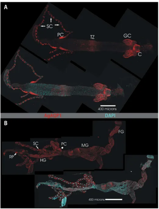
Our available antibody was raised against the N-terminus of AgAQP1 and is predicted to bind to both splice forms. In the larval alimentary canal AgAQP1 staining is evident in the cardia, the gastric caecae, the transitional zone of the midgut, the Malpighian tubule stellate cells, and the proximal Malpighian tubule principal cells (Figure 4A). Analysis of a single plane of the Z stack indicates that AgAQP1 is localized on the basal rather than the luminal membranes, as assessed by its colocalization with basally-located NaK-ATPase [13] (Figure 5).
which has a mechanosensory function to detect sound and is specifically important for courtship behavior [14]. Figure 5E and F show the male JO, which is larger than the female; the female JO also expresses AgAQP1 in a similar distribution (data not shown).
Discussion
We detected two splice variants for AgAQP1, which are differentially expressed in An. gambiae. The structural differences between these two variants are confined to a short portion at the 3’ end that encodes the C-termini of the proteins, presumably located in the cytosolic tails. This small variation may lead to functional differences such as intracellular trafficking [15], mRNA stability [16] or translation [17].
In both larval and adult mosquitoes, we detected AgAQP1 in the stellate cells of the distal region of the Malpighian tubules (MT) as in the previous study [11], but also in the principal cells of the proximal region of the tubules (Figures 4A and 5B). It is known that in the Malpighian tubules, the proximal and distal regions are physiologically distinct [18-21]. The difference in AgAQP1 expression in principal cells between the distal and
proximal regions of the MT adds to these observations and suggests that water movement may be different in different parts of the mosquito renal system. Colocalization of AgAQP1 and ATPase in larval gastric cecae and absence of NaK-ATPase in the MT also indicate the different functions of these two organs (Figure 4B).
A putative ortholog gene in D. melanogaster (CG7777) has three splice variants (FlyBase Genome annotation, release 3.2, 2004), whose variations are also at the 3’ end of the transcripts. Unfortunately, the function(s) of CG7777 have not been studied. Goto et al. identified 3 splice variants for the CG7777 homolog gene, BaAQP1 in the Antarctic midge,
Belgica antarctica, which also differ in the 3’ ends [22], suggesting that the splice variation may be ancestral in lower Diptera.
AgAQP1A and B expressed in X. laevis oocyte membrane showed high water permeability with a similar efficiency (Figure 3). Both isoforms showed reversible sensitivity to Hg2+
inhibition, although AgAQP1 lacks cysteine in the loop E, which is a known site for Hg2+ inhibition [23]. The impermeability of
glycerol suggests that AgAQP1 is a water-specific channel, which is also evidenced by the presence of conserved R/ar Figure 1. Gene and polypeptide structures of AgAQP1A and B. A, genetic structures of the splice variants AgAQP1A and AgAQP1B on Chromosome 3R. The structures are identical from 5’ end to the 4th intron. AgAQP1A consists of 5 exons while AgAQP1B consists of 6 exons. Untranslated regions (UTRs) are shown in red. B, amino-acid sequence alignment by ClustalW2 of the two variants. Predicted transmembrane domains are boxed, while NPA motifs are shaded with grey. Divergent residues at C termini are in white letters in black background. Arrowheads indicate conserved R/ar residues (F53, H177 and R192). * indicates identical residue : indicates similar residue.
doi: 10.1371/journal.pone.0075888.g001
residues, phenylalanine (F), histidine (H) and arginine (R) at positions 53, 177 and 192 (corresponding to F56, H180 and R195 in rat AQP1, Figure 1B), respectively [8]. Mutation of H177 to glycine did not change the permeability of glycerol, which indicated that this residue might not be important for its water/glycerol selectivity (data not shown).
We attempted to raise antibodies against C-termini of AgAQP1 to differentiate the splice variants, but these antibodies had high non-specific binding and were not useful. We successfully raised an antibody against N-terminus [11]. Using the anti-N-terminal antibody, immunofluorescence indicated that AgAQP1 is localized in baso-lateral side of the adult midgut epithelium and larval alimentary canal tissues, which suggests that there is likely another aquaporin expressed on the luminal side. For example, in the silkworm
Bombyx mori, the water-specific AQP-Bom1 (DRIP ortholog) is specifically expressed in the apical (luminal) side of the rectal epithelial cells, whereas another water-specific AQP-Bom3 (CG7777 ortholog) is expressed in the basal membrane of the same cells [24]. Peptides of AGAP008842, another predicted aquaporin gene in An. gambiae, were retrieved from the adult midgut brush-border microvilli proteome [25]. Therefore, we speculate that AGAP008842 may be brush-border specific, while AgAQP1 is baso-lateral specific in the mosquito midgut.
We found that AgAQP1A is almost exclusively expressed in the ovaries of mature female adults at the transcript level, which implicates its function in female reproduction and egg development. However, we found the protein (either AgAQP1A or AgAQP1B) exclusively in the lining of oviduct, but not in ovarian follicles by immunofluorescence (Figure 6C). We attempted to knockdown AgAQP1 in the ovaries by dsRNA Figure 2. H2O permeability of AgAQP1A and B expressed
in Xenopus laevis oocytes. Osmotic water permeability in 70
mOsm hypotonic solution (mean ± SD). Coefficient of osmotic water permeability (Pf in 10-4 cm/s) is shown in the y-axis. HgCl2: incubation in 0.5 mM HgCl2 for 5 min; 2ME: 5 mM
2-mercaptoethanol for 10 min following HgCl2 treatment.
Statistical significance is indicated with asterisks (**: P < 0.01). doi: 10.1371/journal.pone.0075888.g002
injection, but the transcript levels were not affected. Therefore, we can only speculate as to the function of AgAQP1A in the ovary. AgAQP1A may assist reproduction, perhaps by facilitating lubrication of the oviduct during oviposition. Alternatively, the transcript may be stored in the oocytes without translation to be used for embryonic development. qRT-PCR analysis of blood-fed mosquito ovaries showed increased AgAQP1A (throughout egg maturation), which suggests that AgAQP1A has some functions in egg maturation (Figure 3D). AgAQP1B increased transiently at early stages (24 h pbf) of egg maturation, but absolute transcript levels were very low. An orthologous gene in Ae. aegypti (AaAQP2) also increased 24 hrs pbf, which implies a possible conserved function of these orthologs in these two mosquito species [10]. Preliminary analysis of transcript level in the female reproductive system (ovarian follicles vs. oviducts) showed presence of AgAQP1A and AgAQP1B transcript in the oviduct, while follicles exclusively express AgAQP1A (not shown). Nevertheless, we did not detect transcript in embryos. Since sampled eggs were laid overnight and not finely timed, detailed analysis of AgAQP1A expression in embryonic developmental stages may reveal its function in embryogenesis. In D. melanogaster, transcript of the ortholog CG7777 is abundantly found in stage 1-3 embryos, but is quickly degraded, reappearing in stage 13 (Berkeley Drosophila Genome Project [26,27]). This phenomenon may be similar in An. gambiae and the reason for absence of transcript detection in this study.
qRT-PCR indicated that AgAQP1B is abundantly expressed in the adult head. Immunofluorescence indicated that AgAQP1 protein is localized in the Johnston’s organ (JO). The JO is located in the second basal segment of adult antennae and consists of a number of vibration-sensing units called scolopidium (pl. scolopidia), each with two or three sense cells. In Ae. aegypti, a male JO contains 7,000-7,500 scolopidia, which means that a single male mosquito contains ~15,000 scolopidia and ~30,000 sense cells in its paired antennae [28,29]. Recent studies indicate that hearing and adjustment of wingbeat frequency are critical for courtship behavior [13,30]. Harmonic convergence during courtship increases successful copulation [31]. RNAi knockdown of AgAQP1 in the male head was attempted, and while transcript levels were significantly reduced at some time points, results were variable between experimental replicates and there was no change in protein levels by immunofluorescence or Western blot (data not shown). Nevertheless, the presence of aquaporin in confined cells in the JO suggests that it may be important for hearing and successful completion of mating, and could be an intriguing potential target for applied mating disruption mosquito control strategies.
Materials and Methods
Ethics Statement
Mosquito blood feeding was approved under Penn State
IACUC protocol #35790. MosquitoesThe An. gambiae Keele strain was maintained in a walk-in insectary at the Vector Genetics and Control Laboratory at the Pennsylvania State University. Larvae were reared on ground Figure 3. Expression profile of AgAQP1A and AgAQP1B by qRT-PCR (mean ± SD). A, in developmental stages, AgAQP1B is expressed in larval to adult stages with dramatic upregulation in young adults. AgAQP1A is expressed only in older adult females.
B, AgAQP1B is expressed throughout the mature adult body, while AgAQP1A is expressed only in female abdomen. C, Ovaries specifically express AgAQP1A. AgAQP1A (D) and AgAQP1B (E) expression in ovaries from unfed and blood-fed mosquitoes. AgAQP1A is highly upregulated in blood-fed ovaries during egg maturation (≤ 48 h post blood feeding: pbf) returning to baseline 72 h pbf, while AgAQP1B is transiently upregulated at 24 h pbf. Note expression of AgAQP1A is much higher than AgAQP1B (see scales on vertical axes).
doi: 10.1371/journal.pone.0075888.g003
TetraMin® Tropical Flakes (Tetra Holdings Inc., Blacksburg, VA). Adults were maintained on 10% sucrose solution at 27 ± 1°C and 80 ± 5% relative humidity with a 14 h/10 h light/dark cycle.
Expression analysis
For developmental stage analysis, eggs, larvae (first, second, third and fourth instars), male and female pupae, young (<24 hrs after eclosion) and old (~4 days after eclosion) adults were processed for RNA extraction and cDNA synthesis. Figure 4. Whole-mount confocal microscopy of AgAQP1 localization in the larval and adult female alimentary canals. This figure shows two montages of maximum projection images of dissected alimentary canals from larval (A) and adult (B) Anopheles gambiae. Each panel shows TRITC immunofluorescence (red) for AgAQP1 localization (upper figure of each panel) and a merged image with DAPI staining of nuclear DNA (lower figure in each panel). Anterior is to the right. Prominent staining for AgAQP1 in the larval gut (panel A) is evident in the cells of the Cardia (labeled C), the Gastric Caecae (GC) the midgut Transitional Zone (TZ) the proximal Principal Cells of the Malpighian Tubules (PC) and the Stellate Cells (SC) of the Malpighian Tubules. In panel B, the adult alimentary canal shows areas of prominent staining in the Foregut (FG), Midgut (MG), proximal Principal Cells (PC) and Stellate Cells (SC) of the Malpighian tubules, Hindgut (HG) and the Rectal Pads (RP).
For adult body part-specific and female abdominal organ-specific analysis, adults ~5 days after eclosion were used. Organ dissection was performed in PBS and dissected organs were immediately pooled in TRI reagent. For ovary-specific blood-meal response analysis, adult females (5-7 days old) were fed on an anesthetized mouse for 10-15 min. Ovaries were dissected at 0 (within 30 min), 24, 48 and 72 hrs post blood feeding (pbf). RNA extraction was performed immediately after dissection and stored at -80 °C until used.
RNA extraction and cDNA synthesis
Whole mosquitoes or dissected organs were homogenized in TRI reagent (Applied Biosystems, Carlsbad, CA) in a 1.5 mL
microcentrifuge tube. Total RNA was extracted following the manufacturer’s instructions. Extracted RNA was treated with DNase (TURBO DNAfree, Life Technologies, Carsbad, CA) for 1 hr at 37 °C. First-strand cDNA synthesis was performed on 1 µg of total RNA in 20 µL reaction using Accuscript High Fidelity Reverse Transcriptase (Agilent Technologies, Santa Clara, CA).
Rapid Amplification of cDNA Ends (RACE)
Extracted RNA was processed using the GeneRacer Kit (Invitrogen, Carlsbad, CA) with a custom anchor oligonucleotide (Table 1) for cDNA synthesis. RNA was extracted from all life stages (egg, 1st, 2nd, 3rd, 4th instars, Figure 5. Confocal microscopy of AgAQP1 in the larval gastric caecae and Malpighian tubule area indicating basal localization. This figure shows a single larval gut at the region of the Gastric Cacae (GC) (A) and midgut-hindgut junction with Malpighian tubules (B). TRITC (red) staining indicates AgAQP1, Cy5 (blue) indicates the basal membrane marker NaK-ATPase and GSL-1 (green) indicates a plant lectin that binds to various extracellular matrices including the caecal membrane which is internal to the caecal cavity. The top row of each panel shows a maximum projection of several z-planes. Note that in the merge panel at the right end, red and blue signals on the lobes of the cacae co-localize, indicating co-localization of AgAQP1 and NaK-ATPase on the basal membranes of the caecal cells. The lower row of panels showing GC are from the same z-stack but present a single z plane. Again, note that AgAQP1 and the NaK-ATPase colocalize. Note the intense labeling for AgAQP1 in the proximal Principal Cells and the Stellate cells of the Malpighian tubules. As in the GC, AgAQP1 labeling in the Malpighian tubules is on the basal surface of both Principal cells. Scale bar 120 µm.
doi: 10.1371/journal.pone.0075888.g005
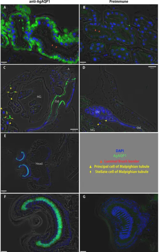
Figure 6. Localization of AgAQP1 in adult and larval mosquitoes by immunofluorescence of sections. Fluorescence micrograph images using anti-AgAQP1 (green). Blue: DAPI (cell nuclei). A, AgAQP1 is expressed on the basal side of the female midgut, (lumen indicated by red arrowheads). B, preimmune. C, AgAQP1 is expressed in oviduct (Od). Immunofluorescence appears to be in posterior portion of the oviduct (A: anus). In this figure, Malpighian tubule principal cells with large nuclei (yellow triangles) with immunolabeled stellate cells (yellow arrows) either traverse or cross section are included. Hindgut rectum with rectal pads is also seen (HG: hindgut; OV: ovary). D, preimmune. E and F, AgAQP1 is expressed in Johnston’s organ (JO). G, preimmune. Shown are the male JO. Scale bar A, B, F, G: 16 µm; C, D, E: 60 µm.
male and female pupae, and young [< 24 hrs after eclosion] and old [~4 days] adults of both sexes) by TRI reagent. RNA processing for RACE was performed with 3 µg RNA according to the manufacturer’s suggested protocol. cDNA was synthesized with the processed RNA and Oligo dT anchor primer using Accuscript High Fidelity Reverse Transcriptase. Gene-specific primers were designed based on the 5’ and 3’ regions of the predicted AGAP008843 in VectorBase. RACE PCR was performed with the gene-specific primers and anchor primer (Roche) or GeneRacer 5’ primer/GeneRacer 5’ nested primer (Invitrogen) (Table 1). RACE PCR products were separated by agarose gel electrophoresis, and bands excised for gel extraction with the QIAquick Gel Extraction kit (QIAGEN, Valencia, CA). Gel-extracted PCR products were cloned into pJET1.2/blunt vector using the pJET PCR Cloning Kit (Fermentas, Glen Burnie, MD). The sequences of the inserted fragments were determined by sequencing.
Gene structure
FancyGene [32] was used to present gene structures on the genome. Alignments of amino-acid sequences were performed using ClustalW [33]. Prediction of transmembrane domains was performed using the TMHMM server (http:// www.cbs.dtu.dk/services/TMHMM/).
Real-time quantitative PCR (qRT-PCR)
Primers were designed against 3’ variable regions of AgAQP1A and B. cDNA was diluted to 1/50 in nuclease-free
H2O and 2.5 µL was used for 10 µL reactions using the
RotorGene Q qPCR system (QIAGEN). Reactions were performed with 5 min at 95 °C, followed by 45 cycles of 95 °C for 5 sec and 60 °C for 10 sec and melt curve analysis from 50 °C to 95 °C. Expression was calculated relative to a housekeeping gene (ribosomal protein S7) by the -2ΔΔCt method
[34]. Primer sequences are listed in Table 1.
Cloning, expression in Xenopus laevis oocytes and swelling assay
AgAQP1A was cloned in the previous study [11]; AgAQP1B was cloned by similar methods. Briefly, coding sequence was amplified using primers containing restriction sites at the 5’ end, and after restriction digestion, PCR products were ligated into the pXβG-myc vector. The purified construct was linearized by XbaI digestion for in vitro complementary RNA (cRNA) transcription using T3 RNA polymerase (Agilent Technologies, Inc., Santa Clara, CA). Synthesized cRNA was purified with the RNeasy Mini kit (QIAGEN). X. laevis oocytes were defolliculated with collagenase I (Sigma-Aldrich, St. Louis, MO) and injected with 5 ng of cRNA or 50 nL of nuclease-free water as control. Injected oocytes were incubated for 3 days in ~200 mOsm Modified Barth’s Solution (MBS: 88 mM NaCl, 1.0 mM KCl, 2.4 mM NaHCO3, 15 mM Tris, 0.8 mM MgSO4, 0.4 mM
CaCl2, 0.3 mM Ca(NO3)2, pH 7.6) for overexpression of
aquaporin in the oocyte plasma membrane, then subjected to swelling assay [5] by transferring to MBS diluted to 70 mOsm. The change of oocyte volume was monitored at room Table 1. primers used for this study.
Primer name Sequence (5’–3’)
For RACE work
Oligo dT-anchor primer* GACCACGCGTATCGATGTCGACTTTTTTTTTTTTTTTTTTTTTTT
PCR anchor primer* GACCACGCGTATCGATGTCGAC
GeneRacerTM 5′ Primer* CGACTGGAGCACGAGGACACTGA
GeneRacerTM 5′ Nested Primer* GGACACTGACATGGACTGAAGGAGTA
GSPAg3F1 AGCTGGGCTCATCACTGGATCTACTGGGCT
GSPAg3F2 ACTGGATCTACTGGGCTGGACCGATCCT
GSPAg3R4 TGTCCGATCGACATCACCACCATGAACAC
GSPAg3R5 CCACCATGAACACGCTCAGCCCGAAT
GSPAg3R6 AATGCCAGCGAGATCAGCGTTTTATCTCCA
For real-time PCR
8843-1qPCR-F TCCAGTACGGAAAACCCCCAAACCGA
8843-1qPCR-R TGATGTGAAGTTGCGGTGCGGATGAGA
8843-2qPCR-F ATGCGTCGACTGGACGGCAAGCA
8843-2qPCR-R ACACACCACAACATTCGACGGGACACA
AGS7UC GTGAGGTCGAGTTCAACAACAAGAA
AGS7DD GGCACCGGCACGTAGATGA
For Xenopus oocyte expression
8843-2F2EcoRI ATAGAATTCCATGGGATACAGCCTAGGAACG
8843-2RXbaI ATATCTAGATTACGCCATATCATGCTTGCC
For * refer to Materials and Methods. GSPAg3 prefixed primers are specific for 3’ (F1 and F2) and 5’ (R4, R5, and R6) of AGAP008843. GSPAg3F2, GSPAg3R5 and GSPAg3R6 are used for nested RACE PCR with PCR anchor primer or GeneRacerTM 5′ Nested Primer. For real-time PCR, 8843-1 primer pair is specific against 3’ region
of AgAQP1A, and 8843-2 primer pair is against 3’ AgAQP1B. doi: 10.1371/journal.pone.0075888.t001
temperature by microscopic video capture for 60 seconds and the relative volume (V/V0) was calculated from the
cross-sectional area at the initial time (A0) and after a time interval
(At): V/V0 = (At/A0)3/2. The coefficient of osmotic water
permeability (Pf) was determined from the initial slope of the time course [d(V/V0)/dt], average initial oocyte volume (V0 = 9 ×
10−4 cm3), average initial oocyte surface area (S = 0.045 cm2),
the molar volume of water (Vw = 18 cm3/mol), and the osmotic
solute gradient (osmin − osmout). Pf = (V0 × d(V/V0)/dt)/(S × Vw ×
(osmin − osmout). A minimum of five individual oocytes were
tested in each group.
To test the effect of Hg2+ on water permeability, oocytes were
placed in 500 µM HgCl2 in MBS for 5 min prior to the swelling
assay. To test reversibility, Hg2+-treated oocytes were
transferred to 5 mM 2-mercaptoethanol in MBS for 10 min. Glycerol permeability was also assessed by replacing diluted MBS with MBS mixed with an equal volume of iso-osmotic (200 mOsm) glycerol solution.
Immunofluorescence detection (whole-mount)
Fourth-instar larvae or 48-hour old adult females were cold anesthetized and dissected in primary fixative (4% p-formaldehyde in 0.1M sodium cacodylate buffer) as previously described [13]. Isolated alimentary canal tissue was incubated in primary fixative over night at 4 °C. Tissues were then washed with Tris-buffered saline (TBS), transferred to pre-incubation medium (PIM) (TBS with 0.1% Triton X-100, 2% normal goat serum and 1% bovine serum albumin) and incubated at room temperature for 2 h. Primary AgAQP1 antibody [11] was diluted in PIM (1:1000) and tissues incubated over night at 4 °C with gentle agitation. AgAQP1 antibody was raised against an N-terminus peptide and binds to both variants of AgAQP1. The following day, tissues were washed first with
TBS and then with fresh PIM. Tissues were then suspended in PIM containing TRITC-conjugated goat anti-rabbit secondary antibody (Jackson Immunoresearch Laboratories, West Grove, PA; diluted 1:500) over night at 4 °C with gentle agitation. The following day, tissues were washed in TBS, incubated in TBS with DAPI and mounted on well-slides with 60% glycerol/TBS containing p-phenylenediamine to inhibit fluorescence quench. Samples were viewed and digital images captured with a Leica SP5 Laser Scanning Confocal Microscope. Images were assembled into figures using CorelDraw 12 software.
Immunofluorescence detection (sections): Adult mosquitoes were paraffin-embedded and sectioned. Slides containing sections were used for immunofluorescence detection using anti-AgAQP1 antibody [11]. Sections were blocked and permeabilized with 10% goat serum in PBS and 0.2% Triton X-100 at 4 °C overnight followed by incubation with anti-AgAQP1 antibody (1:300 in PBS with 10% goat serum) at 4 °C overnight and anti-rabbit IgG secondary antibody conjugated with Alexa 488 (1:1,500 in PBS with 10% goat serum). Sections were washed with PBS four times at room temperature after each step. One drop of ProLong Antifade with DAPI (Invitrogen) was placed on the section-containing slides, covered by a cover slip and sealed by nail polish. Images were obtained on a Nikon 90i upright fluorescence microscope equipped with a digital camera with Volocity imaging software (PerkinElmer, Waltham, MA).
Author Contributions
Conceived and designed the experiments: HT KL PA JLR. Performed the experiments: HT PJL. Analyzed the data: HT KL PJL PA JLR. Contributed reagents/materials/analysis tools: HT KL PJL JLR. Wrote the manuscript: HT JLR.
References
1. WHO (2010) World malaria report 2010.
2. Olayemi IK, Ande AT (2009) Life table analysis of Anopheles gambiae (diptera: culicidae) in relation to malaria transmission. J Vector Borne Dis 46: 295-298. PubMed: 19959856.
3. Stobbart RH (1977) The control of the diuresis following a blood meal in females of the yellow fever mosquito Aedesaegypti (L). J Exp Biol 69: 53-85. PubMed: 908912.
4. Williams JCJ, Hagedorn HH, Beyenbach KW (1983) Dynamic changes in flow rate and composition of urine during the post-bloodmeal diuresis in Aedesaegypti (L.). J Comp Physiol B 153: 257-265. doi:10.1007/ BF00689629.
5. Preston GM, Carroll TP, Guggino WB, Agre P (1992) Appearance of water channels in Xenopus oocytes expressing red cell CHIP28 protein. Science (New York, NY) 256: 385-387. doi:10.1126/science. 256.5055.385. PubMed: 1373524.
6. Nielsen S, Chou CL, Marples D, Christensen EI, Kishore BK et al. (1995) Vasopressin increases water permeability of kidney collecting duct by inducing translocation of aquaporin-CD water channels to plasma membrane. Proc Natl Acad Sci U S A 92: 1013-1017. doi: 10.1073/pnas.92.4.1013. PubMed: 7532304.
7. King LS, Kozono D, Agre P (2004) From structure to disease: the evolving tale of aquaporin biology. Nat Rev Mol Cell Biol 5: 687-698. doi:10.1038/nrm1469. PubMed: 15340377.
8. Beitz E, Wu B, Holm LM, Schultz JE, Zeuthen T (2006) Point mutations in the aromatic/arginine region in aquaporin 1 allow passage of urea, glycerol, ammonia, and protons. Proc Natl Acad Sci U S A 103: 269-274. doi:10.1073/pnas.0507225103. PubMed: 16407156. 9. Pietrantonio PV, Jagge C, Keeley LL, Ross LS (2000) Cloning of an
aquaporin-like cDNA and in situ hybridization in adults of the mosquito
Aedesaegypti (Diptera: Culicidae). Insect Mol Biol 9: 407-418. doi: 10.1046/j.1365-2583.2000.00201.x. PubMed: 10971718.
10. Drake LL, Boudko DY, Marinotti O, Carpenter VK, Dawe AL et al. (2010) The Aquaporin gene family of the yellow fever mosquito, Aedes aegypti. PLOS ONE 5: e15578. doi:10.1371/journal.pone.0015578. PubMed: 21249121.
11. Liu K, Tsujimoto H, Cha S-J, Agre P, Rasgon JL (2011) Aquaporin water channel AgAQP1 in the malaria vector mosquito Anopheles gambiae during blood feeding and humidity adaptation. Proc Natl Acad Sci U S A 108: 6062-6066. doi:10.1073/pnas.1102629108. PubMed: 21444767.
12. Fenton RA, Moeller HB, Zelenina M, Snaebjornsson MT, Holen T et al. (2010) Differential water permeability and regulation of three aquaporin 4 isoforms. Cell Mol Life Sci CMLS 67: 829-840. doi:10.1007/ s00018-009-0218-9. PubMed: 20013023.
13. Xiang MA, Linser PJ, Price DA, Harvey WR (2012) Localization of two Na+- or K+-H+ antiporters, AgNHA1 and AgNHA2, in Anopheles gambiae larval Malpighian tubules and the functional expression of AgNHA2 in yeast. J Insect Physiol 58: 570-579. doi:10.1016/j.jinsphys. 2011.12.009. PubMed: 22206887.
14. Cator LJ, Arthur BJ, Harrington LC, Hoy RR (2009) Harmonic convergence in the love songs of the dengue vector mosquito. Science (New York, NY) 323: 1077-1079. doi:10.1126/science.1166541. PubMed: 19131593.
15. Törnroth-Horsefield S, Hedfalk K, Fischer G, Lindkvist-Petersson K, Neutze R (2010) Structural insights into eukaryotic aquaporin regulation. FEBS Lett 584: 2580-2588. doi:10.1016/j.febslet. 2010.04.037. PubMed: 20416297.
differences in transcript and splice variant abundance. Nucleic Acids Res 39: 556-566. doi:10.1093/nar/gkq790. PubMed: 20852259. 17. Moore MJ (2005) From birth to death: the complex lives of eukaryotic
mRNAs. Science 309: 1514-1518. doi:10.1126/science.1111443. PubMed: 16141059.
18. Sözen MA, Armstrong JD, Yang M, Kaiser K, Dow JA (1997) Functional domains are specified to single-cell resolution in a Drosophila epithelium. Proc Natl Acad Sci U S A 94: 5207-5212. doi:10.1073/pnas. 94.10.5207. PubMed: 9144216.
19. Yang MY, Wang Z, MacPherson M, Dow JA, Kaiser K (2000) A novel Drosophila alkaline phosphatase specific to the ellipsoid body of the adult brain and the lower Malpighian (renal) tubule. Genetics 154: 285-297. PubMed: 10628988.
20. Cabrero P, Pollock VP, Davies SA, Dow JAT (2004) A conserved domain of alkaline phosphatase expression in the Malpighian tubules of dipteran insects. J Exp Biol 207: 3299-3305. doi:10.1242/jeb.01156. PubMed: 15326206.
21. Beyenbach KW, Piermarini PM (2011) Transcellular and paracellular pathways of transepithelial fluid secretion in Malpighian (renal) tubules of the yellow fever mosquito Aedes aegypti. Acta Physiol (Oxf) 202: 387-407. doi:10.1111/j.1748-1716.2010.02195.x. PubMed: 20946239. 22. Goto SG, Philip BN, Teets NM, Kawarasaki Y, Lee RE et al. (2011)
Functional characterization of an aquaporin in the Antarctic midge
Belgica antarctica. J Insect Physiol 57: 1106-1114. doi:10.1016/ j.jinsphys.2011.03.023. PubMed: 21497603.
23. Preston GM, Jung JS, Guggino WB, Agre P (1993) The mercury-sensitive residue at cysteine 189 in the CHIP28 water channel. J Biol Chem 268: 17-20. PubMed: 7677994.
24. Azuma M, Nagae T, Maruyama M, Kataoka N, Miyake S (2012) Two water-specific aquaporins at the apical and basal plasma membranes of insect epithelia: molecular basis for water recycling through the cryptonephric rectal complex of lepidopteran larvae. J Insect Physiol 58: 523-533. doi:10.1016/j.jinsphys.2012.01.007. PubMed: 22285686. 25. Parish LA, Colquhoun DR, Ubaida Mohien C, Lyashkov AE, Graham
DR et al. (2011) Ookinete-interacting proteins on the microvillar surface are partitioned into detergent resistant membranes of Anopheles
gambiae midguts. J Proteome Res 10: 5150-5162. doi:10.1021/ pr2006268. PubMed: 21905706.
26. Tomancak P, Beaton A, Weiszmann R, Kwan E, Shu S et al. (2002) Systematic determination of patterns of gene expression during Drosophila embryogenesis. Genome Biol 3: RESEARCH0088. PubMed: 12537577.
27. Tomancak P, Berman BP, Beaton A, Weiszmann R, Kwan E et al. (2007) Global analysis of patterns of gene expression during Drosophila embryogenesis. Genome Biol 8: R145. doi:10.1186/ gb-2007-8-7-r145. PubMed: 17645804.
28. Boo KS, Richards AG (1975) Fine structure of scolopidia in Johnston’s organ of female Aedesaegypti compared with that of the male. J Insect Physiol 21: 1129-1139. doi:10.1016/0022-1910(75)90126-2. PubMed: 1141704.
29. Boo K (1975) Fine structure of the scolopidia in the Johnston's organ of male Aedes aegypti (L.) (Diptera: Culicidae). Int J Insect Morphol Embryol.
30. Gibson G, Warren B, Russell IJ (2010) Humming in tune: sex and species recognition by mosquitoes on the wing. J Assoc Res Otolaryngol 11: 527-540. doi:10.1007/s10162-010-0243-2. PubMed: 20976515.
31. Cator LJ, Harrington LC (2011) The Harmonic Convergence of Fathers Predicts the Mating Success of Sons in Aedesaegypti. Anim Behav 82: 627-633. doi:10.1016/j.anbehav.2011.07.013. PubMed: 22003255. 32. Rambaldi D, Ciccarelli FD (2009) FancyGene: dynamic visualization of
gene structures and protein domain architectures on genomic loci. Bioinformatics (Oxf, England) 25: 2281-2282. doi:10.1093/ bioinformatics/btp381. PubMed: 19542150.
33. Larkin MA, Blackshields G, Brown NP, Chenna R, McGettigan PA et al. (2007) Clustal W and Clustal X version 2.0. Bioinformatics (Oxf, England) 23: 2947-2948. doi:10.1093/bioinformatics/btm404. PubMed: 17846036.
34. Livak KJ, Schmittgen TD (2001) Analysis of relative gene expression data using real-time quantitative PCR and the 2(-Delta Delta C(T)) Method. Methods (San Diego, Calif) 25: 402-408. doi:10.1006/meth. 2001.1262. PubMed: 11846609.