1299
ESTABLISHING ECHOCARDIOGRAPHIC AND ARTERIAL STIFFNESS MARKERS AS PREDICTORS OF COGNITIVE DECLINE
F. MITU1,MANUELA PADURARIU1, CATALIN JOACABINE1, A. CIOBICA2,3 and
ROXANA CHIRITA1
1“Gr. T. Popa” University of Medicine and Pharmacy, 700115, Iasi, Romania 2 “Alexandru Ioan Cuza” University, Iasi, 700506, Romania
3Center of Biomedical Research of the Romanian Academy, Iasi Branch, Romania
Abstract - Diferent factors seem to contribute to cognitive impairment in the elderly population. It is unclear which car-diovascular risk factors are the most signiicant contributors to cognitive decline. Although there is some recent neuro-pathological evidence that vascular lesions and atherosclerotic occlusion of the cerebral arteries may unmask or strengthen the clinical expression of cognitive decline and dementia, there is still little knowledge about the relevance of echocardio-graphic and arterial stifness markers as predictors for cognitive decline. In the present study we decided to investigate whether and how the severity of cognitive impairment could be related to cerebral hemodynamic impairment, as well as the possible contribution of the alterations in cerebral hemodynamics (as expressed through some echocardiographic and arterial stifness markers) to the progression of cognitive decline in a group of patients with cognitive impairments, as compared to a control group with no cognitive deicits. he main inding of our study indicated signiicant diferences in terms of echocardiographic and arterial stifness markers between the two groups, one composed of patients with cogni-tive impairment and one with normal-cognicogni-tive patients, which suggests an association between these parameters and poor cognitive function. While these functional changes of the cerebral vessel functions could have an important role in the pathogenesis of dementia, the identiication of simple and accurate measures that are acceptable to patients and can serve as indicators of current cognitive impairment or the risk of cognitive decline could be very helpful in developing long-term preventive and therapeutic treatments for these patients.
Key words: Echocardiographic, arterial stifness, markers, cognitive decline
INTRODUCTION
Diferent factors seem to contribute to cognitive im-pairment in the elderly population. A series of clas-sical cardiovascular risk factors has been associated with the impairment of cognitive functions, such as hypertension, cholesterol or diabetes, suggesting a possible role in the etiopathogenic processes of de-mentia (Ciobica et al., 2011a, Joacabine et al., 2012). Still, it is unclear which cardiovascular risk factors are the most signiicant contributors to cognitive
de-cline (Isaac et al., 2011, Vicario et al., 2011, Miralbell et al., 2010).
he current hypothesis that vascular and degen-erative processes may interact in expressing cogni-tive decline is a controversial issue (Langa et al., 2004, Silvestrini et al., 2006). Furthermore, the prevention of cognitive decline, manifested especially as demen-tia, has turned into a major public health challenge, with enormous pressure on identifying older people at risk and/or sufering from cognitive impairment who could beneit from preventive therapeutic in-terventions aimed at slowing down the cognitive de-cline (Hanon et al., 2005).
In the present study we used as a starting point the idea that arterial stifness, which is a conse-quence of atherosclerosis, could be used as a poten-tial marker to predict cognitive decline, considering that the progression of atherosclerosis is involved in cognitive impairment, and that prevention of athero-sclerosis can be efective in this direction, although the exact associations between atherosclerosis and cognitive function have not yet been deinitively de-termined (Fukuhara et al., 2006).
Considering that it is still unclear whether these markers are directly related to the appearance of cognitive decline, in the present report we decided to study whether and how the severity of the cogni-tive impairment could be related to cerebral hemo-dynamic impairment, as well as the possible con-tribution of alterations in cerebral hemodynamics (as expressed through some echocardiographic and arterial stifness markers) to the progression of cog-nitive decline in a group of patients with cogcog-nitive impairments, as compared with a control group with no cognitive deicits.
PATIENTS AND METHODS
Clinical assessments
We enrolled 94 individuals from the patients who presented memory complaints during 2010-2011 at the Socola Psychiatry Hospital in Iasi. Selection of patients was performed by a neurologists and a psy-chiatrist with certiied experience in managing pa-tients with dementia ater careful evaluation of
clini-cal and instrumental examinations.
Patients older than 50 years, regardless of sex, mar-ital or social status were included. he subjects had to be literate and without sensorial impairment. We ex-cluded patients with acute comorbidities, chronic un-stable diseases, except cardiovascular and metabolic dysfunction, as well as patients with other medical conditions that could lead to cognitive deicits. E.g. we excluded patients with chronic pulmonary problems such as TBC and sleep apnea, hypothyroidism, brain infarcts or head injury.
he clinical assessment included medical and family history, physical, neurological and psychiatric examination.
In addition, for BMI determination we divided the weight in kilograms by the height, measured in meters squared. Blood pressure was measured in the right arm ater participants had been seated for ive minutes. Blood samples were obtained in the morn-ing before breakfast, allowed to clot and centrifuged immediately. Sera were aliquoted into Eppendorf tubes and stored at -80°C until determination of se-rum total cholesterol concentrations, which was per-formed in the hospital laboratory.
Neuropsychological assessment
he cognitive testing was performed in the morn-ing, between 10-12 a.m., the patients having been told not to consume any stimulants the morning of the examination, such as cofee or smoking. he neuropsychological assessment was performed by a trained neuropsychologist who was blind to the re-sults of hemodynamic evaluations.
At the same time, the patients were clinically evaluated for cardiovascular risk factors such as al-cohol consumption, smoking and diabetes mellitus.
We selected the Mini-Mental State Examination (MMSE) and the Montreal Cognitive Assessment (MoCA) scales in order to diferentiate the subjects with cognitive impairment (CI group) from those without cognitive impairment. he CI group includ-ed subjects with scores less than or equal to 24 points in the MMSE test and a score less than or equal to 25 in the MoCA test (Folstein et al., 1975).
he hemodynamic parameters were determined through transcranial Doppler ultrasonography and performed by a physician blind to the cognitive eval-uation.
Data analysis
he results for these vascular risk factors were ana-lyzed using one-way analysis of variance (one-way ANOVA). All results are expressed as mean ± SEM. F values for which p<0.05 were regarded as statisti-cally signiicant.
RESULTS
Demographic data
As mentioned above, the mean age of the selected group was 63±9.8 years (i.e. 50-83 years-old) with 58.4% men and 53.1% having a rural background. 69.64 % were in a sexual relationship with only 10% employed and 83.3% being retired. he mean age of education was 8.4±3.4 and all the subjects were liter-ate. he majority of the subjects were cofee consum-ers (83.3%), 40.4% were active smokconsum-ers, while 29% reported chronic alcohol use (Table 1).
Adiposity was estimated by calculating BMI. We found a mean value for BMI of 25.9±2 and the majority of the individuals were overweight or obese (73.41%); 38.46% of the subjects had hyperli-pidemia with a cholesterol mean of 217.5±11.9 mg/ dl and 142.71±83.4 mg/dl for triglycerides; 73.4% were found to have hypertension while 11 individu-als had diabetes mellitus. Regarding the history of stroke, 8.51% of the subjects reported an ischemic stroke more than 1 year before, while 21.27% had an
ischemic heart condition. Twenty-one patients re-ported physical exercise more than twice a week.
Echocardiographic measurements
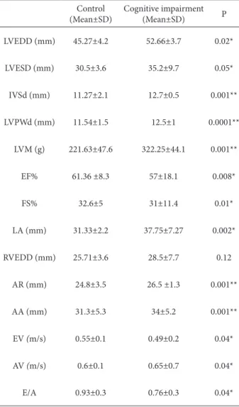
he results of the echocardiographic measurements, which included the let ventricular end-diastolic dimension (LVEDD), let ventricular end-systolic dimension (LVESD), interventricular septal end-diastolic dimension (IVSd), let ventricular end-di-astolic posterior wall dimension (LVPWd), let ven-tricle mass (LVM), ejection fraction (EF), fractional shortening (FS), let atrial dimension (LA), aortic ring (AR), ascending aorta (AA), E velocity (EV), A velocity (AV), are presented in Table 2. As can Table 1. Clinical characteristics of the subjects.
Crt CI NCI
N 74 20
Age (years) 63.8±8.4 56.1±4.7
Sex (%M) 40.5% 54.5%
Urban (%) 54.3% 50%
Marital status (single %) 32.3% 30%
Education (years) 7.93±3.2 10.7±2.1
Smoking (%) 32.3% 55.6%
Alcohol (%) 20% 22.2%
BMI (kg/m2) 27.04±3.9 23.7±4.8
Systolic BP (mmhg) 140.8±17.6 132.4±15.6
Diastolic BP (mmhg) 80.6±10.4 76,3±9.2
Cholesterol (mg/dl) 216,3±46,6 145,4±91,4
Triglyceride (mg/dl) 210,2±45,3 149,5±45
Glycemia (mg/dl) 93.6±25.33 77,9±9
LDL (mg/dl) 86.8±42.9 65.83±29
HDL (mg/dl) 51.1±13.6 49.13±18.3
be seen, signiicant diferences were observed be-tween the cognitive impaired group and the control patients (non-cognitive impaired group) for all the aforementioned echocardiographic factors, except RVEDD (Table 2). his strongly suggests that these factors could be signiicant predictors of decreased cognitive functioning.
Vascular Doppler
Regarding vascular Doppler results, we focused on the following arteriographic indices: brachial aug-mentation index (Aixbr), aortic augaug-mentation index (AIXao), pulse wave velocity (PWV), aortic pulse pressure (PPao), aortic systolic blood pressure (SB-Pao), brachial systolic blood pressure (SBP) and an-kle brachial pressure index (ABPI) (Table 3).
We found very signiicant diferences between the group of patients with cognitive impairment and those without, except in the case of Aixbr and AIXao (Table 3), conirming that these parameters have a fundamental importance as markers for predicting cognitive decline.
his was also conirmed by the results of carotid and vertebral artery analysis (both right and let) for the systolic/diastolic velocity and resistance index (Table 4), which exhibited very signiicant difer-ences between the cognitive-impaired group and the control group, conirming an association between impaired cerebral vessel functionality and an unfa-vorable evolution of cognitive function.
DISCUSSION
Our indings revealed signiicant diferences in terms of echocardiographic and arterial stifness markers between the two groups we studied, one was com-prised of patients with cognitive impairment and one without cognitive impairment. his suggests an asso-ciation between these parameters and poor cognitive function. Our data further suggest a contribution of these vascular factors to the pathogenesis of cogni-tive impairment-related disorders. he results are also consistent with some previous indings suggest-ing that vascular disorders play an important role in cognitive impairment (Hanon et al., 2005).
Johnson et al. (2010) demonstrated in a recent study some signiicant correlations between cogni-tive functions and the ankle-brachial index, which is known as a marker of generalized atherosclero-sis, myocardial infarction and stroke (Heald et al., Table 2. Echocardiographic parameters.
Control (Mean±SD)
Cognitive impairment
(Mean±SD) P
LVEDD (mm) 45.27±4.2 52.66±3.7 0.02*
LVESD (mm) 30.5±3.6 35.2±9.7 0.05*
IVSd (mm) 11.27±2.1 12.7±0.5 0.001**
LVPWd (mm) 11.54±1.5 12.5±1 0.0001**
LVM (g) 221.63±47.6 322.25±44.1 0.001**
EF% 61.36 ±8.3 57±18.1 0.008*
FS% 32.6±5 31±11.4 0.01*
LA (mm) 31.33±2.2 37.75±7.27 0.002*
RVEDD (mm) 25.71±3.6 28.5±7.7 0.12
AR (mm) 24.8±3.5 26.5 ±1.3 0.001**
AA (mm) 31.3±5.3 34±5.2 0.001**
EV (m/s) 0.55±0.1 0.49±0.2 0.04*
AV (m/s) 0.6±0.1 0.65±0.7 0.04*
E/A 0.93±0.3 0.76±0.3 0.04*
2006). Also, a reference study in this area of research by Hanon et al. (2005), which showed a signiicant relationship between arterial stifness and the cogni-tive impairment, suggests that functional changes of the arterial system could be involved in the onset of dementia. Fukuhara et al. (2006) demonstrated that it could be possible to predict cognitive functions based on arterial stifness, as assessed by pulse wave velocity in very old patients. he Rotterdam Study demonstrated that the presence of atherosclerotic plaques or wall thickening of the carotid artery were
signiicantly associated with vascular dementia and Alzheimer’s disease (Hofman et al., 1997).
he aforementioned results could be very impor-tant since these factors are relatively easy to assess and are noninvasive, providing reliable information about the atherosclerotic conditions associated with poor cognitive function. hus, they could represent a low-cost and perhaps efective way of gathering a general indicator of overall cognitive function (Fuku-hara et al., 2006, Johnson et al., 2010).
Table 3. Arteriographic parameters.
N AIxbr (%) AIXao (%) PWVao (m/s) PPao (mmHg) SBPao (mmHg) ABPI dr ABPI stg
Control (Mean±SD)
18.85± 29
47.17± 14.7
9.5± 1
51.2± 8.4
128± 12
0.95± 0.3
1.01± 0.08
Cognitive impairment(Mean±SD)
-2.3±
22.4 19.7±12.3 7.26±1.5 44±15 106.6±21.8 1±0.1 1.06±0.2
P 0.9 0.2 0.01* 0.01* 0.01* 0.008* 0.001**
Data are expressed as means +/-SD, p*≤0.05, p**≤0.001. Aixbr= brachial augmentation index; AIXao=aortic augmentation index; PWV=pulse wave velocity; PPao=aortic pulse pressure; SBPao=aortic systolic blood pressure; SBP=brachial systolic blood pressure; ABPI= ankle brachial pressure index.
Table 4. Carotid and vertebral artery analysis (both right and let) for the systolic/diastolic velocity and resistance index.
Right Common
Carotid Artery
Right Internal
carotid artery
Right External
carotid artery
Right Vertebral
artery
Let Common
Carotid Artery
Let Internal
carotid artery
Let External
carotid artery
Let Vertebral
artery
Systolic
velocity Control 55.3±8.5 43.33±9 59.77±17.8 29±8.19 53.55±11.3 47.22±15.7 47.88±15.2 31.75±3.6
Cognitive
impairment 53.8±11.5 40.66±5.8 52.83±6.2 33.33±12.75 50±11.1 47.5±15.6 48.16±8.4 30.66±9.6
P <0.0001 <0.0001 <0.0001 0.001 <0.0001 0.001 0.001 0.01
Diastolic
velocity Control 17.8±6 18.11±5.7 17.33±7.4 13.5±4.6 8±5.4 22.33±7.2 17.44±7 15.5±2.7
Cognitive
impairment 14.5±2.5 15.16±3.7 14±3.9 15±4.8 5±4.4 20.33±5.2 12.33±4.2 15.33±5.4
P <0.0001 <0.0001 <0.0001 0.01 <0.0001 <0.0001 0.01 0.01
Resistance
index Control 0.67±0.08 0.59±0.08 0.71±0.07 0.55±0.09 0.63±0.08 0.52±0.05 0.63±0.09 0.5±0.1
Cognitive
impairment 0.72±0.05 0.62±0.72 0.74±0.05 0.52±0.08 0.68±0.03 0.56±0.04 0.74±0.08 0.5±0.06
Regarding the possible mechanism implicated in this, some of the cited mechanisms could be rep-resented by cerebrovascular lesions, white matter changes and pre-existent asymptomatic Alzheimer’s brain lesions (Hanon et al., 2005), as well as cerebral amyloid angiopathy, arteriolosclerosis, capillary en-dothelial and basement membrane changes (Silves-trini et al., 2006). With regard to the mechanics, it seems that endothelial lesions in Alzheimer’s disease have a close relationship with senile plaques (Ka-laria et al., 1996). Additionally, the speciic BETA-amyloid compound could be very closely related to the vascular endothelial cells producing free radicals and increasing oxidative stress (homas et al., 1996, Hanon et al., 2005). We previously demonstrated that the central oxidative stress is very important in afecting cognitive functions and most neuropsy-chiatric disorders (Padurariu et al., 2010, Ciobica et al., 2011b,c, 2012, Stefanescu et al., 2012, Irimia et al., 2013). Moreover, it seems that endothelial nitric oxide (NO) is implicated in this matter, which also leads us to possible correlations that might exist be-tween oxidative and nitrosative stress in generating various vascular alterations (Bild et al., 2013a).
Other relevant aspects are represented by the fact that arterial stifness increases in patients with diabe-tes (Airaksinen et al., 1993) and end-stage renal dis-ease (London et al., 1990, Fukuhara et al., 2006). he possibility of inluencing cerebral vessel functions through speciic therapeutics leads to the idea of us-ing angiotensin-convertus-ing enzyme inhibitors to re-duce the cognitive deterioration of subjects. he very relevant European study called Systolic Hypertension in the Elderly in Europe (Syst-Eur), has demonstrat-ed that antihypertensive mdemonstrat-edications could rdemonstrat-educe the incidence of all types of dementia (Forette et al., 2002). Our group has also recently demonstrated that the administration of an angiotensin-converting enzyme inhibitor (captopril) in rats results in signii-cant cognitive improvements and decreased central oxidative stress (Bild et al., 2013b).
CONCLUSIONS
We demonstrate the possibility of obtaining, through
some simple evaluations of cerebral vessel functions, a range of very useful information for the identiica-tion of cognitive decline in patients who are at sig-niicant risk of a rapid and pronounced evolution of dementia. While these functional changes in the arterial system could have an important role in the pathogenesis of dementia, the identiication of sim-ple and accurate measures that are acceptable to pa-tients and can serve as indicators of current cognitive impairment or the risk of cognitive decline could be very helpful in developing long-term preventive and therapeutic strategies for these patients.
REFERENCES
Airaksinen, K.E., Salmela, P.I., Linnaluoto, M.K., Ikaheimo, M.J., Ahola, K. and L.J.Ryhanen (1993). Diminished arterial elasticity in diabetes: Association with luorescent ad-vanced glycosylation end products in collagen. Cardiovasc Res27, 942-945.
Bild, W., Ciobica, A., Padurariu, M. and V. Bild (2013a). he in-terdependence of the reactive species of oxygen, nitrogen and carbon. Journal of Physiology and Biochemistry69, 147-154.
Bild, W., Hritcu, L., Stefanescu, C. and A.Ciobica (2013b). Inhibi-tion of central angiotensin II enhances memory funcInhibi-tion and reduces oxidative stress status in rat hippocampus. Progress in Neuropsychopharmacology & Biological Psy-chiatry43, 79-88.
Ciobica, A., Olteanu, Z., Padurariu, M. and L. Hritcu (2012). he efects of low-dose pergolide on memory and oxidative stress in a 6-OHDA induced rat model of Parkinson’s dis-ease. Journal of Physiology and Biochemistry68, 59-69.
Ciobica, A., Padurariu, M., Bild, W. and C.Stefanescu (2011a). Cardiovascular risk factors as potential markers for mild cognitive impairment and Alzheimer’s disease. Psychiatr Danub23, 340-6.
Ciobica, A., Padurariu, M., Dobrin, I., Stefanescu, C. and R. Do-brin (2011b). Oxidative stress in schizophrenia - focusing on the main markers.Psychiatr Danub23, 237-45.
Ciobica, A., Bild, V., Hritcu, L., Padurariu, M. and W. Bild (2011c). Efects of angiotensin II receptor antagonists on anxiety and some oxidative stress markers in rat. Central European Journal of Medicine 6, 331-340.
Forette, F., Seux, M-L., Staessen, J.A., hijs, L., Babarskiene, M-R., Babeanu, S. et al. (2002). he prevention of dementia with antihypertensive treatment. New evidence from the Sys-tolic Hypertension in Europe (Syst-Eur) study. Arch Intern Med162, 2046-2052.
Fukuhara, M., Matsumura, K., Ansai, T., Takata, Y., Sonoki, K., Akifusa, S. et al. (2006). Prediction of cognitive function by arterial stifness in the very elderly. Circ J70, 756-61.
Hanon, O., Haulon, S., Lenoir, H., Seux, M.L., Rigaud, A.S., Sa-far, M. et al. (2005). Relationship between arterial stifness and cognitive function in elderly subjects with complaints of memory loss. Stroke36, 2193-7.
Heald, C.L., Fowkes, F.G.R., Murray, G.D. and J.F. Price (2006). Risk of mortality and cardiovascular disease associated with the ankle–brachial index: systematic review. Athero-sclerosis189: 61-69.
Hofman, A., Ott, A., Breteler, M.M., Bots, M.L., Slooter, A.J., van Harskamp, F. et al. (1997). Atherosclerosis, apolipoprotein E, and prevalence of dementia and Alzheimer’s disease in the Rotterdam Study. Lancet349, 151–154.
Irimia, R., Ciobica, A., Stanciu, C. and A.Trifan (2013).he rel-evance of oxidative stress in cirrhotic patients with dif-ferent forms of hepatic encephalopathy. Arch. Biol. Sci., Belgrade 65.
Isaac, V., Sim, S., Zheng, H., Zagorodnov, V., Tai, E.S. and M.Chee (2011). Adverse associations between visceral adiposity, brain structure, and cognitive performance in healthy el-derly. Front Aging Neurosci3:12.
Joacabine, C., Padurariu, M., Ciobica, A., Dobrin, R., Popescu, C.D. and F.Mitu (2012). he association between choles-terol levels and brachial/aortic augmentation index versus cognitive status in patients with cardiovascular risk fac-tors. Arch. Biol. Sci., Belgrade 64, 419-426.
Johnson, W., Price, J.F., Rafnsson, S.B., Deary, I.J. and F.G. Fowkes (2010). Ankle-brachial index predicts level of, but not change in, cognitive function: the Edinburgh Artery Study at the 15-year follow-up. Vasc Med15, 91-7.
Kalaria, R.N. and P.Hedera (1996). Beta-Amyloid vasoactivity in Alzheimer’s disease. Lancet347, 1492-1493.
Langa, K.M., Foster, N.L. and E.B.Larson (2004). Mixed demen-tia. Emerging concepts and therapeutic implications. J Am Med Assoc292, 2901-2908.
London, G.M., Marchais, S.J., Safar, M.E., Genest, A.F., Guerin, A.P., Metivier, F. et al. (1990). Aortic and large artery com-pliance in end-stage renal failure. Kidney Int37, 137-142.
Miralbel, J., Soriano, J.J., López-Cancio, E., Arenillas, J.F., Dorado, L., Barrios, M. et al. (2010). Vascular risk factors and cog-nitive performance in patients 50 to 65 years-old. Neuro-logia25, 422-9.
Padurariu, M., Ciobica, A., Hritcu, L., Stoica, B., Bild, W. and C. Stefanescu (2010). Changes of some oxidative stress mark-ers in the serum of patients with mild cognitive impair-ment and Alzheimer’s disease. Neuroscience Letters 469, 6-10.
Roher, A.E., Esh, C., Kokjohn, T.A., Kalback, W., Luehrs, D.C. and J.D.Seward (2003). Circle of Willis atherosclerosis is a risk factor for sporadic Alzheimer’s disease. Arterioscler hromb Vasc Biol23, 2055-2062.
Silvestrini, M., Pasqualetti, P., Barufaldi, R., Bartolini, M., Han-douk, Y., Matteis, M. et al. (2006). Cerebrovascular reac-tivity and cognitive decline in patients with Alzheimer disease. Stroke37, 1010-5.
Stefanescu, C. and A. Ciobica (2012). he relevance of oxida-tive stress status in irst episode and recurrent depression. Journal of Afective Disorders143, 34-8.
homas, T., homas, G., McLendon, C., Sutton, T. and M.Mullan (1996). Beta-Amyloid mediated vasoactivity and vascular endothelial damage. Nature380:168-171.