Sofia Margarida Batista Leite
Novel Approaches for Culturing
Hepatocytes for Drug Testing Applications
Dissertation
presented
to
obtain
the
Ph.D
degree
in
Biochemistry, Biotechnology
Instituto de Tecnologia Química e Biológica | Universidade Nova de Lisboa
Oeiras,
Sofia Margarida Batista Leite
Dissertation presented to obtain the Ph.D degree in
Biochemisry, Biotechnology
Novel Approaches for Culturing Hepatocytes
for Drug Testing Applications
Biochemisry, Biotechnology
Instituto de Tecnologia Química e Biológica | Universidade Nova de Lisboa
First Edition: January 2012
Second Edition: March 2012
Front Cover
By Sofia Batista Leite
Compounds on the left: β-Naphtoflavone; Rifampicin and Acetaminophen
ITQB-UNL/IBET Animal Cell Technology Unit
Instituto de Tecnologia Quimica e Biológica/
Instituto de Biologia Experimental e Tecnológica
Av. da Republica EAN, 2780-157 Oeiras, Portugal
Fax:+351 21 442 11 61, http://www.itqb.unl.pt, http://www.ibet.pt
Copyright © 2011 by Sofia Batista Leite
All Rights Reserved
Dr. Paula M. Alves, Principal Invistigator and Head of the Animal Cell Technology
Unit at Instituto de Tecnologia Química e Biológica (ITQB-UNL) and Executive Director of Instituto de Biologia Experimental e Tecnológica (IBET), Oeiras, Portugal.
Jury:
Dr. Katrin Zeilinger, Principal Investigator at Berlin-Brandenburg Center for
Regenerative Therapies, Head of Bioreactor Technologies for Primary Cells and
Stem Cells Research group at Charité – University Medicine Berlin, Berlin, Germany.
Professor Maria de Lourdes Bastos, Cathedratic Professor and Head of Laboratório
de Toxicologia at Faculdade de Farmácia da Universidade do Porto FFUP, Porto,
Portugal.
Dr. Sandra Coecke, Competence Group Leader of In-House Validation and Training
Laboratory at Validation of Alternative Methods Unit/ECVAM from IHCP of European
Commission Joint Research Center Institute for Health and Consumer Protection,
Ispra, Italy.
Professor Manuel J. T. Carrondo, Cathedratic Professor at Faculdade de Ciências e
Tecnologia- UNL, CEO of IBET and Head of the Engineering Cellular Applications
The present thesis dissertation is the result of four years of research at the Animal
Cell technology Unit of ITQB-UNL/IBET, Oeiras, Portugal, under the supervision of
Dr. Paula Alves. The work described on Chapter V was performed at the Alternative
Methods Unit of Institute for Health and Consumer protection at the EC JRC-Ispra,
Italy. All together, it gave me the opportunity to be introduced to the field of
Biotechnology and Toxicology to culture hepatocyte cells in stirred tank bioreactors
towards the development of alternative methods for animal testing.
This thesis pretends to explore the use of stirred tanks with and without environment
control on the improvement and extended maintenance of liver-specific functions on
hepatocytes cultured as 3D spheroids. For that rat hepatocytes in mono- and
I would like to express my sincere gratitude to all people that direct or indirectly helped during this thesis.
First of all, I would like to thank my supervisor, Dr. Paula Alves, for the inspiration on dedication, to fight for the causes and for showing me the humanized side of Science. For motivating me to embrace the journey of a PhD. For the support, discussion and constant challenges during these 4 years of work. For encouraging me to perform better and being a better student in a whole. For making me see that as good as we can be as individuals we can never be better than a team. For giving me the opportunity to work abroad and became a better researcher and a enriched person. And finally for leading the challenging boat that is the Animal Cell Technology Unit.
To Dr. Sandra Coecke, for all the confidence, for making me believe in myself and giving me great opportunities. For all the friendship, support and inspiration in all the time that I stayed in Ispra.
To Professor Manuel Carrondo, for founding IBET and the Animal Cell Laboratory in particular. For giving me the opportunity to perform relevant science and for all the nice words during my PhD.
To the Animal Cell Technology team, the actual people and the ones that already left, for the hard working and collaboration, for the good environment, specially afterhours. Thank you to Sónia, for teaching me on being precise and accurate in work.
To the hepatocyte team: Joana, Catarina, Marcos, Rui e Sofia R. For all the discussions and help, for the great brainstormings, the perseverance and understanding when experimental work did not want to cope.
To the In Vitro Methods Unit in Ispra, for all the good moments, the challenges and for making me part of the dwarf team. A special thank to Giovanna and David, for being my friends besides colleagues and for being someone that I could always ask for help, Grazie and Gracias. To Gerry, for reviewing the abstract of this thesis in such a stressing period. And very specially to Iwona, for being my partner, for understanding, helping, encouragements and support, Dziękuję.
Thank you to the Ispra people, for making me feel home even being abroad, for all the intercultural knowledge, and for showing me that we are more alike than we think. Special thank to Ana M, Ana F, Aga and Cristina, for being my family, for always being there, because I could not had so much success in work if I did not have your support.
To the financial support provided by Fundação para a Ciência e Técnologia (SFRH / BD / 37102 / 2007).
To my FCUL friends, Manuel, Claudia e Guida for the support and the together evolution and making the university years so significant. And to Filipa Ponte for reading me in a way that no-one else could understand.
To Cientistas-de-pé for being the fresh air when I needed, for stimulating me, and for all the challenges we faced together, thank you Bruno, Cheila, Daniel, David, Ivette, João, Romeu e Joana.
To my ACT good friends, starting with António, Maria João, Guida, Cristina Peixoto and Sofia A. For the help in the lab, for the comprehension and collaboration, for being my friends.
To Tiago V. and Rita, for the good environment inside and outside work, and even after your ACT times. For all the joy and “cry” together, for the stupid moments that made us laugh so much. And to the international lunch crew, for being so supportive in this last period.
To Helena e Cláudia, for being an inspiration, for sharing your lives with me, for always being available to listen to me, guide me and laugh. For our complicity and jokes that no-one else would laugh. Thank you Helena for making me think in life as a whole, full of great details. Thank you Claudia for the great life discussions with a cup of milk in the hands.
To Ana Paula and Barbara, for being my support and complicity so many times, for stimulating my funny side, for trips and for the life discussions.
To Raquel, for being my best friend unconditionally, thank you for being my Thelma when I was Louise and ultimately for reading this thesis.
Á minha familia, tios e primos, por sermos o que somos, por nos provocarmos sempre a ser melhor e por no fundo nunca haver quimico possivel de diluir a cola que nos une. Em especial aos meus avós Mário, Imperatriz e à memória da minha avó Maria, por todo o orgulho e por me fazer ver que nem sempre quando partimos maçãs saímos a perder.
A Rafa, por lo especial que me hiciste sentir en este ultimo año, por toda la seguridad, la comprensión e estímulo. Por la dedicación e por someterse a todo lo que mi año de doctorado me pedía sin quejarte. Por todos los viajes, por lo que hemos compartido, por la mirada, por todo el amor.
Á minha irmã Inês, por me fazer querer ser um exemplo professional e de vida, pela cumplicidade, pelas surpresas e pela paciência. Pelas lágrimas e por todas as risadas que foram muito mais.
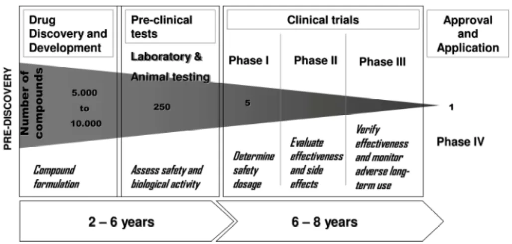
Drug Development is a long and costly process that starts with thousands of
drug candidates and ends with one compound, which often fails in clinical trials. This
high percentage of failure of the drug development process results from the lack of
highly predictable models in the pre-clinical tests.
Being metabolism a bottleneck in in vitro drug testing, special efforts have been made towards the development of metabolic competent liver cell cultures. The
major drawback of standard hepatocyte cultivation systems is the spontaneous cell
dedifferentiation that results in the decrease of metabolic competency and other liver
specific functions and ultimately will compromise the effective biotransformation of
drugs. Therefore, novel culture strategies presenting an improved hepatocyte viability
and functionality for extended periods are required.
Within this context, the main goal of this PhD project was to develop an
efficient strategy for culturing hepatocytes as 3D spheroid structures since this better
mimics the in vivo tissue. Within these 3D structures, cells can re-establish cell-cell interactions and specific microenvironments resulting in a better retention of
important hepatic functions.
The work here depicted has focused on the use of primary cultures of
hepatocytes, since they better retain the liver functional profile. Additionally, the
developed strategy has been applied to a human hepatocarcinoma cell line with the
aim of generating a more physiological model with cells that are easier to obtain then
fresh isolated hepatocytes. Within this context, and also to confirm the robustness of
the 3D culturing strategy, three relevant cellular models were used namely, human
hepatocytes, rat hepatocytes and the HepaRG cell line.
In Chapter I the importance of having alternative methods to animal testing,
regarding both the ethical problems and the inter-species variances, is explained.
Furthermore, the relevance of having metabolic competent methods towards the
accomplishment of the 3R’s is discussed, highlighted the advantages of having 3D
liver cultures for toxicity tests. Special focus is given to the bioreactors used for this
for liver cells, their use still relies on scarce sources. Thus, the novel strategy
developed during the course of this thesis was firstly optimised using rat hepatocytes
as described in Chapters II and III. Moreover, the development of highly predictable
cultures of rat hepatocytes is also a way to reduce and refine the animal testing.
In Chapter II, the identification of critical parameters used to develop the 3D
hepatocyte culture in stirred tanks is described, namely the testing of different stirring
impellers, culture media and cell inoculum. The best strategy – culturing rat
hepatocytes in 3D spheroids using a paddle impeller, William’s E medium with an
inoculum of 1,2x10⁵ cell/ml – has shown to improve specific hepatocyte functions up
to 10x and also extend it to 3 weeks when compared with the 1-2 weeks of the 2D
cultures. Also, the cells were further challenged to perform the physiological
phenomenon of drug clearance.
The system was then tested for further improvement. Aiming at creating a
more physiological milieu, other cell types were introduced into the system, more
specifically fibroblasts were co-cultured with rat hepatocytes as described in Chapter
III. The study started by testing different parameters such as type of fibroblasts, ratio
between the concentration of the two cell types and the total cell inoculum. As
expected, it was confirmed that over the same period of time, co-cultures had a
higher and more stable albumin production as well as improved phase I and II
enzymes activities. The best results were obtained for co-cultures of hepatocytes
with mouse embryonic fibroblasts in a ratio of 1:2 with a total inoculum of 1,2x10⁵
cell/ml. Moreover, CYP induction under different oxygenations (reflecting the
heterogeneous hepatic exposure to oxygen in vivo) was observed, confirming the biotransformation capacity of the cells.
After the optimisation of the system, Chapter IV describes the implementation
of a 3D culture of human hepatocytes in a perfusion system using cells from different
donors. Besides the expected inter-donor variability, culturing cells under the
immunostaining of the cell spheroids has shown that they present an in vivo like structure with biliary structures and polarised arrangement.
Chapter V describes the generation of a more physiological culture of
HepaRG cell lines as an attractive alternative to the use of primary cultures of human
hepatocytes. This cell line has shown a biotransformation activity within the range of
what is observed for human cells (both in vitro and in vivo) and for a longer period, up to 7 weeks (a great improvement when compared to the 3-4 weeks longevity
observed for the 2D HepaRG cultures). Moreover, the application of the system to
screening toxicological studies and to a multidisciplinary integrative model, has been
shown. The test of Acetaminophen toxicity in 3D spheroids generated with a stirred
tank and using 96-well plates, suggests that the developed culture model is a
potential model to be used as a cell-feeder system for high-throughput assays.
Furthermore, the integration of these results in a computational model has shown the
flexibility of the system to integrate with in silico approaches, contributing to the
generation of animal replacement strategies for toxicology applications.
Chapter VI consists of a general discussion integrating all the results
described in the previous chapters and the state-of-the-art of hepatic cultures. The
main achievements of the work are discussed, namely the generation of a more
predictable metabolic model. Final conclusions are then presented as well as
discussing the future outlook towards the accomplishment of the 3R’s policy.
In conclusion, the 3D spheroid hepatocyte culture systems developed in this
work are promising tools that may be used for the establishment of more robust and
O desenvolvimento de fármacos é um processo longo e dispendioso que
começa com milhares de compostos candidatos e termina com uma única, que na
maior parte das vezes é reprovada na fase de ensaio clínico. A elevada
percentagem de insucessos que ocorrem durante a otimização de fármacos resulta,
em grande parte, da inexistência the modelos para teste na fase pré-clinica que
sejam mais fiáveis.
Sendo o metabolismo um ponto fulcral dos testes in vitro, nos ultimos anos tem vindo a ser feito um grande investimento no desenvolvimento the culturas the
células de figado que sejam competentes metabolicamente. O modo tradicional de
cultura de hepatócitos leva à sua desdiferenciação expontânea, por decréscimo das
funções hepáticas, comprometendo a correta biotransformação dos xenobióticos.
Assim sendo, é necessário o desenvolvimento de novas estratégias para a cultura
de hepatócitos, de forma a aumentar a sua viabilidade e manter as suas funções
específicas durante mais tempo.
Neste contexto, o principal objetivo deste trabalho de Doutoramento foi
desenvolver uma estratégia eficaz para cultura de hepatócitos em estructuras 3D
que mimetizam melhor o tecido in vivo. Neste tipo de estruturas, as células podem restabelecer as interações célula-célula e o microambiente específico do fígado,
verificando-se assim uma melhor mimetização do desempenho da função hepática.
O trabalho focou-se essencialmente na ultilização de culturas primárias, tendo
em conta que conseguem reter melhor o perfil funcional do fígado. No entanto, esta
estratégia foi também utilizada para a cultura de uma linha celular de
hepatocarcinoma, de forma a obter um modelo mais fisiológico com células que são
mais fáceis de obter que os hepatócitos provenientes diretamente do tecido vivo.
Assim, neste contexto e também para confirmar a robustez da estratégia de cultura
em 3D, foram usados três modelos celulares relevantes, nomeadamente,
hepatócitos Humanos, hepatócitos de Rato e a linha celular HepaRG.
relevância da existência de modelos metabolicamente competentes no sentido de
concretizar a politica dos 3R’s (Substituir-Reduzir-Refinar, em inglês: Replace-Reduce-Refine), mostrando a vantagem de ter culturas 3D de células de fígado, para testes toxicológicos. É ainda dada especial relevância aos biorreatores
atualmente usados para este propósito, como modelos de cultura controlados e
robustos para manutenção de células.
Apesar das culturas primárias de hepatócitos humanos serem o melhor
modelo celular para mimetização in vitro do fígado, a sua disponibilidade é bastante limitada. Desta forma, a estratégia de cultura de hepatócitos desenvolvida nesta tese
foi primeiramente otimizada usando culturas primárias de hepatócitos de rato
descritas nos Capítulos II e III. Adicionalmente, o desenvolvimento de culturas
rubustas de hepatócitos de rato irá também contribuir para a redução e refinamento
dos testes em animais.
No Capítulo II está descrita a escolha dos parâmetros para o
desenvolvimento das culturas 3D em tanques agitados, nomeadamente o efeito de
diferentes tipos de pás de agitação, o meio de cultura e o inóculo celular. A
estratégia que demonstrou ser a melhor – cultura de hepatócitos em esferoides (3D)
em tanque agitado por pás, usando o meio de cultura William’s E com um inóculo de
células de 1,2x10⁵ cell/ml – demonstrou poder aumentar as funções específicas dos
hepatócitos até 10x comparando com as culturas 2D, ao mesmo tempo que foi
possível aumentar o tempo de cultura de 2 para 3 semanas. No final foi ainda
testada a capacidade das células de desempenhar uma função fisiológica, a
depuração hepática.
O Capítulo III, o melhoramento do sistema de cultura descrito no capítulo
anterior ao introduzir outro tipo de células, neste caso, fibroblastos. O estudo
iniciou-se testando diferentes parâmetros nomeadamente o tipo de fibroblastos, a razão
entre as duas concentrações celulares e o inóculo celular. Como esperado, para o
mesmo período de tempo, os hepatócitos em co-cultura apresentaram uma maior e
em cultura com fibroblastos embrionários de ratinho, numa proporção 1:2 com
inóculo total de células de 1,2x10⁵ cell/ml. Adicionalmente, as células responderam à
indução das CYPs, dependendo a amplitude de resposta da oxigenação (refletindo a
exposição heterogénea a oxigénio característica das células no fígado), confirmando
a capacidade de desempenhar a biotransformação.
Após otimização do sistema, o Capitulo IV descreve a implementação de
culturas 3D de hepatócitos humanos em biorreator de perfusão usando células de
diferentes dadores. Apesar da natural variabilidade entre dadores, a manutenção
das células em tanques agitados como 3D em condições de cultura controladas
demonstrou elevada reprodutibilidade em termos de secreção de albumina e ureia.
Em termos de capacidade de biotransformação as células demonstraram
capacidade de indução e re-indução após 2 a 4 semanas, das CYPs 1A2, 2C9 e
3A4. Adicionalmente, a marcação por imunofluorescência dos esferoides mostrou
uma estrutura semelhante ao fígado in vivo com estruturas biliares e organização polarizada.
O Capítulo V descreve o desenvolvimento de uma cultura mais fisiológica da
linha celular HepaRG, como uma alternativa atrativa ao uso de culturas primárias de
hepatócitos. As células demonstraram atividade de biotransformação dentro do
mesmo intervalo observado pelos hepatócitos humanos (tanto in vitro com in vivo) até 7 semanas (em comparação com as 3-4 semanas previamente obtidas com as
culturas 2D de HepaRG). Foi também possível demonstrar a aplicação das células
em estudos de toxicologia e integração multidisciplinar. O teste de toxicidade de
Acetominofen em esferoides gerados em tanques agitados, usando placas de
96-poços, demonstra a compatibilidade do modelo desenvolvido com testes
high-throughput. A integração dos resultados gerados em mdelos computacionais
demonstrou a flexibilidade do sistema conjuntamente com aplicações in silico no sentido de gerar uma subtituição aos testes toxicológicos em animais.
preditividade; a conclusão geral e as perspetivas futuras.
Em resumo, a cultura de esferoides 3D de hepatócitos desenvolvida neste
1. Miranda JP, Leite SB, Muller-Vieira U, Rodrigues A, Carrondo MJT, Alves
PM. “Towards an extended hepatocyte in vitro culture.” Tissue Engineering: Part C, 15(2) (2009): 157-67
2. Leite SB, Teixeira AM, Miranda JP, Tostoes RM, Clemente JJ, Sousa MS,
Carrondo MJT, Alves PM. “Merging bioreactor technology with 3D
hepatocyte-fibroblasts culturing approaches: enhanced in vitro models for Toxicological Applications” Toxicology in Vitro 25(4):825-32. (June 2011)
[Epub ahead of print]
3. Tostões RM, Leite SB, Brito C, Serra M, Jensen J, Bjorquist P, Carrondo
MJT, Alves PM. “Human liver cell spheroids in extended perfusion
bioreactor culture for repeated dose drug testing.” Hepatology (Accepted)
4. Leite SB, Wilk-Zasadna I, Zaldivar JM, Airola E, Minnecozzi M,
Reis-Fernandes M, Guillouzo C, Chesne C, Alves PM, Coecke S. “3D HepaRG
Abbreviation Full form
2D Two-dimensional / monolayer (cell cultures)
3D Three -dimensional (cell cultures)
3R’s Replace-Reduce-Refine (animal testing)
ADME Absorption, Distribution, Metabolism, Excretion
APAP Acetaminophen
aPKC Atypical Protein Kinase C
BAL Bioartificial Liver Device
BNF β-Naphtoflavone
CDFDA 5-(and-6)-carboxy-2',7'-dichlorofluorescein diacetate
CYP Cytochrome P 450
DAPI 4’,6-diamidino-2-phenylindole
Diff fully Differentiated hepatocytes
DILI Drug Induced Liver Injury
DMSO Dimethylsulfoxide
DMEM Dulbeco’s Minimum Essential Medium Eagle’s
DO Dissolved Oxygen
ECM Extracellular Matrix
ECOD Ethoxycoumarin O-deethylation
EGF Epidermal Grow Factor
EGTA Ethylene Glicol Tetracetic Acid
ELISA Enzyme-Linked Immunosorbent Assay
EMA European Medicines Agency
FBS Foetal Bovine Serum
FDA (US) Food and Drug Administration
GSTA-1 Glutathione S-Transferase Alpha 1
Hep Hepatocytes
Hff Human foreskin fibroblast
HGF Hepatocyte Growth Factor
HNF4α; Hepatocyte Nuclear Factor alfa;
Lac/Glc Lactate formation to Glucose consumption Ratio
LDH Lactate Dehydrogenase
4-MU 4-Methylumbeliferone
MEF Mouse Embryonic Fibroblasts
MEM Minimum Essential Medium Eagle’s
MCmB Multicompartmental bioreactor
PB Phenobarbital
PBS Phosphate Buffered Saline
PBTK Physiologically Based Toxicokinetic models
PFA Paraformaldehyde
Pharma Pharmaceutical Industry
pO2 Partial Pressure of Oxygen
qRT-PCR Quantitative Reverse Transcription Polymerase Chain Reaction
RIF Rifampicin
RWV Rotating Wall Vessel
SEM Standard Error of the Mean
SD Standard Deviation
TTC Threshold of Toxicological Concern
TK Toxicokinetics
UDP Uridine Diphosphate
U-Diff Under-differentiation hepatocytes
UGT UDP-glucuronosyl transferase
Figure 1.1. Page 4 Process of Drug Development.
Figure 1.2. Page 6 ADME schematization of a drug intake
Figure 1.3. Page 13 Hepatocyte organization within the liver and specific architecture in vivo
Figure 1.4. Page 19 Metabolic hepatic functions
Figure 1.5. Page 21 Hepatocyte cultures in 2D
Figure 1.6. Page 34 3D approaches for hepatocyte cultures
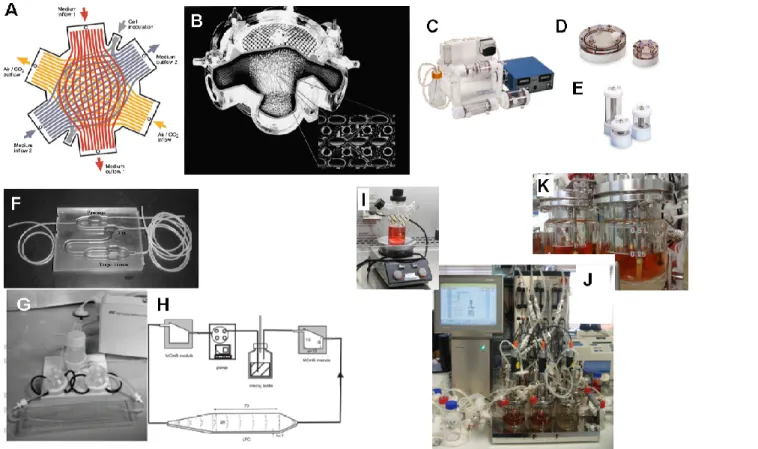
Figure 1.7. Page 40 Bioreactor systems for hepatocyte cultures.
Figure 1.8. Page 46 Schematic representation of the aim of the PhD thesis and strategy taken.
Figure 2.1. Page 67 Minibioreactor apparatus
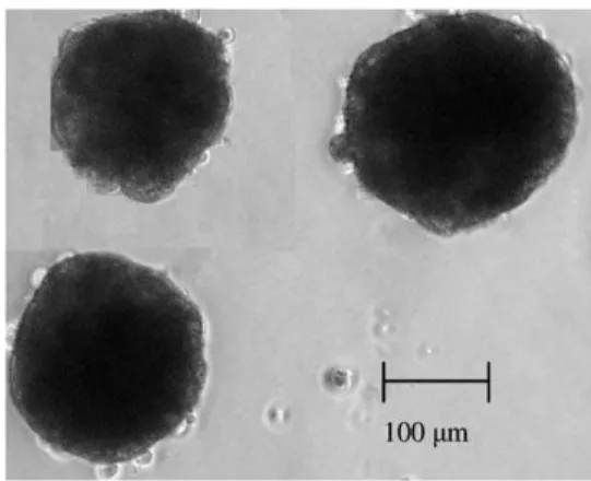
Figure 2.2. Page 72 Phase contrast microscopy of a 3D hepatocyte spheroid, cultured in stirred tank.
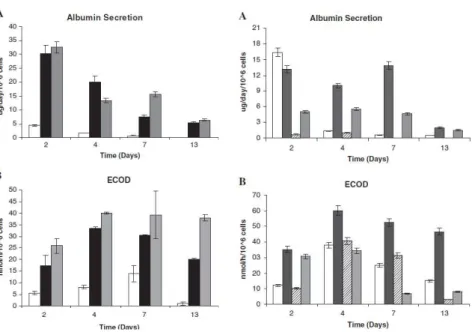
Figure 2.3. Page 74 Effect of culture time in albumin secretion and activity of phase I enzyme ECOD for cells cultured in monolayer 2D and spinner vessels 3D with paddle impeller and different inoculum concentration.
Figure 2.4. Page 74 Effect of culture time on albumin secretion and activity of phase I enzyme ECOD for cells cultured in different culture media in monolayer 2D and spinner vessels 3D.
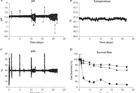
Figure 2.5. Page 75 Profiles of pH, temperature, dissolved oxygen (pO2) and cell survival in the bioreactor culture
hepatocyte spheroids.
Figure 2.6. Page 76 Functional capacity of hepatocytes over long-term cultivation periods was assessed by determining urea production and albumin secretion by the cells cultured in monolayer 2D, bioreactor 3D, and spinner vessels 3D culture systems.
Figure 2.8. Page 78 Metabolic stability of diphenhydramine and troglitazone observed in hepatocyte primary cultures in bioreactor 3D at day 2 and day 21 of culture; and in monolayer 2D at day 2 of culture.
Figure 3.1. Page 100 Experimental set-up used to evaluate the effect of fibroblasts type, hepatocyte:fibroblast ratio and total cell inoculum concentration in hepatocytes functionality.
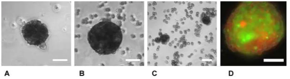
Figure 3.2. Page 101 Cell spheroids collected at day 3 from mono- and co-cultures
Figure 3.3. Page 103 Albumin secretion rate per unit volume along culture time in 3D stirred cultures.
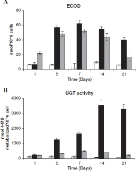
Figure 3.4. Page 104 Non-induced enzymatic activities along culture time in the different stirred 3D cultures.
Figure 3.5. Page 105 Relative ECOD activity in 3D co-cultures performed in bioreactors operated at different oxygen levels, namely 5%, 30% and 70% of air saturation.
Figure 4.1. Page 121 Experimental design of the induction of the CYP450 enzymes in primary cultures of hepatocyte spheroids in the bioreactor.
Figure 4.2. Page 127 Hepatocyte spheroid diameter distribution and cell viability. Bright field pictures of human hepatocyte spheroids. The average diameter of the hepatocyte spheroids and the average cell culture viability.
Figure 4.3. Page 128 Urea and albumin synthesis in bioreactor hepatocyte spheroid cultures.
Figure 4.4. Page 129 Inter-donor variability and time course profiles of phase I and II enzymes in human hepatocyte spheroids bioreactor cultures. Gene expression of the CYP450/phase I enzymes 1A2, 2C9 and 3A4, phase II enzymes GSTA1 and UGT2B7.
weeks of bioreactor culture. HNF4α, Cytokeratin 18, CYP450 isoform 3A.
Figure 4.7. Page 131 Fluorescence microscopy of structural and polarity markers and bile canaliculi function in human hepatocyte spheroids after 2 weeks of bioreactor culture. F-actin; transport of CDFDA to the apical (canalicular) domain; aPKC.
Figure 4.8. Page 132 Albumin quantification and ECOD activity of human hepatocytes in Mono- and Co-culture with human fibroblasts (hff).
Figure 5.1. Page 148 Experimental set-up used for culture 3D
HepaRG in Stirred bioreactors.
Figure 5.2. Page 154 Cell arrangement within HepaRG 3D cultures.
Figure 5.3. Page 155 3D organization of HepaRG spheroid. Details of a spheroid, showing polarization of the Pgp transporter.
Figure 5.4. Page 156 Phase I enzymes inducibility at week 3 in 3D Differentiated cells: determination of the fold increase of CYP 1A2, 2B6, 2C9 and 3A4 activities after 72h incubation with reference compounds.
Figure 5.5. Page 157 Biotransformation activity at selected points.
Figure 5.6. Page 158 Bioctivation capacity of the cells. Dose response curves after addition of APAP at different concentrations at different culture times. Experimental and simulated dose response curves after addition of APAP at different concentrations at different culture times.
Figure 6.1. Page 173 Main outcomes from the studies performed in this thesis
Table 1.1. Page 8 Main Alternative Methods Available.
Table 1.2. Page 16 Human and rodent main CYP P450 isoforms
with the respective inducers and receptors associated
Table 1.3. Page 31 3D Models available
Table 4.1 Page 119 Donor Information
Table 6.1. Page 172 Aimed characteristics of an in vitro method for drug testing applications in Pharma. Industry
Chapter I Introduction 1
Chapter II Rat hepatocytes in 3D
Toward Extended Functional Hepatocyte In Vitro Culture……… 59
Chapter III Rat hepatocytes in 3D co-culture
Merging bioreactor technology with 3D hepatocyte-fibroblasts culturing
approaches: enhanced in vitro models for Toxicological Applications……… 87
Chapter IV Human hepatocytes in 3D
Human liver cell spheroids in extended perfusion 1 bioreactor culture for
repeated dose drug testing……… 113
Chapter V HepaRG in 3D
3D HepaRG: New model toxicological applications……… 141
I
C
h
ap
te
CONTENTS
1. ALTERNATIVE METHODS TO ANIMAL EXPERIMENTATION: assessment of ADME towards the 3R’S policy ... 4
2. METABOLISM AND BIOTRANSFORMATION ... 12 2.1 Phase I of Biotransformation ... 14 2.2 Phase II of Biotransformation ... 17
3. HEPATOCYTE CULTURES ... 18 3.1. Human Hepatocytes ... 22
3.2. Rat hepatocytes ... 23 3.3. Human Hepatic Cell lines ... 25 3.4. Co-Cultures... 26
4. HEPATOTOXICITY ... 28 5. IN VITRO 3D MODELS IN TOXICOLOGY ... 29
5.1. 3D Liver Systems ... 33 5.1.1. Liver slices ... 33 5.1.2. Sandwich ... 35
5.1.3. Cell Spheroids ... 35 5.1.4. 3D Scaffolds ... 36
5.2 Bioreactor Systems ... 37 5.2.1. Multi-compartment 3D perfusion bioreactors ... 41 5.2.2. Rotational Wall Vessel (RWV) ... 41
5.2.3. Microfluidic / Multicompartmental bioreactors ... 42 5.2.4. Stirred culture vessels ... 43
1. ALTERNATIVE METHODS TO ANIMAL EXPERIMENTATION: assessment of ADME towards the 3R’S policy
The development of a medical drug takes several years (5 to 10), involving thousands of compounds and costing millions of dollars (Fig. 1.1) until it is on the
market to be used by patients (innovation.org 2007). The recent advances on genomics, proteomics and computational sciences have given scientists a better understanding of the human body at the molecular level, enforcing the first steps of
the drug discovery process on the producing of more effective compounds. However, there is still a high rate of compound failure on the clinical trial and approval phases
(Service 2004), either for inducing human toxicity or for not producing the desire/expected effect.
Figure 1.1: Process of Drug Development. (adapted from (innovation.org 2007; Mattisson and Matison Faye 2008) )
Besides medical drugs, with the continuous development of the industrialized world, the human body is increasingly exposed to different compounds present in
C
h
ap
te
through different routes reaching distinct targets, and in spite of acute effects being
quickly detected, for many of the compounds, chronic toxicity remains unknown. In this context, the development of methods and strategies that will allow a
better prediction of the impact of the new formulations on human health is of capital importance.
Animal models have been used for a long time in order to access the effect of
compounds as an integrative ADME (Administration, Distribution, Metabolization and Excretion) model. However, despite the advantages of being an in vivo system there
are several reasons that have convinced the scientific community of the importance of developing alternative methods. Besides the ethical implications of sacrificing animals for research purposes and the economical impact involved, animal models
have been shown to produce false positives regarding the prediction of human toxicity specially at the metabolic level (Turpeinen et al. 2009); this is the case with
the catastrophic use of Thalidomide and Valpromide in humans.
In 1959, William Russel and Rex Burch introduced the 3R's concept in The Principles of Humane Experimental Technique (Russel and Burch 1959), that stands
for Refinement, Reduction and Replacement of animal testing. The 3R’s concepts were defined as: Refinement – any method that reduces or eliminates pain and
distress in animals during experiments; Reduction – methods that seek to use fewer animals in an experimental protocol to obtain the same or similar information of scientific value, or use the same number of animals to obtain more scientifically
valuable information; and Replacement – the use of techniques that do not require living animals.
In 1986 a European Directive was established on the protection of laboratorial animals for experimental purposes developing and validation of alternative techniques to animal testing, that latter lead to the creation of ECVAM (European
Centre for the Validation of Alternative Methods). Within this scenario, several industries adopted strategies towards the development of alternative methods.
been gradually introduced on the marketing of cosmetics containing ingredients tested in animals.
However, this year a report prepared for DG SANCO by experts in the field concluded that full animal replacement is still not possible (Adler et al. 2011).
Nevertheless, contrarily to what is many times assumed, the 3R's policy is not a replacement based strategy. The improvement of alternative methods consists on the development of strategies that can better mimic the in vivo system by using less
animal sources. The establishment of this policy is based on a set of concepts that have to be considered in order to achieve methods that better predict human toxicity.
Figure. 1.2: ADME resume of a drug intake, possible paths within the body. ADME (Adapted from Topics on Biotechnology (http://www.biology.iupui.edu/biocourses/Biol540/4pipeline09Full.html) and http://www.humanillnesses.com)
ADME (Fig. 1.2) is a concept that more and more has becoming a fulcral point in Drug Development. Many compounds tested in high-throughput screens, often
C
h
ap
te
that ADME testing has to be moved earlier in the drug discover pipeline (Pelkonen et
al. 2011).
Toxicokinetics (TK) of a compound is the characterization of its ADME,
absorption, distribution, metabolism and excretion in the body. Towards the implementation of the 3R’s policy, TK has become an essential information that is important to determine parameters such as the bioavailability of metabolites towards
the targets; the translation of in vitro nominal concentration to the actual in vivo levels and to understand if the cell or tissue under human exposure conditions is exposed
to the external xenobiotic and/or its metabolite.
With the aim of following the European directives, more reliable methods have been developed and there is already a remarkable set of alternative methods on
C
h
ap
The accomplishment of an approved certified alternative method is a process that includes 7 steps: 1) Research; 2) Development; 3) Prevalidation; 4) Validation; 5)
Independent Review; 6) Regulatory acceptance and 7) Implementation.
Method validation is part of toxicity testing (Pelkonen 2010). Even being a
tedious and time consuming process (5-10 years), the validation of an alternative method, involving several laboratories and a reliable scientific board is a way to ensure the reliability, transferability and robustness of the test method (Pelkonen
2010). This way, the financial and human resources are used more efficiently, having a greater likelihood to meet the expectations of those in the scientific, regulatory and
animal welfare communities on alternative methods benefiting and the human society in general.
One of the major drawbacks to achieve viable alternative approaches is the
lack of efficient conversion approaches of in vitro data, generated at tissue/cell or sub-cellular level, into dose-response information on human body. Within the TK
approach, towards the full replacement models, there are several concepts such as Threshold of Toxicological Concern (TTC), biokinetics, IVIVE and integrative approaches that should be taken into consideration (Adler et al. 2011).
All together, the alternative methods need then to fulfill the needs and questions of human toxicology, using relevant and competent models and
addressing fundamental issues.
When testing the toxicity level of a compound, upfront, is important to determine its TTC value of human exposure: below the TTC there is a very low
probability of an appreciable risk to human health (Adler et al. 2011). This is a concept that is already being used by the food authorities and is the base of further research projects (Adler et al. 2011). If human exposure is above the external TTC it
becomes important to access the biokinetics of the compound – the process that the compound will suffer until it reaches its internal concentration and the concentration
at which it will reach the target organ(s).
C
h
ap
te
substance that in vivo would indeed reach the target organ after passing the
metabolic organs. It is then important to study the mode of toxic action, and the resulting effects on the physiological processes in an organism. In cell culture it is
important to establish strategies that ensure to have enough bioactivated substances in vitro such as the use of cell co-culturing, having in culture both the activating cells
and the target cells.
First, it is important not only to have competent cell cultures, but ensure also that the cells have a behavior which is as close as possible to the organ itself. To
achieve this, several strategies have been developed that will be further explored in the sub-Chapters of Chapter I, but a better predictability will be reached if the cell culture strategy is combined with other approaches and tools. The more extensively
characterized a compound is in terms of metabolism, disposition, bioactivation, cell membrane transposition, interaction with molecular processes and cell structures
and functions, the higher chances of success in the late stages of the drug development.
The use of in silico strategies on kinetic data has been a development
strategy used to bridge the in vitro and in vivo. Today’s softwares, can already simulate chemical properties and interactions. When these are used together with
other computational models, in silico strategies have shown to be the most cost-effective approach (Ekins et al. 2010) and to be able to translate the experimental data using the so called Physiologically based Toxicokinetic models (PBTK). To have
an efficient in vivo in vitro extrapolation (IVIVE) using in silico models it is important to have massive generation of data. Thus, it is important to use down-scale and
automated models that can screen large amounts of compounds. Metabolic data contributes to explain possible toxicities and modes of action and their relation to dose level and route of exposure are described by Toxicokinetics. Moreover, REACH
legislation also requires the testing of thousands of chemicals within a short period of time (few years) (Pelkonen 2010) thus it is of significant importance that the in vitro
through which a high number of compounds and/or conditions can be screened simultaneously.
While full replacement of animal testing is still not yet accomplished, partial replacement and reduction are forthcoming. For this purpose it is important to
develop multidisciplinary strategies that include in vitro, in silico and "omics" approaches. A close collaboration between academia, regulatory authorities and industry is of primordial importance. The creation of consortiums, such as DNT
(Developmental Neurotoxicity), to discuss and set the necessary path to follow are of utmost importance.
Unlike other toxicokinetic steps, there is still a lack of validated in vitro models for metabolization prediction (Adler et al. 2011) which is a result of the complexity of maintaining viable and functionally active Liver in vitro models.
2. METABOLISM AND BIOTRANSFORMATION
To have a competent in vitro toolbox it is important to identify important metabolic pathways of the compound and to provide knowledge on: the metabolic stability of the chemical; the type and structure of metabolites involved. It is also
important to determine any metabolism-dependent toxic effects, test the compound’s capacity to induce or to inhibit the cell enzymes, check for drug-drug interactions and
evaluate its clearance (Coecke et al. 2006; Pelkonen et al. 2008).
Since metabolism is the bottleneck of in vitro toxicology (Coecke et al. 2006), it is important to have competent metabolic methods that can accurately determine
all the parameters involved on the transformation of a compound before it reaches its target. From the pharmaco-/toxicokinetic point of view, liver is the most important organ in the body. Hepatocytes, present in the liver, have an enzymatic machinery
C
h
ap
te
A
Canalicular, apical side
Sinosoid, basoleteral side
Sinosoid,
basoleteral side Space of Disse
Space of Disse HEPATOCYTE
EPITHELIAL CELL
bile flow
sinusoid BILE DUCT
EPITHELIAL CELL
Blood flow HEPATOCYTE
bile canaliculus Space of Disse EPITHELIAL CELL
bile duct
Figure. 1.3. Hepatocyte organization within the liver and specific architecture in vivo (Adapted from (Pelkonen et al. 2008) and (Dunn et al. 1989)). Hepatocytes at the level of functionally important molecules, at the level of tissue architecture, and at the level of kinetic modeling.
Generally, the properties of hepatic cells result from the presence of a
comprehensive set of enzymes that guide compounds through a one or two phase process called biotransformation as well as the presence of multiple transporter
proteins, some of them with polarized distribution, that perform both influx and efflux functions (Fig. 1.3 A) (Pelkonen et al. 2008). The polarized structure is a result of the different hepatic surroundings in the liver (Fig. 1.3 B), the blood flow is on one side of
2.1 Phase I of Biotransformation
During Phase I of biotransformation the compound (xenobiotic) undergoes
oxidative and reduction reactions, carried by monooxygenases becoming more polar. The enzymes involved on phase I are mainly clustered in two groups such as
Cytochrome P450 (CYP450), the largest group of membrane associated heme proteins, and flavine monooxygenases. The human CYPs involved in the majority of marketed drugs are CYP3A4, CYP2D6, CYP2C9, and CYP1A2 (Zanger et al. 2008).
One of the major drawbacks of the development of liver-predictive models is the high inter-individual metabolic variability. The main cause of these differences is
the variation of phenotype and genotype among the enzymes involved in biotransformation, specially the Phase I CYP450 cluster. This is mainly a result of environmental and/or hereditary gene polymorfisms specially on CYPs 2C9, 2C19,
and 2D6 (Hewitt et al. 2007). On the other hand, although gene expression of CYP 1A1, 2E1 and 3A4 is well conserved, variations on their activity levels can still be
observed (Hewitt et al. 2007). In this case, the inter-individual differences are the result not only of genetic predisposition but also of many other factors such as hormonal expression, age, sex, diet, smoking, drug consumption, exposure to
environmental chemicals and disease state. Among all these factors the interference of other drugs by drug-drug interaction, induction or inhibition is the major and more
frequent effect (Hewitt et al. 2007). Moreover, the induction of enzymatic capacity has been used as a quality control property, as a way to access the metabolic competence of hepatocytes in vitro and thus it has been used as a control factor in
validation studies (Richert et al. 2010).
The process of enzymatic induction (CYP induction) is the increase of a specific CYP activity upon the addition of specific compound. The classical definition
of induction is the de novo synthesis of an enzyme (protein) reflecting an increased transcription of the correspondent gene following a specific stimulus. In more detail,
C
h
ap
te
the upstream regulatory elements on CYPs (O’Brien 2004). Among the transcription
factors, the nuclear receptors such as androstane receptor (CAR), the pregnane X receptor (PXR) as well as the aryl hydrocarbon receptor (AhR) can be distinguished
as the main regulators of CYPs activity, acting as sensors for lipophilic xenobiotics, including drugs (Handschin and Meyer 2003; O'Brien et al. 2004).
As explained before, induction is a complex process involving several cellular
machineries, thus the addition of specific known inducer compounds is a good tactic to check the quality of the metabolic competence of a system for more than one day
(Coecke et al. 2006). Furthermore, induction has very specific characteristics such as being tissue-specific, rapid, dose-dependent, and reversible upon removal of the inducer (Handschin and Meyer 2003).
However the capacity to respond to one or other compound as well as the fold increase of the enzyme activity is not only species dependent but also
donor-dependent (Richert 2009).
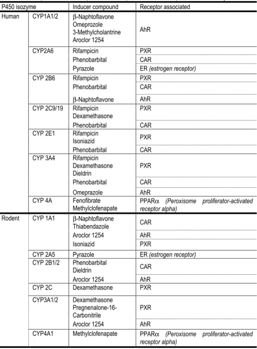
Table 1.2 resumes the main xenobiotic CYP metabolizers in human and rodent species, as well as the specific inducers and receptors involved in the
induction. It can be observed that the same compound does not always induce the same CYP in one or another species. This differences between species is due
Table 1.2: Human and rodent main CYP P450 isoforms with the respective inducers and receptors associated. Adapted from (Handschin and Meyer 2003) and (Ogu and Maxa 2000; Pascussi et al. 2000; Borlak et al. 2002; Brandon et al. 2003; Meredith et al. 2003; Tamrazi et al. 2003; O'Brien et al.
2004; Marek et al. 2005; Dail et al. 2007; Chanda et al. 2009; Richert et al. 2009) P450 isozyme Inducer compound Receptor associated
Human CYP1A1/2 β-Naphtoflavone Omeprozole 3-Methylcholantrine Aroclor 1254
AhR
CYP2A6 Rifampicin PXR Phenobarbital CAR
Pyrazole ER (estrogen receptor) CYP 2B6 Rifampicin PXR
Phenobarbital CAR
β-Naphtoflavone AhR CYP 2C9/19 Rifampicin
Dexamethasone
PXR
Phenobarbital CAR CYP 2E1 Rifampicin
Isoniazid PXR Phenobarbital CAR CYP 3A4 Rifampicin
Dexamethasone Dieldrin
PXR
Phenobarbital CAR Omeprazole AhR CYP 4A Fenofibrate
Methylclofenapate
PPARα (Peroxisome proliferator-activated receptor alpha)
Rodent CYP 1A1 β-Naphtoflavone
Thiabendazole CAR Aroclor 1254 AhR Isoniazid PXR
CYP 2A5 Pyrazole ER (estrogen receptor) CYP 2B1/2 Phenobarbital
Dieldrin CAR
Aroclor 1254 AhR CYP 2C Dexamethasone PXR
CYP3A1/2 Dexamethasone Pregnenalone-16-Carbonitrile
PXR
Aroclor 1254 AhR
C
h
ap
te
2.2 Phase II of Biotransformation
After the oxidative pathway of phase I, the xenobiotic can then be excreted or, if it is still not polar enough, it will undergo the conjugation process – Phase II of
biotransformation. Conjugation path consists of reactions of glucuronidation, sulfation, methylation, acetilation and mercapture formation, at the end of which more soluble metabolites are obtained; these are ready to be eliminated (Gomez-Lechon
et al. 2006).
Phase-II metabolization of a compound is dictated by its chemical properties.
Within phase II reactions, glucoronidation represents more than 35% of the conjugation reactions in human drug metabolism by the UDP-glucuronsyltransferase (UGT) family of enzymes (Trubetskoy et al. 2007). UGT enzymes catalyze the
conversion of substrates (exogenous or endogenous compounds) into more polar glucuronides by covalent linking (conjugation) to UDP-glucuronic acid (UDPGA)
(Chen et al. 2003; Donato et al. 2010).
Recent studies have shown that these enzymes can also be induced by some of the prototypical CYP inducers following the same path of ligand-activated
transcription factors (Donato et al. 2010).
The physiological function of biotransformation is to detoxify the body from foreign compounds. However, the metabolites of biotransformation are often highly reactive and toxic causing hepatotoxicity, being sometimes more toxic than the
parent compound (Gomez-Lechon et al. 2006). For this reason, in a toxicological test it is important to access toxicity of the compound as well as its metabolite.
The biotransformation capacity is dependent not only on the activity of the enrolled enzymes but also on the activity of the membrane transporters with polarized distribution, for instance, apically transport bile acids on the basal site
conduct trafficking of metabolites from the bloodstream. The main superfamily of transporters is the Multidrug resistance-associated superfamily-2 (MRP2) that is a
al. 2002). MRP2 belongs to the ATP-binding cassette (ABC) protein superfamily
together with other relevant transporters such as MRP1, MRP3 and P-glycoprotein
(PGP), the latest also localized on the hepatic biliary side (Borst et al. 2000). As it happens with the CYP enzymes, the pump transporter enzymes also respond with
increased activity upon the addition of inducer compounds such as dexamethasone and Phenobarbital (Courtois et al. 2002).
The role of biotransformation within the organism and the variability
associated to it show how important it is to have, in the drug development process, earlier models that behave as close as possible to what is observed in vivo, with all
the hepatocyte specific functions operating in a reproducible and stable mode. Having good reproducible liver models is an important step forward towards the accomplishment of the 3 R’s policy.
3. HEPATOCYTE CULTURES
Besides biotransformation, hepatocytes are responsible for many other body functions that contribute to the regulation of the body homeostasis and to the detoxification of incoming compounds. Under external stimuli (e.g. hormones,
nutrients, ions) the hepatocytes secrete and degrade a large number of molecules. The main functions of hepatocytes (besides biotransformation), schematically
represented on Figure 1.4 (De Maria et al. 2008) are:
Regulation of the metabolism of carbohydrates, proteins and lipids and their levels in the blood;
Synthesis of important serum proteins such as albumin, protrombin, fibrinogen, etc., (with the exception of immunoglobulins);
Storage of glycogen, triglycerides, iron, copper and liposoluble vitamins;
Catabolism of endogenous substances, such as hormones and serum proteins, to maintain a balanced body concentration;
C
h
ap
te
Figure. 1.4. Metabolic hepatic functions (Adapted from (De Maria et al. 2008))
When in culture, the maintenance of these functions is attempted by culturing, under chemical defined conditions, appropriate culture medium and supplements and
a robust cell culture apparatus. However, it is first of all important to choose the most adequate cell model. Extracellular signals play an important role in the preservation
of differentiated liver-specific functions. Within this context, for years researchers have been trying to decrease the spontaneous de-differentiation of in vitro hepatocytes by mimicking the extracellular signals using different strategies such as
modification of the culture conditions (such as the use of hormonally defined and highly enriched media), addition of soluble factors (such as hormones, growth
factors, cytokines and amino acids) (Feldhoff et al. 1977; Coecke et al. 1999; Schmelzer et al. 2009; Takeba et al. 2011), increase of cell-cell contacts (Pampaloni et al. 2007), addition of matrixes (such as collagen, matrigel, etc.), (Dunn et al. 1989)
Specific medium components such as Serum, Dexamethasone, Insulin, Glucagone and Growth Hormone have shown to have high impact on the
physiological maintenance of hepatocytes in vitro (Coecke et al. 1999; Schmelzer et al. 2009), however some of these factors can also decrease and/or suppress
important hepatic machineries such as CYP activity, transporters or protein synthesis (Coecke et al. 1999). The solution is to try to balance the importance of the addition of each component according to the aim of the study.
When reproducing the in vivo environment in cultured hepatocytes, cell-extracellular membrane (-ECM) interactions, soluble growth factors and cytokines,
physical factors (e.g. stress and strain) and cell-cell communications (Lang et al. 2011) are important elements that need to be controlled.
Oxygen is another sensitive issue in hepatocyte cultures. In the liver, the
oxygen content on the blood flow from the periportal to the perivenous hepatocytes decreases from 8-9% to 3-5% which will affect the enzymatic profiles of the
hepatocytes in each region (Stevens 1965; Jungermann and Kietzmann 1997; Kietzmann and Jungermann 1997; Kidambi et al. 2009; Yan et al. 2010) leading to the so called liver zonated metabolism (Kietzmann and Jungermann 1997; Allen et
al. 2005). Several studies state that regular cellular cultures are over-oxygenated
(Kidambi et al. 2009; Yan et al. 2010) since cells are exposed to the 21% of oxygen
present in the air while in the liver the oxygen levels are much smaller (Kietzmann and Jungermann 1997), and this could cause oxidative stress. On the other hand, some works defend that cells are exposed to an unlimited oxygen gradient as a
consequence of the presence of the hemoglobin in the blood (Nahmias et al. 2006; Kidambi et al. 2009) and thus low oxygen concentrations do not mimic the natural hepatic environment (Cho et al. 2007), so cells oxygenation is a controversial issue.
C
h
ap
te
A
Primary culture of Human Hepatocytes
B
HepG2 cell line
C
HepaRG cell line
3.1. Human Hepatocytes
In the last few years, freshly isolated hepatocytes (Fig. 1.5 A) have been
recognized as one of the most relevant models to study drug metabolism and transporter interactions, since this is the culture model that can best retain liver
functionalities in vitro (Gomez-Lechon et al. 2010). Considered the golden standard of hepatic cultures, the use of freshly isolated hepatocytes is recommended both by the US Food and Drug Administration (FDA) and the European Medines Agency
(EMA) for in vitro liver-related studies, such as induction determination.
Several studies with human hepatocytes have shown that, within the proper
environment, human hepatocytes can retain the biotransformation mechanisms in an integrated form, including transporters and induction (Silva et al. 1999; Courtois et al. 2002; Donato et al. 2010; Rotroff et al. 2010; Perry et al. 2011), reproducing the in
vivo paths and constituting a valuable tool to anticipate potential drug-induced liver
toxicity. Thus, human hepatocytes have been used for hepatotoxicity studies, namely
for: screening cytotoxicity and genotoxicity; characterizing drug-induced lesions, describing toxic mechanisms and determining biotransformation pathways (Coecke et al. 1999; Gomez-Lechon et al. 2010). However, liver-specific markers are hardly
maintained in primary cultures and not at all after the maintenance of isolated human hepatocytes for several weeks. After isolation, hepatocytes undergo a natural
de-differentiation that is characterized by the declining of protein synthesis, leading to a drop in the overall protein content and finally to cell death (Moshage et al. 1988).
To overcome de-differentiation several strategies have been developed to
improve and prolong the human hepatocytes functionalities, these have focused on trying to reproduce in vitro a more physiological environment. For instances, human hepatocytes have shown a good adaptation and improved functionality when cultured
in bioreactors (Schmelzer et al. 2009; Prot et al. 2011; Tostões 2011).
Another characteristic of these cells is the high donor-variability. In spite of
C
h
ap
te
model has shown robustness as it was possible to proceed to inter-laboratory
validation. Reproducible results were obtained in different laboratories with harmonized protocols even for different donor cells (Richert et al. 2010).
As a way to further improve the human hepatocyte model, significant work on improving the efficiency of freezing and platting after thawing techniques has been done (Silva et al. 1999; Hewitt et al. 2007). Cryohepatocytes are freshly isolated
hepatocytes that are maintained at -80ºC until use and have shown to perform similarly to the freshly isolated hepatocytes (Li et al. 1999; Li 2007). This has the
advantage of having the possibility of using cells from same batch at different times. After thawing, they present lower cell viability, but the cells that are viable and able to attach, can perform similarly to fresh hepatocytes in spite of showing lower basal
activities (Abadie-Viollon et al. 2010).
Human hepatocytes can be obtained from different sources such as livers
unsuitable for transplantation, surgical waste material (after reduced-size or split-liver transplantation) and waste material from partial hepatectomy (Richert et al. 2010). However, the human liver tissue is sparsely available, being necessary to develop
and use alternative hepatic culture models.
3.2. Rat hepatocytes
Alternatively to human cells, different mamalian cells have been used for development of in vitro liver models and hepatotoxicity assays since they are easier
to obtain, namely monkey (Silkworth et al. 2005), porcine (Custer and Mullon 1998; Hoebe et al. 2000; Kostrubsky et al. 2000), rat (Guyomard et al. 1996; Shen et al.
2008; Hrach et al. 2011; Schutte et al. 2011) and mouse hepatocytes (Kulkarni and Khanna 2006; Jemnitz et al. 2008; Jaeschke et al. 2010).
Compared to the use of human cell lines, using animal freshly isolated
hepatocytes has the advantage of better reflecting the in vivo hepatic features, having a more active and reliable metabolism. Within the different animal species it is
Currently, the selection of a specie for toxicological assays is mostly based on the knowledge and historical use of that specie, the costs, the amount of hepatocytes
obtained per each individual and the availability (O’Brien 2004), evaluating these parameters makes rat hepatocytes perfect candidates.
Primary cultures of rat hepatocytes are a good alternative often used by the pharmaceutical industry because: (i) they present higher metabolic responses than the common human cell lines and (ii) the inter-donor variability can be minimized by
selecting animals of the same sex, age and similar feeding regimes (Coecke et al. 2000; O'Brien et al. 2004).
Rat hepatocytes, have shown good hepatocyte-specific characteristics such as cell shape and arrangement (Abu-Absi et al. 2002), biotransformation activities, (Kocarek et al. 1990; O'Brien et al. 2004) albumin secretion (Feldhoff et al. 1977),
hormonal regulation (Coecke et al. 2000) and bilary transporters (Nakanishi et al. 2011).
In spite of the interspecies differences between rat and human, rat hepatocytes can be a good tool to explore culture strategies and undisclosed toxic cellular mechanisms. In the specific case of induction studies, although the
differences in the sequences of the ligand domain of the nuclear receptor genes and the CYP lead to different induction responses (Lin 2006), the obtained information
allows to interpret in vivo rodent results and thus conduct to the Refinement and Reducing of animal testing.
Moreover, the strategies developed to improve hepatocytes functionalities can
often be easily applied to primary human hepatocytes. These extrapolations have shown successful results on the use of scaffolds (Bierwolf et al. 2010), 3D strategies (Brophy et al. 2009; Nakanishi et al. 2011), bioreaction (Allen et al. 2005; Miranda et
al. 2009; Nakao et al. 2011), oxygen requirements (Kietzmann and Jungermann
1997; Nahmias et al. 2006; Cho et al. 2007; Kidambi et al. 2009) stem differentiation
(Qihao et al. 2007) and co-culture (Coecke et al. 2000; Tilles et al. 2001).
C
h
ap
te
correlation with what happens in human hepatocytes and with what happens in vivo
(Richert et al. 2009).
3.3. Human Hepatic Cell lines
The main advantage on the use of hepatocyte cell lines, in comparison with freshly isolated hepatocytes is the easiness to culture: (i) is possible to freeze and thaw the cells with no further damage, (ii) cell karyotype can be maintain and (iii)
have almost unlimited growth.
HepG2 is the hepatic cell line being used for a longer period of time, is a
popular and good human cell line used to study a broad variety of toxicity mechanisms (Farkas and Tannenbaum 2005). The cells can be unlimited expanded while conserving the polyhedric hepatocyte-specific morphology in vitro (Fig. 1.5 B).
In spite of being good models for toxicity accessment, on metabolism studies, like CYP induction, these cells have shown low metabolic competency (Farkas and
Tannenbaum 2005).
After HepG2, several hepatic cell lines (HLE, THLE, BC2, Hep 3B) with increased metabolic competencies were developed. For instances, the recently
developed cell line – Fa2N-4 – has shown promising results on its application for CYP3A4 induction during Drug-Drug interactions (Ripp et al. 2006). In spite of
showing punctual relevant competencies, the major limitation of hepatic cell lines is that they do not show an overall metabolic realistic profile thus being of little value for integrative studies.
In 2002, a promising cell line called HepaRG was developed by Gripon et al which performed similar to human hepatocytes (Gripon et al. 2002). HepaRG is an
hepatocarcinoma cell line with limited caryotype alterations, characterized by a surnumerary and remodeled chromosome 7 (Gripon et al. 2002).
After 4 weeks of differentiation including treatment with 2% DMSO, HepaRG
generate two liver cell types: one resembling hepatocytes and the other resembling biliary canaliculi-like structures (Fig. 1.5 C). After detachment both structures can
al. 2007). During the last years, intensive characterization of HepaRG cells has shown that when differentiated these cells express the major cytochrome P450
(CYP), drug-conjugating enzymes, transporter proteins, nuclear receptors and transcription factors as well as other liver-specific proteins at levels close to those
found in primary hepatocytes (Aninat et al. 2006; Guillouzo et al. 2007; Kanebratt and Andersson 2008; Turpeinen et al. 2009; Lubberstedt et al. 2011). These characteristics make HepaRG a good hepatic model for hepatotoxicity studies
(McGill et al. 2011), CYP induction studies (Lambert et al. 2009; Andersson 2010), clearance (Zanelli et al. 2011) and liver pathological situations (Gripon et al. 2002;
Schulze et al. 2011).
Moreover, this cell line has also shown good competency on an upper level of the development of 3R’s ideology, by being compatible with more physiological
models such as bioreactors (Darnell et al. 2011; Hoekstra et al. 2011) and even in vivo engraftment (Jiang et al. 2010).
3.4. Co-Cultures
For studies of metabolism, transporters as well as in hepatoxicity assays, monocultures of hepatocytes are usually preferred to mixed cultures. However, this
scenario does not reflect the in vivo environment and the results can be misleading mainly because of the low hepatic performance in vitro, but also due to the lack of
other cells that can affect the drug interaction with the hepatocyte as it occurs in vivo. Regarding hepatic functionality, different studies have shown that cell-cell interactions, especially heterotypic cell interactions (Bhatia et al., 1998), are more
effective in maintaining hepatocyte functions than ECM configurations (Michalopoulos et al. 1979; Khetani and Bhatia 2008). Significant enhancements in hepatocytes phenotype maintenance and function were described by the use of
feeder cells in co-cultures with hepatocytes such as connective tissue or nonparenchymal cells in several species (Bhatia et al. 1998; Bhandari et al. 2001; Lu
et al. 2005; Khetani and Bhatia 2008). Feeder cells are able to either secrete soluble