Sofia Isabel Marques da Silva
Dissertation presented to obtain the Ph.D degree in Biochemistry
Instituto de Tecnologia Química e Biológica | Universidade Nova de Lisboa
Oeiras,
September, 2011
Sulfate Reducing Bacteria
Formate CO2
H+
SO42
Sofia Isabel Marques da Silva
Dissertation presented to obtain the Ph.D degree in Biochemistry
Instituto de Tecnologia Química e Biológica | Universidade Nova de Lisboa
Oeiras, September, 2011
Sulfate Reducing Bacteria
Second Edition
Work funded by:
Formate oxidation and sulfate reduction by sulfate reducing bacteria.
The photo shows Desulfovibrio vulgaris, the model organism of SRB.
Source: http://failure-analysis.info/2010/06/biological-corrosion-of-metals/
September 8, 2011
President: Supervisor: Opponents:
Professor Carlos Romão
Doctor Inês Cardoso Pereira
Professor Alfons Stams
Professor Christiane Dahl
Professor Maria João Romão Jury
Bacterial Energy Metabolism Laboratory Instituto de Tecnologia Química e Biológica
Avenida da República (EAN)
2780‐157 Oeiras, Portugal
The work described in this thesis was performed under
supervision of Dr. Inês Cardoso Pereira in the Bacterial Energy
Metabolism Laboratory at Instituto de Tecnologia Química e Biológica
of Universidade Nova de Lisboa.
This thesis is divided in 8 chapters. In the first chapter a general
introduction to several aspects of SRB, related to the research carried
out, will be presented, followed by an introduction to the
metalloproteins that were investigated in this work. This first Chapter
has a general character and is complemented by the more specific
information presented in the introduction to each of the other
Chapters. Chapter 1 starts with an historical and evolutive
perspective of SRB and biological sulfate reduction, including a
phylogenetic perspective of sulfate reducing organisms, followed by a
description of the main characteristics of the Desulfovibrio genus,
and two of its most studied species, Desulfovibrio vulgaris and
Desulfovibrio desulfuricans, which were the microorganisms studied
in this thesis. In the following sections several aspects concerning SRB
energy metabolism will be discussed, with emphasis on formate,
lactate, pyruvate and hydrogen metabolism, as well as sulfate
reduction. Subsequently, the metalloenzymes formate
dehydrogenases and hydrogenases and their soluble electron carriers
of formate in interspecies metabolite transfer. The role of SRB in the
environment and health will also be discussed, followed by an
overview about the human pathogen B. wadsworthia.
Chapters 2 to 6 consist of original research, of which the work
in chapters 2, 5 and 6 has already been published. The bioinformatic
work described on Chapter 5 is part of a broader analysis of energy
metabolism genes in sulfate reducing organisms, and supplementary
information regarding this work can be found on Chapter 8.
Concluding remarks and future perspectives are presented on
Chapter 7.
I would like to express my gratitude to several people that
directly or indirectly contributed to my work, allowing me to pursue
my objectives and get through this long walk...
Dr. Inês Cardoso Pereira, my supervisor, who received me in
her lab and introduced me to the fascinating world of anaerobic
bacteria. For her friendship, many advices, scientific discussions, and
support to overcome the difficulties that emerged during my work.
Prof. Gerrit Voordouw, for his collaboration and for providing
the formate dehydrogenase mutants. Also for welcoming me in his
lab during my short stay at the University of Calgary.
Joahanna Voordouw for constructing the formate
dehydrogenase mutants and, together with Prof. Gerrit Voordouw,
for helping me to get around in Calgary during my first days under a
friezing (-30ºC!) Canadian winter.
Also to all the people in Prof. Gerrit Voordouw’s lab specially
Phoebe who helped me with RNA extractions, Shwana for the guided
tour in the University and help in the lab, Sasha for guidance with
my first weeks in the lab, and for the first experiments with Mo and
W that were the basis of part of the work presented in this thesis.
Sofia Venceslau, who first welcomed me in the lab, for her
friendship, for the guidance in my first anaerobic growths with
Bilophila and all the scientific discussions during these years at the
lab.
Isabel Pacheco, for her friendship and for welcoming me at
ITQB I, a long time ago, and for teaching me how to use the
anaerobic chamber, which eventually became my main workspace.
Catarina Pimentel from Genomics and Stress Laboratory for her
collaboration in real-time PCR experiments, her help and guidance in
molecular biology work, and for the contagious optimism.
Prof. Claudina Pousada for welcoming me in her lab to perform
the real-time PCR experiments.
All my former and present colleagues of Bacterial Energy
whom I could always count for a little bit of reagent, or a technical
advice.
João Carita, from the ITQB Fermentation Unit, for the countless
anaerobic growths, for all the help and support.
The people from the ITQB analytical services, mainly Cristina
Leitão for the HPLC analysis of countless samples, and Manuela
Regalla for N-terminal determinations.
João, my husband, with whom I share my life, for his love and
support, for all the times he accompanied me when I needed to work
until late hours in the lab or check out cell growths during the
weekend. And also for being my ‘’informatics genius’’ and helping me
out with ‘’computer things’’. Thank you with all my love.
My parents, without whom I would not have come this far, for
their love and support in every moment of my life.
The work presented in this thesis was financially supported by a
PhD fellowship (SFRH/BD/24312/2005) awarded by Fundação para
Sofia M. da Silva, Sofia Venceslau, Cláudia V. Fernandes, Filipa M. A
Valente, Inês A.C. Pereira (2008). Hydrogen metabolism in the human
pathogen Bilophila wadsworthia. Antonie Van Leeuwenhoek 93,
381-390.
Sofia M. da Silva, Catarina Pimentel, Filipa M. A. Valente, Claudina
Rodrigues-Pousada, Inês A. C. Pereira (2011). Tungsten and
molybdenum regulation of formate dehydrogenase expression in
Desulfovibrio vulgaris Hildenborough. Journal of Bacteriology 193,
2909-2916.
Inês A. C. Pereira, Ana Raquel Ramos, Fabian Grein, Marta Coimbra
Marques, Sofia M. da Silva, Sofia Santos Venceslau (2011). A
comparative genomic analysis of energy metabolism in sulfate
reducing bacteria and archaea. Frontiers in Microbial Physiology and
Metabolism 2 (69).
Sofia M. da Silva, Joahanna K. Voordouw, Cristina Leitão, Gerrit
Voordouw, Inês A. C. Pereira (2011). Function of formate
dehydrogenases in Desulfovibrio vulgaris Hildenborough energy
transfer between formate dehydrogenase and cytochromes c in
Desulfovibrio desulfuricans ATCC27774. In preparation.
Publication not included in this thesis:
Shwana Johnston, Shiping Lin, Phoebe Lee, Sean M. Caffrey, Janine
Wildschut, Johanna K. Voordouw, Sofia M. da Silva, Inês A. C. Pereira,
Gerrit Voordouw (2009). A genomic island of the sulfate-reducing
bacterium Desulfovibrio vulgaris Hildenborough promotes survival
under stress conditions while decreasing the efficiency of anaerobic
Sulfate reduction is a very ancient metabolic process and it is
responsible for more than 50% of carbon mineralization in anaerobic
marine sediments. Sulfate-reducing organisms (SRO) are able to
couple the reduction of sulfate to the oxidation of organic
compounds, such as lactate or formate, or molecular hydrogen (H2),
in order to obtain energy for cell synthesis and growth. Despite
recent significant advances, a lot remains to be known about the
mechanisms for energy conservation in SRO, and the specific
components involved in those mechanisms. Formate and hydrogen
are two abundant metabolites in SRO habitats, usually formed as
fermentation products of other organisms. However, while the role
of hydrogen and hydrogenases in anaerobic metabolism has been
intensively studied over the years, formate has not received the same
attention as an equally important metabolite. That situation has
changed recently though, and formate has been increasingly
recognized as a key metabolite in several processes. The importance
of hydrogen in sulfate-reducing bacteria (SRB) metabolism is well
patent in the fact that most of these organisms possess multiple
hydrogenases. One of the enzymes implicated in formate metabolism
is formate dehydrogenase (FDH), which is also present in multiple
numbers in most SRB, also suggesting an important role in their
insight into SRB metabolic pathways, namely in formate metabolism,
by studying the key enzymes, formate dehydrogenases. The role and
expression regulation of FDH was addressed in Desulfovibrio vulgaris,
a model organism of SRB, whose genome codes for three FDHs. The
possible energy metabolism pathways were further examined by
testing electron transfer between FDH and [NiFe]-hydrogenase and
several c-type cytochromes from Desulfovibrio desulfuricans. In order
to have a broader perspective on the potential energy conserving
pathways in different sulfate reducers, a comparative analysis of
periplasmic FDH, hydrogenase and c-type cytochrome genes was
performed in 25 available genomes of SRO.
The study of the energy metabolism of Bilophila wadsworthia, a
recognized opportunistic pathogen phylogenetically related to SRB,
was also undertaken in this thesis.
Molybdenum (Mo) and tungsten (W) are two elements with
very similar properties. Formate dehydrogenase was the first enzyme
to be shown to naturally incorporate W, when this element was
considered to be mostly an antagonist to Mo. Since then many
tungstoenzymes have been isolated and characterized, mainly, but
not exclusively, from archaeal organisms. Some molybdoenzymes
were shown to be able to also incorporate W and retain their activity,
while others become completely inactive. Formate dehydrogenases
containing Mo or W in their active site have been reported in SRB. In
by Mo and W in D. vulgaris. The replacement of Mo by W had a
profound effect in FDH activity when formate or H2 were the electron
donors. Surprisingly, the effect of W was even more pronounced in
the presence of H2 than formate. We showed by activity stained gels,
Western-blot analysis, real-time qPCR and protein isolation that
different isoenzymes are expressed during growth with either Mo or
W, indicating the presence of a metal-dependent regulatory
mechanism for D. vulgaris FDHs. For FdhABC3, an enzyme containing
a cytochrome c-like subunit, a high selectivity for Mo incorporation is
observed, while for FdhAB, an heterodimeric enzyme, the process of
metal incorporation is not so specific or tightly regulated since both
metals can be incorporated.
In order to further study the role of the two main FDHs of D.
vulgaris, mutants for each soluble FDH were generated and the effect
of both mutations in growth with different electron donors was
addressed. Both soluble formate dehydrogenases are important for
growth on formate in the presence of Mo, whereas in W only the
FdhAB plays a role, due to the repression of fdhABC3. Both ΔfdhAB
and ΔfdhABC3 display defects in growth with lactate/sulfate providing
evidence for the involvement of formate cycling in this process. In
contrast, both mutants grew similarly to the wild-type in
hydrogen/sulfate. In the absence of sulfate, the D. vulgaris cells
produced formate when supplied with H2/CO2, which resulted from
sulfate reducing bacteria by hydrogen oxidation coupled to CO2
reduction in syntrophy with organisms that consume formate and are
less efficient in H2 utilization.
The electron transfer pathways involving FdhABC3 from D.
desulfuricans and several c-type cytochromes were also investigated.
FdhABC3 was shown to transfer electrons efficiently to every
cytochrome tested, although the higher rates were obtained for the
monoheme cytochrome c553, previously reported as its physiological
partner. FdhABC3 was also able to reduce with high rates the Type I
cytochrome c3 (TpIc3) and the split-Soret cytochrome. Furthermore,
FdhABC3 was able to transfer electrons directly to each of these
proteins and the presence of catalytic amounts of c553 or TpIc3 did not
have a significant influence in the reduction rates. FdhABC3 is also
able to slowly reduce NhcA, a cytochrome associated with a
membrane redox complex, and more slowly Dsr, a membrane
complex containing a c-type cytochrome which does not belong to
the c3 family.
The comparative genomic analysis revealed the presence of
multiple FDHs in most SRO. All organisms, with exception of two
archaea and a member of Clostridia, contain at least one periplasmic
FDH. Concerning c-type cytochromes, there are two distinct groups:
one composed by Deltaproteobacteria and Thermodesulfovibrio
yellowstonii, which are characterized by a large number of multiheme
numbers or totally absent. The cytochrome c-containing group also
have a higher number of periplasmic hydrogenases and formate
dehydrogenases, and membrane-associated redox complexes
suggesting a higher flexibility in energetic pathways.
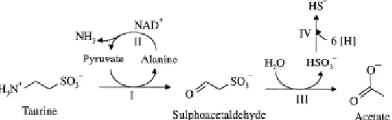
Bilophila wadsworthia is an important opportunistic pathogen
isolated from several anaerobic infections. It is a most interesting
bacterium as it belongs to the Desulfovibrionaceae family, although is
not able to reduce sulfate. This organism performs an interesting
type of respiration in which taurine serves as a source of sulfite, the
final electron acceptor, for the oxidation of short-chain fatty acids,
such as lactate or formate, produced by fermentative organisms
present in the human gut. We showed that H2 is also used as energy
source in a very efficient way by B. wadsworthia, which expresses up
to five hydrogenases in the presence of different electron donors. H2
is a very important source of energy for bacteria in the human gut
and the ability to use it efficiently most likely constitutes a virulence
A redução de sulfato é um processo metabólico muito antigo e
é responsável por mais de 50% da mineralização de carbono nos
sedimentos marinhos anaeróbios. Os organismos redutores de
sulfato (ORS) têm a capacidade de acoplar a redução de sulfato à
oxidação de compostos orgânicos, tais como o lactato ou o formato,
ou ainda ao hidrogénio molecular, de modo a obterem energia para a
síntese celular e crescimento. Não obstante os mais recentes
progressos, os mecanismos que permitem a conservação de energia
nos ORS continuam essencialmente desconhecidos. O formato e o
hidrogénio são dois metabolitos abundantes nos habitats dos ORS,
geralmente formados como produtos de fermentação por outros
organismos. No entanto, enquanto o papel do hidrogénio e das
hidrogenases no metabolismo anaeróbio tem sido estudado
intensivamente ao longo dos anos, o formato, por outro lado, não
tem sido alvo da mesma atenção enquanto metabolito igualmente
importante no metabolismo anaeróbio. Todavia, a situação tem
vindo a alterar-se, e o formato tem vindo cada vez mais a ser
reconhecido como um metabolito fundamental em vários processos
biológicos. A importância do hidrogénio para as bactérias redutoras
de sulfato (BRS) está bem patente no facto da maioria destes
microrganismos possuir múltiplas hidrogenases. Uma das enzimas
maioria das BRS, indicando assim que desempenham também um
papel importante no seu metabolismo.
O principal objectivo do trabalho apresentado nesta tese foi
contribuir para a clarificação das vias metabólicas das BRS,
principalmente do metabolismo do formato, através do estudo das
formato desidrogenases, enzimas fundamentais neste processo. A
função e a regulação da expressão das FDHs foi estudada em
Desulfovibrio vulgaris, um organismo modelo das BRS, cujo genoma
codifica para três FDHs. As potenciais vias do metabolismo
energético foram ainda estudadas através de experiências em que se
testou a transferência electrónica entre a FDH ou a hidrogenase de
[NiFe] e vários citocromos c de D. desulfuricans. De modo a perceber
quais as potenciais vias de conservação de energia que envolvem as
desidrogenases periplásmicas em diferentes ORS, foi feita uma
análise comparativa dos genes correspondentes às FDHs,
hidrogenases e citocromos c periplásmicos, em 25 genomas de ORS.
Nesta tese foi ainda estudado o metabolismo energético de Bilophila
wadsworthia, um reconhecido patogénio oportunista
filogeneticamente relacionado com as BRS.
O molibdénio (Mo) e o tungsténio (W) são dois elementos com
propriedades muito semelhantes. A formato desidrogenase foi a
primeira enzima em que se mostrou a presença de W, numa altura
em que este metal era apenas considerado um antagonista do Mo.
pertencentes ao domínio Archaea. Algumas enzimas de Mo têm a
capacidade de incorporar W e manter a sua actividade, enquanto
outras, ao substituir o Mo por W, são inactivadas. Formato
desidrogenases contendo Mo ou W no seu centro activo têm sido
reportadas em BRS. No trabalho apresentado nesta tese mostrámos
pela primeira vez evidência directa de regulação transcricional ou
pós-transcricional pelo Mo e W nas três FDHs de D. vulgaris. A
substituição de Mo por W no meio de cultura tem um efeito bastante
pronunciado na actividade da FDH, sobretudo quando o formato ou o
hidrogénio são usados como dadores de electrões.
Surpreendentemente, o efeito do W é ainda mais pronunciado na
presença do H2 do que do formato. Neste trabalho, usámos géis de
actividade, Western-blot, PCR em tempo real e isolamento das
proteínas, para mostrar que diferentes isoenzimas são expressas
durante o crescimento com Mo ou W, sugerindo a existência de um
mecanismo de regulação dependente dos metais para a expressão
das FDHs de D. vulgaris. Observou-se que para a FdhABC3, uma
enzima que contém uma subunidade citocromo c, existe uma elevada
selectividade para a incorporação de Mo, enquanto que para a
FdhAB, uma enzima heterodimérica, o processo de incorporação do
metal não é tão estritamente regulado, uma vez que tanto o Mo
como o W podem ser incorporados.
Com o objectivo de esclarecer a função das duas principais
diversos dadores de electrões foram analisados. As duas formato
desidrogenases solúveis são importantes para o crescimento em
formato na presença de Mo, enquanto que com W apenas a FdhAB
desempenha uma função, devido à repressão do gene fdhABC3. Os
dois mutantes, ΔfdhAB e ΔfdhABC3, apresentam deficiências no
crescimento em lactato/sulfato, o que constitui uma evidência do
envolvimento do ‘ciclo do formato’ neste processo. Por outro lado, os
dois mutantes cresceram de forma semelhante ao tipo selvagem em
hidrogénio/sulfato. Na ausência de sulfato, e na presença de H2/CO2,
as células de D. vulgaris produziram formato que resultou da redução
de CO2 pelas enzimas periplásmicas. Nós propomos que este
processo pode ser ambientalmente significativo, pois permite o
crescimento das bactérias redutoras de sulfato, através da oxidação
do hidrogénio acoplada à redução de CO2, em sintrofia com
organismos que consomem formato e são menos eficientes na
utilização do H2.
As vias de transferência electrónica envolvendo a FdhABC3 de
D. desulfuricans e vários citocromos c foram também estudadas. Os
resultados mostram que a FdhABC3 transfere electrões de forma
eficaz para todos os citocromos testados, embora as taxas de
redução mais elevadas tenham sido obtidas com o citocromo
monohémico c553, previamente designado como o parceiro fisiológico
da FDH. As taxas de redução do citocromo c3 do tipo I (TpIc3) e do
proteína testada e a presença de quantidades catalíticas de c553 ou
TpIc3 não mostraram ter uma influência significativa nas taxas de
redução. Neste trabalho mostrámos também que a FdhABC3 tem a
capacidade para reduzir lentamente o NhcA, um citocromo associado
a um complexo redox membranar, e ainda mais lentamente o Dsr,
um complexo membranar que contém um citocromo c que não
pertence à família do c3.
A análise genómica permitiu confirmar que a presença de
múltiplas FDHs é comum na maioria das BRS, e todos os organismos,
com excepção de duas archaea e um membro dos Clostridia, contém
pelo menos uma FDH periplásmica. Em relação aos citocromos c,
podemos reconhecer dois grupos distintos, um composto pelas
Deltaproteobacteria e pelo Thermodesulfovibrio yellowstonii, que se
caracteriza pela presença de um elevado número de citocromos c
multihémicos, e um segundo composto por membros das Archaea e
Clostridia, que possuem muito poucos ou nenhuns citocromos c. O
grupo dos organismos que contém citocromos c também possui um
maior número de hidrogenases e formato desidrogenases
periplásmicas, assim como complexos redox membranares, o que
sugere uma maior flexibilidade das vias energéticas.
A B. wadsworthia é um importante patogénio oportunista
isolado a partir de várias infecções anaeróbias. É uma bactéria
singular porque, embora pertença à família Desulfovibrionaceae, não
fonte de sulfito, o aceitador final de electrões, para a oxidação de
ácidos gordos de cadeia curta, como o lactato ou o formato,
produzidos por microrganismos fermentativos que habitam o
intestino humano. Neste trabalho mostrámos que o H2 é também
usado como fonte de energia de modo muito eficiente pela B.
wadsworthia, que expressa até cerca de cinco hidrogenases na
presença de diferentes dadores de electrões. O H2 é uma fonte de
energia muito importante para as bactérias que compõem a flora
intestinal humana e a capacidade para o usar eficientemente
constitui provavelmente um factor de virulência para a B.
ADH Alcohol dehydrogenase
ANME Anaerobic methanotrophic archaea
AOM Anaerobic oxidation of methane
AOR Aldehyde ferredoxin oxidoreductase
APS Adenosine phosphosulfate
ATP Adenosine triphosphate
BCA Bicinchoninic acid
BCIP 5-bromo-4-chloro-3-indolyl phosphate toluidine salt
C553 Monoheme cytochrome
ca. circa, approximately
CoA Coenzyme A
Cys Cysteine
ΔG⁰’ Gibbs free energy change under physiological standard conditions
DMB 3,3’-dimethoxybenzidene dihydrochloride
DMSO Dimethylsulfoxide
cDNA Complementary deoxyribonucleic acid
Dsr Dissimilatory sulfite reductase
DTT Dithiothreitol
EDTA Ethylenediaminetetraacetic acid
e.g. exempli gratia, for example
ETC Electron transport chain
FDH Formate dehydrogenase
Fd Ferredoxin
FHL Formate hydrogen lyase
FMDH Formylmethanofuran dehydrogenase
HmcA Cytochrome subunit from Hmc (High molecular mass cytochrome
c) complex
HPLC High performance liquid chromatography
IBD Inflammatory bowel diseases
ICP-MS Inductively coupled plasma mass spectrometry
i.e. id est, that is
IMG Integrated Microbial Genomes
MCD Molybdopterin cytosine dinucleotide
MGD Molybdopterine guanosine dinucleotide
Moco Molybdenum cofactor
MTC Maximum tolerable concentration
NADH Nicotinamide adenine dinucleotide, reduced form
NAD(P) Nicotinamide adenine dinucleotide (phosphate), oxidized form
NBT Nitro blue tetrazolium salt
PFL Pyruvate formate lyase
PFOR Pyruvate ferredoxin oxidoreductase
pI Isoelectric point
Pi Inorganic phosphate
PPi Inorganic pyrophosphate
PMSF phenylmethanesulfonil fluoride
PVDF Polyvinylidene fluoride
qRT-PCR Quantitative real time polymerase chain reaction
rRNA Ribossomic ribonucleic acid
SCFA Short chain fatty acid
Sec Selenocysteine
SDS-PAGE Sodium dodecyl sulfate polyacrylamide gel electrophoresis
SLP Substrate level phosphorylation
sp. / spp. Species (singular / plural)
SRB Sulfate reducing bacteria
SRO Sulfate reducing organisms
Tat Twin arginine translocation pathway
Tc Technetium
TCA Trichloroacetic acid
TMAO Trimethylamine N-oxide
TpIc3 Type I cytochrome c3
TpIIc3 Type II cytochrome c3
Tuco Tungsten cofactor
UV Ultraviolet
WP Widdel-Pfennig
ºC Degree Celsius
g Gram
h Hour
KDa Kilodalton
l litter
mg Milligram
min Minute
ml Milliliter
mM Millimolar
mol Mole
μM Micromolar
nM Nanomolar
nmol Nanomole
pM Picomolar
C
C
h
h
a
a
p
p
t
t
e
e
r
r
1
1
Sulfate Reducing Bacteria...11.1. An overview ...3
1.2. Taxonomy...5
1.2.1. Desulfovibrio spp. ...6
1.3. Energy metabolism ...8
1.3.1. Sulfate reduction ...10
1.3.2. Formate...14
1.3.3. H2...16
1.3.4. Lactate and pyruvate ...17
1.4. Metalloenzymes and electron carriers in SRO...20
1.4.1. Formate dehydrogenase...27
1.4.2. Hydrogenase ...32
1.4.3. Periplasmic c-type cytochromes...37
1.5. Syntrophic lifestyle ...40
1.6. Health, environmental and biotechnological impact ...45
1.7. A human pathogen related to Desulfovibrio spp.: Bilophila
wadsworthia...51
1.8. References ...53
C
C
h
h
a
a
p
p
t
t
e
e
r
r
2
2
Tungsten and Molybdenum regulation of Formatedehydrogenase expression in Desulfovibrio vulgaris Hildenborough 77
2.1. Summary ...79
2.3.1. Culture media, growth conditions and preparation of
cellular extracts ... 83
2.3.2. Activity and kinetic assays... 84
2.3.3. Sulfate reduction rates... 85
2.3.4. Gel electrophoresis ... 85
2.3.5. Protein and metal quantification ... 85
2.3.6. Western-blot analysis... 86
2.3.7. Quantitative Real-Time PCR ... 86
2.3.8. Protein purification ... 88
2.3.9. N-terminal determination ... 89
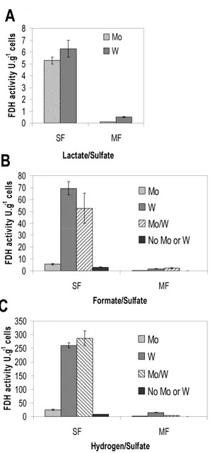
2.4. Results ... 89
2.4.1. Effect of Mo and W on D. vulgaris growth and FDH
activities ... 89
2.4.2. Analysis of FDHs by activity-stained native gels and
Western-Blot ... 93
2.4.3. FDH gene expression analysis by qRT-PCR... 95
2.4.4. Purification of the three FDHs and metal analysis... 99
2.5. Discussion... 103
2.6. Acknowledgments... 108
2.7. References... 109
C
C
h
h
a
a
p
p
t
t
e
e
r
r
3
3
Function of formate dehydrogenases in Desulfovibriovulgaris Hildenborough energy metabolism... 115
3.1. Summary ... 117
3.3.1. Construction of mutant strains...122
3.3.2. Culture media, growth conditions and preparation of
soluble fraction ...124
3.3.3. Analytical procedures ...124
3.3.4. Formate quantification in cell suspensions ...125
3.4. Results...126
3.4.1. Growth in formate/sulfate ...126
3.4.2. Growth in lactate/sulfate...129
3.4.3. Growth in H2/sulfate...131
3.4.4. Formate quantification in cell suspensions ...132
3.5. Discussion ...136
3.6. Acknowledgments ...144
3.7. References ...145
C
C
h
h
a
a
p
p
t
t
e
e
r
r
4
4
Periplasmic electron transfer between formatedehydrogenase and cytochromes c in Desulfovibrio desulfuricans
ATCC27774...151
4.1. Summary ...153
4.2. Introduction ...154
4.3. Materials and methods...158
4.3.1. Protein purification...158
4.3.2. Enzymatic measurements...159
4.3.3. Reductions with D. desulfuricans ATCC 27774 formate
hydrogenase... 160
4.4. Results and discussion... 160
4.5. Acknowledgments... 169
4.6. References... 170
C
C
h
h
a
a
p
p
t
t
e
e
r
r
5
5
A comparative genomic analysis of periplasmic electrontransfer pathways in Sulfate Reducing Organisms ... 175
5.1. Summary ... 177
5.2. Introduction... 177
5.3. Periplasmic formate dehydrogenases... 179
5.4. Periplasmic hydrogenases... 181
5.5. c-type cytochromes... 183
5.6. Conclusions... 188
5.7. Acknowledgments... 188
5.8. References... 189
C
C
h
h
a
a
p
p
t
t
e
e
r
r
6
6
Hydrogen as an energy source for the human pathogenBilophila wadsworthia... 193
6.1. Summary ... 195
6.2. Introduction... 196
6.3. Materials and methods ... 198
6.3.1. Cell growth and preparation of crude, soluble and
membrane extracts ... 198
6.3.2. Analytical methods... 200
6.3.5. Activity-stained gels...203
6.3.6. Western blot analysis...203
6.4. Results and discussion ...204
6.4.1. Growth experiments...204
6.4.2. Quantification of hydrogenase, formate
dehydrogenase and pyruvate oxidoreductase activities in cell
extracts ...207
6.4.3. Detection of hydrogenase and formate dehydrogenase
isoenzymes...211
6.4.4. Partial purification ...215
6.5. Conclusions ...216
6.6. Acknowledgments ...217
6.7. References ...218
C
C
h
h
a
a
p
p
t
t
e
e
r
r
7
7
Concluding remarks...223C
!
1.1. An overview. ...3
1.2. Taxonomy...5
1.2.1. Desulfovibrio spp.. ...6
1.3. Energy metabolism. ...8
1.3.1. Sulfate reduction. ...10
1.3.2. Formate...14
1.3.3. H2...16
1.3.4. Lactate and pyruvate. ...17
1.4. Metalloenzymes and electron carriers in SRO. ...20
1.4.1. Formate dehydrogenase...27
1.4.2. Hydrogenase. ...32
1.4.3. Periplasmic c-type cytochromes...37
1.5. Syntrophic lifestyle. ...40
1.6. Health, environmental and biotechnological impact. ...45
1.7. A Human pathogen related to Desulfovibrio spp.
Bilophila wadsworthia. ...51
1.1. An overview
The first sulfate-reducing organism (SRO) named Spirillum
desulfuricans was isolated more than a hundred years ago by
Martinus Beijerinck from a Dutch city canal in Delft [1]. Sulfide had
been previously acknowledged as a product of biological sulfate
reduction in aquatic habitats, but Beijerinck was able to enrich a
culture and obtain isolated colonies in agar whose distinctive
characteristic was the formation of a black iron precipitate in their
surroundings. By adding aerobic bacteria to the growth medium,
which consume the oxygen available, he was able to obtain
reproducible growth for the SRO, disseminating the idea that these
bacteria were strict anaerobes. Since Beijerinck time to our days
many discoveries have been made about this important group of
microorganisms and their ecological, physiological and metabolic
diversity [2-4]. The development of genomic, biochemical, genetic
and biogeochemical tools provided new insight into the diversity of
SRO, allowing the isolation of new members from previously
unknown habitats and revealing previously unknown energetic
strategies [4].
Sulfate reduction is a very ancient anaerobic metabolism,
whose first evidence dates back to 2.7 billion years ago, in the
Archean. About this time low concentrations of atmospheric O2 from
oxygenic photosynthesis caused the appearance of oxidized species
in certain zones, creating restricted sulfate-rich environments that
promoted biological sulfate reduction. However, only in Proterozoic,
2.3 billion years ago, high amounts of sulfide started accumulating
driven by an increasing sulfate concentration, and consequent higher
rate of biological sulfate reduction, establishing a sulfidic ocean until
about 0.5 billion years, when a major oxidation event in the Earth’s
atmosphere lead to deep-water oxygenation [5-8].
SRO have been found in a wide diversity of anaerobic habitats
from marine and freshwater sediments to the gastrointestinal tract
of animals, including humans [9-12]. They are particularly abundant
in marine sediments were sulfate concentration is high (28mM), and
they play a major role in linking the sulfur and carbon cycles by their
ability to use sulfate in a dissimilatory way. More than 50% of the
organic matter mineralization in marine sediments is due to sulfate
reduction [13-15]. SRO are able to couple the reduction of sulfate to
the oxidation of organic compounds or molecular hydrogen in order
to obtain energy for cell synthesis and growth [4]. Sulfate reduction
metabolism results in the production of high amounts of sulfide,
which serves as electron donor to a variety of aerobic or anaerobic
chemotrophic and anoxygenic phototrophic microorganisms that
oxidize it to elemental sulfur and sulfate, creating a cycle of sulfur
species transformations [16, 17]. In man-made environments where
SRO also thrive, accumulation of sulfide may constitute a problem
due to the toxic and corrosive nature of this compound. Biocorrosion
poses a significant threat to many industries, mainly oil industry [18].
Sulfide, produced by sulfate-reducing bacteria (SRB) present in the
human larg intestine, has also been implicated in inflammatory bowel
diseases because of its toxic effects to epithelial colonic cells [19].
Nevertheless, SRO are drawing increased attention because of their
potential role in bioremediation, due to their ability to grow with
diverse pollutant compounds [2]. Given the economical, health and
environmental importance of SRO it is essential to further
understand their metabolism in order to control their undesired
activity or to take advantage of all their potential.
1.2. Taxonomy
The group of SRO is heterogenous since it gathers members
from different phylogenetic lineages and with a wide metabolic
diversity, which have in common the ability to use sulfate as terminal
electron acceptor. As mentioned before sulfate reduction is a very
ancient metabolism, and several events of lateral gene transfer can
help explain the dispersion of this ability by phylogenetic unrelated
microorganisms [20]. The sets of genes coding for two key enzymes
of sulfate reduction, adenosine phosphosulfate (APS) reductase and
dissimilatory sulfite reductase (Dsr), were probably laterally
transferred among different lineages in several moments of SRO
Comparative analysis based on 16S ribosomal RNA (16S rRNA)
yields seven phylogenetic lineages in SRO: Deltaproteobacteria
(families Desulfovibrionaceae, Desulfomicrobiaceae,
Desulfo-bacteriaceae, Desulfohalobiaciae, Desulfobulbaceae and
Syntrophobactereacea), Nitrospirae (genus Thermodesulfovibrio),
Thermodesulfobacteria (genus Thermodesulfobacterium), Clostridia
(genera Desulfotomaculum, Desulfosporosinus and
Desulfos-poromusa), Thermodesulfobiaceae (genus Thermodesulfobium) and
Archaea (genus Archaeoglobus from Euryarchaeota; genera
Caldivirga and Thermocladium from Crenarchaeota). SRO belonging
to Deltaproteobacteria are Gram-negative, usually mesophilic
bacteria while Clostridia comprise Gram-positive, spore-forming
bacteria. Genera Thermodesulfovibrio, Thermodesulfobacterium and
Thermodesulfobium include only thermophilic bacteria [3, 23].
Several genomes of SRO have been recently sequenced, mostly
from Deltaproteobacteria [24-27]. In fact, most of SRO described
until now, belong to this class.
1.2.1. Desulfovibrio spp.
Research on SRO metabolism has been carried out mostly with
the genus Desulfovibrio, since these organisms are easily
manipulated in laboratory conditions. Desulfovibrio spp. are
mesophilic Gram-negative Deltaproteobacteria, belonging to the
ability to form spores and which are characterized by the presence of
desulfoviridin, a type of dissimilatory sulfite-reductase. Desulfovibrio
spp. use organic acids, alcohols or molecular hydrogen as substrates
for growth with reduction of sulfate, and are incomplete oxidizers
since they are not able to oxidize acetate. It is the only genus among
the SRO that can be found in the digestive tract of animals and
humans, and two species, D. piger and D. fairfieldensis, have actually
never been isolated from habitats outside the human body [28-30].
Desulfovibrio spp. have been considered as strict anaerobes, but they
are often found in temporary oxic zones possessing the necessary
enzymes to cope with oxygen toxicity. It was recently shown that a
strain is able to grow at atmospheric oxygen levels and it was
previously documented that in D. vulgaris and D. desulfuricans
oxygen reduction can be coupled with proton translocation and
energy conservation [31-34].
D. vulgaris Hildenborough is a model organism for SRO. It is a
well studied strain of Desulfovibrio and the first SRO to have its
genome sequenced [35]. Its genome codes for several periplasmic
enzymes like hydrogenases, one of the most studied enzymes, and
also several formate dehydrogenases (FDH), which have received less
attention. Given the number of FDHs, formate metabolism may have
a central role in energy-conserving mechanisms for D. vulgaris, and
understanding more about the role played by each FDH would be of
D. desulfuricans ATCC27774 is another well studied strain of
Desulfovibrio spp., whose genome was also recently sequenced. It is
one of the few strains able to use nitrate in a dissimilatory way,
besides sulfate [36, 37].
Desulfovibrio spp. are characterized by possessing a high
number of c-type cytochromes, and several types are widespread in
members of this genus, of which the most abundant and well studied
is the type I cytochrome c3 (TpIc3) [38]. However, some c-type
cytochromes have only been isolated from D. desulfuricans, making it
an interesting organism to study the alternative electron transfer
pathways that occur in the periplasm, linking substrate oxidizing
enzymes and membrane-bound electron carriers [29, 39, 40].
1.3. Energy metabolism
Diverse SRO can oxidize more than a hundred different
substrates, which include, among others, lactate, formate, ethanol,
malate, sugars, amino acids, aromatic hydrocarbons or alkanes and,
in addition to sulfate, they may also reduce sulfite, thiosulfate, sulfur,
nitrate, iron (III), fumarate and even oxygen. Molecular hydrogen (H2)
can also be used as sole electron donor with CO2 as the only carbon
source (autotrophy), or together with acetate
(chemolithoheterotrophy) [3, 4, 41]. Some SRO are complete
oxidizers, being able to degrade organic matter completely to CO2,
acetate oxidation can be accomplished by two ways: a modified citric
acid cycle or via the acetyl-coenzyme A (CoA) pathway [42]. The
acetyl-CoA or Wood-Ljungdhal pathway in SRO is similar to the one
described for methanogenic and acetogenic organisms, and it can
function to fix CO2 in autothrophic growth or it can be reversed in
order to oxidize acetate and obtain energy [43].
The SRO respiratory chain and the components involved in
energy conservation are still poorly understood, although several
membrane complexes have been isolated that probably contribute to
this function during sulfate respiration [44]. One striking
characteristic of the SRO respiratory chain is the cytoplasmic
localization of all terminal reductases, which prevents their direct
involvement in the establishment of a proton gradient across the
membrane [29]. Given the diversity of SRO, it is likely that several
respiratory chains exist, according to the electron donor used, which
makes it hard to propose one single model for electron transport,
except for the last reactions of sulfate activation and reduction which
will be common to all pathways [4].
Studies regarding energy conservation in SRO, and the proteins
involved in the process, have been carried out mainly in Desulfovibrio
spp. In the following sub-sections the last steps of sulfate reduction,
and the electron transfer chains involving formate, H2 and lactate as
1.3.1. Sulfate reduction
Sulfate (SO42⁻) is transported across the membrane by symport
with protons in freshwater species, or with sodium ions (Na⁺) in
marine species [45-47], although there is at least one exception since
Desulfomicrobium baculatum, a freshwater sulfate reducer was
shown to transport sulfate with Na⁺ ions and not H⁺ [48]. SRO express
a constitutive electroneutral transport system with a cation:sulfate
symport ratio of 2:1. However, when growing under sulfate
limitation, marine and freshwater SRO express a high-accumulating
and electrogenic sulfate uptake sytem with a symport ratio of 3:1
[49-51]. Because two protons leave the cell with the end product of
sulfate reduction, hydrogen sulfide (H2S), by simple diffusion, the net
ATP consumption will be ⅓ATP, if a stoichiometry of 3H⁺/ATP is
assumed [47]. In Na⁺/sulfate symport the Na⁺ gradient is generated
by a Na⁺/H⁺ antiporter [52].
Sulfate is a very stable compound which first needs to be
activated by reaction with ATP, before it can be used by the cell. This
first reaction is common to dissimilatory and assimilatory sulfate
reduction and is catalyzed by ATP sulfurylase in the cytoplasm,
forming adenosine phosphosulfate (APS) and inorganic
pyrophosphate (PPi) (Eq. 1). Because PPi formation is
thermodynamically unfavourable the reaction is completed by a
pyrophosphatase that hydrolyzes PPi (Eq. 2) [2, 29, 53, 54]. In a few
SRO, a membrane-bound proton-pumping pyrofosfatase is present,
Eq.1 ATP + SO42⁻ → APS + PPi ΔG⁰’ = +46 kJ/mol
Eq.2 PPi + H2O → 2Pi ΔG⁰’ = -22KJ/mol
APS is the first electron acceptor in the respiratory chain being
reduced by APS reductase to sulfite (SO32⁻), which is subsequently
reduced to sulfide (S2⁻) by dissimilatory sulfite reductase (DsrAB)
[29]. APS reductase is a soluble protein extremely abundant in the
cell, constituting 2 to 3% of all soluble proteins in SRO of
Desulfovibrio genus. It was already isolated from several
Desulfovibrio spp. and also from Archaeoglobus fulgidus showing high
similarity in its physical and chemical properties [56-60]. A membrane
complex, quinone-interacting membrane-bound oxido-reductase
(Qmo) is believed to be the electron donor to APS reductase,
transferring electrons from the membrane menaquinone pool to the
cytoplasm [61, 62]. The last step of sulfite reduction involves the
transfer of six electrons to form sulfide. It was proposed previously
that this reduction did not occur directly but instead in three steps
with the formation of thiosulfate and trithionate as intermediates
[63, 64]. Sulfite reduction, as mentioned before, is catalyzed by
sulfite reductase. There are four major types of dissimilatory sulfite
reductases, distinguished by spectroscopic and molecular
characteristics. Desulfoviridin, a green protein characterized by
in Desulfovibrio spp. and a few other organisms [4]. Recently, the
structure of D. vulgaris desulfoviridin (DsrAB) was solved, showing
the involvement of a third protein, DsrC, in sulfite reduction, and it
was proposed a four-electron reduction, instead of six, occurs with
the formation of a sulfur (S0) intermediate that is transferred to DsrC
to form a persulfide. Once DsrC is released from DsrAB the persulfide
is reduced forming sulfide (S2⁻) and DsrC oxidized, which can be again
reduced by the membrane complex DsrMKJOP in a cyclic reaction
(Figure 1.1) [65]. It is thought that this membrane complex transfers
two electrons for sulfite reduction through DsrC, possibly
contributing to proton translocation while the other four electrons
1.3.2. Formate
In anaerobic environments formate is a common growth
substrate and its reversible oxidation to CO2 is catalyzed by FDH (Eq.
3).
Eq.3 Formate ↔ CO2 + H+ + 2e⁻
Formate is an important interspecies electron transfer
metabolite in anaerobic mixed communities [3] where it is formed by
fermentative organisms and used for growth by SRO or methanogens
(see section 1.5 on Syntrophic lifestyle).
Formate can be produced by CO2 reduction, a reaction
catalyzed by FDH, or as an end product of bacterial fermentation
[67]. Anaerobic facultative microorganisms, capable of glucose
fermentation, such as Escherichia coli, produce formate as an end
product in a reaction catalyzed by pyruvate-formate lyase (PFL),
yielding also acetyl-CoA used for substrate-level phosphorylation
(SLP) [68]. Formate is also an intermediate in the acetyl-CoA pathway
for acetate oxidation to CO2, or in the reverse way for biosynthesis of
purines, aminoacids and acetate, in bacteria that use
tetrahydrofolate as C1 carrier [69]. In methanogens and
sulfate-reducing Archaea, however, formate is not an intermediate in C1
metabolism because these organisms lack tetrahydrofolate, although
formate may be used for growth and as a source of CO2, which is
Most SRO have periplasmic FDHs that oxidize formate.
Energetically, as an electron donor for sulfate reduction (Eq.4),
formate is equivalent to H2 since the redox potential of the couples
2H⁺/H2 and HCO3⁻ /HCOO⁻ is similar (E⁰ ≈ -0.41V) [4].
Eq.4 4HCO2⁻ + SO42⁻ → 4HCO3⁻ + HS⁻ ΔG⁰’= -146.6 KJ/reaction
With formate oxidation a proton gradient is established and the
resulting electrons are transferred through periplasmic electron
carriers and membrane-bound complexes to reduce sulfate in the
cytoplasm (for a more detailed discussion on the periplasmic electron
transfers in SRO involving FDH see Chapter 5 of this thesis). Because
formate oxidation and sulfate reduction occur in different sides of
the membrane, energy is conserved through this process. Formate is
also an intermediate in the acetyl-CoA pathway for SRB that oxidize
acetate to CO2 or that grow autotrophically with CO2 as carbon
source (e.g. Desulfotomaculum acetoxidans and Desulfobacterium
autotrophicum) [70-72]. In D. vulgaris, a ‘formate cycling’ model was
suggested, analogous to H2 and CO cycling, as an energy conserving
mechanism in face of the genome sequence that encodes a PFL and
three periplasmic FDHs [35, 73, 74].Formate metabolism in SRB,
mainly D. vulgaris will be further discussed on Chapter 3 of this
1.3.3. H2
Hydrogen is a major energy source for SRO in their natural
habitats and is also an intermediate in several of their metabolic
pathways. It is used as energy source, with acetate and CO2 as carbon
source (chemolithoheterotrophy) or only with CO2 (autotrophy). H2
formation as a product of fermentative metabolism makes it an
important interspecies electron transfer in anaerobic syntrophic
communities. The reversible oxidation of H2 is catalyzed by
hydrogenases, a very intensively studied group of enzymes in SRO,
mainly in Desulfovibrio genus [4, 29].
H2 is also a major fermentation product in the human gut and
the ability of some pathogenic bacteria, such as Helicobacter pylori or
Salmonella enterica to efficiently use H2 as an energy source
constitutes a virulence factor [75, 76]. This subject is further
discussed in Chapter 6, where the energy metabolism of the
opportunistic pathogen B. wadsworthia was addressed.
SRO can obtain energy from H2 oxidation with dissimilatory
sulfate reduction (Eq. 5) by vectorial electron transport.
Eq. 5 4H2 + SO42⁻ + H⁺ → HS⁻ + 4H2O ΔG⁰’= -151.9 KJ/reaction
In this mechanism the eight electrons, resulting from the
oxidation of four H2 molecules in the periplasm, are used for
chemiosmotic ATP production, while the electrons are transferred to
those protons is required for electrogenic symport of sulfate to the
cytoplasm (see section 1.3.1 on Sulfate reduction), only seven remain
available for ATP. Considering a ratio of 3 H⁺/ATP, seven protons
would then yield 2⅓ mol of ATP, from which 2ATP are required for
sulfate activation, giving a net ATP gain for H2 oxidation of ⅓ mol ATP
[46]. Some growth experiments have contradicted this value giving a
higher net gain of ATP for each mol of sulfate reduced [4]. This
suggested the existence of an additional mechanism for proton
translocation like vectorial proton transport through proton-pumping
redox proteins or energy-conserving redox loops involving
menaquinones. Several membrane complexes have been isolated
and proposed to be involved in menaquinone cycling like Dsr and
Qmo from D. desulfuricans, or more recently the quinone-reductase
complex (Qrc) from D. vulgaris [61, 66, 77].
1.3.4. Lactate and pyruvate
Lactate is a metabolic product of several fermentative bacteria
and it can be used as a substrate for growth and energy by most SRO
in natural conditions. Pyruvate, on the other hand, is unlikely to be a
major product of fermentative metabolism but is a cellular
intermediate in lactate oxidation and other biosynthetic pathways,
and is widely used for SRO cultivation in laboratory. These two
organic acids can be oxidized completely to CO2 by some SRO or
L- and D-lactate oxidation to pyruvate is catalyzed by NAD(P)+
-independent lactate dehydrogenases (LDH), enzymes that are
membrane-associated and whose activity was shown in several
Desulfovibrio spp. [4, 78, 79]. D-LDH was purified from D.
desulfuricans and Archaeoglobus fulgidus, a hyperthermophilic SRO
[80, 81]. Pyruvate is further oxidized by pyruvate:ferredoxin
oxidoreductase (PFOR) to acetyl-CoA. This ‘energy-rich’ compound is
then converted to acetate by phosphotransacetylase and acetate
kinase, allowing SLP [82, 83]. Because one ATP is formed by lactate
oxidation, and two molecules of lactate are oxidized by each reduced
sulfate molecule, the net gain by substrate-level phosphorylation is
zero, since two ATPs are needed to activate sulfate. So, in order to
obtain energy from lactate oxidation with sulfate, an additional
mechanism is necessary [4]. Regarding this question Odom and Peck,
in 1981, proposed the hydrogen-cycling model for growth on lactate
[84]. In this model, electrons generated by oxidation of lactate and
pyruvate are converted to hydrogen by a cytoplasmic hydrogenase
and released in the periplasmic space were hydrogen is again
reoxidized with electrons being transferred to the cytoplasm, thus
forming a proton gradient which is used for additional ATP synthesis.
The main criticicisms to this theory are the non existence of a
cytoplasmic hydrogenase in many SRO and the fact that hydrogen
formation from lactate oxidation is an energetically unfavourable
process [29, 79, 85]. Another model was proposed by Lupton et al.
cytoplasm are transported through membrane carriers to sulfate
reduction while protons are translocated to the periplasm (vectorial
electron transport). In this case a cytoplasmic hydrogenase, or other
enzyme, serves only to regulate electron flow and the levels of
reduced electron carriers (e.g. ferredoxins), which results, as a side
reaction, in the production of H2 [86, 87]. Recently, evidence was
provided in D. vulgaris for cycling of another reduced intermediate,
CO, when the organism grows with lactate and sulfate [74]. The
formation of H2 from lactate oxidation can also be important for
syntrophic associations and it can be an adaptation of Desulfovibrio
spp. to survival in methanogenic environments with low or no
sulfate. In this case, the removal of H2 by other organisms lowers the
gas partial pressure making H2 production from lactate oxidation a
thermodynamically favorable process [85] (see section 1.5 on
1.4. Metalloenzymes and electron carriers in SRO
The wide diversity of reactions catalyzed by proteins cannot be
accounted only by the different reactive groups of amino acid
residues, but resides also in the presence of cofactors, organic and/or
inorganic groups that bind to the proteins and take part in catalysis
[88]. Metal ions are the most common cofactors since almost half of
known enzymes require their presence [89]. The use of specific
metals by enzymes reflects not only their chemistry but also their
evolutionary history. The biggest shift in metal availability happened
2.3 billion years ago with the rise of atmospheric O2 which influenced
the use of trace metals for biological activities. That use can be seen
by analyzing genomes and proteomes of present organisms from
each domain of life [89, 90]. Iron (Fe) was shown to be the
predominant redox metal in biological systems, which is probably
due to its high concentration in the ocean during the early evolution
of living organisms [89]. When the ocean was still an anoxic
environment, iron was found in its soluble form and available for
biological use, but deep water oxygenation caused iron to precipitate
lowering its concentration. On the other hand, other metals (e.g.
zinc) that were scarce became more abundant and readily usable by
organisms that appeared latter in the course of evolution [90, 91]. Fe
and molybdenum/tungsten (Mo/W) are among the most common
metals in oxidoreductases whose structure is known [89], and
hydrogen metabolism, their main features will be briefly discussed
below.
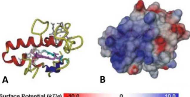
Fe may be found associated with inorganic sulfur and cysteine
residues forming [Fe-S] clusters, which are one of the oldest and
more versatile cofactors, ubiquitously present in living organisms,
and being able to perform several functions: as electron carriers
between the enzymes active site and their redox partner, as catalysts
and even as O2 or Fe sensors [92].
[Fe-S] clusters can have different
compositions and the low
potential [2Fe-2S]2+/+ and
[4Fe-4S]2+/+ clusters (Figure 1.2) are
the most common, and present at
90% of analyzed unique FeS protein folds, while high potential
clusters like the [3Fe-4S]1+/0 are less represented [93]. Although these
simple inorganic cofactors can be assembled spontaneously in vitro,
in the cell the process is catalyzed by other proteins and rather
complex, involving several possible different systems in prokaryotes
and eukaryotes [94]. [Fe-S] clusters are thought to precede life itself
and to be at the origin of the first chemical reactions that drove the
origin of life in the primitive, anoxic ocean [95]. Besides being
associated with S to from [Fe-S] clusters, Fe can also be found as part
of heme, a cofactor that exists bound to several proteins with
essential roles in energy-transduction processes, like respiration and
photosynthesis, gas transport and storage, catalysis or gene
regulation. Heme is composed by a ferrous iron (Fe2⁺) coordinated by
a porphyrin macrocycle, and depending on the nature of the
substituents different types of hemes can exist. The most common
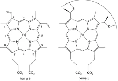
are hemes b and c, very similar to each other but, while heme b
bounds non-covalently to the protein, heme c bounds through two
covalent thioether bonds formed between cysteine side chains and
the heme vinyl groups (Figure 1.3) [96]. The covalent attachment of
heme may be important to confer greater stability to the protein and
to allow a more dense packaging of hemes, facilitating rapid electron
transfer [97]. One of the largest families of heme-containing proteins
is the cytochrome c family. These proteins are mainly involved in
electron transfer or enzyme catalysis, they may contain multiple
heme groups and many times they are found assembled with other
subunits forming protein complexes. Some organisms possess an
incredible high number of cytochromes c, like Geobacter
sulfurreducens or to a less degree, Desulfovibrio spp., which is
thought to confer to these organisms an increased energetic
Mo and W are transition metals that have similar chemical and
physical properties but, while Mo involvement in fundamental
biological processes is known for many years, W role in biology is a
more recent discovery [100]. Mo is distributed among all domains of
life, being an essential trace metal for microorganisms, plants and
animals, while W is present only in some bacteria and archaea (Figure
1.4) [101]. The enzymes containing Mo or W in their active site have
essential roles in carbon, nitrogen and sulfur metabolism [102].
The ability of Mo and W to be redox-active under physiological
conditions, ranging from oxidation states VI and IV, allows the Mo or
W-containing enzymes to
catalyze
oxidation-reduction reactions with
transfer of an oxygen
atom and an exchange of
two electrons at low
potential [103]. However,
there are exceptions,
such as in the case of
acetylene hydratase, a
W-containing enzyme, that does not catalyze a redox reaction but
instead the hydration of acetylene to acetaldehyde, or the cases of
FDH and formylmethanofuran dehydrogenase (FMDH) that catalyze
reactions which do not involve transfer of oxygen atoms [101, 104].
Mo and W are present at the active site of enzymes
coordinated to an organic cofactor, pyranopterin, originating the Mo
cofactor (Moco) or W cofactor (Tuco). The basic structure of
pyranopterin consists of a modified pterin containing a dithiolene
moiety, which quelates the metal ion with its two sulfur atoms. In
eukaryotes the pyranopterin is found in the simple monophosphate
form, while in prokaryotes the phosphate group is linked to a
nucleotide, usually cytosine (molybdopterin cytosine dinucleotide –
MCD) or guanosine (molybdopterin guanosine dinucleotide – MGD)
[104]. Mo/W containing enzymes are divided in four families (Figure
1.5): 1) xanthine oxidase family, 2) sulfite oxidase family, 3)
dimethylsulfoxide (DMSO) reductase family and 4)
aldehyde:ferredoxin oxidoreductase (AOR) family, according to the
type of reaction catalyzed and the structure of the active site [104,
105]. From these families only the third includes molydo- and
tungstoenzymes, while the enzymes belonging to the first two always
contain Mo and the forth, also called the family of ‘’true
tungstoenzymes’’, includes the five AORs from Pyrococcus furiosus
[101].
The fact that some enzymes incorporate only Mo, while others
only W, and some incorporate both metals, probably reflects the
availability of these elements in nature and their specific chemical
properties, such as the lower redox potential of the W(IV)/W(VI)
couple that favors reduction of substrates instead of oxidation [100,
101]. In the primitive Earth, before the major oxygenation event that
caused deep-water oxygenation, the ocean was predominantly
sulfidic due to the accumulation of H2S from biological and
chemical/geological sources (see above, section 1.1). In this
primordial ocean, Mo and W were present in the form of sulfides,
MoS42- and WS42-, respectively. However, while W sulfides are
relatively soluble, Mo sulfides are highly insoluble, which determined
that in the primitive, anaerobic ocean W was more bioavailable than
Mo [91]. As oxygen began to accumulate in the oceans, Mo and W
sulfide salts were oxidized and gradually replaced by the respective
oxoanions, MoO42- and WO42-. The very high solubility of Mo
oxoanion caused a major increase in Mo availability, while the W
concentration remained low, similar to the values found in the
primitive ocean. The changing bioavailability of both metals has
certainly conditioned protein evolution, and it is thought that
W-containing enzymes are more ancient than their Mo counterparts. W
is found in the active site of enzymes mainly in anaerobic bacteria
and archaea, and is present at very low concentrations in marine
environments. The exception occurs at anaerobic, sulfide-rich marine
habitats, such as hydrothermal vents and black smokers, where W is
mainly found in the form of sulfide, being more soluble, and thus
more available for microorganisms that live in this environment [100,


![Figure 1.8. Ribbon diagram of the three-dimensional structure of [NiFe]-hydrogenase from D](https://thumb-eu.123doks.com/thumbv2/123dok_br/15770057.641193/65.748.356.605.498.703/figure-ribbon-diagram-dimensional-structure-nife-hydrogenase-d.webp)