Ciências
Gellan microspheres application for capture or
purification of plasmid DNA vaccine
Diana Vanessa Duarte Gomes
Dissertação para obtenção do Grau de Mestre em
Biotecnologia
(2º ciclo de estudos)
Orientadora: Profª. Doutora Ângela Maria Almeida de Sousa
Coorientador: Prof. Doutor Luís António Paulino Passarinha
“Anything is possible when
you have the right people
there to support you.”
Misty Copeland
To my amazing family.
Acknowledgements
Firstly, I would like to express my profound and sincere gratitude to Professor Doctor Ângela Sousa and Professor Doctor Luís Passarinha for all the support, patience, and guidance, for the scientific learning that you were able to provide me. You have always been ready to lend a hand, good advisors, so I need to say that it was a great pleasure and an enormous satisfaction to work with you this past year. And also, to Professor Doctor Diana Costa for kindly providing the PEI used in this project.
I would also like to acknowledge the Health Sciences Research Centre and the University of Beira Interior, for providing the adequate conditions that allowed me to develop this work. Secondly, I would like to thank the Biotechnology and Biomolecular Sciences group, especially to Margarida Grilo, Jorge Ferreira and Margarida Almeida for their help, advice and knowledge shared with me.
To my friends since the beginning of my journey here in Covilhã, Leandro Madureira, Rita Vieira da Costa, Carolina Batista, Ana Nunes, Tiago Pereira, João Diogo, João Valente, Sofia Oliveira, André Cunha, Alexandre Santos a big thanks for the amazing years spent here, all the talks, funny moments, laughs, arguments, friendships, big decisions and especially to give me a second home here in Covilhã.
A big thanks to my lab colleagues and friends from this past year, Adriana Pinto, Diana Pereira, Raquel Neves, Rita Carapito, Micaela Riscado, Rita Proença, and the new ones, Pedro Vicente, Sofia Oliveira and Pedro Ferreira for all the advices, support, the talks about nothing and everything at the same time, all the shared breaks and also the “not so good moments”, because this has made us a family that I know I can always count on. THANKS!
Finally, to my amazing family for all the support, help provided and also for always believe that I was capable. To my parents, Clara Martins and Rui Gomes, my siblings, Catarina Gomes and Rui Gomes and also my grandparents, Adosinda Duarte e António Martins a huge thank you for always being there for me, for making every effort possible to give me the best, for all dedication and love. I would not have accomplished everything I have without you, I’m so grateful. I love you!
Resumo Alargado
O vírus do papiloma humano (HPV) é um vírus sexualmente transmissível e a persistência da sua infeção é considerada a maior causa para o desenvolvimento do cancro do colo do útero. Este potencial oncogénico do HPV está diretamente relacionado com a expressão das oncoproteínas E6 e E7, visto que estas têm a capacidade de interferir na desregulação do ciclo celular, indução da apoptose, entre outros fenómenos biológicos. O cancro do colo do útero corresponde à 4º maior causa de morte nas mulheres a nível mundial. No entanto, com as evoluções alcançadas ao nível científico tem sido possível melhorar e desenvolver novas terapias à base de DNA para tratar vários problemas de saúde como cancro e doenças genéticas. Sendo uma delas, as vacinas de DNA, uma vez que estas têm a capacidade de despoletar todos os tipos de imunidade desejada, através da resposta celular e resposta humoral, evitando a evolução da doença, o que é uma vantagem em relação às vacinas convencionais. Os vetores de DNA plasmídico (pDNA) codificantes de determinados antigénios têm sido muito explorados como vacinas de DNA, uma vez que apresentam baixa toxicidade e são mais simples de desenvolver. O processo biotecnológico de preparação do biofármaco de pDNA compreende etapas sequenciais de produção, clarificação e purificação com objetivo de obter a isoforma superenrolada (sc) com o grau de pureza recomendado pelas agências reguladoras, já que é considerada a conformação de pDNA biologicamente ativa. Contudo, este processo é bastante dispendioso para a indústria farmacêutica e apresenta um impacto ambiental acentuado, devido ao uso de elevadas quantidades de solventes orgânicos (isopropanol) e sais caotrópicos (sulfato de amónio e sulfato de sódio). Desta forma, é fundamental desenvolver novas estratégias de captura ou purificação do pDNA sc de modo a simplificar a amostra e evitar o uso de determinados reagentes, tornando o processo mais “green” e económico.
A goma gelana é um exopolissacárido microbiano aniónico que tem a capacidade de, em determinadas condições (presença de catiões, concentração de polímero, temperatura), alterar a sua estrutura conformacional e formar uma rede tridimensional dando origem a um gel termorreversível com diferentes características estruturais e propriedades, consoante o objetivo desejado. A gelana apresenta diversas aplicações na indústria alimentar (espessante, gelificante), farmacêutica (formulações oftálmicas), cosmética (loções, cremes), biotecnológica (substituinte do agar), e devido, a características como a sua biocompatibilidade, hidrofilicidade, porosidade e versatilidade tem vindo a ser explorada nos últimos anos como matriz cromatográfica. Recentemente foi explorada e otimizada por desenho experimental a formulação de microsferas de gelana através do método de emulsão água-em-óleo e reforço com iões divalentes para capturar proteínas em função da sua carga ou afinidade.
Assim, o objetivo central deste trabalho foi formular as microsferas de gelana e desenvolver uma estratégia de captura de pDNA sc a partir de um lisado bruto de Escherichia coli (E. coli) aplicando o método de batch. Para tal, a amostra de lisado foi obtida através de uma fermentação em E. coli, com posterior lise alcalina. As microesferas de gelana foram preparadas pelo método de emulsão água–em–óleo, reforçadas com alguns iões divalentes, cobre, níquel, zinco, cobalto, e algumas formulações foram posteriormente funcionalizadas com o polímero de polietilenoimina (PEI). O menor diâmetro das formulações foi obtido com 1.41 % de uma solução aquosa de gelana, previamente aquecida a 90 ºC que foi gotejada através de uma seringa para uma solução de óleo aquecida anteriormente a 100 ºC sob constante agitação de 750 rpm a uma velocidade de 75 µL/min. Ambas as formulações foram caracterizadas relativamente ao diâmetro médio (microscopia de semiótica), à morfologia (SEM), à carga global (potencial zeta) e à composição elementar (EDX e FTIR). Dos resultados obtidos quanto à morfologia, ambas as topologias de microesferas apresentam uma forma esférica e consistente. A estabilidade das duas formulações foi avaliada pela medição do diâmetro médio ao longo de vinte dias e ambas se apresentam estáveis ao longo do período em análise. Em relação ao diâmetro médio, as microesferas funcionalizadas com PEI apresentam um menor diâmetro que as microesferas reforçadas com cobre, o que pode ser devido à formação de uma ligação de coordenação entre o PEI e o cobre tornando a sua estrutura mais compacta. Esta ligação pode ter também um efeito na composição elementar pois, as microesferas reforçadas com cobre apresentam cerca de 9 % de cobre e as que foram funcionalizadas com PEI, apresentam 17 % de aminas e apenas 0.62 % de cobre. As microesferas reforçadas com cobre apresentam uma carga superficial de cerca de – 5 mV e as que que foram funcionalizadas com PEI, apresentam uma carga superficial de cerca de + 5 mV.
As estratégias de captura desenvolvidas tiveram por base a interação entre os iões metálicos reticulados nas microesferas e o pDNA presente no lisado celular, uma vez que a carga negativa do polímero de gelana repele os ácidos nucleicos. Após inúmeros estudos de otimização, as melhores condições de ligação do pDNA sc às microesferas de gelana reticuladas com cobre foram obtidas a pH 5.0 e a eluição com 200 mM NaCl em 10 mM Tris-HCl, 1 mM EDTA, pH 8.0, permitindo assim recuperar 15.61 % de pDNA sc com 2.42 % de pureza. Adicionalmente, outra estratégia desenvolvida para melhorar a captura de pDNA consistiu na precipitação do lisado de E. coli com 2,5 M de sulfato de amónio. Ao eliminar a maior parte das impurezas (RNA e proteínas) a quantidade de pDNA sc capturado aumentou para 32.41 % com um grau de pureza de 12.43 %. Por fim, as microesferas de gelana foram funcionalizadas com PEI, com objetivo de melhorar a captura de pDNA, tendo em conta o aumento de grupos funcionalizados para interagirem com DNA na superfície das microesferas. Nas microsferas funcionalizadas com PEI, o passo de ligação foi realizado com um tampão de 10 mM MES a pH 5.0 e a eluição com 200 mM NaCl em 10 mM Tris-HCl, 1 mM EDTA, pH 10.5. Nestas condições, foi possível a captura total do pDNA presente na amostra de lisado e uma recuperação/eluição 88.09 % de pDNA sc. Contudo, também ocorreu a retenção de grande parte do RNA da amostra que resultou num
decréscimo do grau de pureza para 3.18 %. Assim sendo, se o objetivo principal for a captura total de pDNA de lisados brutos sem recorrer a sais ou solventes orgânicos, a estratégia que apresenta grande potencial é aquela em que as microesferas foram funcionalizadas com PEI. Por outro lado, se o objetivo principal for a captura de pDNA com um grau de pureza mais elevado, é recomendado realizar um passo prévio de tratamento com sulfato de amónio antes do passo de captura, sendo que neste se pode aplicar as microesferas reticuladas com cobre ou também as funcionalizadas com PEI. Concluindo, estas estratégias simples e de baixo custo permitiram a clarificação do lisado sem ser necessário recorrer a solventes orgânicos como o isopropanol, visto que o lisado de E. coli normalmente apresenta cerca de 1.07 % de pDNA.
Palavras-chave
Infeção por HPV, método de batch, microesferas de gelana, passo de clarificação,
Abstract
Cervical cancer is the 4th cause of death among women worldwide and is profoundly associated
with HPV infection, due to apoptosis inhibition and uncontrolled cell proliferation caused by oncoproteins E6 and E7 action. At the moment, some prophylactic vaccines are available in the market, but that are only capable of preventing HPV infection. Thus, the development of effective treatment for HPV-infected individuals is fundamental. DNA vaccines emerged as a promising way to prevent and treat several diseases since it can stimulate both cellular and humoral immune responses. The biotechnological process for obtaining plasmid DNA (pDNA) includes several steps, which present an environmental impact and makes it quite expensive to the pharmaceutical industry. Therefore, it is crucial to explore new alternatives. In this work, copper-crosslinked gellan microspheres were produced through a water-in-oil emulsion in order to capture pDNA directly from the Escherichia coli (E. coli) lysate seeking a reduction in the recovery and clarification-associated costs. The lowest diameter of gellan microspheres was achieved with 1.41 % of an aqueous gellan gum solution, previously heated at 90ºC, and dripped through a syringe to the oil solution formerly heated at 100 ºC with constant stirring of 750 rpm at a flow rate of 75 µL/min. Afterwards batch method optimization, the gellan microspheres captured 15.61 % of pDNA with 2.42 % of purity by a strategy based on immobilized metal affinity chromatography (IMAC), by manipulating the pH and ionic strength of binding and elution buffers. Another strategy was developed in order to increase the pDNA capture by precipitating the E. coli lysate with ammonium sulfate. The elimination of major impurities improved the recovery percentage to 32.41 % and the purity degree to 12.43 %. Moreover, copper-crosslinked gellan microspheres were functionalized with polyethylenimine (PEI), in order to increase the pDNA capture by increasing the functional groups in the microspheres surface. This allowed an improvement in the recovery percentage to 88.09 %, but the same did not happen to the purity percentage, 3.18 %.
Thus, if the central aim is total pDNA capture from crude lysates without resorting to salts or organic solvents, the strategy in which the microspheres were functionalized with PEI showed great potential. On the other hand, if the main objective is to capture pDNA with higher purity, it is recommended to perform a prior step to the capture with ammonium sulfate, where copper-crosslinked microspheres may be applied or also the ones functionalized with PEI. In conclusion, these simple, fast and low-cost strategies allow lysate clarification since an E. coli lysate usually has 1.07 % of pDNA.
Keywords
HPV infection, pDNA vaccine, clarification step, gellan gum microspheres, batch method, polyethylenimine.
Index
CHAPTER 1-INTRODUCTION ... 1
1.1HUMAN PAPILLOMAVIRUS ... 1
1.1.1 HPV life-cycle and molecular biology ... 1
1.1.2 E6 Oncoprotein ... 3
1.1.3 E7 Oncoprotein ... 4
1.1.4 Preventive and therapeutic vaccination... 5
1.2DNA-BASED THERAPY ... 5
1.2.1 Gene Therapy ... 6
1.2.2 DNA vaccines... 7
1.2.3 DNA delivery systems ... 9
1.2.3.1 Viral Vectors ... 9 1.2.3.2 Non-viral vectors ... 11 1.3PLASMID DNA ... 14 1.3.1 Upstream process ... 15 1.3.2 Downstream process ... 16 1.3.2.1 Clarification methods ... 17 1.3.2.1.1MEMBRANE PROCESSES ... 17 1.3.2.1.2 Precipitation ... 19 1.3.2.1.3 Liquid-Liquid Extraction ... 20 1.3.2.2 Chromatographic methods ... 21
1.3.2.2.1 Size Exclusion Chromatography (SEC) ... 21
1.3.2.2.2 Ion Exchange Chromatography (IEC) ... 22
1.3.2.2.3 Hydrophobic Interaction Chromatography (HIC) ... 23
1.3.2.2.4 Affinity Chromatography (AC) ... 24
1.3.2.2.4.1 Immobilized Metal Ion Affinity Chromatography (IMAC) ... 25
1.3.2.3 Batch Method ... 25 1.4GELLAN GUM ... 26 1.4.1 Molecular Structure ... 26 1.4.2 Properties ... 28 1.4.3 Applications ... 30 CHAPTER 2 - OBJECTIVES ... 35
CHAPTER 3 – MATERIALS AND METHODS ... 37
3.1MATERIALS ... 37
3.2.1 Bacterial growth conditions and plasmid production ... 37
3.2.2 Plasmid extraction ... 38
3.2.3 Gellan microspheres production through water-in-oil emulsion ... 38
3.2.3.1 Semi-optical analysis ... 38
3.2.3.2 FTIR analysis ... 38
3.2.3.3SEM ANALYSIS ... 39
3.2.3.4 EDX analysis ... 39
3.2.3.5 Global charge analysis through zeta potential ... 39
3.2.4 Batch method for plasmid DNA capture ... 39
3.2.4.1 Agarose gel electrophoresis ... 39
3.2.4.2 Plasmid DNA quantification ... 40
3.2.4.3 Total protein quantification ... 41
CHAPTER 4 – RESULTS AND DISCUSSION ... 43
4.1GELLAN MICROSPHERES PRODUCTION... 43
4.2STRATEGIES FOR PLASMID DNA CAPTURE THROUGH A BATCH METHOD ... 44
4.2.1 Screening of the batch method conditions ... 44
4.2.1.1 Influence of different metal ions as crosslinkers ... 44
4.2.1.2 Influence of pH and buffer... 48
4.2.2 Characterization of copper-crosslinked gellan microspheres ... 52
4.2.2.1 Gellan microspheres stability analysis ... 53
4.2.2.2 FTIR analysis ... 54
4.2.2.3 SEM analysis ... 57
4.2.2.4 EDX analysis ... 57
4.2.2.5 Global charge analysis through zeta potential ... 58
4.2.3 Study of the impurities influence by analysis of a pre-clarified E. coli lysate ... 58
4.2.4 Study of the pDNA capture through functionalized microspheres ... 60
4.2.5 Characterization of PEI-functionalized gellan microspheres ... 64
4.2.5.1 Gellan microspheres stability analysis ... 65
4.2.5.2 SEM analysis ... 66
4.2.5.3 EDX analysis ... 66
CHAPTER 5 - CONCLUSIONS AND FUTURE PERSPECTIVES ... 69
List of figures
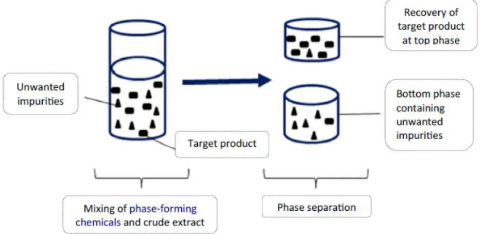
Figure 1 – HPV genome (adapted from (Durzynska, Lesniewicz et al. 2017)). ... 2 Figure 2 – Schematic representation of HPV infection (adapted from (Durzynska, Lesniewicz et al. 2017)). ... 3 Figure 3 - Schematic representation of the action mechanism of E6 oncoprotein (adapted from (Viarisio, Gissmann et al. 2017)). ... 4 Figure 4 - Schematic representation of the action mechanism of E7 oncoprotein (adapted from (Viarisio, Gissmann et al. 2017)). ... 4 Figure 5 - Mechanism of antigen presentation (a): Antigen presentation mediated directly by transfected myocytes; (b): Transfection of professional APCs; (c): Cross priming (adapted from (Hasson, Al-Busaidi et al. 2015)). ... 8 Figure 6 - Number of patents in the field of DNA vaccines (adapted from (Abdulrahman and Ghanem 2018)). ... 9 Figure 7 - Models of non-viral nucleic acid delivery. (adapted from (Slivac, Guay et al. 2017))… ... 12 Figure 8 - Representation of upstream and downstream process of large-scale purification of supercoiled plasmid DNA (adapted from (Guilherme N.M. Ferreira 2000, Van Alstine, Jagschies et al. 2018)). ... 15 Figure 9 - Plasmid DNA expression vector (adapted from (Armando Tejeda-Mansir 2008)). .... 16 Figure 10 – Different types of membranes and the respectively retained solute (adapted from (Aires-Barros and Azevedo 2017)). ... 18 Figure 11 – Normal flow filtration and tangential flow filtration processes (adapted from (Aires-Barros and Azevedo 2017, Liderfelt and Royce 2018)). ... 19 Figure 12 –Representation of precipitation methods - A: Elimination of proteins and high MW RNA by salt precipitation; B: Concentration of pDNA by addition of alcohols (adapted from (Thatcher 2018)). ... 19 Figure 13 - Schematic representation of target product recovery by ATPS (adapted from (Phong, Show et al. 2018)). ... 20
Figure 14 - Schematic representation of size exclusion chromatography (adapted from (Sciences 2015)). ... 22 Figure 15 - Schematic representation of ion-exchange chromatography (adapted from (Sciences 2016)). ... 23 Figure 16 - Schematic representation of hydrophobic interaction chromatography (adapted from (Sciences 2006)). ... 23 Figure 17 - Schematic representation of affinity chromatography (adapted from (Sciences 2016)). ... 24 Figure 18 - Representation of batch method steps. ... 26 Figure 19 - Chemical structure of native (A) and deacetylated (B) gellan gum (adapted from (Osmalek, Froelich et al. 2014)). ... 27 Figure 20 - Representation of the gelation process (adapted from (Morris, Nishinari et al. 2012)). ... 29 Figure 21 - Calibration curve obtained from the sc pDNA with a range of concentrations from 1 to 100 g/mL. ... 40 Figure 22 – A: Chromatographic profile of the E. coli lysate sample in the CIMacTMpDNA analytical
column. B: Chromatographic profile of the pDNA elution…. ... 40 Figure 23 - Calibration curve obtained with BSA standards (0-1200 μg/mL). ... 41 Figure 24 -Schematic representation of microspheres production. ... 44 Figure 25 – Agarose gel electrophoresis of the supernatants recovered from gellan microspheres prepared with different divalent ions… ... 46 Figure 26 - Agarose gel electrophoresis of the supernatants recovered in the 2nd assay…. .... 49
Figure 27 – Representative image of copper-crosslinked gellan microspheres after its production obtained in the semiotic microscope (5x). ... 52 Figure 28 - Representative images of the copper-crosslinked gellan microspheres obtained in the semiotic microscope (5x). A – Snap taken at 0 days; B - Snap taken at 3 days; C - Snap taken at 10 days; D - Snap taken at 20 days. ... 53 Figure 29 - FTIR spectra: A - Gellan gum spectrum; B - Microspheres crosslinked with Cu2+
Figure 30 - FTIR spectra: A – Second derivative of the FTIR spectrum of the microspheres crosslinked with copper after the binding step; B - Microspheres crosslinked with Cu2+ spectrum.
... 56 Figure 31 - Representation of the copper-crosslinked gellan microspheres, visualized in SEM at a magnification of x70 (A) and x200 (B). ... 57 Figure 32 - Agarose gel electrophoresis of the supernatants recovered from the 3rd assay… .. 59
Figure 33 – Zeta potential of the PEI-functionalized microspheres at different pH values. .... 61 Figure 34 - Agarose gel electrophoresis of the supernatants recovered from the 4th assay… .. 62
Figure 35 - Image of (A) microspheres crosslinked with copper and (B) microspheres functionalized with PEI. ... 64 Figure 36 – Image of PEI-functionalized gellan microspheres after its production obtained in the semiotic microscope (5x). ... 64 Figure 37 - Images of the PEI-functionalized gellan obtained in the semiotic microscope (5x). A – Snap taken at 0 days; B - Snap taken at 3 days; C - Snap taken at 10 days; D - Snap taken at 20 days. ... 65 Figure 38 - Representation of the PEI-functionalized gellan microspheres, visualized in SEM at a magnification of x70 (A) and x200 (B). ... 66
List of tables
Table 1 – Viral protein functions and features (adapted from (Viarisio, Gissmann et al. 2017, Gupta, Kumar et al. 2018)). ... 2 Table 2 – Approved gene therapy products (adapted from (Ginn, Amaya et al. 2018)). ... 7 Table 3 - Advantages and disadvantages of viral vectors (adapted from (Ibraheem, Elaissari et al. 2014, Carter and Shieh 2015)). ... 10 Table 4 - Description of the different physical methods applied in gene delivery. ... 13 Table 5 - Chemical composition of different types of gellan (adapted from (loannis Giavasis 2000)). ... 27 Table 6 - Conventional applications of gellan gum in food. ... 31 Table 7 - Utility of gellan gum in the medical field through oral, nasal or ophthalmic formulations (adapted from (Prajapati, Jani et al. 2013, Osmalek, Froelich et al. 2014)). .... 32 Table 8 – Assessment of the sc pDNA concentration, total sc pDNA amount…. ... 47 Table 9 - Description of the equilibrium and elution conditions applied in the 2nd assay. ... 49
Table 10 - Assessment of the sc pDNA concentration, total sc pDNA amount… ... 50 Table 11 - Total protein amount of the E. coli lysate and supernatants recovered after the binding and elution step of the 1st and 2nd assays. ... 51
Table 12 – Copper-crosslinked gellan microspheres mean diameter considering the stability over time (n=5). ... 54 Table 13 - Chemical characterization of gellan microspheres crosslinked with copper through EDX. ... 58 Table 14 - Total protein amount of the crude E. coli lysate from 1st assay and of E. coli lysate
precipitated with 2.5 M ammonium sulfate from the 3rd assay. ... 58
Table 15 – Assessment of the sc pDNA concentration, total sc pDNA amount… ... 60 Table 16 - Assessment of the sc pDNA concentration, total sc pDNA amount…. ... 63
Table 17 – PEI-functionalized gellan microspheres mean diameter considering the stability over time (n=5). ... 65 Table 18 - Chemical characterization of PEI-functionalized gellan microspheres through EDX. ... 67
List of acronyms
3D Three-dimensional
AAV Adeno-associated viruses
AC Affinity chromatography
Al3+ Aluminum (III) ion
APCs antigen-presenting cells
APOBEC3 Apolipoprotein B mRNA editing enzyme catalytic polypeptide 3 ATPS Aqueous two-phase systems
ATR Attenuated total reflection
Ba2+ Barium (II) ion
BAK Bcl-2-antagonist killer
BCA Bicinchoninic acid
BSA Bovine serum albumin
Ca2+ Calcium (II) ion
Cd2+ Cadmium (II) ion
CDK2 cyclin dependent kinase 2
Cgas-STING Cyclic GMP-AMP synthase-Stimulator of interferon genes Co2+ Cobalt (II) ion
CTAB Cetyl trimethylammonium bromide Cu2+ Copper (II) ion
DLS Dynamic light scattering
DMAEMA n,n-dimethylaminoethyl methacrylate
DNA Deoxyribonucleic acid
E. coli Escherichia coli
E2F Transcription factor E2F1-3 Subclass of E2F factors E2F4-5 Subclass of E2F factors E6AP E6 associated protein
EDTA Ethylenediamine tetraacetic acid EDX Energy-dispersive X-ray spectroscopy
EMA European Medicines Agency
EP Electroporation
EPS Exopolysaccharides
FDA Food and Drug Administration Fe3+ Iron (III) ion
FTIR Fourier-transform infrared spectroscopy
GG Gellan gum
HIC Hydrophobic interaction chromatography HPMC Hydroxypropylmethylcellulose
HPV Human papillomavirus
HR HPV High-Risk HPV HSV Herpes simplex virus
IEC Ion Exchange Chromatography
IFNα Interferon α
IMAC Immobilized Metal Ion Affinity Chromatography IRF3 Interferon regulatory factor 3
K+ Potassium ion
Kbp Kilobase pair
KDa Kilo Dalton
LB Luria-Bertani
LCR Long region control
ln Linear isoform
MES 4-Morpholineethanesulfonic acid
MF Microfiltration
Mg2+ Magnesium ion
MHC Major histocompatibility complex MMP-9 Matrix metallopeptidase 9
Mn2+ Manganese ion
MW Molecular Weight
MWCO Molecular Weight cut-off
Myc intermediary proto-oncogene protein
Na+ Sodium ion
NF Nanofiltration
NFF Normal flow filtration
Ni2+ Nickel ion
Nm Nanometer
O/W emulsion Oil-in-water emulsion oc Open circular isoform
OD Optical density
P300/CBP complex protein
CREB binding protein
PAMAM Poly(amidoamine)
PBAE Poly(β-amino ester)s
Pd2+ Palladium ion
PEG Polyethylene glycol
PEI Polyethylenimine
PLGA Poly(lactic-co-glycolic acid)
PLL Poly-l-lysine
polyDADMAC Polydiallyldimethylammonium chloride pRb retinoblastoma protein
RNA Ribonucleic acid
RO Reverse Osmosis
Rpm Revolutions per minute
sc Supercoiled isoform
SCRIB scribbled planar cell polarity protein
SD Standard deviation
SDS Sodium dodecyl sulfate
SEC Size Exclusion Chromatography SEM Scanning electron microscope
siRNA Silencing RNA
Sp1 Specificity protein 1
Sr2+ Strontium ion
TAE Tris-acetate-EDTA
TB Terrific Broth
TFF Tangential Flow Filtration TLR9 Toll-like receptor 9
Tris Tris(hydroxymethyl)aminomethane
UF Ultrafiltration
VLPs Virus-like particles W/O emulsion Water-in-oil emulsion
w/v % Weight per volume percentage WHO World Health Organization
XRCC1 X-ray repair cross-complementing protein 1
List of communications
Oral communication:
• XIV Annual CICS-UBI Symposium, Covilhã, July 2019: Diana Gomes, Luís A. Passarinha, Ângela Sousa, Plasmid DNA capture from E. coli lysates applying gellan microspheres through a batch method.
• 13th International Meeting of the Portuguese Carbohydrate Group, Porto, September 2019: C. Gonçalves, D. Gomes, M. Grilo, A. Sousa, L.A. Passarinha, Gellan microsphere formulation for direct capture of Soluble catechol-O-methyltransferase from a
Komagataella pastoris lysate.
• 13th International Meeting of the Portuguese Carbohydrate Group, Porto, September 2019: J. Coelho, D. Eusébio, D. Gomes, F. Frias, L.A. Passarinha, A. Sousa, Gellan exopolysaccharide biosynthesis and recovery to formulate microspheres optimized by Design of experiments.
Poster presentation:
• 13th International Meeting of the Portuguese Carbohydrate Group, Porto, September 2019: D. Gomes, L.A. Passarinha, A. Sousa, A new application of gellan microspheres for plasmid DNA capture from E. coli lysates through a batch method.
• III International Congress in Health Sciences Research: Towards Innovation and Entrepreneurship - Trends in Aging and Cancer, November 2019: Diana Gomes, Luís A. Passarinha, Ângela Sousa, A new insight of gellan microspheres application for E. coli lysates pDNA capture through a batch method.
List of publications
Coelho, J., D. Eusebio, D. Gomes, F. Frias, L. A. Passarinha and A. Sousa (2019). "Biosynthesis and isolation of gellan polysaccharide to formulate microspheres for protein capture." Carbohydr Polym 220: 236-246.
C. Gonçalves, D. Gomes, M. Grilo, A. Sousa, L.A. Passarinha. “Strategies for direct capture of Soluble catechol-O-methyltransferase from a Komagataella pastoris lysate with gellan microspheres" (in preparation)
Chapter 1 - Introduction
1.1 Human Papillomavirus
Human papillomavirus (HPV) causes about 5% of all human cancers, (Martinez-Ramirez, Carrillo-Garcia et al. 2018) and it is responsible for 99% of cervical cancer cases, which is the fourth cause of cancer death among women worldwide. HPV is also involved in non-cervical malignancies such as vulvar, vaginal, penile, head and neck cancers (Lowy 2016, Gupta, Kumar et al. 2018). According to Globocan (Global Cancer Observatory), in 2018 there were 750 new cases and 340 deaths due to cervical cancer in Portugal.
Over 200 genotypes of human papillomavirus are known and they can be divided according to their potential to induce cellular transformation in high, intermediate or low risk and can also be classified phylogenetically (Durzynska, Lesniewicz et al. 2017, Viarisio, Gissmann et al. 2017). The alpha, beta and gamma genera contain the majority of the HPV types (Viarisio, Gissmann et al. 2017). The alpha genus causes mucosal and cutaneous lesions and the beta and gamma genus only cause cutaneous lesions. High-risk types, mainly found in the alpha genus, cause pre-and malignant lesions and the low-risk types cause benign lesions (Humans 2007). The most common and carcinogenic are HPV16 and HPV18, found in 60% and 15% of cervical cancer cases, respectively (Martinez-Ramirez, Carrillo-Garcia et al. 2018). Therefore, it is important to develop new therapeutic approaches against HPV-associated cancers, in order to reduce its incidence and mortality rates.
1.1.1 HPV life-cycle and molecular biology
HPVs are member of the papillomaviridae family and consist in small double-stranded circular DNA viruses with a length of about 8 Kb. HPV genome, represented in figure 1, is composed by a long control region (LCR), which contains cis-elements that are required for the replication and transcription of viral DNA, early genes (E1-E7), needed for viral replication and late genes, (L1-L2) that are necessary for virion assembly (Humans 2007, Durzynska, Lesniewicz et al. 2017, Viarisio, Gissmann et al. 2017, Martinez-Ramirez, Carrillo-Garcia et al. 2018).
The viral E proteins are transcribed from the early promoter and the L proteins from the late promoter. The functions of these proteins (E and L) are described in table 1.
Table 1 – Viral protein functions and features (adapted from (Viarisio, Gissmann et al. 2017, Gupta, Kumar et al. 2018)).
Viral Protein Functions and Features
E1 Forms a heterodimer complex with E2 and controls viral replication; Helicase function.
E2 Regulates early gene promoter and, together with E1, viral DNA replication; Genome segregation and encapsulation.
E3 Unknown function; present in only a few HPVs.
E4 May mediate the release of viral particles by destabilizing the cytokeratin network; Binds to cytoskeletal protein.
E5 Stimulates mitogenic signals of growth factors; Interaction with EGF/PDGF receptors.
E6 One of the major viral oncoproteins; interacts with and inactivates many cellular proteins.
E7 One of the major viral oncoproteins; interacts with and inactivates many cellular proteins.
L1 The major capsid protein; interacts with L2; the component of the HPV prophylactic vaccine.
L2 The minor capsid protein; involved in viral DNA encapsulation, facilitates viral entry and trafficking.
In figure 2 it is shown the establishment of HPV infection and neoplasia of cervical epithelium. The viral life cycle starts with the arrival of viral particles to the basal layer of the squamous epithelia through micro-abrasions allowing the replication of the HPV genome. At the beginning of the infection, low levels of E1, E2, E6, and E7 proteins are expressed, suspending normal
Figure 1 – HPV genome (adapted from (Durzynska, Lesniewicz et al. 2017)).
keratinocyte differentiation. Then, E2 recruits E1 in order to increase the number of copies of viral episomes, which continue increasing upon epithelium differentiation. In the upper differentiating epithelial layers, E6 and E7 are highly express leading to uncontrolled cell proliferation and when L1 and L2 proteins are expressed in the uppermost layer of the epithelium the viral life cycle is completed. So, the viral genome is encapsulated and occurs the release of mature virions (Mittal and Banks 2017, Martinez-Ramirez, Carrillo-Garcia et al. 2018). Although many women acquire an HPV infection, the majority are cleared or suppressed by the immune system within 1–2 years of exposure (Schiffman, Castle et al. 2007, Viarisio, Gissmann et al. 2017).
Figure 2 – Schematic representation of HPV infection (adapted from (Durzynska, Lesniewicz et al. 2017)).
Development of cervical lesions is connected with the establishment of a persistent infection, where the accumulation of DNA damage due to interactions of HR HPV E6 and E7 with p53 and pRb cause apoptosis inhibition and uncontrolled proliferation, which after a long time, may lead to the alteration of chronically infected cells into cancer cells (Viarisio, Gissmann et al. 2017, Martinez-Ramirez, Carrillo-Garcia et al. 2018).
1.1.2 E6 Oncoprotein
E6 is a protein constituted by 160 amino acids, two zinc-binding motifs and is essentially found in the nucleus-cell. It interacts with several host cell proteins and thus presents different functions, such as, escaping cell death (downregulation of p53 protein and Bak protein), deregulation of cell cycle (downregulation of P300/CBP complex protein and miR34a), immune system modulation (downregulation of IRF3 and IFNα), cell immortalization (upregulation of Myc and Sp1) genomic instability (upregulation of APOBEC3 and downregulation of XRCC1) and cell invasion (downregulation of SCRIB and miR-23b) (Estevao, Costa et al. 2019).
The most important function of the E6 oncoprotein is escaping cell death, through degradation of the p53 protein, which is represented in figure 3. It is known that p53 is a tumor suppressor
and acts in response to DNA damage or cellular stress, activating cellular processes in order to repair or eliminate cells with a damaged genome. In human cancers, this gene is often mutated (Zhang and Lozano 2017). In the particular case of cervical cancer, E6 interacts with the LXLL motif of E6 associated protein (E6AP), an E3 ubiquitin ligase that works as a connecting bridge between E6 and p53, inducing p53 degradation through proteasome pathway and consequently blocking p53 dependent apoptosis (Mittal and Banks 2017, Viarisio, Gissmann et al. 2017, Martinez-Ramirez, Carrillo-Garcia et al. 2018).
Figure 3 - Schematic representation of the action mechanism of E6 oncoprotein (adapted from (Viarisio, Gissmann et al. 2017)).
1.1.3 E7 Oncoprotein
E7 is a phosphoprotein with approximately 100 amino acid residues and three conserved regions, CR1, CR2, and CR3. Conserved region 2 contains the LXCXE (Leu-X-Cys-X-Glu) motif which is essential in the association with its targets and CR3 forms a zinc finger structure (Songock, Kim et al. 2017). This oncoprotein is found in the nucleus and interacts with several targets, presenting diverse functions, like, deregulation of cell cycle (pRB protein and p107/p130 downregulation), immune system modulation (blocking action of TLR9 and Cgas-STING), cell invasion (upregulation of MMP-9) and genomic instability (upregulation of CDK2) (Estevao, Costa et al. 2019).
One of the most important protein regulation mechanisms by E7 is through the retinoblastoma (pRb)/E2F system, which is presented in figure 4 (Mittal and Banks 2017, Songock, Kim et al. 2017).
Figure 4 - Schematic representation of the action mechanism of E7 oncoprotein (adapted from (Viarisio, Gissmann et al. 2017)).
pRb family proteins are known as “pocket proteins,” function through binding to the activating E2F1-3 transcription factors and recruiting transcriptional inhibitory complexes to promoters (Songock, Kim et al. 2017). Association of E7 with pRb and the recruitment of the cullin2-RING ubiquitin ligase promotes pRb degradation via the proteasome pathway. As a consequence, this disrupts the interaction of pRb with E2F transcription factors, stimulating the G1/S phase cell cycle transition (Mittal and Banks 2017, Estevao, Costa et al. 2019).
In addition, E7 interacts with p105, p107 and p130 proteins through the same motif (LXCXE) that E7 interacts with pRb. p107 and p130 proteins function through binding to the inhibitory E2F4–5 transcription factors. In this way, occurs the degradation of p105 and p107, which control cell cycle entry in the basal layer, as well as p130, which is involved in cell cycle re-entry in the upper epithelial layer (Mittal and Banks 2017, Viarisio, Gissmann et al. 2017). So, this ensures that the cell remains in an S-phase-competent state which is vital for the viral life cycle.
1.1.4 Preventive and therapeutic vaccination
HPV infections worldwide continue to be extremely common, so the development of effective treatments is crucial in order to control existing HPV infections and their associated diseases (Yang, Jeang et al. 2016). Two prophylactic vaccines are available commercially for preventing HPV infection, Gardasil and Cervarix (Hung, Ma et al. 2008, Ken Lin 2010, Yeo-Teh, Ito et al. 2018). Both vaccines are made using virus-like particles (VLPs) that consist of L1 (Dochez, Bogers et al. 2014). Expression of L1 protein, the major component of the capsid, results in self- assemble of virus-like particles (VLPs), which are similar in shape and size to HPV virions but don’t have viral DNA (Ken Lin 2010). Gardasil is a quadrivalent vaccine containing recombinant L1 VLPs antigens for HPV types 6, 11, 16 and 18 whereas the bivalent vaccine, Cervarix, contains L1 VLPs antigens for HPV-16 and 18 (Ken Lin 2010, Dochez, Bogers et al. 2014, Yeo-Teh, Ito et al. 2018). These prophylactic vaccines have only been effective when administered in healthy patients, preventing the HPV infection and there is no evidence that they present therapeutic effects in treating or clearing established infections (Yang, Jeang et al. 2016).
Therefore, is still necessary the development of therapeutic vaccines to prevent and also treat existing HPV infections. Nowadays there are several groups developing and testing in preclinical and clinical trials different types of therapeutic HPV vaccines, in which the main target are E6 and E7 oncoproteins (Hung, Ma et al. 2008, Yang, Jeang et al. 2016, Yeo-Teh, Ito et al. 2018).
1.2 DNA-based therapy
DNA-based therapy has been evolving in the past century from an experimental technology into a sustainable strategy for developing therapeutics for several human disorders (Siddhesh D. Patil 2005). Since the discovery of the DNA double helix structure by Watson and Crick that it was possible to achieve a better comprehension of biological systems (Zimdahl 2015).
The discovery and understanding of the human genome made possible the identification of human genes involved in some diseases, which could help in the development and improvement of DNA-based drugs for gene therapy or DNA vaccines (Siddhesh D. Patil 2005). DNA-based therapeutics comprise plasmids for gene therapy or DNA vaccines, oligonucleotides, ribozymes, DNAzymes, aptamers and small interfering RNAs (siRNAs).
1.2.1 Gene Therapy
Nowadays, gene therapy is being studied and applied as a promising treatment option for a number of human diseases, such as inherited disorders, some types of cancer, and certain viral infections (Biswas, Kumar et al. 2018, Dunbar, High et al. 2018, Ginn, Amaya et al. 2018). Gene therapy is designed to introduce genetic material into target cells to replace the function of an abnormal or dysfunctional gene with a functioning variant (Sridharan and Gogtay 2016, Grace Hampson 2018, Vermeulen, Brans et al. 2018).
Normally, to deliver the gene has been used a vector, which is typically a virus, and this can be achieved through two approaches, the germline gene therapy, and somatic gene therapy. The first one implicates the introduction of the gene into the germ cells which passes through to the next generations whereas the latter consists of the transfer of the target gene to the somatic cells that are not inheritable (Sridharan and Gogtay 2016). A diversity of vectors and delivery procedures have been applied in gene therapy. The DNA vector function as a therapeutic gene expression cassette, which contains a promoter (guides the transcription of the gene), the gene of interest and a termination signal (finishes the transcription). The most commonly used are the viral vectors, however, the application of nonviral vectors has been increasing (Sridharan and Gogtay 2016, Ginn, Amaya et al. 2018).
The main challenge of gene therapy consists of the successful and safe target gene delivery due to the host’s immune response directed against the vector and the transgene product. The research in the field of gene therapy has been increasing according to the frequent reports of therapeutic efficacy and recently approved products for the treatment of blindness by in vivo gene transfer and also for cancer (2017). All the approved products are presented in table 2 (Biswas, Kumar et al. 2018, Ginn, Amaya et al. 2018).
Table 2 – Approved gene therapy products (adapted from (Ginn, Amaya et al. 2018)).
Tradename Approval date
Approving agency Indication Manufacturer
Genidicine
(Adenoviral vector)
October 2003
State Food and Drug Administration of China
Head and neck squamous cell carcinoma Shenzhen SiBiono GeneTech (Shenzhen, China) Glybera® (alipogene tiparvovec) (adeno-associated viral vector) November 2012 European Marketing Authorization (EMA) Lipoprotein lipase deficiency uniQure (Amsterdam, Netherlands) Strimvelis™ (ϒ-retroviral vector)
June 2016 EMA Adenosine desaminase deficiency (ADA-SCID) GlaxoSmithKline (Midlesex, United Kingdom) Kymriah™ (tisagenlecleucel) (lentiviral vector) August 2017
Food and Drug Administration (FDA) Acute lymphoblastic leukemia Novartis Pharmaceuticals (Basel, Switzerland) Yescarta™ (axicabtgene ciloleucel) (ϒ-retroviral vector) October 2017
FDA B-cell lymphoma Kite Pharma, Incorporated (Santa Monica, California, USA) Luxturna™ (voretigene neparvovec-rzyl) (adeno-associated viral vector) December 2017
FDA Retinal dystrophy (biallelic RPE65 mutation) Spark Therapeutics, Inc. (Philadelphia, Pennsylvania, USA)
1.2.2 DNA vaccines
According to the World Health Organization (WHO), “a vaccine is a biological preparation that improves immunity to a particular disease” and the most licensed, since 1796, are conventional vaccines (Hasson, Al-Busaidi et al. 2015). This kind of vaccines include inactivated or live-attenuated microorganisms and (Hasson, Al-Busaidi et al. 2015, Kardani, Bolhassani et al. 2016) have been applied to prevent, treat, and even eradicate diseases such as smallpox, diphtheria, tetanus, poliomyelitis, leading to higher life quality and expectancy (Hasson, Al-Busaidi et al. 2015). Unfortunately, these vaccines present several problems related to the lack of safety and difficulty in the cultivation of the pathogenic microorganism, causing adverse immune reactions (Hasson, Al-Busaidi et al. 2015, Kardani, Bolhassani et al. 2016). In addition, they have the limitation of inducing only a prophylactic immune response being useful as a preventive vaccine but not as a therapeutic one.
Therefore, DNA vaccines emerged as a promising way to overcome the problems referred above by offering new approaches for the prevention and therapy of numerous diseases, since it can stimulate both cellular and humoral immune responses against parasites, bacteria and disease-causing viruses (Hasson, Al-Busaidi et al. 2015, Rappuoli and De Gregorio 2016, Lee, Arun Kumar et al. 2018). DNA vaccines are based on bacterial plasmids used to produce particular proteins (antigens) from a disease provoking pathogen (Ghanem, Healey et al. 2013, Hobernik and Bros 2018). The main advantage of such an approach is that the antigen is processed in a similar way to a viral infection. Thus, the antigen can be presented by three different pathways (figure 5): (a) plasmid DNA is internalized, then expressed by somatic cells (myocytes) and antigen presentation to CD8 T cells is made through MHC class I complex; (b) transfection of professional APCs, in which antigen presentation to T cells is accomplished through MHC class I and II complexes; (c) somatic cells transfected with pDNA are phagocytosed by APCs, resulting in cross-priming and antigen presentation to both CD4 and CD8 T cells (Hasson, Al-Busaidi et al. 2015, Lee, Yang et al. 2016, Lee, Arun Kumar et al. 2018).
Figure 5 - Mechanism of antigen presentation (a): Antigen presentation mediated directly by transfected myocytes; (b): Transfection of professional APCs; (c): Cross priming (adapted from (Hasson, Al-Busaidi et al. 2015)).
Besides triggering either humoral or cellular immune responses, DNA vaccines can achieve this without the requirement of live vectors or complex biochemical manufacture procedures (Hasson, Al-Busaidi et al. 2015). In addition, DNA vaccines present other advantages such as improved stability in transportation and storage, stronger immune responses than traditional vaccines and longer-lasting production of the antigenic protein (Ghanem, Healey et al. 2013, Babiuk 2014, Lee, Arun Kumar et al. 2018). Moreover, it is possible to insert multiple variants of an antigen into a single plasmid vaccine, which reduces the number of vaccinations that must be administered (Hasson, Al-Busaidi et al. 2015). Thus, and as shown in figure 6, the number of patents in the field of DNA vaccines has been increasing (Abdulrahman and Ghanem 2018) which demonstrates the greatest interest in this new approach in therapy of cancer, allergies, autoimmune and infectious diseases (Hobernik and Bros 2018).
Figure 6 - Number of patents in the field of DNA vaccines (adapted from (Abdulrahman and Ghanem 2018)).
1.2.3 DNA delivery systems
Cellular environment is not favorable to DNA-based therapeutics entrance and its intracellular pathway until the cell-nucleus due to the extra and intra-cellular barriers. So, this inefficient internalization calls into question the exploration of the welfares of DNA-based therapeutics (Siddhesh D. Patil 2005). Therefore, to ensure that DNA can reach the nucleus without degradation it is necessary to use a delivery system that can protect it (Ibraheem, Elaissari et al. 2014). Delivery systems can be applied in the delivery of DNA vaccines, as well as in gene therapy and they can be divided into viral vectors and non-viral vectors.
1.2.3.1 Viral Vectors
The virus is composed of genetic material such as DNA and RNA, a protein coat that protects the genes and a lipid envelop around the protein coat (Yang, Liu et al. 2014). Through the virus-life cycle and its ability to reach the cell-nucleus of the host, express and replicate its own genetic material, they were used, as carriers, to delivery and guarantee protection to the therapeutic gene (Ibraheem, Elaissari et al. 2014). So, to make a virus a carrier of the therapeutic gene, it must be adapted by genetic engineering. This change consists in the removal of most genes coding for viral proteins and replacement by the desired gene (Giacca and Zacchigna 2012, Ibraheem, Elaissari et al. 2014).
There are several viruses that are used as viral vectors (retroviruses, adenoviruses, adeno-associated viruses (AAV), lentivirus, and herpes simplex virus) (Ibraheem, Elaissari et al. 2014, Foldvari, Chen et al. 2016). In table 3, are presented the main used, as well as their advantages and disadvantages (Ibraheem, Elaissari et al. 2014, Carter and Shieh 2015).
Table 3 - Advantages and disadvantages of viral vectors (adapted from (Ibraheem, Elaissari et al. 2014, Carter and Shieh 2015)).
Viral vectors are the most frequently used because they have shown the capability to reach a high level of transgene delivery in in vivo models and clinical trials, in spite of their weaknesses. These weaknesses can be summarized in (Ibraheem, Elaissari et al. 2014, Yang, Liu et al. 2014, Foldvari, Chen et al. 2016):
- The uncertainty of provoking an immune response that could be fatal;
- Problems with large-scale production of viral vectors and difficulty of packing large molecules, like nucleic acids;
Vector Advantages Disadvantages
Adenovirus High transduction efficiency ex vivo and
in vivo
Transduces many cell types Transduces proliferating and nonproliferating cells
Production easy at high titers
Remains episomal Transient expression Requires packaging cell line
Immune-related toxicity with repeated administration
Potential replication competence No targeting
Limited insert size: 4-5 kb
Adeno-associated virus (AAV)
Integration on human chromosome 19 (wild-type only) to establish latent infection
Prolonged expression
Transduction does not require cell division
Small genome, no viral genes
Not well characterized No targeting
Requires packaging cell line Potential insertional mutagenesis Limited insert size: 5 kb
Herpes simplex virus (HSV)
Large insert size: 40-50 kb Neuronal tropism
Latency expression
Efficient transduction in vivo Replicative vectors available
Cytotoxic No targeting
Requires packaging cell line
Transient expression, does not integrate into genome
Lentivirus Transduces proliferating and nonproliferating cells
Transduces hematopoietic stem cells prolonged expression
Safety concerns: from human immunodeficiency virus origin Difficult to manufacture and store limited insert size: 8 kb
Clinical experience limited Retrovirus Integration into cellular genome
Broad cell tropism
Prolonged stable expression
Requires cell division for transduction Larger insert size: 9-12 kb
Inefficient transduction Insertional mutagenesis
Requires cell division for transfection Requires packaging cell line
No targeting
- The limited size of the genetic materials that can be delivery by the virus; - Transgene mis-insertion risks and resistance to repeated administration.
The use of viral vectors as delivery systems has been restricted and led to research on vectors that could be safer, cheaper and that could overcome the toxicity issues in delivery. Consequently, non-viral vectors have arisen as a promising alternative (Ibraheem, Elaissari et al. 2014, Yang, Liu et al. 2014, Slivac, Guay et al. 2017).
1.2.3.2 Non-viral vectors
Problems related to the safety and small cargo of therapeutic DNA of viral vectors made it necessary to research an alternative, and thus, have emerged vectors not based on viral systems (Ginn, Amaya et al. 2018). Non-viral vectors may provide quite a few advantages related to the safety profile, cost-effective manufacturing and they can carry large therapeutics and be repeatedly administered without provoking a noticeable immune response (Siddhesh D. Patil 2005, Foldvari, Chen et al. 2016).
Naked plasmid vectors are the simplest and popular non-viral delivery systems used in clinical trials because when injected directly in some tissues, are able to produce significant levels of gene expression (Ginn, Amaya et al. 2018). However, naked DNA has difficulty in pass through the extracellular environment due to the action of the nuclease and cross the cell membrane owing to the negative charge repulsion, which results in poor transfection efficiency. So, to overcome this limitation, non-viral delivery systems are developed through different methods and they can correspond to complexes or nanoparticles with the nucleic acid and a single or a mixture of materials (cationic lipids, surfactants, peptides, polysaccharides, metals) and synthetic polymers (PLGA, PEI, PAMAM) (Foldvari, Chen et al. 2016).
The delivery methods of DNA-based therapeutics can be divided into two groups (figure 7) (Ibraheem, Elaissari et al. 2014, Slivac, Guay et al. 2017):
1- Physical methods: these involve a physical force that disrupts the cell membrane to enable localized delivery of the naked DNA into the nucleus. They include needle injection, electroporation, gene gun, and ultrasound delivery.
2- Chemical methods: these can be prepared by electrostatic interaction between particulate carriers (polycationic derivatives) that bind to DNA forming condensed complexes or by encapsulation of DNA within biodegradable spherical structures leading to micro and nanoparticles or through DNA adsorption.
Figure 7 - Models of non-viral nucleic acid delivery. The nucleic acid delivery can be achieved in two ways: nucleic acid interacts with a particulate agent through mostly electrostatic interaction, forming a condensed complex capable of access the cells and the other method correspond to the delivery of naked DNA or RNA into the cells by using an external physical force that causes temporary permeabilization in the cell membrane. Then, the nucleic acid is introduced in the body and after reaching the target tissue will modify cells (adapted from (Slivac, Guay et al. 2017)).
Physical methods use mechanical, ultrasonic, electric, hydrodynamic or laser-based energy with the objective of creating momentary weak points in the target cell membrane, allowing the entrance of DNA by diffusion (Ibraheem, Elaissari et al. 2014, Slivac, Guay et al. 2017). Table 4 describes the fundamentals, advantages and disadvantages of the physical methods.
Table 4 - Description of the different physical methods applied in gene delivery.
Chemical methods are proposed with the objective of improving DNA delivery into the cell-nucleus (Ibraheem, Elaissari et al. 2014, Slivac, Guay et al. 2017). In this way, they mask the negative charge of DNA, compact the DNA molecule to make it minor, and protect it against degradation caused by intracellular nucleases. In order to achieve these purposes the DNA is packed by electrostatic interaction among anionic DNA and polycations, or through encapsulation with biodegradable polymers, or, even, by adsorption (Ibraheem, Elaissari et al. 2014). Exploring the electrostatic interaction between DNA negatively charged and positively charged lipid or polymer, leads to the formation of a positively complex known as a lipoplex or polyplex, respectively (Abdelhamid Elaissari 1995). In lipoplexes, the cationic lipid is constituted by a hydrophobic anchor, a hydrophilic positively charged head and a spacer/linker, and it is used to protect DNA from nuclease and simplify endocytosis. They are formed when the lipid condenses with anionic DNA and the charge density, as well as the spacer of the lipid, have an important role in the delivery of DNA (Ibraheem, Elaissari et al. 2014, Yang, Liu et al. 2014). Besides the low efficiency in vivo, cytotoxicity caused by the positive charge of lipoplex is the main problem related to its application (Ibraheem, Elaissari et al. 2014).
Physical Methods Fundamentals Advantages/ Disadvantages References Microneedle technique
Direct injection of DNA into the desired tissue through a syringe.
Localized DNA delivery; (Slivac, Guay et al. 2017)
Biobalistic (Gene gun)
DNA delivery into the target cell/tissue through metal nanoparticles (gold, tungsten, silver).
More efficient cellular uptake;
Low efficiency;
Sometimes requires surgery; May cause physical damage;
(Ibraheem, Elaissari et al. 2014, Carter and Shieh 2015, Jorritsma, Gowans et al. 2016, Slivac, Guay et al. 2017)
Electroporation
Application of an electric field causing permeabilization of the cell membrane, allowing DNA to easily enter the cell.
Improves DNA uptake; Leads in trials on DNA vaccination against viral infections;
Ability to transfect cells in many different in vivo and in
vitro environments;
(Carter and Shieh 2015, Jorritsma, Gowans et al. 2016, Slivac, Guay et al. 2017) Sonoporation Application of ultrasound waves to cause membrane permeabilization, allowing the DNA entrance into cells.
Safe; Non-invasive;
Can reach internal organs without the necessity of surgery;
(Ibraheem, Elaissari et al. 2014, Slivac, Guay et al. 2017)
Polymers have shown to be a promising alternative to use as a vector for DNA delivery due to their potential for structural diversity and flexible functionality (Yang, Liu et al. 2014). Polyplexes consist of the use of a cationic polymer to condense the negatively charged DNA into a small (nano-sized) complex through electrostatic interactions. By decreasing size, it facilitates DNA entrance into the cell, which leads to an increase in transfection efficiency (Zhang, Xu et al. 2004). However, this approach presents some disadvantages related to poor efficiency, toxicity, and safety, which differ due to the structures of polymers (Yang, Liu et al. 2014). Among the most studied cationic polymers is polyethyleneimine (PEI), with high amino group density (Foldvari, Chen et al. 2016, Slivac, Guay et al. 2017), which has a strong buffering potential (Slivac, Guay et al. 2017), and presents higher nucleic acid binding and delivery capacity. Additional polymers explored are poly-l-lysine (PLL), n,n-dimethylaminoethyl methacrylate (DMAEMA), and poly(ß-amino esters) (Foldvari, Chen et al. 2016). At present, available polymer-based DNA carriers do not fulfill the necessities of DNA delivery for clinical applications, which demands continuous improvements (Shi, Zheng et al. 2017, Xiang, Oo et al. 2017).
The encapsulation of DNA with biodegradable polymers (e.g. PLGA, PBAE, and protonated polyamines) (Luten, van Nostrum et al. 2008, Jorritsma, Gowans et al. 2016) is a promising alternative to DNA condensed with polycations and can overcome limitations of the latter. This method consists in the formation of nano or micro-sized circular structures of hydrolytically degradable polymers incorporating DNA. It presents advantages such as easiness in removal from the body and great DNA protection (Ibraheem, Elaissari et al. 2014). Nevertheless, also presents some drawbacks like poor encapsulation efficiency and partial release of DNA, which causes low DNA bioavailability (Fu, Pack et al. 2000).
In the case of biodegradable polymers, DNA adsorption combines two procedures referred above, electrostatic interaction and encapsulation. It is based on the use of biodegradable particles containing cationic polymers adsorbed to its surface, and, therefore the anionic DNA can be linked through electrostatic interactions (Manmohan Singh 2000). It is possible to improve the bioavailability of DNA, but as DNA is adsorbed to the surface of the particle it is exposed to enzymes that could damage it (Kasturi, Sachaphibulkij et al. 2005, Munier, Messai et al. 2005).
1.3 Plasmid DNA
The interest in pDNA for therapeutics, such as vaccination and gene therapy, has been increasing in the past years leading to the improvement of biopharmaceutical pDNA production methods (Blom, Bennemo et al. 2010). Plasmids are highly negatively charged, circular double-stranded DNA molecules and the size can vary from 1 to 200 Kbp, allowing large amounts of DNA to be loaded (Alex Xenopoulos 2014, Tolmasky 2017, Abdulrahman and Ghanem 2018). They are usually produced in recombinant Escherichia coli (E. coli) host and can pass from one
2013, Abdulrahman and Ghanem 2018). The pDNA has different conformations, open-circular (oc), linear (ln), supercoiled (sc) and denatured conformations, which present different charge distribution, size and base exposure. The supercoiling phenomenon of the sc isoform increases its base exposure and its charge in comparison with other isoforms, however, the sc isoform may convert into other pDNA isoforms depending on the physical and chemical procedures applied (Diogo, Queiroz et al. 2005, Abdulrahman and Ghanem 2018). The isoform that has interest in research is sc pDNA since several reports proved that this isoform has better biological performance and is more efficient for transfection than the other isoforms because the latter could present damage in the expression cassette (Valente, Sousa et al. 2014, Valente, Sousa et al. 2018).
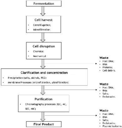
The manufacturing of pDNA comprehends two main steps, an upstream process, and a downstream process, which are represented in figure 8 (Aires-Barros and Azevedo 2017).
Figure 8 - Representation of upstream and downstream process of large-scale purification of supercoiled plasmid DNA (adapted from (Guilherme N.M. Ferreira 2000, Van Alstine, Jagschies et al. 2018)).
1.3.1 Upstream process
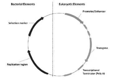
The upstream process consists in the construction, production, and isolation of the desired plasmid. First, occurs the plasmid construction and the selection of a specific strain that allows the cloning process (Abdulrahman and Ghanem 2018, Lindskog 2018). The plasmid should have the necessary elements to propagate in the host and to express the interest gene. Therefore, it must contain bacterial elements (selection marker and replication region) and eukaryotic elements (promoter, transgene, and a transcriptional terminator) as shown in figure 9 (Bower and Prather 2009). The selection marker allows selective growth of E. coli cells normally in the
presence of an antibiotic (such as Kanamycin) and the replication region replicates the plasmid independently of the host machinery increasing its number of copies per cell. The eukaryotic elements are composed of an expression cassette in which the promoter drives the transgene expression and the transcription terminator finishes the transgene expression (Armando Tejeda-Mansir 2008, Carson, Miller et al. 2019).
Figure 9 - Plasmid DNA expression vector (adapted from (Armando Tejeda-Mansir 2008)).
Escherichia coli is still the most used system in biotechnology because it is well characterized
and due to its safety, ease of handling and high productivity (Eva K. Lindskog 2018). Thus, the pDNA replication occurs by a process called fermentation, in which the conditions should be carefully selected since pDNA production is influenced by the plasmid type/size, E. coli strains, growth conditions, culture type and medium composition (Islas-Lugo, Vega-Estrada et al. 2016).
1.3.2 Downstream process
The major goal of the downstream process is elimination of cell impurities and pDNA obtaining in conditions that meet the pharmaceutical specification (Abdulrahman and Ghanem 2018). This process is normally divided into three steps: recovery, intermediate purification, and polishing.
The main objective of the first step is the target product release, the solid compounds removal and the sample concentration (Aires-Barros and Azevedo 2017). So, at the end of pDNA production, the E. coli cells are harvested usually by centrifugation or microfiltration (Alex Xenopoulos 2014, Besnard, Fabre et al. 2016, Abdulrahman and Ghanem 2018). At laboratory scale is mainly used centrifugation, but at large scale, the use of centrifugation could be costly and hard to perform, so the use of filtration has been increasing (Nunes, Morão et al. 2012, Padilla-Zamudio, Guerrero-German et al. 2015, Besnard, Fabre et al. 2016). After bacterial harvest, the cells are lysed to liberate the pDNA. The lysis techniques applied could be mechanical, physical, alkaline or thermal (Voß 2007, Chunsheng, Qinglin et al. 2011, Baumann and Hubbuch 2017, Abdulrahman and Ghanem 2018), however in pharmaceutical plasmid
1979, Voß 2007, Chunsheng, Qinglin et al. 2011, Carson, Miller et al. 2019). Alkaline lysis causes cell membrane disruption through the use of high pH, a detergent (SDS) and a strong base like NaOH, leading to pDNA release as well as all cell components (Alex Xenopoulos 2014, Carson, Miller et al. 2019). The E. coli extract presents several impurities that need to be eliminated, like cell debris, RNA, gDNA, endotoxins, proteins and also the desired pDNA that only corresponds to a very small percentage of the extract (Voß 2007, Ghanem, Healey et al. 2013, Alex Xenopoulos 2014). Thus, normally the solid-liquid separation is achieved through cross-flow filtration or tangential-cross-flow filtration (TFF) and also bag filters at industrial scale (Alex Xenopoulos 2014, Besnard, Fabre et al. 2016). After the removal of solid debris, it is performed a reduction of sample volume (sample concentration) by using alcohols as nucleic acid precipitating agents. And finally, a clarification step is normally applied for the impurity precipitation usually with chaotropic salts, like ammonium sulfate or with other precipitating agents like PEG and calcium chloride, in order to remove impurities such as gDNA, high molecular weight RNA, proteins and endotoxins (Luechau, Ling et al. 2009, Alex Xenopoulos 2014). The pDNA fraction could be increased before the purification with an adequate chromatography step (Sousa, Sousa et al. 2012). And finally, the goal of the polishing step consists in the removal of the remaining impurities (Aires-Barros and Azevedo 2017). However, this represents a big challenge because supercoiled pDNA is very similar in size, structure, and charge to RNA, gDNA and open-circular pDNA (Ghanem, Healey et al. 2013, Alex Xenopoulos 2014).
1.3.2.1 Clarification methods
Clarification consists in the removal of undesirable materials, such as cells or cell debris, allowing the recovery of the desired product (Besnard, Fabre et al. 2016, Aires-Barros and Azevedo 2017). Despite the efficiency of the clarification step has a direct influence on the purification performance, this step is often undervalued. Diverse technologies are used for different operations due to the diversity of products and in order to achieve the desired clarification (Besnard, Fabre et al. 2016).
1.3.2.1.1 Membrane Processes
Membrane technology has been used in recent years for biomolecule separation and concentration of process fluids, due to its flexibility, simplicity, being environmentally friendly and cost-effective (Aires-Barros and Azevedo 2017). In the process, membrane acts as a barrier retaining the bigger molecules and allowing the small ones to pass through the pores. The membrane processes, represented in figure 10, could be divided according to their pore size in microfiltration (MF), ultrafiltration (UF), nanofiltration (NF), reverse osmosis (RO).
Figure 10 – Different types of membranes and the respectively retained solute (adapted from (Aires-Barros and Azevedo 2017)).
The microfiltration process is used for suspended material retention and works with a pore size range from 0.1-10 µm. Thus, large particles like cell debris will not pass through the membrane, whereas smaller particles like proteins, vitamins, and salts will pass. The membranes can be symmetric or asymmetric, the most used are the ones with a pore size of 0.1, 0.22, and 0.45 µm. Ultrafiltration allows nucleic acids, proteins and colloids retention and the membrane pore size range varies from 1 to 50 nm, but usually, the membranes are characterized by the molecular weight cut-off (MWCO). Commercially there are available membranes with MWCO from 3 to 500 KDa. Ultrafiltration membranes can only be asymmetric and generally contain an ultrathin active layer on the top of a sublayer. Nanofiltration membranes are asymmetric with pore size range from 0.2 to 10 nm, allowing retention of sugars, vitamins and polyvalent ions (Aires-Barros and Azevedo 2017, Liderfelt and Royce 2018). Reverse osmosis is mainly used in the desalinization of seawater, since almost all substances are retained, only allowing water and a few organic molecules passage (Liderfelt and Royce 2018).
Initially, the membrane processes were performed in dead-end-mode (normal flow filtration), this means that the feed flows perpendicularly through the membrane, but since it could occur deposition of particles, sharp pressure drops and precipitation of molecules on the surface or in the filter pores, leading to a cake formation, nowadays is more used tangential flow filtration (TFF), figure 11. Tangential flow filtration or cross-flow filtration gained more attention in the industry because the feed flows tangentially to the membrane, reducing the cake formation to a thinner layer (Aires-Barros and Azevedo 2017, Liderfelt and Royce 2018). However, the membranes used in filtration present some problems related to membrane clogging/fouling (Prather, Sagar et al. 2003) and also the supernatant can be poorly clarified when there are large amounts to be processed (Van Alstine, Jagschies et al. 2018).