UNIVERSIDADE DE LISBOA FACULDADE DE CIÊNCIAS
DEPARTAMENTO DE BIOLOGIA VEGETAL
Creation of a Danio rerio mutant
using CRISPR-Cas9
as a model system to study Primary Ciliary Dyskinesia
(PCD)
Margarida Oliveira Almeida Rasteiro
Mestrado em Biologia Molecular e Genética
Dissertação orientada por: Doutora Susana Lopes Professora Doutora Rita Zilhão
II
1.1. Abstract
On the surface, vertebrates seem to have a bilateral symmetry. However, the disposition of the internal organs, such as the heart and liver says otherwise. The left-right axis differentiation is preceded by asymmetries in the gene expression pattern in the tissues around the left-right organiser (LRO).
The LRO (called Kupffer’s vesicle in zebrafish) is a transient structure localized at the end of the notochord that is formed by cells contain a cilium protruding from its apical membrane. Most of these cilia are motile and their beating movement creates a fluid flow towards the left side of the LRO. In a way not yet fully understood, the cells surrounding the LRO sense the fluid directionality and trigger asymmetric expression of nodal and cerl2. The expression of nodal on the left side of the LRO triggers the Nodal-Lefty-Pitx2 pathway that leads to the typical positioning of the internal organs recognised as
situs solitus.
Besides the laterality establishment, the respiratory epithelium, the ependymal cells lining the brain ventricles, the oviducts epithelium and the sperm cells also relay on motile cilia to function properly.
When motile cilia function is impaired, this sequence of events is not guaranteed and patients may suffer from heart congenital malformations bronchiectasis and infertility that characterize primary ciliary dyskinesia (PCD). PCD is caused by mutations in a vast number of genes that codify for cilia components. One of those genes is called CCDC40 and codifies for a protein necessary for the assembly of the inner dynein arms (IDA) and the nexin-dynein regulatory complexes (N-DRC).
In this work, we generated a ccdc40-/- zebrafish mutant (116 aa) using a CRISPR-Cas9 approach. Due to the long zebrafish maturation period, it was not possible to study the mutant phenotype. Instead, we studied the phenotype of embryos injected with a translation-blocking morpholino (MO). We noted that the organ situs was altered in 40 to 70% of the cases, (depending on the amount of MO-injected) and the developing of tail malformations (not related to the injected MO amount).
The ccdc40-/- zebrafish mutant was designed to replicate a human mutation and can be used to test gene editing approaches in an attempt to develop a PCD treatment.
Keywords: Motile cilia, Primary ciliary dyskinesia (PCD); CCDC40, CRISPR-Cas9, Zebrafish
1.2. Resumo alargado
Os vertebrados apresentam uma organização corporal complexa com três eixos: eixo antero-posterior, o eixo dorso-ventral e o eixo esquerda-direita. As assimetrias nos eixos antero-posterior e dorso-ventral são facilmente identificáveis exteriormente. No entanto, no que diz respeito ao eixo esquerda direita, as diferenças são notadas unicamente na disposição interna dos órgãos. No Homem, o coração está deslocado para o lado esquerdo, assim como o estômago e o baço, enquanto que o fígado e a vesícula biliar ficam do lado direito. Também os pulmões, devido à posição do coração, apresentam assimetria no número de lóbulos. Esta disposição (situs solitus) é determinada cedo durante o desenvolvimento embrionário e mantem-se conservada nos vertebrados. A diferenciação do eixo esquerda-direita é precedida por assimetrias na expressão de genes no organizador esquerda-direita (LRO, na sigla em inglês). O LRO é uma estrutura transiente localizada no final da notocorda, que no peixe-zebra é chamada de vesícula de Kupffer (KV).
Esta vesícula é formada por células, cada uma com um cílio diferenciado na sua membrana apical. A maioria destes cílios são móveis e com o seu movimento coordenado produzem um fluxo direcionado para o lado esquerdo do LRO. Esta assimetria no fluxo é depois transferida para uma assimetria na expressão de genes nos tecidos à volta do LRO.
III Inicialmente há dois genes que são expressos de forma simétrica à volta do LRO: nodal e cerl2. A forma como o fluxo consegue controlar a expressão de nodal e cerl2 ainda não é conhecida, mas foram propostas duas hipóteses que a tentam explicar. A primeira propõe que o fluxo gerado pelos cílios móveis transporta substâncias secretadas pelo LRO e as concentra do lado esquerdo, levando à assimetria na expressão nodal e cerl2 verificada posteriormente. A outra hipótese propõe que os cílios móveis criam o fluxo de fluído que, no lado esquerdo, vai causar a deflação dos cílios imóveis, estimulando os seus mecanorecetores e induzindo uma via de ativação dependente de Ca2+ que culmina
na expressão assimétrica de nodal e cerl2. No entanto, apesar dos esforços, ambas as hipóteses têm sido questionadas e o exacto mecanismo de transdução da informação permanece desconhecido.
Após a ação do fluxo, que é sentido pelas células que rodeiam o LRO, a expressão de cerl2 é inibida no lado esquerdo. A inibição da expressão de cerl2 (inibidor de nodal) permite uma maior expressão de
nodal e consequentemente a ativação da cascata de sinalização Nodal-Lefty-Pitx2. Pitx2 é a proteína
efetora da cascata iniciada por Nodal, sendo apontada como a responsável por induzir o fenótipo “esquerdo” nas células do lado esquerdo da mesoderme lateral (L-LPM).
Os cílios, que podem ser móveis ou imóveis (primários), estão presentes em quase todos os tipos células nos vertebrados e por isso, são determinantes quer no período de desenvolvimento embrionário quer na idade adulta. Quando o seu funcionamento fica comprometido pode desencadear várias doenças, como por exemplo a doença do rim poliquístico (PKD, na sigla em inglês) ou síndromes somáticas associadas a polidactilia, malformações neurológicas ou obesidade, no caso de alterações associadas aos cílios primários. Quando são os cílios móveis afetados, as alterações manifestam-se através de infertilidade, infeções respiratórias recorrentes e situs inversus que caracterizam a uma condição chamada discinesia ciliar primaria (PCD). A PCD afecta 1 em cada 10 000 nascimentos e é uma das causas de defeitos cardíacos congénitos. A presença de heterotaxia, condição na qual, pelo menos um órgão está no lado oposto ao que seria de esperar, aumenta 200 vezes a prevalência destes defeitos congénitos.
A PC pode ser causada por mutações que alterem ou inibam a produção de qualquer um dos componentes necessários à montagem das estruturas essenciais para o movimento dos cílios, como o par central, os raios, o complexo regulador da nexina, os braços interno e externo de dineínas ou outras proteínas citoplasmáticas que participam na montagem de componentes ciliares.
Este trabalho foca-se na proteína ccdc40, que é um dos componentes do braço interno de dineínas. Esta proteína é também responsável pela poliglutamilação dos microtúbulos contribuindo para a sua estabilização. Além disso, a proteína Ccdc40 forma um complexo com outra, a Ccdc39, e atua como uma régua molecular. Este complexo permite que os raios dos cílios apenas se liguem no local devido ao longo dos microtúbulos, isto é, a cada 96 nm. Na sua ausência, estes raios continuam a ligar-se aos microtúbulos, mas em locais onde isso não era suposto acontecer.
Em pacientes com PCD portadores de alterações neste gene, foram identificados locais da proteína onde as mutações são mais frequentemente encontradas: as mutações c.248delC e c.3129delC alteram a grelha de leitura enquanto que as outras (c.2440 C>T, c.961 C>T e c.1345C>T) produzem proteínas truncadas.
Neste trabalho, pretendeu-se criar um peixe-zebra mutante que produza a proteína Ccdc40 truncada e que possa servir como modelo de doença para estudar o impacto das mutações mais perto do terminal N da proteína (c.248delC e c.961 C>T). Estas mutações próximas da extremidade N representam cerca de 50% dos casos de mutações identificadas neste gene e produzem proteínas sem o domínio helicoidal (coiled-coil).
Para produzir este mutante usou-se o sistema CRISPR-Cas9. Este sistema foi adaptado a partir de um mecanismo de defesa descoberto em bactérias e permite direcionar a Cas9 (DNase) para um local específico do genoma, usando uma sequência guia de RNA complementar ao local escolhido. Uma vez clivado o DNA, a célula inicia o processo de reparação através da junção das extremidades não
IV homólogas (NHEJ), processo este que é propenso à inserção ou remoção de bases azotadas, levando à ocorrência de mutações que podem inativar a proteína.
Neste caso, o local escolhido como alvo da Cas9 foi a zona do genoma que codifica para o aminoácido 116 da proteína ccdc40 no peixe zebra. Esta região é homóloga da região do genoma humano onde ocorre a mutação c.961 C>T. Uma mutação nonesense ou uma alteração da grelha na leitura neste local impede totalmente a tradução do domínio em hélice, o que causa alterações na montagem da estrutura interna dos cílios e consequentemente na sua mobilidade.
De forma a poder estudar e futuramente validar o fenótipo resultante da injeção do morfolino bloqueador da tradução, foram analisados a posição do coração, fígado e pâncreas assim como defeitos na cauda e aparecimento de edema cardíaco, em embriões injetados com o morfolino. Os resultados mostraram que, independentemente da concentração injetada, os embriões apresentam defeitos relativos à posição do coração (coração à direita ou ao centro), fígado e pâncreas, assim como curvaturas na cauda e aparecimento de edema por volta dos 3dpf. Estes resultados, irão futuramente ser comparados com o fenótipo observado no mutante produzido, validando ou não este morfolino.
Além disso, este mutante permitirá estudar as alterações estruturais e funcionais dos cílios em que a proteína Ccdc40 apresenta uma mutação próxima da extremidade N e compará-las com a estrutura dos cílios de outa linha mutante já existente, lok, em que a proteína é mutada próximo da extremidade C e ainda possui parte do domínio helicoidal.
Futuramente, este mutante poderá ser útil para testar terapia génica direcionada à correção desta mutação que poderá mais tarde ser reproduzida em células humanas recolhidas de pacientes com PCD com o intuito de serem reimplantadas, possibilitando uma forma de tratamento que possa minimizar os efeitos da PCD no aparelho respiratório.
V
1.3. Contents
1.1. ABSTRACT ___________________________________________________________________________ II 1.2. RESUMO ALARGADO __________________________________________________________________ II 1.3. CONTENTS __________________________________________________________________________ V 1.4. LIST OF FIGURES _____________________________________________________________________ VII 1.5. LIST OF TABLES ______________________________________________________________________ VII 1.6. ABBREVIATIONS LIST ________________________________________________________________ VIII 1. INTRODUCTION ________________________________________________________________________ 1 1.1. ASYMMETRY AXIS FORMATION DURING EMBRYOGENESIS ___________________________________________ 1 1.2. L-R ASYMMETRY:NODAL-LEFTY-PITX2 PATHWAY _______________________________________________ 1 1.3. HOW FLOW-RECEIVING CELLS SENSE FLOW: TWO-CILIA VS MORPHOGEN MODEL ___________________________ 2 1.4. MORPHOGEN GRADIENT MODEL ___________________________________________________________ 3 1.5. TWO-CILIA MODEL ____________________________________________________________________ 3 1.6. CILIARY STRUCTURE ____________________________________________________________________ 4 1.7. CILIARY STRUCTURE: MOTILE VS IMMOTILE CILIA _________________________________________________ 5 1.8. CILIOPATHIES ________________________________________________________________________ 6 1.9. PRIMARY CILIARY DYSKINESIA (PCD)(MIM244400) ____________________________________________ 6 1.10. COILED-COIL DOMAIN-CONTAINING PROTEIN 40(CCDC40) _______________________________________ 8 1.11. CRISPR-CAS9 SYSTEM AS A TOOL FOR GENE EDITING _____________________________________________ 9 1.12. ZEBRAFISH AS ANIMAL MODEL FOR PCD _____________________________________________________ 10 1.13. PROJECT GOAL ______________________________________________________________________ 11 2. METHODS ____________________________________________________________________________ 12 2.1. ZEBRAFISH MAINTENANCE ______________________________________________________________ 12 2.2. ZEBRAFISH EUTHANASIA ________________________________________________________________ 12 2.3. CRISPR-CAS9 TARGET SEQUENCES -DESIGN OF SGRNA _________________________________________ 12 2.4. SGRNA ANNEALING AND CLONING INTO PDR274 USING BSAI RESTRICTION SITE __________________________ 13
2.5. E. COLI (DHΑ5) TRANSFORMATION AND IDENTIFICATION OF POSITIVE COLONIES __________________________ 14
2.6. IN VITRO TRANSCRIPTION OF SGRNAS USING T7 PROMOTER _______________________________________ 15
2.7. CAS9 MRNA PRODUCTION ______________________________________________________________ 15 2.8. MICRO CO-INJECTION OF CAS9 MRNA AND SGRNA INTO ONE-CELL STAGE ZEBRAFISH EMBRYOS _______________ 15 2.9. PRIMERS’ DESIGN AND OPTIMIZATION ______________________________________________________ 16 2.10. GENOMIC DNA EXTRACTION ____________________________________________________________ 17 2.11. HETERODUPLEXES DETECTION USING PAGE ANALYSIS ____________________________________________ 17 2.12. MICROINJECTION OF ZEBRAFISH EMBRYOS WITH MORPHOLINO _____________________________________ 18 2.13. HEART AND GUT SCREENING _____________________________________________________________ 18 3. RESULTS _____________________________________________________________________________ 19 3.1. CRISPR-CAS9 PREPARATION ____________________________________________________________ 19 3.2. SGRNA CLONING INTO PDR274 AND SGRNA SYNTHESIS _________________________________________ 19 3.3. PRIMERS OPTIMIZATION ________________________________________________________________ 20 3.4. CO-INJECTION OF CAS9 MRNA AND SGRNA AND CRISPR-CAS9 EFFECTIVENESS ANALYSIS BY PAGE ASSAY _______ 21 3.5. SEARCH FOR FOUNDERS: ZEBRAFISH SCREENING AND GENOTYPING ___________________________________ 22 3.6. MORPHOLINO ANTISENSE TECHNOLOGY - PHENOTYPE CHARACTERIZATION ______________________________ 25 4. DISCUSSION __________________________________________________________________________ 27 5. CONCLUSION AND PROSPECTS ___________________________________________________________ 29 6. BIBLIOGRAPHY ________________________________________________________________________ 29
VI
7. APPENDICES __________________________________________________________________________ 35 7.1. HUMAN- ZEBRAFISH CCDC40 ALIGNMENT ____________________________________________________ 35 7.2. PDR274 STRUCTURE __________________________________________________________________ 37 7.3. PLASMID SEQUENCING RESULTS SHOWING THE DETAILED SEQUENCE __________________________________ 38 7.4. CRISPANTS SEQUENCING _______________________________________________________________ 39 7.5. CCDC40 CLONING PRIMERS ______________________________________________________________ 40
VII
1.4. List of Figures
FIGURE 1.1–TEMPORAL AND SPATIAL DISTRIBUTION OF NODAL,PITX2 AND LEFTY2. ______________________ 2 FIGURE 1.2–MORPHOGEN GRADIENT MODEL AND TWO CILIA MODEL. __________________________________ 3 FIGURE 1.3–TRANSVERSAL VIEWS OF THE AXONEME ORGANISATION. __________________________________ 5 FIGURE 1.4– CILIA ULTRASTRUCTURE AND DYNEIN PLACEMENT IN THE AXONEME IN A TRANSVERSAL AND
LONGITUDINAL VIEW. ____________________________________________________________________ 6 FIGURE 1.5–NORMAL SITUS AND SITUS ABNORMALITIES. ____________________________________________ 7 FIGURE 1.6–TYPICAL AXONEME DEFECTS IN DIFFERENT MUTATED GENES IN PCD. ________________________ 7 FIGURE 1.7–PROTEINS KNOWN TO BE ASSOCIATED TO EACH AXONEME COMPONENT IN HUMANS. ____________ 8 FIGURE 1.8–RELATIVE POSITION OF THE CCDC40 PROTEIN WITHIN THE MOTILE CILIA. ____________________ 8 FIGURE 1.9–CCDC40 LOCATION INSIDE THE CELL. _________________________________________________ 9 FIGURE 1.10–KNOCKOUT GENERATING PROCESS USING CRISPR-CAS9 SYSTEM. _________________________ 9 FIGURE 1.11–HUMAN AND ZEBRAFISH CCDC40 CDNA SEQUENCE ALIGNMENT. _________________________ 11 FIGURE 3.1–COLONY PCR RESULTS. _______________________________________________________________ 19 FIGURE 3.2 – PDR274 LINEARIZATION WITH HINDIII. ______________________________________________ 19 FIGURE 3.3–SEQUENCING RESULTS FROM COLONY 10(SGRNA#2) AND COLONY 14(SGRNA#1). ___________ 20 FIGURE 3.4–PRIMER-PAIRS OPTIMIZATION FOR ZEBRAFISH GENOMIC DNA. ____________________________ 20 FIGURE 3.5–DEFORMITIES FOUND IN ZEBRAFISH LARVAE (3DPF) AFTER CO-INJECTION WITH CAS9 MRNA AND SGRNA. _______ 21 FIGURE 3.6–HETERODUPLEXES DETECTED IN PAGE-BASED ANALYSIS OF CRISPR-CAS9 SYSTEM EFFICACY IN
TGBAC(CFTR-GFP) EMBRYOS. ____________________________________________________________ 21 FIGURE 3.7–HETERODUPLEXES DETECTED IN PAGE-BASED ASSAY FOR TESTING CRISPR-CAS9 EFFICACY IN AB,
TG(FOXJ1A:GFP) AND TG(SOX17-GFP) EMBRYOS INJECTED WITH CAS9 MRNA(75 NG/ΜL) AND SGRNA#1 (37.5 NG/ΜL).__________________________________________________________________________ 22 FIGURE 3.8 –HETERODUPLEXES DETECTED IN A PAGE-BASED ASSAY USED TO TEST TGBAC(CFTR-GFP)♂23
AND FOX:GFP♂3 OFFSPRING. ___________________________________________________________ 23 FIGURE 3.9–SANGER SEQUENCING RESULTS FOR WILDTYPE (WT) AND HETERODUPLEX (HTD) BAND ISOLATED
FROM ♂23BTGBAC(CFTR-GFP) AND ♂3BTG(FOXJ1A:GFP). _______________________________ 24 FIGURE 3.10–MORPHOLOGICAL DEFECTS OBSERVED IN CCDC40 TRANSLATION-BLOCKING MO INJECTED
EMBRYOS. ____________________________________________________________________________ 25 FIGURE 3.11.-.PHENOTYPE OF LAVAE (3DPF) INJECTED WITH CCDC40 TRANSLATION-BLOCKING MO(6 NG)
SHOWING CARDIAC OEDEMA AND TAIL CROOKS. ______________________________________________ 25 FIGURE 3.12–GUT SITUS FROM TG(SOX17-GFP) EMBRYOS INJECTED WITH TRANSLATION-BLOCKING CCDC40MO __________ 25 FIGURE 3.13–HEART AND GUT SITUS COMBINED. _________________________________________________ 26 FIGURE 3.14– DIAGRAM SHOWING THE PREDICTED ZEBRAFISH CCDC40 PROTEIN DOMAINS IN WT, LOK MUTANTS
AND CCDC40 GENERATED MUTANT USING CRISPR-CAS9 SYSTEM. _______________________________ 29 FIGURE 7.1– PDR274 PLASMID STRUCTURE ______________________________________________________ 37 FIGURE 7.2–SEQUENCING RESULTS FOR COLONY 10(SGRNA2) ______________________________________ 38 FIGURE 7.3–SEQUENCING RESULTS FOR COLONY 14(SGRNA2) ______________________________________ 38 FIGURE 7.4–COMPLETED SANGER SEQUENCING RESULTS FOR WILDTYPE (WT) AND HETERODUPLEX (HTD)
BANDS ISOLATED FROM ♂23TGBAC(CFTR-GFP) AND ♂3TG(FOXJ1A:GFP). ____________________ 39
1.5.
List of Tables
TABLE 2.1– SGRNA SEQUENCES _______________________________________________________________ 13 TABLE 3.1– SGRNA1 AND 2 NANODROP QUANTIFICATION __________________________________________ 20 TABLE 7.1–PRIMERS TESTED TO AMPLIFY ZEBRAFISH CCDC40 CDNA _________________________________ 40
VIII
1.6. Abbreviations list
aa Amino acid
ATP Adenosine triphosphate Ca2+ Calcium ion
CCDC39/40/65 Coiled-coil
domain-containing protein 39/40/65 CHD Chronic heart disease CRISPR-Cas9 Clustered regularly
interspaced palindromic repeats associated with caspase 9
Dand5/Cerl-2 DAN domain family member 5/Cerberus-like protein 2
DNAH5 Dynein axonemal heavy chain 5
DNAI Dynein intermediate chain DNAL Dynein light chain
dNTP Deoxynucleotide triphosphate
dpf Days post fertilization GRP Gastrocoel roof plate
h Hour(s)
hpf Hours post fertilization IDA Inner dynein arm
iv Inversus viscerum
Kif3A/B Kinesin-Associated Protein 3A/B
KO Knockout
KS Kartagener syndrome KV Kupffer’s vesicle
LB medium Luria-Bertani medium LPM Lateral plate mesoderm
L-R Left-right
lrd Left-right dynein
LRO Left-right organiser min Minute(s)
MO Morpholino
MTOC Microtubule organising centre
N-DRC Nexin-dynein regulatory complex
O/N Overnight ODA Outer dynein arm
PAM Protospacer adjacent motif PCD Primary ciliary dyskinesia PCR Polymerase chain reaction Pitx2 Pituitary homeobox 2
PKD Polycystic kidney disease PKD1L1 Polycystin 1 Like 1
PKD2 Polycystin 2 RA Retinoic acid
rNTP Ribonucleotide triphosphate rpm Rotations per min
RS Radial spoke RT Room temperature Shh Sonic hedgehog
TGF Transforming growth factor V Volts
1
1. Introduction
1.1. Asymmetry axis formation during embryogenesis
Most of the living beings, specifically the vertebrates, have a complex body organization. The axis differentiation is crucial in the process of shaping multicellular organisms. Several existing pathways, spatially and temporally coordinated, cooperate to establish antero-posterior, dorso-ventral, and L-R (left-right) axis, breaking the embryo’s initial radial symmetry.
The embryo’s initial radial symmetry is only apparent, the ovule goes through several maturation steps that result in an asymmetrical environment inside this cell. The dorso-ventral axis formation is dependent on the establishment of animal and vegetal poles during oogenesis. The maternal dorsalizing factors are located in the vegetal pole and immediately after fertilization, together with zygotic factors, induce the dorso-ventral axis establishment by acting on Wnt /β-catenin, BMP (Bone morphogenic protein), Nodal, and FGF (fibroblast growing factor) pathways (reviewed by Langdon and Mullins 2011 [1]).
The formation of the anterior-posterior axis ultimately establishes the tail, trunk and head. This body segmentation results from the interaction of three signalling pathways: BMP, Nodal and Wnt [2]. The tail forms when all these three are activated, if only Nodal is on, that region will be the trunk, and the head develops at the end where all three pathways are off [3].
The third and last asymmetry axis formed is the one that establishes sidedness (L-R). This asymmetry is first noticed in gene expression in the left-right organiser (LRO) and then spreads to the LPM (lateral plate mesoderm). Ultimately, this information is translated into an asymmetric organ disposition [4].
1.2. L-R asymmetry: Nodal-Lefty-Pitx2 pathway
At first glance, most vertebrates show bilateral symmetry. However, there is an asymmetry within the body. This asymmetry, referred to as situs solitus, is highly conserved across species: the heart, stomach, pancreas and spleen lay on the left side of the longitudinal axis, while the liver and gallbladder are on the right. Additionally, the lungs also show asymmetries in the number of lobes. These morphological asymmetries are preceded by asymmetries in gene expression on the LRO during early embryogenesis [5].
The LRO is a transient embryonic organ (cavity) positioned at the end of the notochord and accordingly to the animal model it is given a different designation: in zebrafish is called Kupffer’s vesicle, in mouse is referred to as the node, in chick as Hensen’s nodea and in Xenopus as gastrocoel
roof plate [6]. Even though these structures are morphologically diverse, the mechanisms behind the L-R asymmetry establishment seem to be conserved across vertebrates [7].
The exact mechanisms that led to the onset of the L-R asymmetry axis are not fully understood, but it is known that a directional fluid flow on the LRO is essential in its establishment [8]. The cells surrounding the LRO have motile cilia protruding from their apical membranes that rotate generating a leftward flow [9]. This asymmetry in the flow is then transcribed into asymmetries in gene expression in the LRO surrounding cells.
a The Hensen’s node does not have ciliated epithelium. Instead, in chick, the left-side expression of Nodal is achieved by the
LRO displacement to the left side of the L-R axis during early gastrulation. The notochord forms on the right side of the LRO restricting Nodal expression only to the left side without requiring flow [113].
2 There are two specific genes, nodal and
dand5 (nodal inhibitor also known as cerl2), that
are initially symmetrically expressed on the LRO. When the flow downregulates the expression of dand5 on the left side of the LRO, it promotes the Nodal-signalling activation on this side [10]. As a consequence nodal expression becomes limited to the left side lateral plate mesoderm (Figure 1.1 A), the same side towards which the embryonic heart loops [5] while its antagonist, Dand5 is restricted to the right side of the LRO [11].
Nodal is a member of the TGFβ (transforming growth factor beta) family that, when lateralized on the LRO, spreads throughout the left-LPM where it induces its own expression as well as Lefty1, Lefty2 and Pitx2. Lefty1 and Lefty2, which are also members of the TGFβ family, compete with Nodal for the receptors preventing its action. Additionally, Nodal is only active as a dimmer while Lefty1 and Lefty2 are monomers, this lets them diffuse faster and farther, inhibiting the Nodal activity on the surrounding tissues. The lefty1 expression is restricted to the left side of the ventral neural tube, where it acts as a midline barrier preventing the left-side establishing factors from crossing to the right left-side [12], while lefty2 is expressed in the left LPM [13] (Figure 1.1 B).
Meno et al. demonstrated that, in lefty2 mutants, Nodal expression lasts longer and can diffuse over to the right side [14]. Therefore, Lefty2 on itself does not decide which is going to be the left side, it functions as a regulator that restricts the duration of Nodal expression.
Finally, Pitx2 is the effector of Nodal signalling and is the most likely candidate for “informing” the cells on the left LPM that they are to adopt a leftward morphology [4]. However, there are some asymmetries such as the visceral looping that seem to be independent of Pitx2-signalling as observed in zebrafish [15].
1.3. How flow-receiving cells sense flow: two-cilia vs morphogen
model
The asymmetric expression of Nodal is established when the motile cilia rotate and generate a fluid flow in the LRO [16]. The crown cells sense this leftward flow and trigger the Nodal-Lefty-Pitx2 pathway in a Ca2+ induced way [17]. This principle is supported by studies showing that mutants lacking
motile cilia do not generate the required flow and thus have defects regarding gene expression and organ laterality [18]. Also, mutants having cilia with motility impairments display similar phenotype [19,20] making it clear that the defects are due to lack of movement and not because of the lack of cilia. The unequivocal connection between the cilia-generated flow in the LRO and the L-R asymmetry establishment was done by Nonaka et al. when they proved that the Nodal-Lefty-Pitx2 pathway asymmetry could be disturbed in WT mouse embryos or normalized in mutants showing laterality defects by inducing an artificially generated flow [16].
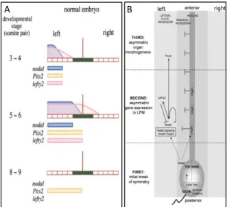
Figure 1.1 – Temporal and spatial distribution of Nodal, Pitx2 and Lefty2.
Lefty2 limits nodal diffusion by migrating faster and competing for the same receptors. Lefty1 is expressed in the midline (green) preventing Nodal from crossing to the right side. Pitx2 is expressed were Nodal is.
Horizontal aligned blocks stand for the LPM, the vertical brown line stands for the midline and blue, pink and yellow indicate the presence of Nodal, Lefty2 and Pitx2, respectively, along the LPM. Adapted from Meno et al. 2001 and Peeters et al. 2006 .
3 However, the exact mechanism on how the LRO senses the flow and transcribes it into gene expression is not yet clear. In an attempt to understand how this cilia-generated flow is translated into an asymmetry in molecular markers, two hypotheses were put forward.
In a summarized way, according to the mechanosensory hypothesis, cilia react to the flow’s mechanical force. When the flow passes through the cilia, they bend and trigger the Nodal-Lefty-Pitx2 pathway [21]. On the other hand, the chemosensory hypothesis proposes that the Nodal-Lefty-Pitx2 pathway is triggered by a morphogen gradient created by the flow.
1.4. Morphogen gradient model
This model assumes that when beating, the motile cilia create a unidirectional flow towards the left side carrying secreted morphogens along with it. This assures that the left side of the organiser has a higher morphogen concentration than the right side and consequently the gene expression on the left side will diverge from that on the right [22].
However, these hypothetic free secreted morphogens capable of directly inducing the Nodal-Lefty-Pitx2 pathway have not yet been identified [9].
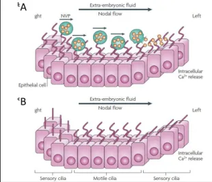
Along with this, it has also been proposed that some of these potential morphogens (such as Shh, RA) could instead be encapsulated inside vesicles (nodal vesicular parcels) that once caught in the fluid flow are transported towards the left side where they eventually break down near the LRO wall setting the morphogens free (Figure 1.2A) [23].
However, unlike expected, slowing or accelerating the flow does not alter the situs [16]. Furthermore, mutants lacking motile cilia (kif3A/B) on the LRO show bilateral expression or absence of the Nodal-Lefty-Pitx2 pathway [18] while in mutants with motionless cilia (lrd, iv) the Nodal-Lefty-Pitx2 pathway can be expressed on the left side, on the right side, bilaterally or be totally absent [24].
According to the morphogen model, it was expected that both types of mutants presented the same phenotype, as they both have no fluid flow in the LRO [21]. Additionally, some other factors such as PKD2 [25] that are not involved in generating the flow, but seem to be important in sensing it, have been described to have a role in the gene asymmetric expression on the LRO.
1.5.
Two-cilia model
In an attempt to justify the phenotype differences between mutants lacking motile cilia on the LRO (Kif3A/B) and mutants with cilia but immotile (iv and Lrd), it was demonstrated that the LRO has two types of cilia: motile cilia, those which generate the flow, and immotile cilia, likely those that may sense it (Mcgrath and Brueckner 2003).
This model assumes that immotile cilia on the left side of the LRO encounter a flow stronger than those on the right. The model advocates that the flow can bend the cilia stimulating the
A
Figure 1.2 – Morphogen gradient model and two cilia model.
A: The morphogen model predicts that secreted morphogens, free or encapsulated in vesicles (NVP), are transported within the flux crating a concentration gradient that is sensed by the cilia in the left side triggering the Ca2+
release on the left side. B: The two -cilia model proposes that motile cilia create the flow while immotile cilia sense it. The flow detection occurs when the fluid pressure bends the sensory cilia triggering the L-R asymmetry breaking in a Ca2+ dependent way. Adapted from Fliegauf et al. 2007
B A
4 mechanoreceptors to induce a Ca2+-dependent signalling pathway (Figure 1.2B) leading to the
asymmetric release of signalling molecules [22].
Mutants with motionless cilia are incapable of generating the flow but preserve the sensing capacity, meaning that external stimuli can randomly activate the mechanosensors. On the other hand, mutants lacking motile cilia loose both abilities rendering them unable to generate or sense the flow [21]. This offers one explanation to the different phenotypes observed.
The explanation backing the mechanosensation connects PKD1 and PKD2 to the early L-R signalling. PKD1 and PKD2 were first identified in polycystic kidney disease. These are membrane proteins present in the kidney cell’s primary cilia [26] that physically interact to change the conformation of PKD2, a Ca2+ channel, and allow it to open [27].
PKD1L1 (PKD1-related) was found to be the important protein, being restricted to the LRO cilia both in mouse [28] and in medaka[25].
Praetorius & Spring showed that bending the primary cilia in kidney culture cells led to an increase of Ca2+ into the cytoplasm that spreads to the nearby cells through gap-junctions [29] bringing up the
possible role of the PKD1L1-PKD2 complex as a mechanosensor in the LRO cilia. Furthermore, both mutants in PKD2 and PKD1L1 do not succeed in establishing asymmetric gene expression at the LRO and LPM, displaying right lung isomerism regardless of the fact that both LRO morphology and cilia motility are apparently normal [28] [25]. PKD2 mice mutants also show several laterality defects [30] similar to those seen in mutants lacking motile cilia on the LRO [18].
These results connect the PKD1L1-PKD2 complex (located in all cilia) and Ca2+ levels to the L-R
axis establishment [17][28]. In addition, Ca2+ signalling levels are typically higher on the left side of the
LRO and become randomized in PKD2 mutants [30].
It has recently been described that PKD1L1 is not necessary to Nodal activation, instead, it acts as PKD2 regulator limiting Nodal expression to the left side [31]. Therefore, PKD1L1 acts as an intermediate between the flow and the Ca2+ signalling cascade activated by PKD2 [31].
In spite of Ca2+ asymmetries and flow response likely being connected, the exact mechanisms how
they induce the signalling cascade that leads to gene expression asymmetries still needs to be clarified [32]. Furthermore, recently there has been some evidence against this hypothesis. The mechanosensation was recently questioned by measuring the Ca2+ influx in response to the flow in primary cilia in vivo
[33]. The results showed that no Ca2+influxes were observed in flow stimulated nodal cilia, suggesting
that, if cilia are to act as mechanosensors, they must induce a non-Ca2+ related signalling pathway.
Nevertheless, the calcium indicator used by these researchers [33] was also questioned at subsequent commentaries and meetings and other alternatives and better calcium probes are being developed in labs from different countries. We shall have to wait for the new results to conclude if the mechanosensation is still feasible in the LRO.
1.6. Ciliary structure
The majority of cells only have one cilium, while others have several, such as the multiciliated respiratory epithelium [34].
Cilia are cellular hair-like structures projected from the plasma membrane that are built when cells get into a differentiated form and reabsorbed when they re-enter the cell cycle [35]. Cilia can be divided into sub-compartments consisting of basal body, transition zone, axoneme and ciliary tip.
Two similar proteins mainly form cilia: α-tubulin and β-tubulin [36]. These two proteins come together to form dimers that eventually aggregate into a polymer. This polymer rolls itself up to create a tube that serves as rails for the intraflagellar transport (IFT) [37]. The basal end of the microtubule is anchored to the microtubule organising centre (MTOC), which is derived from the centriole. The other end is let free so tubulin dimers can be added or removed accordingly to the cell’s state.
5 In cells that have cilia or flagella, the MTOCb is called basal body. Cilia and flagella have similar structures, with the difference that cilia are shorter while flagella tend to be longer.
A cilium is not in continuity with the cytoplasm. All the building blocks necessary to its structure are produced in the cytoplasm and selectively transported through the transition zone (minus end) to the ciliary tip (plus end). Also, when the cilia need to be removed, all the components must be brought back to the cytoplasm.
Aside from building blocks, the motor proteins can also transport signalling molecules. This transport is done by kinesins (or KIFs) (anterograde) and cytoplasmic dyneins (retrograde) that shuttle in and out of the cilia respectively [38]. In humans, there are 46 kinesins [39] and 17 dyneins [40] identified with different motor domains handling different types of transport.
The distal tail domains of the motor proteins bind selectively to cargo while the motor domains interact with the microtubules. These motor domains have the ability to hydrolyze ATP using the generated energy to move forward along the microtubule [41].
In the absence of these motor proteins such as the KIF3 complex (anterograde transporter), the cilia are not correctly assembled. As a result, even though the basal bodies are present, Kif3B [18] and Kif3A [19] mice mutants have no cilia in the LRO leading to the randomization of the L-R asymmetry breaking.
1.7. Ciliary structure: motile vs
immotile cilia
Cilia can be categorised as motile or immotile accordingly to their morphological structure. Immotile cilia (also called primary), present in almost every cell type in vertebrates, are usually short and motionless. Motile cilia are longer and have the ability to beat thanks to the presence of dynein arms in their ultrastructure.
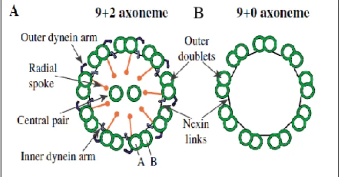
Most motile cilia’s axonemes exhibit a 9+2 arrangement (Figure 1.3A) with 9 doublets hold together by nexin fibres (N-DRC) and a central pair, as those generating the flow in zebrafish KV [42]. Each doublet is composed of two tubules, A (complete) and B (incomplete) where tubule A has two dynein arms attached: inner dynein arm (IDA) and outer dynein arm (ODA) [34]. Apart from holding the microtubules together, N-DRC also influences the axonemal bending by controlling the IDA’s attachment to the A-tubule and mediating the communication between the central pair and the dynein arms through the RS [43].
Cilia with 9+0 arrangement (Figure 1.3 B) lack the central pair and dynein arms and are mostly found in immotile cilia. However, there are 9+0 cilia that have dynein arms and are capable of beating (such as those inducing nodal flow in mouse [44] or medaka [45]).
bThe centrosome is also a type of MTOC and acts as an anchor to microtubules during mitosis.
Figure 1.3 – Transversal views of the axoneme organisation. A: The 9+2 structure is commonly found in motile cilia, showing 9 microtubule doublets, a central pair, radial spokes and dynein arms. B: The 9+0 structure is often associated with immotile cilia that have no dynein arms. From Dawe, et al.2006) [116].
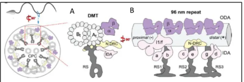
6 IDA and ODA (Figure 1.4) are made up of axonemal dyneins (DNAH, DNAI, DNAL) that hydrolyse ATP and transform it into kinetic energy. The IDA and ODA touch the neighbour B-tubule and cause the two adjacent doublets to glide over each other, generating cilia beating pattern [46].
In humans, ODA and IDA are made of 12 and 15 dyneins respectively (NCBI).When axonemal dyneins such as DNAH1 [48] and DNAH5 [49] are mutated or absent, the ciliary beating function is impaired and can give rise to primary ciliary dyskinesia symptoms.
1.8. Ciliopathies
Cilia can be found in almost every cell type [50]. Primary cilia have a major role in signalling pathways throughout development and in tissue homeostasis. They can act as antennae capturing signalling molecules (Shh, TGF), can react to light or odours in the eye or olfactory epithelium, react to fluid pressure in the kidney and sense pressure in the bone (reviewed by [34]).
Motile cilia also retain sensory skills, but while primary cilia are ubiquitous in almost all cell types (except red blood cells), in adults, motile cilia are localized in sperm, epithelial cells in the bronchi and oviducts, and ependymal cells lining the brain ventricles [51].
Due to this ubiquity, several human diseases and developmental disorders can arise when cilia are defective [52]. These disorders can range from PKD (polycystic kidney disease), pancreatic cancer, or somatic syndromes (characterised by loss of vision and audition, polydactyly, obesity and neurological malformations) when primary cilia are affected. When the problem lies in motile cilia, primary ciliary dyskinesia arises and may involve infertility, upper and lower respiratory impairment, hydrocephaly and
situs inversus.
1.9. Primary ciliary dyskinesia (PCD) (MIM 244400)
A relationship between cilia and L-R axis establishment had been suspected since the discovery of Kartagener syndrome (KS) [53]. Kartagener described a condition causing situs inversus, otitis, sinusitis and bronchiectasis and later Afzelius group added the lack of dynein arms [54] and consequent male sterility [55] to the collection of indicators.
KS is a particular case of PCD. When a patient presents situs inversus totalis, they are said to suffer from KS, which happens in 50% of PCD cases [9].
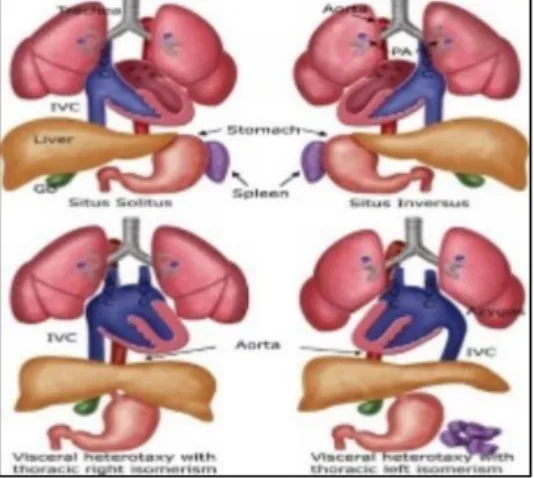
While the perfect inversion of the situs solitus arrangement, called situs inversus totalis, carries little medical consequences [56], the problem lies in the heterotaxy spectrum cases [57] (Figure 1.5).
Figure 1.4 – Cilia ultrastructure and dynein placement in the axoneme in a transversal and longitudinal view.
A shows the relative positions of the duplet, IDA (pink), N-DRC (yellow), ODA (purple) and RS (grey).in a transversal view. B shows the relative position of the same components as A in a longitudinal view, evidencing the axonemal unit repeating at every 96 nm.
Adapted from J. Lin et al. 2014 [117]. B A
7 In humans, laterality defects can be manifested in distinct ways. All organs can be placed in their normal relative position (situs solitus) but in a mirrored image (situs inversus), what is estimated to occur in 1 out of 6 000 – 8 000 newborns [56]. Another situation, known as heterotaxy or situs ambiguous, occurs when at least one organ is misplaced along the L-R axis.
Heterotaxy syndrome is responsible for several congenital cardiac and gastrointestinal malformations [58] found in PCD patients [59] and it is thought to affect about 1 in 10 000 newborns or 1 in every 5 000 – 7 000 of live births with a chronic heart disease (CHD) [57].
According to Kennedy et al., 45% of patients diagnosed with PCD had situs solitus, 47.7% situs inversus, and 6.3% showed heterotaxy. Among this 6.3 %, the majority had cardiovascular malformations [60]. The same authors also reported that the prevalence of congenital heart diseases among patients diagnosed with heterotaxy is 200-fold higher in PCD-suffering patients than in the general population (1:50 vs.1:10 000) [60].
To complicate matters, heterotaxy often comes associated with isomerism. When isomerism is present, one organ that normally would be asymmetric presents itself symmetrically (Figure 1.5). Left isomerism is linked to polysplenia while right isomerism is associated with asplenia [61] but its prevalence in PCD patients is not known [60]. However, sometimes patients present anatomical uncommon arrangements, making it difficult to classify it as left or right isomerism [62].
PCD can arise as the result of several inherited mutations in the cilia-motor machinery such as ODA [49] IDA, radial spokes or central pair components [59].
Mutations in DNAH1, DNAH5 and DNAH11 make up for 25% of all PCD cases [56], but so far more than 35 genes have been linked to PCD (with 70% of patients testing positive for biallelic mutations) [63]. However, a defect in any other one of the about 120 proteins that make up the cilia or in the cytoplasmic proteins [64] responsible for transporting or assembling cilia components can potentially affect the cilia’s ultrastructure and cause PCD. Figure 1.6 shows the most common mutated genes and the consequent effect on cilia’s axoneme structure.
Figure 1.6 – Typical axoneme defects in different mutated genes in PCD.
It shows the list of PCD-related genes that when mutated result in normal axoneme structure, absence of ODA, IDA, or central pair are absent, or axoneme disorganisation. There are still two identified genes that when mutated impair the cilia formation. Adapted from Ferkol 2017 [63].
Figure 1.5 – Normal situs and situs abnormalities. GB, gallbladder; IVC: inferior vena cava; PA: pulmonary artery; SVC: superior vena cava From: https://radiologykey.com/ultrasound-evaluation-of-the-fetal-hear
8
1.10. Coiled-Coil Domain-Containing Protein 40 (CCDC40)
Among the genes responsible for PCD there is Coiled-Coil Domain-Containing Protein 40 (CCDC40) (HGNC: 26090) [65–67]. When mutated, CCDC40 does not completely render the cilia immotile, however, they were found to beat in a fast, flickering way within a reduced amplitude [68].
CCDC40 stands for Coiled-Coil Domain-Containing Protein 40. These mentioned coiled-coil domains are common in proteins [69], they are made of two to five amphipathic α-helices that twist around each other to form a supercoil. Coiled-coil domains are implicated in homodimerization and are present in proteins involved in intracellular transport, molecular recognition, signal transduction and movement regulation [70].
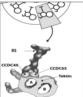
CCDC40 (also known as FAP172, KIAA1640 or CILD15) is necessary for cilia motility, as it is needed for the correct assembly of the N-DRC and IDA [71,72] (Figure 1.7).
It localizes to the area where IDA and RS attach to the A-tubule [73] (Figure 1.8) and is also essential for tubulin polyglutamylation at the proximal extremity of the cilium [74], a post-translational modification that regulates the microtubule’s stability [75].
CCDC40 does not act alone, it has been shown that mutations in CCDC39 or CCDC40 result in indistinguishable phenotypes [67]. Also, in a 2013 study,
CCDC40, together with CCDC39, when mutated were
found to be the cause of PCD in 69% of the identified patients, especially among those previously diagnosed with “radial spoke defect” (loss of IDAs and axonemal disorganization) [76]. Furthermore, there are clues that the destabilization of CCDC40 (along with CCDC65) leads to the disassembly of the major structural components of the axoneme [73].
CCDC40, together with CCDC39, form a complex that was identified as a molecular ruler in cilia, responsible for forming a 96 nm-length gap between RS [77]. When the complex CCDC39/40 is absent, there is an inconsistent number of RS that bind along the A-tubule, meaning that the binding of the RS to the tubule does not depend on the CCDC39/40 complex. On the contrary, in normal cilia, it is thought that the complex blocks the radial spoke binding regions leaving available only the suitable ones at every 96 nm [77].
Antony et al. noted that RS components are detected in cilia from patients carrying CCDC40/39 mutation however, there is no indication that they are correctly assembled or localized [76]. Therefore, the so-called “radial spoke defect” may not be due to the loss of RS, but instead due to its mislocalization or inability to attach to the microtubules.
Figure 1.7 –Proteins known to be associated with each axoneme component in humans.
Ccdc40 encodes a protein necessary for the assembly of dynein regulatory complex (DRC) and inner dynein arm (IDA) complexes.
From Pereira et al. 2015 [118].
Figure 1.8 – Relative position of the CCDC40 protein within the motile cilia.
CCDC40 locates where the IDA, RS and N-DRC attach to the A-tubule.
Adapted from Werner-Peterson & Sloboda 2013 [73].
9 Additionally, CCDC39/40 also regulates IDA and the N-DRC attachment to the A-tubule [77] making it a crucial element in the assembly of axonemal structures and spatial integrity.
Several studies seem to attest this notion by linking CCDC40 mutations with IDA absence and axonemal disorganisation [65,67,76,78].
CCDC40 has been identified in several organismsc. According to
AmiGO2 database, it is located on cilia (respiratory epithelia, sperm flagellum, LRO) and also in the cytoplasm as shown in Figure 1.9.
In humans, it is located in the chromosome 17 (ENSG00000141519) and has 16 transcripts identified with at least 7 producing a protein.
It has two annotated domains: BRE1 (E3 ubiquitin ligase) [79] and SMC N terminal domain. The BRE1domain role is, in this case, not yet known [76], the SMC domain is larger and is believed to be involved in microtube transporting [72]. In zebrafish, ccdc40 is located on chromosome 6 (ENSDARG00000100584), has only two transcripts annotated with only one resulting in a protein.
In PCD patients, some of this gene’s (CCDC40) locations seem to be more prone to mutations (“hot spots”). The most frequent mutations are c.248delC and c.3129delC, that modify the reading frame [71] and c.2440 C>T, c.961 C>T and c.1345C>T that produce truncated proteins [76].
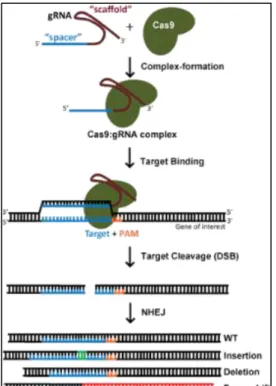
1.11. CRISPR-Cas9 system as a tool for gene editing
The CRISPR/Cas9 (clustered regularly-interspaced palindromic repeats associated with Cas9) is based on a defence system evolved by bacteria to protect themselves against viral infections [80,81].
When a virus infects a bacterium, it stores a fragment of the viral DNA (spacer) in between PAM (Protospacer adjacent motif) sequences [80].
When the virus strikes again, the bacterium produces two types of RNA: crRNA (CRISPR RNA) and tracrRNA (trans-activating crRNA) [82]. The crRNA holds a sequence that is complementary to the spacer, while the tracrRNA facilitates the connection between crRNA and Cas9.
crRNA and tracrRNA form a complex with the Cas9 which acts as a helicase and nuclease. When the complex finds the matching sequence to the crRNA, the Cas9 makes a double-stranded break (DSB) in the DNA neutralising the virus.
There are at least three types of CRISPR/Cas systems [83] but type II is most used in genome editing [84]. This system was engineered to allow scientists to cut any DNA strand at a particularly chosen location by changing the
c http://amigo.geneontology.org/amigo/search/annotation?q=ccdc40
Figure 1.9 – CCDC40 location inside the cell.
CCDC40 is found in
throughout the cytoplasm, were it co-localizes with microtubules, and in cilia. White triangle shows a cilium.
Image available at:
www.proteinatlas.org/ENSG00000141519-CCDC40/cell
Figure 1.10 – Knockout generating process using CRISPR-Cas9 system.
After the Cas9 makes a DSB and the cell repairs the cut using the NHEJ pathway. This pathway is susceptible to insert or remove bases what can result in protein truncation or alterations in the reading frame. From https://www.addgene.org/crispr/guide/
10 sequence in the guide RNA (merging of crRNA and tracrRNA) [84] as long as it is followed by a PAM sequence (Figure 1.10).
This PAM sequence consists of a 5'-NGG-3' sequence (where "N" can be any base) and is fairly common in the genome [85] therefore the CRISPR-Cas9 system can virtually cut any site along the genome.
This guide RNA (sgRNA) can be injected into a cell or embryo together with cas9 mRNA or the Cas9 protein itself. Once inside the nucleus, the complex sgRNA-Cas9 anchors to the PAM, the Cas9 unzips the DNA and matches it to the target sequence in the sgRNA. If the match is successful, the Cas9 makes a DSB in the DNA. The cell reacts by trying to repair the cut using the non-homologous end joining (NHEJ)d pathway, however, this process is error-prone, leading to base insertions, deletions or
frameshift mutations that can silence the gene [86].
The insertions or deletions produced are random, nevertheless, it is also possible to replace a mutated gene with a healthy copy. This can be achieved by injecting a DNA sequence carrying the correct sequence together with the sgRNA and Cas9. In this situation, after the Cas9 cuts the DNA, the cell can use the given DNA sequence as a template, when repairing the cut, following the homology-directed repair (HDR)e pathway [86].
CRISPR-Cas9 system can be used as knocking down tool allowing to study a gene’s role in a cell or organism. Unlike previous methods, CRISPR-Cas9 can be used to target multiple genes at once [87], what makes it useful for studying processes or diseases regulated by several genes working together.
1.12. Zebrafish as animal model for PCD
An organism or cell culture that displays all or some of the physiological signs that can be observed in the actual disease can be considered a good disease model. It is important that it allows to understand the disease’s physiological consequences, develop new diagnostic approaches or test new lines of treatment.
Zebrafish presents itself as o good model to study PCD. It has external embryonic development and the embryo is transparent enough to allow KV inspection and manipulation without killing the fish.
The presence of the KV is another reason favouring the use of zebrafish. It is vesicle full of liquid surrounded by ciliated cells. The cilia on these cells are one per cell, being 80% motile and 20% immotile but they all present a 9+2 structure [88].
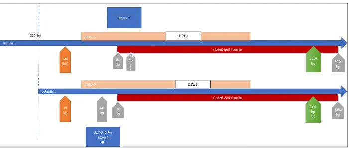
Additionally, among all the genes identified in the human genome, around 70% have at least a homologous gene within the zebrafish genome [89]. In what concerns the CCDC40 gene, when aligning the human sequence with that of the zebrafish by using EMBOSS Needle alignment, they show a similarity of 43.9% (see 7.1). However, if we only consider the genomic sequence against which the sgRNAs were created, the similarity between zebrafish and human is 59.1%.
Importantly, this target region on zebrafish relates to the one that spans across the site where a common nonsense mutation was identified (961, C>T) in patients suffering from PCD [76].
d The NHEJ is a common mechanism used by cells to repair DSB. It is called this way because it does not relay on a template.
Instead, the repairing enzymatic complex revises the ends by removing or adding bases and re-join the resulting extremities without checking for possible introduced errors [114].
e The HDR repair mechanism uses a similar undamaged sequence of DNA as a template. The repairing enzymatic complex
interlace the damage and undamaged strands, get them to exchange sequences of nucleotides and fill in the missing gaps in an error-free way [115].
HDR is less common than NHEJ, as it can only occur in the presence of a template sequence. This template can only be accessed in diploid cells in S phase or when a donor sequence is artificially introduced into the cell.
11
1.13. Project goal
The main aim of this project was to generate a ccdc40 zebrafish mutant using the CRISPR-Cas9 system to be studied as a disease model.
The reason behind this choice lays on the fact that mutations in this gene have been identified in patients diagnosed in our laboratory as well as in some published articles that associate it with PCD [67,90,91].
There is already a ccdc40 zebrafish mutant line (lok) expressing a truncated protein. This mutant expresses an early terminated protein (non-sense mutation at 778 aa) containing almost the totality of the coiled-coil domain.
The mutant we generated expresses a truncated protein up to the 116 a.a. excluding completely the coiled-coil domain.
The CRISPR-Cas9 system appears as an alternative to the Morpholino (MO) technology, a knockdown method, that has been questioned lately [92,93]. The MO’s have long been a popular method to study gene function, particularly in zebrafish. However, they can induce toxicity and have unpredictable off-targets. Kok et al. compared MO-induced and mutant phenotypes and concluded that, in 70% of the analysed genes, the results were not equivalent [93]. Therefore, when possible, MO-induced phenotypes should be verified by gene-editing methods such as CRISPR-Cas9.
To do so, the sgRNAs were designed to guide the CRISPR-Cas9 complex to the region of the zebrafish genome codifying for the orthologous human region containing one of these “hot spots” (Figure 1.11). After the designing, they were cloned into a pDR274 vector containing a gRNA scaffold that takes the role of tracrRNA and interacts with Cas9. The vector was replicated in competent bacteria and used as a template to produce RNA in vitro. This sgRNA produced included the gRNA (scaffold) and the complementary sequence to the target region.
In an attempt to cause a loss of function mutation, we injected sgRNA together with Cas9 mRNA into zebrafish embryos and tested some of those embryos to check the CRISPR-Cas9 system efficacy, while the majority was let to grow.
We could identify crispant fish (founders) and now have their progeny (F1) growing until they are ready to be genotyped. However, as zebrafish is an organism that takes about three months to reach maturity, obtaining the homozygous mutant (ccdc40-/-) was not viable in this work’s time span.
Figure 1.11 – Human and zebrafish ccdc40 cDNA sequence alignment.
Non-scaled diagram showing human and zebrafish ccdc40 cDNA sequence alignment EMBOSS Needle alignment algorithm. The CRISPR-Cas9 system was prepared to target the region around the position 341 bp in zebrafish, leading to a truncated protein without the coiled-coil domain. The lok mutation causes the production of a shortened Ccdc40 protein closer to the C-terminal.
12 So, the second aim was to compare the CRISPR-Cas9 to the MO-induced phenotype. I, therefore, knocked down ccdc40 by using a transcription blocking MO. The MO was tested by injecting several concentrations (as explained in 2.12 in page 18). After the MO injection, parameters such as mortality, heart and gut situs, cardiac oedema and tail defects were evaluated to future comparison with ccdc40-/- mutant zebrafish.
2. Methods
2.1. Zebrafish maintenance
In this work were used wild-type AB lines (ZFIN ID: ZDB-GENO-960809-7), Tg(sox17:GFP) (ZFIN ID: 070117-57) Tg(foxj1a:GFP) (ZFIN ID: ZDB-TGCONSTRCT-131120-3) and TgBac(cftr-GFP) (ZFIN ID: ZFIN ID: ZDB-TGCONSTRCT-130423-1). These fish were kept in the CEDOC fish facility under a controlled environment approved by the Direção Geral de Alimentação e Veterinária (DGAV).
Adult fish were kept in fresh water (28o C, 700 µS, pH 7) under a 14:10 h light:dark cycle. Embryos
were obtained by natural mating and incubated at 25oC or 28ºC up to five days post-fertilization (dpf)
after which they were transferred to a tank in the main system or sacrificed.
The natural mating was stimulated by placing a male and one or two females inside a mating box with a transparent divider allowing visual contact. The fish were removed from the main system and placed in the mentioned mating boxes in the evening and left separated, as described, during the night. In the morning, the dividers were removed and the water level lowered allowing the courtship behaviour between male and female and consequently the external fertilization of the eggs. This method allows for the synchronization of the embryonic development.
In each mating box, there was a sieve preventing the fish from eating the eggs. After the eggs were laid the adult fish were again placed in the same main system tank. The eggs were collected by filtering the water remaining in the mating box and then were placed in a Petri dish immersed in embryonic medium with methylene blue (5mM NaCl, 0.2mM KCl, 0.3mM CaCl2, 0.3mM MgSO4 and distilled
water H2O, pH 6.5). The embryonic medium renewal and the removal of dead embryos were done daily
to prevent bacterial and fungal infections.
The embryonic staging was done according to what is described in Kimmel et al. [94]
2.2. Zebrafish euthanasia
Larvae up to 5dpf were sacrificed by being placed in a bleach solution. Larvae older than 5dpf and adult fish were euthanized using an overdose of tricaine (MS-222) solution in a 250 mg/L concentration [95,96].
2.3. CRISPR-Cas9 target sequences - Design of sgRNA
To study the functionality of ccdc40 gene (ENSDARG00000100584) in zebrafish, the CRISPR-Cas9 technique was used to induce DNA breaks. The CRISPR-CRISPR-Cas9 target sequences inside the ccdc40 gene were determined using the crisprscan tool available online (crisprscan.org). This tool scans the gene-of-interest coding sequence (ENSDART00000169752) and gives back a list of potential targets sites ranked accordingly to its predicted efficacy. All the 20 nucleotide-long sequences presented are followed by an NGG motif also called PAM (protospacer adjacent motif) that is required for the Cas9
13 recognition. The designed sgRNA oligonucleotides are expected to induce the cutting at any sequence following the formula 5′-GG- N18 -NGG-3’. The GG-N18 (underlined) is included in the transcribed sgRNA while the NGG integrates the genomic DNA only (not present in the sgRNA). If the sequence has a mismatch in one of the first two GG’s it is called non-canonical but the Cas9 can still work properly [97].
The provided list shows information concerning the score assessed by the tool, the sequence’s position in the chromosome, its canonical state and the number of off-targets.
From the referred list, we chose two oligonucleotides that along with having the highest score (higher scores mean higher predicted efficacy), were closer to the initial ATG (to create a mutation as early as possible), these had no off-targets (those with no off targets minimize induced cuts in other genome sites) and were considered canonical (no mismatches in the first GG providing a stronger binding to the target site). The capital letters shown indicate the sequence that must be present in the sgRNA and transcribed in vitro.
To allow the sgRNA cloning into a vector, for each target site were designed two complementary oligonucleotides exhibiting single-stranded overhanging ends (bold) (ordered from Stab Vida; Oeiras, Portugal) (Table 2.1).
Table 2.1 – sgRNA sequences
ccdc40 sgRNA #1 oligonucleotides - 5’>3’ sgRNA #2 oligonucleotides - 5’>3’ Forward (Fw) TAGGTAGTAGTTGAGAAGGCAG TAGGCCGAGGACCACCTGTATG Reverse (Rv) AAACCTGCCTTCTCAACTACTA AAACCATACAGGTGGTCCTCGG
2.4. sgRNA annealing and cloning into pDR274 using BsaI
restriction site
Both pairs of (forward and reversed) designed oligonucleotides were dissolved in miliQ H2O (100
μM) and annealed. To perform the annealing reaction, 1.5 μl of the forward oligonucleotide (100 μM) and 1.5 μl of the reverse oligonucleotide (100 μM) were added to 23 μl of miliQ H2O and 24 μl of
annealing buffer (10 mM Tris, pH7.5-8.0, 50mM NaCl, 1m EDTA). This solution was then heated to 95ºC for 5 min and let cool down to RT.
Simultaneously, pDR274 plasmid vector (Addgene #42250) (see 7.2) was incubated with BsaI (NEB #R0535) and brought to a linear conformation. The linearization was accomplished by mixing 5 μg of the circular plasmid, 2 μl of the enzyme, 5 μl of NEBuffer™ 3.1 (NEB # B7203S) and 38 μl of miliQ water and incubating the resulting solution O/N at 37ºC. To check the linearization success, 1μl of the reaction product was loaded into a 0.8% TBE-agarose gel and compared with the “non-insert” control ligation reaction. The remaining volume was purified using a DNA clean & concentrator kit (Zymo research #D4003) and quantified by NanoDrop™ 2000.
The pDR274 plasmid contains three useful features: a T7 promoter found upstream of a partial gRNA sequence and a domain conferring kanamycin resistance. The partial gRNA sequence allows the sgRNA sequence and Cas9 interaction. The annealed oligonucleotides were inserted between the T7 promoter and the gRNA scaffold where the BsaI restriction site is found. To ensure this, the oligonucleotides (10 μM) and the linearized plasmid (100 ng) were incubated O/N at 16ºC with 0.5 μl T4 ligase (Sigma-Aldrich #9015-85-4), 5 uL T4 buffer (2x) (Sigma-Aldrich #KEM0046B) and 2.17 μl miliQ water.
14 In the forward strand, BsaI cuts immediately after the recognition sequence 5’-GGTCTCN-3’ whereas in the reverse strand it cuts only four nucleotides ahead (3’-CCAGAGN(N)4-5’) creating
non-compatible sticky ends that match the overhanging ends added to the designed oligonucleotides. This directional restriction cut by BsaI not only forces the correct insertion of the annealed oligonucleotides into the plasmid backbone but it also prevents the plasmid from self-ligating.
2.5. E. coli (DHα5) transformation and identification of positive
colonies
The ligation product was used to transform competent E. coli bacteria. In this case, DHα5 was the elected strain, rendering a transformation efficiency >1x10⁶ colony forming units for every mg (Thermo
Fisher #18265017).
The bacteria, stored at -80ºC, were thawed on ice for 20 min. Then, a 50 μl aliquot of DHα5 together with 10 μl of the ligation product were subjected to a thermal shock (30 min on ice, 1 min at 42ºC, 2 min on ice) to boost the plasmid uptake. Next, followed an incubation at 37ºC and 250 rpm for 1h 30 min maximizing air-liquid interface. The incubation step was intended to let the bacteria recover from the thermal shock and assure the plasmid stability inside the cells. Subsequently, the culture was centrifuged (5000x g, 10min, RT), to increase cell density. These transformed bacteria were streaked in a LB-agar plate treated with kanamycin (30μg/μl) and incubated once again at 37ºC O/N with the agar layer facing upwards.
Because the cloning plasmid has a domain conferring resistance to kanamycin, only the bacteria that up-took it were able to subsist. However, some bacteria could have assimilated the plasmid without the oligonucleotides inserted. This oligonucleotide chain is too small to be resolved on an agarose gel, so to avoid this predicament, colonies were screened by PCR (colony PCR). This technique relied on the use of M13R primer (5’- TGTAAAACGACGGCCAGT-3’), that anneals with the plasmid backbone, and the forward designed oligonucleotide for the corresponding Cas9 decided-target. When amplification occurs, it meant the colony-inducing bacterium integrated the plasmid with the insert. However, when no amplification was detected it could be because the cloning process was unsuccessful or the PCR failed.
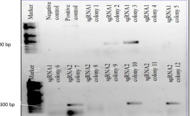
To detect the true positive colonies, we picked around six colonies for each sgRNA-dedicated agar plate were directly used as template in a colony PCR reaction and streaked in a new agarose plate. Each of these colony PCR reactions was prepared by combining 19.55μl of miliQ water, 2.5μl of buffer (10x, provided with the enzyme), 1.25μl of MgCl2 (50mM, provided with the enzyme), 0.5μl of dNTPs
(10mM),0.2μl of NZYTaq DNA polymerase (Nzytech MB00101) and 0.5μl of each primer: M13R (10 μM) and forward sgRNA oligonucleotide (10 μM), adding up to a total volume of 25μl. The colony PCR followed the steps from a common PCR i.e. initial denaturation (95ºC, 10min), denaturation (95ºC, 1min), annealing (51ºC for sgRNA#1 and #2, 1min), elongation (72ºC, 1min) and final elongation (72ºC, 10min) for 30 cycles. The M13R primer is tolerant to a wide range of temperatures, therefore the annealing conditions were adjusted to the designed oligonucleotides structure.
The positive colonies were inoculated in primary liquid cultures (LB medium, kanamycin 30μg/μl) and grown O/N at 37ºC and 250 rpm. These cultures sustain high bacteria densities, essential to a good plasmid yield.
The plasmids were isolated using a commercial kit from Zymo Research (ZR Plasmid Miniprep™ #D4015) and then Sanger sequenced by Stab Vida using M13R primer.
All the described bacteria manipulations, including agar plate preparation, were done in a sterile environment.
15
2.6. In vitro transcription of sgRNAs using T7 promoter
The plasmids (for sgRNA #1 and sgRNA #2) isolated from the positive colonies and confirmed by Sanger sequencing were linearized downstream of the cloning site using a “unique cutter” enzyme. The linearization was achieved by incubating together 1.25 μl of HindIII (NEB # R0104), 5 μl of NEBuffer
TM 2.1 (NEB # B7202S), 5μg of plasmidic DNA and miliQ water (up to 50 μl) for 10h at 37ºC followed
by storage at 4ºC. The then linear DNA was purified using a Zymo Research commercial kit (Zymo Research #D4003) and since it contained a T7 promoter, it was used as a template to produce RNA.
Besides the purified DNA template with a promoter, the RNA in vitro transcription reaction required triphosphate ribonucleotides (rNTP) (mix containing 10 mM of ATP, UTP, CTP and GTP), transcription buffer 5x (MgCl2, 1 M; NaCl, 5 M; Tris-HCl, 1 M pH8), dithiothreitol (DTT) (50 mM), T7 RNA
polymerase (NEB #M0251S) and miliQ water adding up to a total volume of 50 μl.
DTT is a redox reagent that stabilizes the sulfhydryl groups within the T7 protein and without it, the enzyme efficiency drops considerably.
First, the water, 10μl of transcription buffer, 5 μl of DTT and 5 μl of the rNTP mix were added together and incubated for 5 min. Secondly, the DNA template was added (1.5 μg) and after a 1-minute incubation, 1 μl of RNA inhibitor was joint to the solution, followed by another 1-minute incubation. Ultimately, T7 RNA polymerase was joined. First, 2 μl preceded a 2h-incubation and then another 1 μl was followed by a 1h-incubation.
Lastly, 1 μl of DNase (provided with RNA clean & concentrator kit by Zymo Research #R1013) was incubated for 30 min with the solution to remove the template DNA initially added and make sure just RNA remained. All the incubation periods were performed at 37ºC.
The generated RNA was purified with an RNA clean & concentrator kit by Zymo Research (#R1013) and stored at -20ºC. The produced RNA was not 5′-capped or 3′-polyadenylated as it was not to be translated. This protocol makes the RNA more prone to degradation, however as it is to be promptly used, the quality decay is minimal.
This resulting RNA was injected together with Cas9 mRNA into one-cell stage zebrafish embryos.
2.7. Cas9 mRNA production
For Cas9 mRNA synthesis, the plasmid pCS2-ncas9n (Addgene, #47929) was linearized using NotI, a “unique cutter) enzyme.
The in vitro transcription reaction done here was very similar to the one done to synthesize the sgRNAs with the exception that here the reaction also included 5ul of G(5')ppp(5')G RNA Cap Structure Analog (20mM)(NEB #S1407S). Therefore, as expected from a mRNA, the Cas9 mRNA is 5′-capped. This feature is crucial as the Cas9 protein needs to be produced by the cell after co-injection with sgRNA for the CRISPR-Cas9 system to work.
Raquel Jacinto, a lab’s PhD student performed the synthesis of this mRNA.
2.8. Micro co-injection of Cas9 mRNA and sgRNA into one-cell
stage zebrafish embryos
After the collection, the fertilized eggs were lined up against a glass slide inside a petri dish, without embryonic medium and injected using a thin glass needle attached to a Narishigi pico-pump injector. The needle was calibrated using a graticule (10mm/0.1mm Graticule Ltd., Tonbridge, Kent) in a way that each pulse delivered 1.4 nL into the yolk.