Pedro Rafael Martins de Almeida Sampaio
Degree in Molecular and Cell Biology
Using cilia mutants to study left-right
asymmetry in zebrafish
A thesis submitted in fulfillment of the requirements for the degree of the Masters in
Molecular Genetics and Biomedicine
Supervisor: Susana Santos Lopes, PhD, CEDOC-FCM
Internal supervisor: Ana Madalena Ludovice, PhD, FCT/UNL
Using cilia mutants to study left-right asymmetry in
zebrafish
Submitted by
Pedro Rafael Martins de Almeida Sampaio
A thesis submitted in fulfillment of the requirements for the degree of
Masters in Molecular Genetics and Biomedicine
Using cilia mutants to study left-right asymmetry in zebrafish
Copyright Pedro Rafael Martins de Almeida Sampaio, FCT/UNL, UNL
Acknowledgments
I would like to start saying that in this last year I had the privilege to work and learn from so many and good scientists, without whom this work would not have been possible. For those who were part of it, I want to express my deepest gratitude.
Moreover, I want to thank especially to:
Susana Lopes, my supervisor, for letting me to be part of her “ship”; for mentoring and guiding me during this exciting ride; for promoting and supporting my scientific growth; and most important of all for believing me. I am honestly thankful and privileged for having worked with you!
Petra and Bárbara for all the patience and great support showed to me since the beginning. You were really the cornerstones of this work, for all you taught me.
Cláudia Pereira, Petra, “Princesa do Mal”, Queiroga, Sofia, “Tsabimbi”, “Amélia” and Telmo for the friendship and all the good moments and support. I really feel lucky for having met all of you!
Adán Guerrero for teaching and discussing with me so many things that helped me a lot for the success of this work and my growth as a scientist; for showing so much support and being there when I needed. I believe you are a great scientist and I wish you all the best in the coming years. Muchas gracias, un gran abrazo!
My closest friends, for being there when I needed them.
Abstract
In vertebrates, internal organs are positioned asymmetrically across the left-right (L-R) body axis. Events determining L-R asymmetry occur during embryogenesis, and are regulated by the coordinated action of genetic mechanisms. Embryonic motile cilia are essential in this process by generating a directional fluid flow inside the zebrafish organ of asymmetry, called Kupffer’s vesicle ﴾KV). A correct L-R formation is highly dependent on signaling pathways downstream of such flow, however detailed characterization of how its dynamics modulates these mechanisms is still lacking. In this project, fluid flow measurements were achieved by a non-invasive method, in four genetic backgrounds: Wild-type (WT), deltaD-/- mutants, Dnah7 morphants (MO) and control-MO embryos. Knockdown of Dnah7, a heavy chain inner axonemal dynein, renders cilia completely immotile and depletes the KV directional fluid flow, which we characterize here for the first time. By following the development of each embryo, we show that flow dynamics in the KV is already asymmetric and provides a very good prediction of organ laterality.
Through novel experiments, we characterized a new population of motile cilia, an immotile population, a range of cilia beat frequencies and lengths, KV volumes and cilia numbers in live embryos. These data were crucial to perform fluid dynamics simulations, which suggested that the flow in embryos with 30 or more cilia reliably produces left situs; with fewer cilia, left situs is sometimes compromised through disruption of the dorsal anterior clustering of motile cilia. A rough estimate based upon the 30 cilium threshold and statistics of cilium number predicts 90% and 60% left situs in WT and deltaD -/-respectively, as observed experimentally. Cilia number and clustering are therefore critical to normal situs via robust asymmetric flow. Thus, our results support a model in which asymmetric flow forces
registered in the KV pattern organ laterality in each embryo.
Resumo
Em vertebrados, os orgãos viscerais estão posicionados assimetricamente ao longo do eixo esquerdo-direito (E-D) do organismo. Esta assimetria ocorre durante a embriogénese sendo determinada pela acção coordenada de mecanismos genéticos.
Cílios móveis embrionários participam neste processo ao gerarem um fluxo direcional do fluido interno do órgão de assimetria do peixe-zebra, denominado Vesícula de Kupffer (VK). A correcta formação do eixo E-D depende de vias de sinalização a jusante de tal fluxo, no entanto carece uma caracterização detalhada de como essa dinâmica modula estes mecanismos.
Neste projecto, medições do fluxo do fluido foram obtidas em quatro genótipos: embriões selvagens (WT), mutantes deltaD-/-, morfolino-injectados (MO) Dnah7 e MO-controlo. Dnah7 é uma dineína de cadeia pesada situada no lado interno dos microtúbulos do axonema e a sua ausência tornou os cílios imóveis quebrando o fluxo direcional do fluido da VK. Acompanhando o desenvolvimento de cada embrião, mostramos que a dinâmica do fluido na VK é assimétrica, fornecendo uma previsão da lateralidade dos órgãos.
Neste trabalho caracterizamos uma nova população de cílios móveis, uma população imóvel, uma variedade de frequências de batimento e comprimentos ciliares, volumes da VK e número de cílios em embriões vivos. Subsequentes simulações de dinâmicas de fluido sugerem que o fluido em embriões com 30 ou mais cílios produzem situs solitus; com menos cílios, a posição correcta dos órgãos pode ser comprometida através da disrupção do agrupamento de cílios móveis presente na região dorsal anterior. Estimativas aproximadas baseadas num limiar de 30 cílios predizem 90% e 60% situs solitus em WT e deltaD-/-, respectivamente, como observado experimentalmente. O número e agrupamento ciliar são por isso decisivos para formar um situs correcto através de um robusto fluxo assimétrico. Assim, os nossos resultados suportam um modelo em que forças de fluxo assimétricas presentes na VK padronizam a lateralidade dos órgãos em embriões.
General index
Acknowledgments ... vii
Abstract ... ix
Resumo ... xi
General index xi
Figures index xv
Tables index xvii
1. Introduction ... 1
1.1. Cilia and Flagella ... 2
1.1.1. Cilia classification ... 4
1.1.2. Dynein-based ciliary motility ... 6
1.2. Cilia defects and ciliopathies ... 7
1.2.1. Primary cilia dyskinesia ... 8
1.3. Left-right patterning asymmetry ... 10
1.3.1. An asymmetric cascade of signals ... 10
1.3.2. The role of the node in establishing L-R asymmetry in early mouse development 11 1.3.3. The two left-right models ... 13
1.3.4. Left-Right asymmetry in zebrafish ... 15
1.3.4.1. Zebrafish Kupffer’s vesicle ... 15
1.3.4.2. Motile cilia generated flow in Kupffer’s vesicle ... 17
1.3.4.3. Fluid hydrodynamics ... 18
1.3.4.4. Directional flow ... 19
2. Materials and Methods ... 23
2.1. Zebrafish mating... 24
2.2. Recording of cilia beat frequencies in the Kupffer’s Vesicle ... 24
2.2.1. Mounting zebrafish live embryos for KV imaging... 24
2.2.3. Microscope setup ... 25
2.2.4. Recording KV cilia – image acquisition ... 25
2.2.5. Image processing and kymograph design ... 26
2.2.6. CBF spectral analysis ... 27
2.2.7. Kupffer’s vesicle fluid flow velocity measurements ... 28
2.3. In silico experiments - Mathematical modeling ... 29
2.4. Molecular study of the motility gene dnah7 ... 30
2.4.1. Morpholino antisense oligonucleotides knockdown ... 30
2.5. Generating transparent zebrafish ... 31
2.5.1. Zebrafish heart and gut laterality screening... 32
2.6. Imaging KV cilia by immunostaining ... 32
2.7. Cilia length measurements ... 33
2.8. Cilia localization inside the Kupffer’s vesicle ... 33
2.9. Kupffer’s vesicle volume measurements ... 33
2.10. Whole-mount in situ hybridization ... 34
2.11. Statistical analyses ... 35
3. Results ... 37
3.1. Cilia Beat Frequency studies ... 38
3.1.1. CBF analysis in WT and deltaD-/- embryos identifies three cilia populations ... 38
3.2. Molecular study on dnah7 motility gene downstream of DeltaD ... 42
3.2.1. dnah7 knockdown in zebrafish rendered immotile cilia ... 43
3.2.2. Immunofluorescence experiments in dna7-MO injected embryos... 45
3.2.3. Time-course study of the expression pattern for dnah7 ... 46
3.2.3. Zebrafish heart and liver laterality screening ... 48
3.3. Kupffer’s vesicle fluid flow velocity measurements ... 50
3.3.1. deltaD-/- mutant embryos and dnah7 morphants generated a range of flow patterns ... 50
3.4. In silico flow studies ... 56
3.4.1. Cilia number variations affect KV fluid flow ... 58
4. Discussion ... 63
References ... 71
Annex I ... 79
Figures index
Figure 1.1 - Schematics of a 9 + 2 configuration cilia 2
Figure 1.2 - Diversity of cilia number, length and disposition 3
Figure 1.3 - Chlamydomonas reinhardtii flagella 3
Figure 1.4 - Intraflagellar transport machinery 4
Figure 1.5 - The architecture of primary and motile cilia 5
Figure 1.6 - Organs affected in human Ciliopathies 8
Figure 1.7 – Organ laterality defects in PCD human patients 9
Figure 1.8 – Nodal pathway activity in the determination of L–R asymmetry 11
Figure 1.9 – Scanning Electron micrographs of a mouse node 12
Figure 1.10 - Schematic representation of the two L-R models 14
Figure 1.11 – Adult zebrafish 15
Figure 1.12 – Images of zebrafish KV and cilium 16
Figure 1.13 - Pumping flow with motile cilia at low Reynolds numbers 19
Figure 2.1 - Snapshot image of a KV beating cilium in a live embryo 27
Figure 2.2 - Kymograph showing a cilium beating pattern 27
Figure 2.3 – Characterization of KV cilium motility 28
Figure 3.1 – WT cilia population with single CBFs 39
Figure 3.2 – WT cilia population with double CBFs 40
Figure 3.3 – deltaD-/- cilia population with single and double CBFs. 41
Figure 3.5 - Characterization of the wild-type and deltaD-/- KV cilia populations 42
Figure 3.6 – dnah7-MO cilia population with single CBFs 44
Figure 3.7 – Immunofluorescence experiment for visualizing KV cilia 45
Figure 3.8 - Cilia length measurements in WT and dnah7 morphants 45
Figure 3.9 – WISH experiment for dnah7 gene in WT bud and 8th somite stage embryos 46
Figure 3.10 - WISH experiment for dnah7 gene in WT 16 hpf 47
Figure 3.11 - WISH experiment for dnah7 gene in WT 24 hpf 47
Figure 3.12 - WISH experiment for dnah7 gene in WT 48 hpf hpf 48
Figura 3.13 - Screening of heart and liver orientation in WT and deltaD-/- embryos 49
Figure 3.14 - Screening of heart and liver orientation in dnah7 morphant and MO-Control embryos 49
Figure 3.15 - Representative KV flow maps 51
Figure 3.16 - Gut laterality at 50 hpf 51
Figure 3.17 - Genetically generating a range of fluid flow speeds 52
Figure 3.18 - Experimental flow measurements 54
Figure 3.19 - Cilia length distribution in WT and deltaD-/- 56
Figure 3.20 - Cilia number distribution in WT and deltaD-/- 57
Figure 3.21 - Experimental measurements for KV volume of WT and deltaD-/- mutant embryos 58 Figure 3.22 - Generating a detailed computational mesh of Kupffer’s Vesicle 59
Tables index
Table 2.1 - WISH experiment 35
CHAPTER 1
1.1. Cilia and Flagella
Cilia and flagella are hair-like organelles that project from the cell surface, having been implicated in a diversity of cell functions such as cellular motility, signal transduction and embryonic development (Fliegauf et al., 2007). They are evolutionarily ancient organelles, the structure and function of which is highly conserved from unicellular organisms to higher vertebrates (Ibañez-Tallon, 2003). Typical cilia and flagella are motile, but they vary in number per cell, length and in the patterns of motility that they produce. The first light microscope observations of beating cilia were done by Anton van Leeuwenhoek on ciliated protozoa in 1675 (reviewed by Gibbons, 1981).
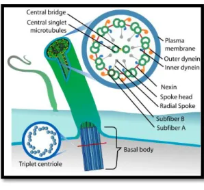
Their typical main structure is the axoneme, a cylinder of nine doublet microtubules surrounding a central pair of microtubules covered by the cell membrane, referred to as a “9+2” configuration (Mitchell, 2007). As shown in Figure 1.1, each doublet microtubule consists of A and B tubules: the A tubule is a complete microtubule with 13 protofilaments, while the B tubule contains 10 protofilaments. Moreover, the nine doublets assemble from a much shorter cylinder of nine triplet microtubules, a modified centriole named basal body, which is anchored to the cell surface and stabilized in the cytoplasm by other cytoskeletal elements (Vincensini et al., 2011).
Figure 1.1 – Schematics of a 9 + 2 configuration cilia. Representation of a basal body and a cross section of a 9+2 axoneme. Adapted by Bloodgood et al. (2010)
Historically, the flagellum axonemal structure has been studied using Chlamydomonas as model organism which has predominated in these analyses for over 30 years. Chlamydomonas reinhardtii, a unicellular, biflagellate green alga, presented unique advantages for studying eukaryotic flagella and basal bodies (Figure 1.3). These cells use flagella for swimming and for cell-cell recognition during mating, and by being situated on the surface of the cell allows them to be easily isolated for biochemical analysis (Silflow and Lefebvre, 2001).
From these studies we found that eukaryotic flagella are composed of more than 200 proteins which most of the components are also present in mammalian cilia (Ibañez-Tallon et al., 2003). Moreover, this model organism along with other ciliates, such as Paramecium and Tetrahymena, were especially important to understand processes involved in ciliogenesis and motility processes (Silflow and Lefebvre, 2001; Vincensini et al., 2011).
Ciliogenesis is a multistep process that is tightly coordinated with cell cycle progression and differentiation. The ciliary axoneme extends from the basal body which initially migrates to and docks onto the apical plasma membrane as cells enter growth arrest. Meanwhile, the axoneme elongation is controlled via intraflagellar transport (IFT), a bidirectional transport system that follows along the polarized microtubules of the axoneme, and which is required for assembly of cilia and flagella (Mitchel, 2007). This transporting process occurs between the doublets and the membrane, involving the rapid movement of particle components toward the tip (anterograde movement) or the base (retrograde movement) of the cilia, powered by motor proteins crucial for cilia assembly and protein trafficking in this cellular compartment (Vincensini et al., 2011). In the anterograde direction, IFT is
Figure 1.2 – Diversity of cilia number, length and disposition. Scanning electron micrograph of: a - Chlamydomonas reinhardtii
cell with two motile flagella with 12 μm in length (Silflow and Lefebvre, 2001); b -
Paramecium tetraurelia covered by motile cilia (Hayes et al., 2007); c – Motile cilia in mammalian trachea (Badano et al., 2006).
Figure 1.3 - Chlamydomonas reinhardtii
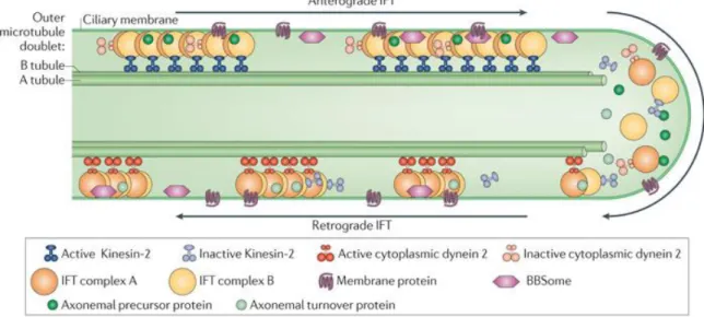
powered by kinesins of the kinesin2 family while in the retrograde direction, power is provided by dyneins of the cytoplasmic dynein 2 family (Figure 1.4).
Figure 1.4 – Intraflagellar transport machinery. The canonical anterograde intraflagellar transport (IFT) motor, heterotrimeric Kinesin-2, transports IFT complexes A and B, axonemal proteins and cytoplasmic dynein 2 to the tip of the cilium. During this anterograde motion, Kinesin-2 is active and the retrograde motor, cytoplasmic dynein2, is kept inactive to allow smooth processive anterograde movement. At the tip of the cilium, anterograde IFT trains release axonemal proteins and rearrange their conformation for retrograde IFT. Cytoplasmic dynein 2 is activated and transports retrograde IFT trains to the cell body. Figure from (Ishikawa and Marshall, 2011).
Moreover, despite their overall structural similarities, the specialization of cilia for particular functions has resulted in significant variations of structure and regulation.
1.1.1. Cilia classification
Cilia and flagella are virtually equivalent in structure and function (hereafter called cilia, there being no consistent difference between organelles with these two designations) and can be divided into two categories: primary and motile cilia (Figure 1.5).
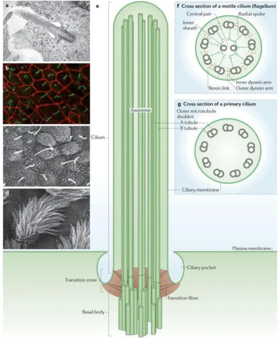
Figure 1.5 – The architecture of primary and motile cilia. a – Transmission electron micrograph of the primary cilium of retinal pigment epithelial cells; b – Immunofluorescence image of primary cilia in inner medullary collecting duct cells. The cilium (green) is produced once per cell and extends from the basal body (magenta). Cell-cell junctions are shown in red; c,d –
Scanning electron micrographs of mouse nodal cilia (c) and mouse tracheal motile cilia (d); e – Schematic diagram of the primary cilium; f,g – Cross section diagrams of a typical motile cilium (f) and a non-motile primary cilium (f) and a non-motile primary cilium (g). Figure and legend from (Ishikawa and Marshall, 2011).
Cilia were initially thought to be vestigial, but recent findings failed this hypothesis and linked primary cilia to a major cell signaling function (reviewed by Singla and Reiter, 2006) due to presence of a variety of receptors, such as ion channels and transporter proteins, as well as some of their downstream effector molecules, in the cilium and basal body.
include mechanical stimulation (transduction of the fluid flow forces through cilium bending) and chemosensation (recognition of a specific ligand, growth factor, hormone or morphogen). This significance of primary cilia in signaling became clear with the implication of its defective function on specific organ diseases and developmental disorders, commonly referred to as ciliopathies.
In contrast to the typical axonemal organization of doublet microtubules, primary cilia have absence of the central pair of microtubules (“9+0” axoneme, Figure 1.5g) along with other interconnecting multiprotein complexes, contributing this way to its non-motility (Singla and Reiter, 2006). However, these cilia are far more widespread than the motile type. For example, in humans, only a few cell types have motile cilia, namely sperm, epithelial cells in the bronchi and oviducts, and ependymal cells that line brain vesicles. In contrast, virtually all other cells have a primary cilium.
Motile cilia are normally found at high density at the surface of the cells, beating in a synchronized manner to direct fluid movement (Vincensini et al., 2011). Most of these cilia present a “9+2” axoneme structure (Figure 1.5f) and within the microtubule core there are a number of multiprotein complexes that interconnect the different components (Ibañez-Tallon, 2003). Among these are radial spokes, nexin links, central sheath and dynein arms (Figure 1.1). The dynein arms, which are divided in outer dyneins (ODA) and inner dyneins (IDA), are attached to the peripheral microtubules with certain periodicity and generate motion by ATP-dependent reactions. The other components, mainly the central apparatus and radial spokes, provide the structural interface for conducting regulatory signals to the dynein arms (Porter and Sale, 2000).
1.1.2. Dynein-based ciliary motility
To appreciate the molecular architecture of the cilium is crucial to elucidate the beating mechanism produced by this complex organelle. However, the current known structure of the cilium has not yet been fully correlated with the complex composition and localization of ciliar components needed for this process.
Mediating motility requires cilia to have an intrinsic structure axoneme, which consists in dynein arms, the molecular motors essential for the beating of these organelles. Dyneins are composed of multiple subunits thought to be preassembled in the cytoplasm before they are transported into the cilium (Fowkes and Mitchell, 1998). These called dynein arms are large protein complexes (Kobayashi and Takeda, 2011) comprising several heavy (HC of 400–500 kDa), intermediate (IC of 45–110 kDa) and light chains (LC of 8–55 kDa). Within these multiprotein assemblies, the ATPase activity that resides in the HC molecules provides the energy to produce the sliding movement between microtubules, which results in the beating of the cilium (Porter and Sale, 2000). Moreover, the capability of dynein arms to function as microtubule-based molecular motors requires the integrity of many dynein components.
positioned proximally to the cilia membrane while the inner dynein arms (IDA) are proximal to the central apparatus and have a more diverse composition.
Generation of ciliary beating involves multiple different dynein motors acting in a concerted way to specify the waveform and beat frequency (King and Kamiya, 2009).
From analyses of Chlamydomonas ODA and IDA mutants, ODAs appear to be more important for high frequency cilia beating, whereas IDAs are required for the proper formation of ciliary waveforms (Brokaw and Kamiya, 1987; Kamiya et al., 1991). In Chlamydomonas, the IDA and ODA are spaced by specific length intervals of 96 nm and 24 nm, respectively, along the axis of each MT pair (Goodenough and Heuser, 1985). Furthermore, these motors must exhibit an integrated response to various signaling factors. This requirement for coordinated action necessitates a complex control mechanism with a response time on the order of 20 ms or less. Additionally, the nexin link, which was identified as the dynein regulatory complex (DRC) (Heuser et al., 2009; Piperno et al., 1992), connects adjacent MT doublets, and the radial spokes connect the outer MT doublets to the central pair (Fig. 2). Both of these structures are thought to regulate ciliary motility.
The motor activity of dyneins is also regulated by several other componentes (Supatto and Vermot, 2011; Heuser et al., 2012): the radial spokes, which are projections originating from each peripheral doublet and directed towards the central pair; the projections associated with the central pair; and the dynein regulatory complex, which is constituted by the nexin links. The basis for axonemal movement is the sliding of microtubules relative to one another (Supatto and Vermot 2011). Dynein-generated forces that cause sliding between pairs of doublet microtubules produce axonemal bending because axonemal microtubules are anchored to the cell via the basal bodies (Supatto and Vermot 2011; Heuser et al., 2012). Despite of all the advances, the dynamics of the dynein motor itself need to be better understood.
The loss of proteins involved in this process results in cilia dysmotility. Studies of human ciliopathies have contributed to our understanding of the cilia beating process.
1.2. Cilia defects and ciliopathies
Ciliated cells can be found in various tissues throughout the human body. These include the eye, the trachea, the kidney, the reproductive tract, the intestines, the heart, and many others. In each of these tissues, the cilia perform a significant role in allowing proper function of the tissues.
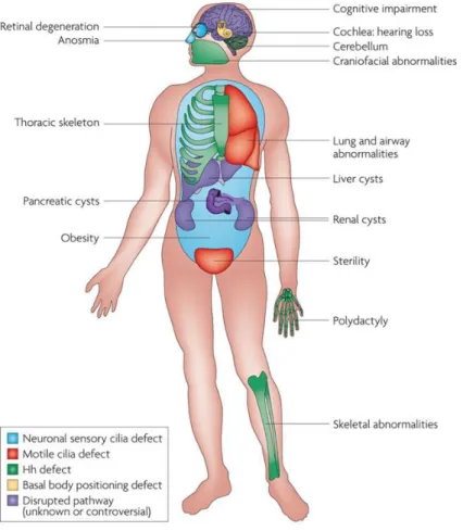
Figure 1.6 - Organs affected in human Ciliopathies. Numerous pleiotropic human disorders have been attributed to defects in cilia formation. Some aspects of these syndromes have been attributed to defective hedgehog (Hh) signalling. Other attributes of human disorders result from defective specialized cilia. Infertility observed in patients with ciliopathies is the result of defective sperm flagella and motile oviduct cilia. Figure adapted from (Goetz and Anderson, 2010).
The role of motile cilia in a number of physiological processes has been long accepted and thus the consequences of motile cilia dysfunction have four major manifestations in mammals: early embryonic death due to failure of embryonic turning; respiratory dysfunction; reproductive sterility; and hydrocephalus (Badano et al., 2006) (Figure 1.6). Ciliary dysfunctions involving motile cilia also result in body laterality defects (McGrath, 2003), like situs inversus (Afzelius, 1976). However, not all ciliopathies are related to motile cilia, some are based in defects occurred in primary cilia, like PKD (Polycystic Kidney Disease), BBS (Bardet−Biedl Syndrome), Joubert Syndrome, Oral-Facial-Digital Syndrome, Alström Syndrome or Meckel Gruber Syndrome (Fliegauf et al., 2007; Vincensini et al., 2011). In this study we will focus on motile cilia and on motile cilia ciliopathies.
1.2.1. Primary cilia dyskinesia
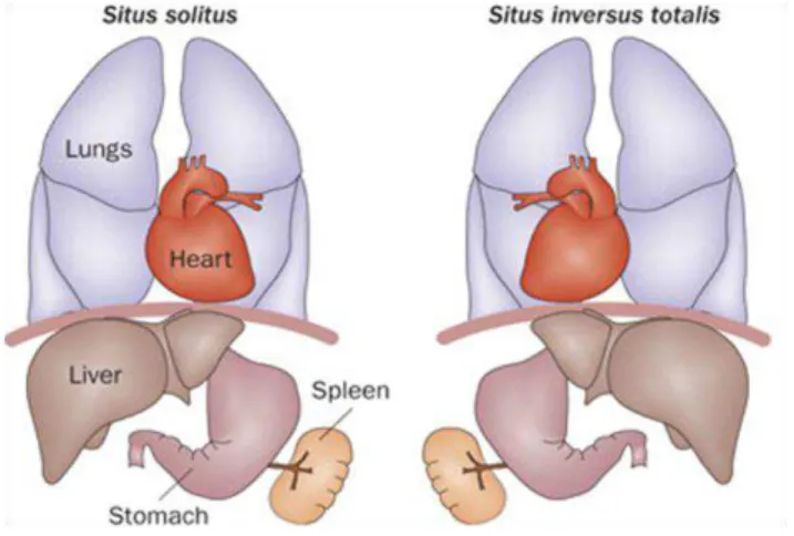
infertility. Approximately 50% of patients with PCD also have situs inversus, a condition in which the major visceral organs are reversed from their normal positions (Ibanez-Tallon et al., 2003), thereby defining the Kartagener Syndrome.
Figure 1.7 – Organ laterality defects in PCD human patients. Normal left-right asymmetry (situs solitus) and situs inversus. Figure from (Patel and Honoré, 2010).
PCD is a rare genetic condition, usually autosomal recessive, affecting approximately 1 in 15,000 people (Bush et al, 2007; Afzelius, 2004;).
One of the typical causes of motility defects is due to a range of ultrastructural ciliary axoneme defects, more than 70% involving loss of the outer dynein arms (Papon et al., 2010). Inner dyneins arms defects also have been involved in this disease where isolated absence of the IDA is reported to cause between 10 and 29% of PCD cases (Chilvers et al., 2003; Carda et al., 2005).
Ciliary motility seems to be linked to the type of ciliary defects: most of the cilia become immotile when both dynein arms and outer dynein arms are affected while the ciliary beating pattern seems atypical with reduced amplitude in case of inner dynein arm defects (Chilvers et al., 2003). Recently, Shoemark et al. (2013) also showed that depletion of inner dynein arms can also render completely immotile cilia.
The diagnosis of PCD is based on the identification of functional and structural abnormalities of cilia by TEM or/and by abnormal ciliary function (Bush et al., 2007; Armengot et al., 2012). In most patients with PCD, all the cilia share the same ultrastructural defect, as expected for a congenital disease.
However, depending on the patients, cilia have been shown to carry various axonemal abnormalities. In 10–20% of patients with PCD and situs inversus, no ultrastructural defects can be found through conventional electron microscopy, although cilia are immotile (Afzelius, 2004). For these reasons, the diagnostic validity of the ultrastructural study is limited (Bush et al., 2007), and has been complemented by high-speed videomicroscopy (Armengot et al., 2012).
abnormal. At present, guidelines and algorithms have been developed to standardize diagnostic procedures (Bush et al., 2007) in order to improve the PCD diagnostics.
The ciliary genome is highly conserved throughout the phylogenetic spectrum from simple unicellular eukaryotes to mammals, which supported the identification of the causative genes in PCD. Studies carried out in the green algae Chlamydomonas reinhardtii established the link between cilia and several genetic diseases (Vincensini et al., 2011). By the end of the 20th century, DNAI1, the human ortholog of the Chlamydomonas reinhardtii gene IC78, was identified as the first gene involved in PCD (Pennarun et al., 1999), thus emerging a novel field of research to interpret the pathophysiology of this complex disorder. Also, despite the respiratory insufficiency of these patients could be correlated with ultrastructural defects in their airway motile cilia (Afzelius, 1976), the rising question of how ciliary abnormalities are responsible for the incorrect positioning of visceral organs opened a subject of intense research interest.
1.3. Left-right patterning asymmetry
The external body plan of vertebrates is bilaterally symmetric, but several internal organs, including the heart, digestive organs and regions of the brain, display highly conserved left-right (L-R) orientations that are crucial for their functions.
Strikingly, however, these gross anatomical asymmetries arise in early embryos that are bilaterally symmetrical along the mediolateral axis. A couple decades ago, nothing was known about the molecular or genetic underpinnings of left-right asymmetric morphogenesis, and no genes with left-right asymmetric expression had been identified. Since then several discoveries have led to an emerging picture of how left-right asymmetry is initiated, stabilized, propagated, and translated into asymmetric organogenesis during development of vertebrate embryos.
1.3.1. An asymmetric cascade of signals
members of the TGFβ family that competitively bind to a class of Nodal receptors. Biochemically, Lefty2 exists as a monomer, in contrast to Nodal, which functions as a dimer. This property allows Lefty2 to diffuse faster and farther than Nodal, thereby limiting the Nodal activity to the left side. On the other hand, Pitx2, a paired-like homeodomain transcription factor, acts as the effector of Nodal signaling.
Thereby, Pitx2 has been proposed as a controller of the subsequent asymmetric morphogenetic events by regulating the gene expression mechanism essential for left-sided morphogenesis (Hamada et al., 2002).
It is known that members of the nodal and lefty cell-signaling families and pitx2 also display similar asymmetric expression patterns in the lateral plate mesoderm in chick, frog and zebrafish embryos, but how does the asymmetric pattern of Nodal pathway genes get established in the first place?
1.3.2. The role of the node in establishing L -R asymmetry in early
mouse development
The fact that the earliest asymmetrically expressed genes are at the node increased the interest for this structure as a region where the initial left-right decision might be made.
Several studies conducted in mouse indicated that the node plays an important role in the establishment of L-R asymmetry (Nonaka et al., 1998; Okada et al., 2005), being the reason why this organ is also called the L-R organizer. The mouse node (Figure 1.9) is found at the rostral end of the primitive streak. It consists dorsally of epiblast and ventrally of the most caudal aspect of the notochordal plate (Brennan et al., 2002). The node generates midline structures such as the
notochord and floor plate that act as a midline barrier required for preserving the correct laterality (reviewed in Brennan et al., 2002).Remarkably, the cells on the ventral surface of the mouse embryo node each have a motile monocilium projecting from their apical surface that have a clockwise rotator beating pattern.
Figure 1.9 – Scanning Electron micrographs of a mouse node. a – The node is at the apex of the egg cylinder, and the head process, which give rise to the notochord, extends anteriorly. b – Close-up showing the cup-like shape of the node. The cilia motility generates a leftward fluid flow (red arrow). The anterior (A) is oriented to the top. Figure adapted from (Vogan and Tabin, 1999).
The strongest arguments for a role for nodal flow in the establishment of L-R asymmetries come from cultured mouse embryos in which externally applied rightward fluid flow is capable of reversing L-R patterning in wild-type (WT) embryos and externally applied leftward flow is able to rescue L-R patterning in mutants that would normally have inverted L-R orientation (Nonaka et al., 2002). Also, submitting iv mutant embryos to external flow restored their normal situs, which otherwise would have developed randomized asymmetry. But how do a directional nodal flow functions in establishing L-R asymmetry?
1.3.3. The two left-right models
Since the discovery of nodal flow, two hypotheses have evolved to explain the mechanism by which L-R is transferred to the LPM.
The first of these was named the ‘morphogen hypothesis’. It proposes that directed motion of the nodal cilia leads to a unidirectional transport of a secreted morphogen to the left side of the node (Nonaka et al., 1998; Okada et al., 1999). This simple model suggests that ciliary beating guarantees that one side of the node preferentially receives greater concentration of a morphogen than the other side (figure 1.10a). Thus, the asymmetry in the distribution of the morphogen then triggers signaling events that strengthen the asymmetry in the developing embryo. However, this hypothesis raises several questions, primarily among them being the identity of the morphogen itself. Later evidences came in this direction from the work of Tanaka et al. (2005). This group observed flowing objects inside the node cavity. These particles, which they termed nodal vesicular parcels (NVPs), were seen to be released into the flow and to fragment upon contact with the ciliated surface, thereby releasing their contents on the left side of the node. But which is the nature of this putative morphogen? Tanaka et al. (2005) also found that Sonic Hedgehog (Shh) and Retinoic acid (RA) are associated with the NVPs, and are released into the nodal flow in a fibroblast growth factor (FGF)-signaling-dependent manner. Based on these findings, Cartwright and colleagues (Cartwright et al., 2006) modeled the movement of NVPs across the mouse node and verified that the flow could definitely cause them to accumulate on the left side of the node, which would be needed for symmetry breaking. However, based on the biophysical properties of node such as high viscosity of the fluid, they disagreed that the morphogens could be delivered to the surrounding cells by their mechanical rupture either by the action of cilia or the flow. Alternatively, they hypothesize that if there is rupture it must be induced by a biochemical mechanism not yet discovered (Cartwright et al., 2006).
Regardless of these fascinating findings, the NVP model of the ‘morphogen hypothesis’ has not been further supported. Most importantly, genetic analysis of Sonic Hedgehog and the Retinoic acid pathways do not supply convincing support of their roles as nodal flow morphogens (Vermot et al., 2005; Zhang et al., 2001).
dividing the ‘nodal flow’ process into two steps: generating the flow, and responding to the flow by two distinct kinds of cilia.
The work that suggested cilia could be implicated in the generation and also the sensation of nodal flow came from the known function of the polycystic kidney disease 1 (Pkd1) and polycystic kidney disease 2 (Pkd2) genes in the kidney tubule cells - the proteins encoded by these genes form a functional mechanosensory complex that detects urine flow and gives rise to a Ca2+ signal response (Nauli et al., 2003).
Although Pkd1 is not required for L-R determination and is not expressed in the node (Tanaka et al., 2005), Pkd2 mutant mice exhibited many features that illustrate abnormal L–R development, thus involving Ca2+ signaling in the establishment of L–R asymmetry (Pennekkamp et al., 2002).
Recently it was discovered that Pkd1l1 is the functional partner of Pkd2 in the L-R organizer being conserved in medaka and mouse (Field et al., 2011). Additional support of this possibility appeared from the examination of Pkd1l1-Pkd2 localization on nodal cilia. While Lrd, the dynein protein required for ciliary motility, localized to the motile cilia on the central cells of the node pit, Pkd2 was present on the motile cilia as well as the immotile Lrd-negative cilia on the perinodal cells that surround the node (McGrath, 2003).
Furthermore, in the Kupffer’s vesicle (KV) of medaka, which is the fish homologue of the mouse node, where all cilia are visibly motile, it was also proposed that cilia have a double role in producing the characteristic fluid flow and interpreting it through Pkd1l1-Pkd2 sensor-channel complexes expressed in all cilia (Kamura et al., 2011). In zebrafish it is still not known if pkd1l1 plays a role in L-R determination, while Pkd2 role is definitely conserved.
Nonetheless, since the molecular sensors of nodal flow are still unknown, both chemosensory and mechanosensory hypotheses are plausible in the L-R organizer.
1.3.4. Left-Right asymmetry in zebrafish
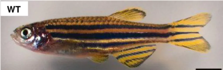
Zebrafish (Danio rerio) is a small teleost fish (Figure 1.11) and has emerged as one of the leading models for studying development. As a vertebrate organism, this fish presents many organs and cell types similar to that of mammals (reviewed in Lieschke and Currie, 2007).
Figure 1.11 – Adult zebrafish– wild-type female zebrafish. Scale bar = 4mm. Figure from (Parichy et al., 2009).
The zebrafish embryos are transparent (Figure 1.12) and also develop rapidly (Kimmel et al., 1995), thus, allowing real-time imaging of developing stages in embryos and larvae. In addition zebrafish proved to be more advantageous over previous model organisms in many ways. For example early developmental processes are less accessible in the mouse because they occur in utero.
Moreover, an individual female can produce hundreds of eggs in each clutch, enabling huge numbers of progeny to be generated, facilitating the detection of rare mutations.
Through the 1980s, the development of zebrafish genetic techniques, such as ‘cloning’, mutagenesis, transgenesis and mapping approaches, underpinned the use of zebrafish to apply invertebrate-style forward genetics to questions of vertebrate development (reviewed in Lieschke and Currie, 2007).
Over the past decade, the zebrafish has proven to be very useful regarding genetic manipulation of laterality. Studies using zebrafish have also provided insights into how the Kupffer’s vesicle, the fish homologue of the mouse node, relays L-R asymmetry information to the lateral plate mesoderm (Long et al., 2003; Wang and Yost, 2008).
1.3.4.1.
Zebrafish Kupffer’s vesicle
Kupffer’s vesicle (KV) is the equivalent to the mouse node ciliated organ of asymmetry in the zebrafish embryo that initiates L-R development of the brain, heart and gut (Essner, 2005). First described in 1868 by Kupffer, KV is a conserved structure among teleost fishes. In zebrafish, KV is
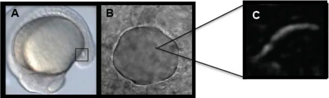
formed from the dorsal surface epithelial cells, known as dorsal forerunner cells (DFCs), a group of approximately two-dozen cells that migrate at the edge of the embryonic shield at the beginning of gastrulation. DFCs move to the vegetal pole in close contact with the overlying enveloping layer margin and become transformed into an epithelial vesicle (KV). During subsequent somite stages, KV formation is completed with the generation of motile monocilia on the apical membranes of KV cells facing the lumen (Figure 1.12). It constitutes a small and distinctive epithelial closed vesicle containing fluid, located mid-ventrally posterior to the yolk cell, and is transiently present during most of the segmentation period (Amack et al., 2007; Kimmel et al., 1995; Kramer-Zucker, 2005).
Figure 1.12 –Images of zebrafish KV and cilium. A - Localization of the KV (squared region) in the body of the zebrafish embryo at 14 hpf. B - Snapshot image of a Kupffer’s vesicle of a live embryo filmed from the dorsal side. C - Snapshot image of a KV beating cilium in a live embryo.
The monociliated cells lining the epithelium of the KV have a 9+2 arrangement (Kramer-Zucker et al., 2005; Kreiling et al., 2007) and generate a counterclockwise fluid flow. This flow promotes intracellular Ca2+ elevation in cells localized on the left side of KV (Francescatto et al., 2010; Sarmah et al., 2007) and is involved in establishing and maintaining the L-R asymmetry of the body axis (Essner, 2005; Kramer-Zucker, 2005). Even so, the presence of sensory cilia is still a subject of discussion in the zebrafish (Borovina et al., 2010; Okabe et al., 2008).
At present, it is assumed that KV is analogous to the mouse node in terms of L-R patterning (Essner, 2005). While the ciliated surface of the mouse node is reasonably flat, KV is a hollow sphere containing cilia, projecting both from dorsal roof and ventral floor (Kreiling et al. 2007; Amack et al., 2007). Kramer-Zucker et al. (2005) reported that cilia rotate counterclockwise when observed from the apical side in the KV, which is contrary to what is observed in the mouse node.
bilaterally in KV cells at the 6-somite stage, but its expression changes to a right-sided asymmetric pattern at the 8 to 10 somite stages in a fluid flow-dependent manner (Lopes et al., 2010). Since Charon binds to Spaw and antagonizes Spaw functions (Hashimoto et al., 2004), the rightward gradient of Charon around KV tends to inhibit Spaw strongly in the right side. Due to the opposing gradients of activator (Spaw) and inhibitor (Charon), Spaw cannot stimulate expression of spaw on the right side of the LPM. On the other hand, Spaw induces its own expression on the left-side LPM by positive feedback regulation which indicates that opposed gradients between Spaw and Charon around KV may contribute to initiating spaw expression in the left-side LPM.
Okabe et al. (2008) hypothesized that the fluid flow in zebrafish KV is analogous to the flow in the mouse node and medaka fish in terms of L-R patterning, although the ciliated cell arrangement appear to vary in structure. According to Kamura et al. (2011), all medaka KV cilia are motile, whereas in the mouse node McGrath et al. (2003) showed the presence of two populations of cilia, one motile and other immotile. In zebrafish, it is still not clear if all cilia are motile.
Although a unidirectional fluid flow is evident in the mouse node, the zebrafish KV has a more intricate internal arrangement of cilia contributing to the generation of a more complex flow that needs to be investigated.
1.3.4.2. Motile cilia generated flow
in Kupffer’s vesicle
Ciliary beating is characterized by a series of bends, originating at the base of the structure and propagated toward the tip. High-speed microscopy allows the waveform of the beat to be seen and it is currently used as an important tool for diagnose of ciliopathies such as PCD by studying the cilia beat frequency (CBF).
An important factor dictating cilia-mediated hydrodynamics is the type of beat they generate. Beating can be planar or three-dimensional and it can be described by its amplitude, wavelength, and frequency. The bends push against the surrounding fluid, propelling the cell forward or moving the fluid across a fixed epithelium.
The beat pattern appears to be related with the inner organization of cilia but many unsolved questions remain regarding the correlation of structure and cilia beat in different developing organs.
The nexin link, which was first identified as the dynein regulatory complex (DRC), connects adjacent MT doublets, and the radial spokes connect the outer MT doublets to the central pair. In humans and zebrafish, mutants for the radial spoke heads showed to affect cilia motion (Castleman et al. 2009) and that DRC is critical for proper cilia motility in zebrafish (Colantonio et al. 2009). These two structures are considered to regulate ciliary motility. In addition, motility requires axoneme-associated dynein arms to generate the sliding of microtubules and thus motion (reviewed in Vincensini et al., 2011).
led to the assumption that the absence of the CP apparatus confers the rotary pattern of beating to the node cilia. However, some studies suggest that this interpretation is unlikely to be correct. Teleost fishes such as the zebrafish and medaka also make use of ciliary motility to establish L–R asymmetry (Essner et al., 2005; Kramer-Zucker et al., 2005). In these species, motile cilia exist in in Kupffer’s vesicle (KV). Although medaka KV cilia are 9+0 and beat in a rotary manner (Okada et al., 2005), the zebrafish has CP-containing KV cilia (9+2 structure), and yet they too demonstrate rotational motion (Kramer-Zucker et al., 2005).
Similarly, contrary to the conventional view, it has been recently reported that the mouse node cilia, which have a circular motion, also contains cilia with CP (Caspary et al., 2007). Thus, the presence or absence of the CP appears not to decide the beating pattern of the cilium.
Moreover, genetic confirmation does favor the hypothesis that the CP is dispensable for nodal cilia motility. In mice and humans, mutation of genes that are necessary for the assembly or function of the CP do not have an affect on laterality, while the planar motility of different 9+2 cilia (such as those in the airways) is strongly affected (Lechtreck et al., 2008; Olbrich et al., 2012). Together, these results demonstrate the mechanisms that confer rotary beat pattern to the nodal cilia remain an unresolved question.
Another challenging problem is how rotational movement of the cilia could be linked to directional flow.
1.3.4.3. Fluid hydrodynamics
To understand the role of beating in producing fluid flow it is important to first have a look on how does fluid behave at microscales in a confined compartment such as the KV.
Fluid dynamics are ruled by laws that are complex at this microscale and the resulting fluid flow exhibits properties that are not obvious when used to human scale (Supatto and Vermot, 2011).
In fluid mechanics, the Reynolds number (Re) is a dimensionless number that defines the nature of a fluid flow and the relative contribution of inertia and viscous dissipation. The cilia-driven flow involved in zebrafish development shows characteristic scales, L < 100μm and U < 100 μm s-1
(L = length; U = typical velocity) (Supatto and Vermot, 2011). Using the kinetic viscosity of water (ν ≈ 106 μm2 s-1), the resulting Re is < 10-2. The flow produced by beating cilia is described by a low Re (Re << 1) environment, and it is governed by Stokes equations - being referred as ‘Stokes flow’ (Smith et al., 2012).
and the time reversibility of ‘Stokes flow’ equation presents fundamental properties of the flow that can be generated by motile cilia (Supatto and Vermot, 2011).
1.3.4.4. Directional flow
Most cilia and flagella generate flow by an unidirectional power stroke, which is planar or almost planar, and in the direction of the flow (planar beating), while nodal cilia rotate in 3D.
Based on ‘Stokes flow’ equation, the first obstacle to the generation of a directional flow is the difficulty to achieve a net flow (Supatto and Vermot, 2011). The absence of inertia results in a velocity that is proportional to the force applied to the fluid (Supatto and Vermot, 2011; Smith et al., 2012). This means that a specific mechanism is required to produce a directional fluid flow from the rotating cilia because the simple circling movement of the nodal cilia would only produce a vortex instead of biased flow (Buceta et al., 2005). According to previous studies (Cartwright et al., 2009; Smith et al., 2012), in order to produce a directional flow at low Re, a beating cilium needs an asymmetry either in space or shape.
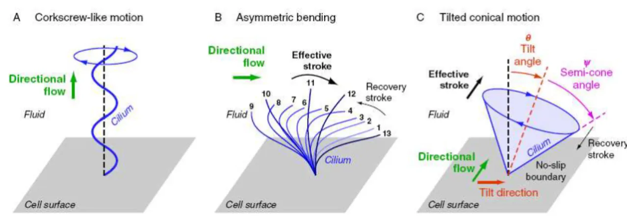
Most likely due to the cilia ultrastructure, length, and/or orientation, three types of spatially asymmetric cilia beating patterns have been proposed theoretically and observed experimentally in different organs of developing embryos: the ‘corkscrew-like motion’ (Kramer-Zucker, 2005), the ‘asymmetric bending’ (Scheweicbert et al., 2007), and the ‘tilted conical’ motion (Okada et al., 2005), represented in Figure 1.13. From these three the ‘tilted conical’ motion was observed experimentally in mouse, rabbit, and fish L-R organizer. This specific type of cilia beating pattern is characterized by having a directional flow generated by cilia that exhibits a symmetrical circular motion without asymmetric shape as in the two previous cases.
In this case, theoretical fluid dynamics simulations of nodal cilia rotation could foretell that a linear directional flow will result if the rotational axes of the cilia are tilted (Cartwright et al., 2009).
Here, the cilium interaction with the cell surface plays a critical role (Cartwright et al., 2006; Smith et al., 2008; Smith et al., 2010; Smith et al., 2012;): such a tilt will make sure that the effective stroke (towards the left side) will be much more efficient than the recovery stroke (towards the right side), because the latter will need to move fluid much closer to the cell surface, where viscosity is higher (no-slip boundary condition). Thus, the direction of the net flow generated depends on the cilium tilt and on the cell surface, and must be perpendicular to the direction of the cilium tilt and parallel to the surface.
Experimental authenticity of this prediction initially came from a analysis of the dynamics of ciliary beat. Using high-speed videomicroscopy, two different groups found that the rotational axes of the mouse nodal cilia are indeed tilted towards the posterior (Nonaka et al., 2005; Okada et al., 2005) promoting a directional flow toward the left direction with cilia rotating clockwise. It was also observed that the surface of the nodal cells was conspicuously convex, and that the position of basal body, the structure to which cilia are anchored at the cell surface, switched to the posterior side from an initial central location. Even more remarkably, Nonaka et al. (2005) demonstrated that motorized ‘artificial cilia’ could indeed drive a net leftward flow of viscous liquid silicone when their rotational axes were posteriorly tilted.
On the other hand in zebrafish it is noted that the beating pattern of KV cilia is different (counterclockwise motion) from the mouse nodal cilia, which presumably is because of different ultrastructures. This implies the counterclockwise fluid flow circulating around the dorsal-ventral axis within the KV cannot be due to a posterior tilt, but due to a dorsal tilt (Supatto and Vermot, 2011). In fact this has been already observed in vivo in previous studies. Kreiling et al. (2007) showed that cilia are concentrated in the dorsal-anterior region of the vesicle and that those cilia tilt is towards the already-established posterior and dorsal axes. This, thus, allows the viscous interaction of fluid with the cell surface to produce the continuous counterclockwise flow (Smith et al., 2012; Supatto and Vermot, 2011).
In recent years, several models and simulations have been developed based on mouse node experimental data. In these theoretical studies, the cilium is modeled either as an microscopic sphere rotating in its place (Cartwright et al., 2006), a small sphere moving on a fixed trajectory in the surrounding area of a planar surface (Vilfan et al., 2012) or a slender body (Smith et al., 2008). Whatever the complexity of the model used, they are a great advantage to understand how biological processes occur, by explaining new observations, raising specific predictions, and suggesting the next set of experiments.
1.4. Project goals
The main goal of this Master project was the characterization of the fluid flow dynamics in the Kupffer’s vesicle (KV) of zebrafish embryos. It was our objective to understand how fluid flow dynamics could modulate the mechanisms responsible for the establishment of the left-right asymmetry.
To answer this question we decided to tackle the problem by using two different approaches: experimental characterization of the KV fluid flow in zebrafish embryos, and development of mathematical simulations for the KV flow in collaboration with David Smith group from Birmingham.
A tissue-specific screen previously done in deltaD-/- mutants in our lab underpinned the beginning of my Master project. These mutants were previously reported by Lopes et al., (2010) to present left-right defects and also KV fluid flow abnormalities. With this screen we found several differential expressed genes in deltaD-/- mutants, compared to wild-type embryos. Having identified some motile cilia-related genes, a validation study of gene expression was realized to 3 of these genes by a previous lab colleague. The validated genes were dnah7, rsph3 and foxj1a. With this Master project we wanted to test if deltaD-/- mutants had cilia defects and whether that could partially explain the defects in L-R patterning reported (Lopes et al. 2010). So considering what could be the best candidate responsible for the KV fluid flow problems seen in the mutants we decided to focus our analysis in the dnah7 gene. Zhang et al. (2002) identified Dnah7 as an inner dynein arm component of human cilia important for its motility, and reported cilia from a PCD patient with mutations in Dnah7. Therefore we aimed to dissect the contribution of dynein Dnah7 deregulation to cilia defects and consequently in the KV fluid flow, by making use of the anti-sense technology of morpholinos in zebrafish embryos.
The first part of the project consisted in characterizing the fluid flow dynamics. Considering that we proposed to evaluate different parameters that inherently contribute to the fluid flow generation: cilia beat frequencies, by using high-speed videomicroscopy; and cilia lengths, through immunofluorescence assays. These measurements were conducted in three different genetic backgrounds: Wild-type (WT) and deltaD-/- mutant embryos and dnah7 morphants. After we studied the cilia-generated fluid flow inside the zebrafish KV by tracking native particles and calculating their velocity in a non-invasive assay. We intended to analyze different situations where the KV fluid flow was disrupted so we used the 3 different genetic backgrounds that allowed us to get a range of motile cilia inside the KV.
To have an insight of dnah7 mRNA distribution throughout embryogenesis, we also evaluated the dnah7 gene expression pattern throughout several stages of embryo development using whole-mount in situ hybridization (WISH).
The second part of this project consisted in characterizing several parameters needed to input into the mathematical model that was being developed: KV volumes and cilia numbers in live embryos, with the help of two lab colleagues, Barbara Tavares and Petra Pintado.
CHAPTER 2
2.1. Zebrafish mating
General maintenance, collection and staging of zebrafish were carried out at the IGC zebrafish facility (Oeiras, Portugal) as described by Westerfield, (2000).
Zebrafish fertilization and embryo isolation outlined the basic steps to get embryos for use under the zebrafish line needed for each experiment. Wild type (AB strain) and deltaD-/- mutant embryos were staged according to Kimmel et al. (Kimmel et al., 1995). The development stages are given in hour post-fertilization (hpf) and day post-fertilization (dpf) according to morphological criteria. For somite-stage embryos, the number of somites was used as a proxy of developmental time.
Adult male and female zebrafish selected from the main-tank system were put together in a top compartment of a mating-tank and stayed in the dark overnight (O/N). To ensure that male fish would not mate with the females during this period, fish of the opposite sex were separated by a divider. At the onset of the light cycle, zebrafish would normally initiate breeding behavior that results in the laying and fertilization of eggs (Westerfield, 2000). So in the beginning of the fish light cycle the dividers were removed allowing the mating process. The females laid eggs, which were externally fertilized by the males, and fell to the lower compartment of the tank through a sieve, preventing the egg cannibalism by the parents. Afterwards the adult fish were moved back to their home tank and the collected eggs were sorted into a Petri dish with embryo medium (5 mM NaCl, 0,2 mM KCL, 0,3 mM CaCl2, 0,3 mM MgSO4, ddH2O – pH 7,2). These eggs stayed in temperature-controlled (28ºC)
incubators until the embryos reached the desired stage. May be say here that in order to perform imaging at 8-10 somite-stages in the next morning the incubation temperature was optimized to 25ºC.
2.2.
Recording of cilia beat frequencies in the Kupffer’s Vesicle
The use of light microscopy coupled with high-speed imaging in cilia beat frequency studies yields useful data that allows to identify altered ciliary beat patterns that may be related to specific ultrastructural defects. To characterize cilia motility in the L-R organizer of WT and zebrafish mutants, we analyzed the CBF of several individual cilium by filming live embryos with high-speed videomicroscopy.
2.2.1. Mounting zebrafish live embryos for KV imaging
(FluoroDishTM, WPI Inc, China). This design allowed to mount and maintain live embryos still with the KV dorsal roof facing the bottom of the mould.
At room temperature, live embryos between 6-7th somite-stages were gently dechorionated with sharped forceps and placed in the agarose mould filled with embryo medium to keep the embryos moist. With the help of a microloader pipette tip, embryos were placed correctly inside the mold wells with the dorsal roof of the KV facing down.
2.2.3. Microscope setup
Mounted embryos at 8-10th somite-stages were set under the 100x/1.30NA oil immersion objective lens on a Nikon Eclipse Ti-U inverted brightfield light microscope at room temperature. After finding and focusing the KV, images were recorded with a high speed FASTCAM MC2 camera (Photron Europe, Limited) and controlled with PFV (Photron FASTCAM Viewer) software.
2.2.4. Recording KV cilia
–
image acquisition
Kupffer’s vesicle was scanned through the different focal planes along the dorsal-ventral axis in order to observe as many cilia as possible. Since this spherical organ has cilia projecting from all the cells lining the fluid filled space (Okabe et al., 2008), these can be visible from multiple different orientations. Once a region of interest inside the KV was chosen, focused cilia were recorded and its anterior-posterior and left-right position was annotated, allowing to map each cilium inside the KV.
The recorded videos were produced by the collection of consecutive images known as frames, taken at a properly selected rate expressed in frames per second (fps), for a finite time period. Accurate measurements of cilia beat frequencies were dependent on the frame rate at which images were acquired. The Nyquist-Shannon sampling theorem implies that to measure the frequency F0of
cilia, image acquisition should be performed at a rate more than twice as fast (Jaffe et al., 2010), which is a necessary condition to avoid aliasing (motion artifacts). Therefore acquisitions at even faster rate are highly recommended (typically, F > 3-4* F0). This higher acquisition rate compensates
Each movie was produced by a total number of 1000 frames which corresponds to a 2 second movie at a 500 fps frame rate. To validate that 2 second movie was a significant time window to correctly characterize the cilia beating pattern during the 8-10th somite stages we decided to do a live time course imaging with an embryo during these developing stages. A focal plane of the KV was chosen from one zebrafish embryo at 8th somite stage and the cilia focused on this focal plane were recorded every 10 minutes during 1 hour at room temperature (approximate time to reach 10 somites) with the same experimental setup described before. The six movies collected during the time course showed that during the 8-10th somite stages cilia had a consistent cilia beat pattern (see Figure S1 in Annex II).
In summary, for this project the KV cilia were recorded during 2 seconds at a 500 fps rate. This experimental procedure allowed us to measure 5 to 45 Hz CBF, which is the frequency range previously reported, providing novel information on zebrafish KV cilia motility.
2.2.5. Image processing and kymograph design
Ciliary movement was recorded for each pixel within the image resolution, resulting in a set of temporal waveforms with as many members as there were pixels. Each waveform depicted the pixel intensity as a function of time, displaying the periodicities of the ciliary movement (Olm et al. 2011) in a graph called kymograph (Figure 2.2).
CBF analysis had to follow a previous movie image filtering procedure using ImageJ program
(http://imagej.nih.gov/ij/) to extract the resulting kymographs, before passing by the Fourier analysis in
Figure 2.1 -Snapshot image of a KV beating cilium in a live embryo –cilia image after background subtraction and image improved contrast for posterior kymograph analysis.
Figure 2.2 -Kymograph showing a cilium beating pattern– orthogonal view of a cilium beating at a single fundamental frequency, during a time of 2 seconds (1000 frames).
2.2.6. CBF spectral analysis
Ciliary movement was recorded for each pixel within the drawn line in the kymograph, resulting in a set of temporal of waveforms which reflected the cilium beat pattern. The aim of the spectral analysis was to estimate, based on this set of waveforms, the proportion of each CBF value in relation to the overall movement of the cilia within the drawn line, accomplished through Fourier analysis.
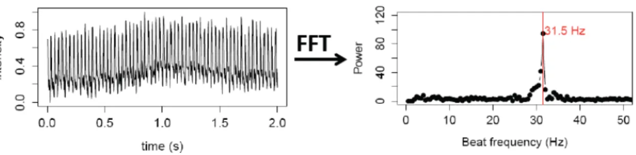
As described previously, a list of plot values was obtained from the resultant waveform of each cilium kymograph, and was imported to R program. In collaboration with Dr. Adán Guerrero (Instituto Gulbenkian da Ciência) we developed a script for R program, which applies the FFT algorithm to KV cilia data (“FFT script function” in Annex).
The FFT script function resolves a time waveform (signal) into its sinusoidal components, meaning that this function takes the time-domain data, which is represented by the list of values obtained previously, and returns the Fourier spectrum of the data. This spectrum interprets the ciliary movement by describing how the power of the signal is distributed with frequency.
Figure 2.3 –Characterization of KV cilium motility - Time series over 2 seconds for the same beating cilium, and the resulting power spectrum after analysis using Fast Fourier Transform (FFT) for the same cilium showing that it has only one fundamental frequency of 31.5 Hz.
Significant cilia beat frequencies were selected from their Fourier spectra based on local maxima criteria using a frequency window of 4Hz. Only local maxima that were beyond 2Hz and above a 13 power limit were considered in order to remove the influence of any signal noise. These parameters made the algorithm very robust, allowing a confident spectral analysis.
2.2.7.
Kupffer’s ve
sicle fluid flow velocity measurements
For all the genetic backgrounds we measured the KV fluid flow by a non-invasive method described as in Lopes et al. (2010). This method consists in tracking naturally occurring particles that move with the fluid flow.
Differently to the protocol described for cilia imaging here we used a frame rate of 60 fps in a 2000 frames time window (~33 seconds). Particle movement was recorded after selecting several focal planes of the entire KV where one or more particles were observed.
The main difference in this methodology to that used in Lopes et al. (2010) occurred in the posterior movie analysis. In ImageJ program all the videos analyzed were initially rotated in a way that the anterior region of each KV was oriented upwards. Afterwards, we used the ImageJ plugin MTrackJ to follow each individual particle (http://www.imagescience.org/meijering/software/mtrackj/) in a 0.2 second interval allowing to measure instant velocity of the particle movement.
Particles were tracked manually and the ImageJ plugin returned a list of coordinates for each track point (x and y) and a list of distances between two consecutive point tracks. These data were imported to an Excel datasheet and particle velocity was calculated.
each second a different color (‘Macro’ function in Annex), being red the color used to identify the first second of the movie.
The quantification of such flow speed was achieved by created boxplots that represented the different flow speed contributions in each region of the KV, and heat maps that visually showed local flow speed (‘Boxplot script function’ in Annex).
This procedure was then used to analyze the entire data correspondent to WT, deltaD-/- and dnah7 morphants embryos and provide an overall view of the fluid flow dynamics for each genetic backgroung.
It is important to refer that our experimental observations and mathematical calculations were performed in the midplane of the KV. The microscope setup allowed us to track particles in a depth of field below 1μm. Elementary mathematical analysis shows that if the z-component Vz is small, it makes a contribution to the velocity magnitude which is very well approximated by -(1/2)*(Vz^2/sqrt(Vx^2+Vy^2)). Essentially a small component Vz changes the velocity magnitude by a very small amount, which allows us to say that our speed measurements are correct and present a negligible error.
Moreover, each filmed embryo was transferred to a 12 well test plate (TPP Techno Plastic Products AG, Switzerland) in embryo medium and incubated at 28ºC for later individual heart and gut scoring.
2.3. In silico experiments - Mathematical modeling
In order to better understand the flow fields involved we mathematically modeled KV fluid flow. In collaboration with Drs. Andrew Smith, Thomas Montenegro-Johnson and David Smith, we computed the most realist mathematical model of the KV using experimental data previously collected for KV volume, CBF, cilia number and cilia length.
To interpret the effect of cilium number and distribution on the flow generated within KV, we used a modified version of the computational model of Smith and Johnson et al. (Smith et al., 2012) described in detail in the Annex II section. These modifications allowed for arbitrary placement of cilia, multiple beat frequencies and variable lengths. Cilia were modeled as whirling rods, with diameter 0.3 μm, programmed to perform a conical rotational motion with dorsal roof and ventral floor cilia tilted posteriorly and 'equatorial' cilia tilted dorsally.