Ana Rita Pereira Oliveira
Diet and exercise – impact on lipid
metabolism and involvement of
epigenetic mechanisms
Ana Rita Pereira Oliveira
Diet and exercise – impact on lipid
metabolism and involvement of
epigenetic mechanisms
Master thesis
Applied Biochemistry – Biomedicine
Work performed under the orientation of:
Prof. Dr. Cristina Pereira Wilson
Prof. Dr. Maria João Martins
Nome: Ana Rita Pereira Oliveira
Endereço eletrónico: ana.rpo@hotmail.com Telefone: 912028476
Número do Cartão de Cidadão: 13986221 8 ZZ5
Título da tese: Diet and exercise – impact on lipid metabolism and involvement of epigenetic mechanisms
Orientadores: Professora Doutora Cristina Pereira-Wilson; Professora Doutora Maria João Martins.
Ano de conclusão: 2015
Mestrado em Bioquímica Aplicada, Ramo de Biomedicina.
DE ACORDO COM A LEGISLAÇÃO EM VIGOR, NÃO É PERMITIDA A REPRODUÇÃO DE QUALQUER PARTE DESTA TESE/TRABALHO
Universidade do Minho: 30 de janeiro de 2015
Queria deixar aqui o meu sincero agradecimento à Professora Doutora Cristina
Pereira-Wilson, mentora deste projeto, pela amabilidade com que me orientou, aconselhou e ajudou a ser
sucedida nesta etapa. Muito obrigada por sempre ter acreditado nas minhas capacidades.
Queria agradecer também à Professora Doutora Maria João Martins por toda a sua
disponibilidade e simpatia, sem a sua colaboração este trabalho não seria possível.
O meu maior obrigada às meninas do
meu
grupo de trabalho no laboratório:
Dalila, pelo terno acolhimento, por todos os ensinamentos, conselhos e ajuda desde que
comecei este trabalho, e acima de tudo pela bondade com que sempre aceitaste as minhas perguntas
inexperientes.
Cristina, que mesmo não querendo mostrar tens um coração mole, obrigada pela introdução
ao “mundo” do PCR, por todas as dicas, pelos momentos de descontração e pela partilha de
conhecimento.
Carlinha, pela tua amizade, todos os abracinhos e gargalhadas, que proporcionaram o
melhor dos ambientes de trabalho, mas principalmente o meu grande obrigada pela tua opinião,
ajuda e conselhos, sempre amavelmente pronto. Claro, que dizer-te obrigada por isto nunca será
suficiente, mas ainda assim aqui fica a minha tentativa.
Cátia, comigo desde o primeiro passo desta jornada, obrigada pela partilha na dificuldade e
superação.
Ao LBA/LCCA melhor laboratório do Departamento, que cativa também as melhores
pessoas, desde os superiores, em especial, Marisa, Ana, Suellen e Artur, aos colegas mestrandos que
retiraram o árduo ao trabalho diário, e tornaram a dificuldade em aprendizagem, alegria, partilha,
animação, companheirismo e entreajuda, em especial à Cidália, Tatiana, Rita, Patrícia e Cátia. A
todos vocês um muito obrigada.
Obrigada a todas as pessoas com quem diariamente me cruzei nos corredores, desde os
funcionários aos docentes, sem esquecer os colegas. Porque na vida aprende-se por “osmose”.
os dias especiais. Tenho o prazer de ter pessoas assim na minha vida e por isso vos agradeço imenso.
Quem está no meu coração sabe-o perfeitamente, sou privilegiada pelas pessoas que me rodeiam.
Por fim, mas de todo não menos importante: ao meu irmão Célio e aos meus queridos Pai e
Mãe – a família perfeita, que sempre me incentivaram para ser melhor, que me apoiam
incondicionalmente e que me mostram todos os dias que na vida é preciso lutar, mas que lutando
tudo se consegue. Eles são os responsáveis pelo que sou hoje, por tudo o que alcancei até aqui e por
tudo o que anseio alcançar no futuro.
Obrigada a toda a minha família mais chegada, mesmo os que já não estão aqui presentes,
em especial à minha avó Felismina pelo exemplo extraordinário de garra, superação, conselho sábio
e boa disposição sempre presente.
“To work is elevating, to accomplish is superb.” – Mr. Selfridge.
epigenetic regulation
Abstract
A sedentary lifestyle and consumption of high-caloric and micronutrient pour diets are strong risk factors associated with the development of cardiovascular diseases, obesity, dyslipidemia and colon cancer, where deregulation of cholesterol metabolism plays an important role. This thesis is divided in two experimental parts: Part A: Effects of exercise on cholesterol metabolism in high-fat fed rats: involvement of epigenetic mechanisms; and Part B: Effects of naturally occurring dietary flavones on liver cholesterol metabolism, in vitro.
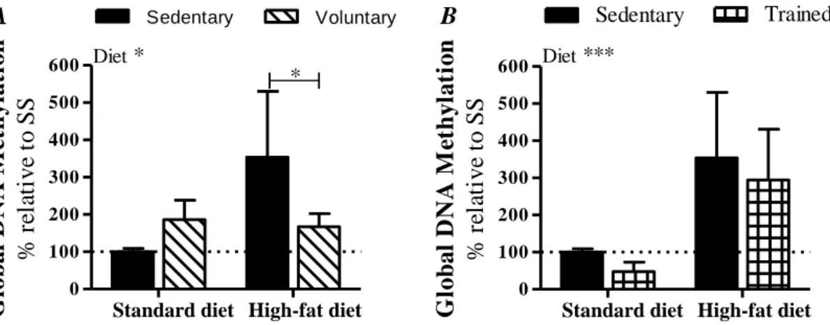
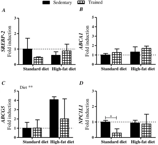
In our study (Part A), were analysed colon samples from Sprague-Dawley rats submitted to 2 iso-hipercaloric diets: standard diet (35 % of calories derived from fat), and high-fat diet (71 % of calories derived from fat), and each group further divided in 3 sub-groups according to the exercise regime: sedentary, voluntary physical activity and endurance training. The consumption of a high fat diet significantly increased global methylation levels. However, in both diets DNA methylation seem to be normalized by voluntary exercise, while endurance training had no effect. Our qRT-PCR analysis did not show differences in SREBP-2 and ABCA1 expression. However, high-fat consumption increased gene expression of the efflux transporter of neutral sterols, ABCG5, which was reverted by voluntary physical activity. Our study diets seem not have an effect on NPC1L1 expression, but voluntary exercise in both diets, and endurance training in standard diet, decreases NPC1L1 expression, decreasing cholesterol re-absorption capacity.
In our study (Part B), we assessed the effect of Luteolin (L) and its major naturally occurring glycosylated form, Luteolin-7-O-Glucoside (L7G), on cholesterol metabolism regulation in vitro using human hepatocellular carcinoma HepG2 cells. Our work show that L reduces both SREBP-2 activation and HMGCR gene expression, similarly to simvastatin, revealing an effect on cholesterol endogenous synthesis regulation. In addition, both L and L7G seem to increase PPARα protein levels. The results suggest the potential effect of flavones, L and L7G, in the prevention of abnormal cholesterol metabolism. The combination of these flavones consumption, with regular physical activity and reduced fat intake may prevent metabolic related diseases.
Dieta e exercício – impacto no metabolismo lipídico e envolvimento de
regulação epigenética
Resumo
Um estilo de vida sedentário e o consumo de uma dieta hipercalórica e pobre em micronutrientes são fortes fatores de risco para o desenvolvimento de doenças cardiovas-culares, obesidade, dislipedimea e cancro colorectal, nas quais a desregulação do meta-bolismo do colesterol tem um papel importante. Esta tese está dividida em duas partes experimentais: Parte A: Efeitos do exercício físico no metabolismo do colesterol em ratos alimentados com altos teores de gordura: envolvimento de mecanismos epigenéticos; e Parte B: Efeitos de flavonas naturais presentes na dieta, no metabolismo do colesterol, in vitro..
No nosso estudo (Parte A), foram analisadas amostras de colon de ratos Sprague-Dawley submetidos a 2 dietas iso-hipercalóricas: dieta padrão (35 % das calorias deriva-das de gordura), e dieta rica em gorduras (71 % deriva-das calorias derivaderiva-das de gordura), cada grupo foi ainda dividido em 3 sub-grupos de acordo com o regime de exercício: sedentá-rios, atividade física voluntária e treino de resistência. O consumo de uma dieta rica em gorduras aumentou o nível de metilação global. No entanto, nas duas dietas, a metilação do DNA parece ser normalizada pela prática voluntária de exercício físico. Na nossa aná-lise, por qRT-PCR, não foram encontradas diferenças na expressão de SREBP-2 e ABCA1. Contudo, o consume de muita gordura aumentou a expressão do transportador de efluxo de esteróis neutros, ABCG5, efeito que foi contrariado pela prática de atividade física voluntária. As dietas deste estudo não parecem afectar a expressão de NPC1L1, mas o exercício voluntário nas duas dietas, e treino de resistência na dieta padrão reduziram a expressão de NPC1L1, diminuindo a capacidade de re-absorção de cholesterol.
No nosso estudo (Parte B), foram analisados os efeitos da Luteolina (L) e a sua forma glicosilada com maior ocorrência natural, Luteolina-7-O-Glicosideo, no metabolismo do colesterol in vitro, usando células HepG2 de carcinoma hepatocelular humano. O nosso trabalho mostrou que L reduz tanto a clivagem do SREBP-2, mas também a expressão de HMGCR, de forma similar à sinvastatina, revelando um efeito na regulação da síntese endógena do colesterol. Ainda, tanto a L como L7G parecem aumentar os níveis de proteína do PPARα. Este trabalho mostrou o potencial de flavonas, L e L7G, na prevenção de doenças relacionadas com o metabolismo do colesterol. A combinação do consumo de flavonas, com a prática regular de exercício e redução da ingestão de gordura podem prevenir doenças metabólicas.
Agradecimentos ... iii
Abstract ... v
Resumo ... vii
Contents ... viii
List of abbreviations ... x
List of tables ... xii
List of figures ... xii
Objectives and scope of the thesis ... xiv
Chapter 1 - General Introduction ... 1
1.1 - Lifestyle Related Diseases ... 3
1.1.1 - The Metabolic Syndrome ... 3
1.1.2 - Non-alcoholic Fatty Liver Disease ... 5
1.2 - Lipid Metabolism and Regulation ... 6
1.2.1 - Cholesterol Synthesis and Absorption ... 7
1.2.2 - Sterol Regulatory Element-Binding Proteins ... 12
1.2.3 - Liver X Receptor and Farnesoid X Receptor ... 15
1.2.4 - Peroxisome Proliferator-Activated Receptor ... 18
1.3 - Dietary Changes ... 19
1.3.1 - The Mediterranean Diet Approach ... 20
1.3.2 - Dietary Flavonoids, Specifically Flavones ... 21
1.4 - Physical Activity ... 24
1.5 - Epigenetic Regulation of Gene Expression ... 26
Chapter 2 - Experimental Part A: Effects of Exercise on Cholesterol Metabolism in High-fat Fed Rats: Involvement of Epigenetic Mechanisms ... 31
2.1 - Chapter Introduction ... 33
2.2 - Material and Methods ... 33
2.3 - Results ... 39
2.4 - Discussion ... 44
Chapter 3 - Experimental Part B: Effects of Naturally Occurring Dietary Flavones on Liver Cholesterol Metabolism, in vitro ... 49
3.1 - Chapter Introduction ... 51
3.2 - Material and Methods ... 52
3.3 - Results ... 58
3.4 - Discussion ... 64
Chapter 4 - General Conclusions and Future Perspectives ... 69
References ... 75
ABC – ATP Bindig Cassete – ABC;
ACAT – Acyl-CoA: Cholesterol Acyltransferase; ACC – Acetyl-CoA Carboxylase;
AHA – American Heart Association;
AMPK – 5-Adenosine Monophosphate-Activated Protein Kinase; BSA – Bovine Serum Albumin;
CHD – Coronary Heart Disease;
CPT1 – Carnitine Palmitoyltransferase 1; CVD – Cardiovascular Diseases;
CYP7A1 – Cytochrome P450 Enzyme Cholesterol 7 α-hydrolase; DMSO – Dimethyl Sulfoxide;
DNMT – DNA Methyltransferases; EDTA – Ethylenediaminetetraacetic Acid; FA – Fatty Acids;
FAS – Fatty Acid Synthase; FBS – Fetal Bovine Serum; FF – Fenofibrate;
FXR – Farnesoid X Receptor; HAT – Histone Acetyltransferases; HDAC – Histone Deacetylases; HDL – High Density Lipoproteins;
HMGCR – 3-Hydroxy-3-Methylglutaryl-Co Enzyme A Redutase; HMGCS – 3-Hydroxy-3-Methylglutaryl-Co Enzyme A Synthase; HS – High-fat diet, sedentary;
HT – High-fat diet, endurance training; HV – High-fat diet, Voluntary exercise; IDF – International Diabetes Federation; INSIG – Insulin Induced Gene;
L – Luteolin;
L7G – Luteolin-7-O-Glucoside; LXR – Liver X receptor;
MEM – Minimum Essential Medium Eagle; MetS – Metabolic Syndrome;
MOPS – 3-(N-Morpholino)propanesulfonic acid;
MTT – 3-(4,5-Dimethylthiazolyl-2)-2,5-diphenyltetrazolium bromide; MTTP – Microsomal Triglyceride Transfer Protein;
NAFLD – Non-alcoholic Fatty Liver Disease; NASH – Non-alcoholic Steatohepatitis;
NCBI – National Centre for Biotechnology Information; NHLBI – National Heart, Lung, and Blood Institute; NPC1 – Niemann-Pick C1;
NPC1L1 – Niemann-Pick C1-Like 1 – NPC1L1; PBS – Phosphate-buffered saline;
PEPCK – Phosphoenolpyruvate Carboxykinase; PPAR – Peroxisome Proliferator-Activated Receptor;
qRT-PCR – quantitative Real Time – Polymerase Chain Reaction; RXR – Retinoid X Receptor;
SAH – S-Adenosylhomocysteine; SAM – S-Adenosylmethionine;
SCAP – SREBP Cleavage Activating Protein; SIMV – Simvastatin;
SR-BI – Scavenger Receptor class B type I;
SREBP – Sterol Regulatory Element Binding Protein; SS – Standard diet, sedentary;
SSD – Sterol Sensing Domain;
ST – Standard diet, endurance training; SV – Standard diet, voluntary exercise; T2DM – Type II Diabetes Mellitus; TCC – Total Cellular Cholesterol;
Chapter 1
Table 1.1 – Criteria that define Metabolic Syndrome. According to (Alberti et al., 2009). ... 4 Table 1.2 – Flavonoids groups, structures, sources and example of specific compounds.
Information retrieved from (Egert and Rimbach, 2011; Kumar and Pandey, 2013). ... 22
Chapter 2
Table 2.1 – Primers sequences used in qRT-PCR. ... 34
Chapter 3
Table 3.2 – Primers sequences used in qRT-PCR. ... 53
Supplementary Information
Table S.1 – Description of diets constituents of Lieber DiCarli diets. ... 89 Table S.2 – Blood analysis after 17 weeks of treatment. Retrieved from (Gonçalves et
al., 2014a). ... 90
List of figures
Chapter 1
Figure 1.1 – Process of intestinal and hepatic cholesterol absorption and secretion Adapted from (Park, 2013; Wang, 2007). ... 9 Figure 1.2 – Explanatory scheme of SREBP-2 and SREBP-1c transcription factor
activation in response to cholesterol and insulin levels, respectively. ... 14 Figure 1.3 – Representation of gene expression control through LXR/RXR. ... 16 Figure 1.4 – Regulatory interplay between synthesis of fatty acid in cytosol, and fatty
acid β-oxidation in mitochondria. ... 17 Figure 1.5 – DNA methylation of CpG dinucleotide, by DNMTs. Retrieved and adapted
Chapter 2
Figure 2.1 – Experimental design according to diet and exercise regimen. ... 36 Figure 2.2 – Global DNA methylation, in percentage of control, in colon samples. A –
Effect of voluntary physical activity. B – Effect of endurance training. Results are presented as % relative to SS group. ... 40 Figure 2.3 – Analysis of SREBP-2 (A), ABCA1 (B), ABCG5 (C) and NPC1L1 (D) gene
expression using qRT-PCR technology, relatively to sedentary vs voluntary physical activity, in both diets.. ... 41 Figure 2.4 – Analysis of SREBP-2 (A), ABCA1 (B), ABCG5 (C) and NPC1L1 (D) gene
expression using qRT-PCR technology, relatively to sedentary lifestyle vs adhesion to endurance training, in both diets. ... 42 Figure 2.5 – Analysis of RNA quality extracted from colon samples in a denaturing 1.2
% agarose gel using formaldehyde.. ... 43
Chapter 3
Figure 3.1 – Chemical structure of Luteolin (A), Luteolin-7-O-Glucoside (B), Simvastatin (C) and Fenofibrate (D). ... 52 Figure 3.2 – Effect on cell viability of HepG2 cells of L, L7G, SIM and FF, upon 48 h of
incubation.. ... 59 Figure 3.3 – Quantification of total cellular cholesterol. A – HepG2 cells incubated with
L, L7G or SIMV, for 24 h. B – HepG2 cells incubated with L and L7G alone or in co-incubation with SIMV.. ... 60 Figure 3.4 – Comparison of SREBP-2 cleaved form levels in HepG2 cells with western
blot.. ... 61 Figure 3.5 – Analysis of HMGCR (A), LDLr (B) and ABCA1 (C) gene expression using
qRT-PCR technology.. ... 62 Figure 3.6 – Comparison of PPARα levels in HepG2 cells with western blot.. ... 63 Figure 3.7 – Analysis of CPT1 gene expression using qRT_PCR technology.. ... 63 Figure 3.8 – Comparison of p-ACC protein levels relative to total ACC in HepG2 cells
Objectives and scope of the thesis
This master thesis focus on aspects of lifestyle, such as diet and physical activity, on health. Diet and physical activity are important features that we can control and adapt according to our needs and preferences. Additionally, they are an useful tool in the pre-vention and treatment of pathologies, namely metabolic diseases. Cardiovascular dis-eases, type II diabetes mellitus and non-alcoholic fatty liver disease, are pathologies very influenced by lifestyle factors, and collectively referred to as components and associated features or consequences of the Metabolic Syndrome. The incidence numbers are rising, and Metabolic Syndrome related diseases are currently a huge public health challenge.
This work is divided in two distinct experimental parts.
In Chapter 2 – Experimental Part A: Effects of exercise on cholesterol metabolism in high-fat fed rats: involvement of epigenetic mechanisms, we aim to understand the ef-fect of a high-fat diet combined with different types of exercise (voluntary and endurance) on intestinal cholesterol transport and metabolism. Briefly, using colon samples from Sprague-Dawleyrats submitted to different diets and exercise regimens, we analysed the expression of transporters related to cholesterol absorption and secretion, and the main regulators of cholesterol synthesis. Epigenetic marks are affected by lifestyle and envi-ronmental factors, and may be a key factor to define susceptibility to diseases. Therefore, we also intended to evaluate epigenetics changes, namely total DNA methylation.
In Chapter 3 – Experimental Part B: Effects of naturally occurring dietary flavones on liver cholesterol metabolism, in vitro, we proposed to evaluate the effect of natural compounds on lipid metabolism, using an in vitro approach. We assessed the effect of Luteolin and its major natural occurring form, Luteolin-7-O-Glucoside, in vitro using hu-man hepatocellular carcinoma HepG2 cell line. Luteolin is a flavonoid and a member of the flavone sub-family and has been associated to a great number of biological activities, which includes lipid metabolism modulation. We intended to evaluate these compounds since they are abundant in fruits and vegetables, which consumption is easily added (or increased) in the human diet. Achieving a regulation of lipid metabolism would be essen-tial as a preventive measure against metabolic diseases, and specifically non-alcoholic fatty liver disease.
1.1 - Lifestyle Related Diseases
Our health is highly influenced by our way of living: what we eat, how we behave and how active we are, combined with individual genetic characteristics, determine our susceptibility to diseases. Metabolic diseases are an example of pathologies in which pre-vention includes a balanced diet and an improvement of physical activity (Kirchner et al., 2012; WHO/FAO, 2003).
However, a sedentary lifestyle and a non-varied and hypercaloric diet are typical of the Western world. The economic development with industrialization and market glob-alization improved the standards of living by increasing access to services and food avail-ability. But these changes led to an increase of food consumption, side by side with an increase of diets high in fat and low in unrefined carbohydrates, vegetables and fruits. Also led to modifications in the kind of work, which, currently, is less physically demand-ing contributdemand-ing to a sedentary pattern. In combination, all these changes have contributed to the increase of chronic diseases such as obesity, type II diabetes mellitus (T2DM), cardiovascular diseases (CVD), hypertension, stroke and some types of cancer, as colo-rectal cancer (WHO/FAO, 2003). In particular CVD has been classified as the most im-portant cause of morbidity and mortality worldwide, being a major public health chal-lenge. It is important to notice that the majority of the risk factors for the development of CVD, such as high body weight, hypertension, insulin resistance and dyslipidaemia, con-stitute the Metabolic Syndrome (MetS) and are also related to the chronic diseases men-tioned (Afzali et al., 2013).
1.1.1 - The Metabolic Syndrome
The features grouped as MetS share interconnected physiological, biochemical, clinical, and metabolic factors, which include, as mentioned above, visceral adiposity ac-cumulation, hypertension, dyslipidaemia and glucose intolerance, leading to an increased risk for development of CVD and T2DM. Since the first time that MetS was defined, in 1920 by Kylin, many criteria have been used to define this term, being necessary to achieve a consensus (Kaur, 2014). In Table 1.1 are described the criteria brought together by an initiative of major health organizations, including International Diabetes Federation (IDF), National Heart, Lung, and Blood Institute (NHLBI) and American Heart Associa-tion (AHA), to be considered in the diagnosis of MetS (Alberti et al., 2009). The
occur-circumference as a measure for central obesity is ethnic/racial specific, since the norm for body weight distribution varies with ethnicities and nationalities (IDF, 2006; Kaur, 2014). Pharmacological treatment to control raised triglycerides and reduced high-density lipo-proteins (HDL) cholesterol, or previously diagnosed hypertension or T2DM weights the same in definition as the abnormal values (Alberti et al., 2009; Eckel et al., 2010). Ac-cording to the IDF definition (2006) it was estimated that 25 % of the world’s adult pop-ulation has MetS. However in some regions of the globe the number can be 84 % (Cornier et al., 2008; Kaur, 2014).
Table 1.1 – Criteria that define the Metabolic Syndrome. According to (Alberti et al., 2009).
Criteria Cut points
Central obesity Waist circumference ≥ 94 cm for Europid men; ≥ 80 cm for Eu-ropid women
Triglycerides ≥ 150 mg/dL (1.7 mmol/L)
HDL cholesterol Men < 40 mg/dL (1.03 mmol/L); Women < 50 mg/dL (1.29 mmol/L)
Blood pressure Systolic ≥ 130 mm Hg or Diastolic ≥ 85 mm Hg
Fasting plasma glucose ≥ 100 mg/dL (5.6 mmol/L)
Several studies have been assessing the correlation between MetS and prediction of CVD and T2DM. And a higher number of abnormal factors in the MetS definition was strongly related with diabetes incidence (Ford, 2005; Ford et al., 2008). A systematic review and meta-analysis conducted by Mottillo and collaborators, in 2010, using 87 pub-lished prospective observational studies identified an association of MetS diagnosis with a 2-fold increase risk for CVD and severe complications, myocardial infarction and stroke, and also a 1.5-fold increase for all-cause mortality. The greater risk for CVD is not entirely dependent on the impairment of glucose metabolism in the definition of MetS (Mottillo et al., 2010). Despite an evident correlation between CVD and T2DM with MetS, there is a controversy about whether the diagnosis of MetS, as a cluster of risk factors, worsens the prognosis of metabolic related diseases relatively to the sum of risks of the individual factors (Ford et al., 2008; Mottillo et al., 2010).
1.1.2 - Non-alcoholic Fatty Liver Disease
Specifically in liver, MetS manifests as an increased input of free fatty acids (FA) into the liver, leading to fat accumulation and therefore fatty liver disease. FA could be originated from adipose tissue release, de novo biosynthesis and also from diet (Cohen et al., 2011). This condition is known as non-alcoholic fatty liver disease (NAFLD), and it is only diagnosed if there is no historic of heavy alcohol consumption: < 20 g/day in men and < 10 g/day in women, as well as absence of other liver diseases, such as Hepatitis B and C (Cauchy et al., 2014; Kim and Younossi, 2008).
Fatty liver, although not included, so far, in MetS definition, has been considered the first manifestation of MetS in the liver and the most common injury affecting the liver, being mainly associated with the increased obesity and T2DM development. Reaching about 30 % of the population in Western countries, NAFLD covers a range of conditions from simple steatosis to progressively worse diagnosis, such as non-alcoholic steatohep-atitis (NASH), cirrhosis, and hepatocellular carcinoma (Cauchy et al., 2014; Cohen et al., 2011; Hassan et al., 2014; Kim and Younossi, 2008; Muhidin et al., 2012; Neuschwander-Tetri, 2005). The severity of NAFLD is distinguished by the liver parenchyma alterations in non-alcoholic fatty liver or simple steatosis - more than 5 % of hepatocytes containing fat - where there is no significant inflammation or liver fibrosis. And NASH, in which fat accumulation occurs in association with hepatic necroinflammatory changes, hepatocel-lular ballooning and fibrosis (Baran and Akyüz, 2014; Neuschwander-Tetri, 2005).
The progression to more severe states of NAFLD have been associated with risk factors encompassed in MetS, but fatty liver contributes itself to intensify the impairment of glucose metabolism, increased inflammatory response and insulin resistance. Despite association with other disorders directly related to liver, NAFLD has been also associated with colorectal cancer and CVD as an independent risk factor (Liu and Lu, 2014; Muhidin et al., 2012). This may be due to the drop in adiponectin level - an adipokine with anti-inflammatory properties -seen in NAFLD patients, which increases insulin levels wors-ening insulin resistance and promoting proliferative signals. The drop in the levels of adiponectin also contributes to the intensification of the pro-inflammatory state promot-ing angiogenesis what may contribute to tumour-cell proliferation in many cancers (Muhidin et al., 2012).
Insulin resistance leads to a greater lipolysis in adipocytes increasing plasma free FA. However, even in situations of insulin resistance, insulin does not seem to lose the
(SREBP)-1c, as discussed later. Thus, the increased rate of FA synthesis leads to higher malonyl-CoA levels that inhibit FA β-oxidation leading greater fat accumulation in liver. Insulin resistance triggered by fat accumulation in the liver can be sufficient to induce dyslipidaemia, which is one of the criteria for MetS diagnosis. The liver exposure to free FA promotes chronic oxidative stress which may result in the degradation of apolipopro-tein apoB100, necessary to very-low density lipoproapolipopro-teins (VLDL) assembly, and there-fore reducing VLDL secretion and aggravating the hepatic steatosis (Gusdon et al., 2014; Koo, 2013).
Metabolic diseases have similar risk factors associated: physical inactivity, insulin resistance, hyperlipidemia, obesity, hypertension, and aging. Because of that, the diagno-sis of a metabolic disease increases the likelihood of developing other related diseases since the pathological mechanisms are similar. In fact, aging is the only factor which we could not fight, although health-promoting behaviours, such as exercise and more diver-sified diet, lead to a healthier aging (WHO/FAO, 2003). As for MetS lifestyle related factors, nature of MetS has been associated to primary care for most of NAFLD patients, including dietary changes and exercise targeting significant weight loss. That is because the prevention of obesity also prevents MetS. Particularly, IDF recommends a moderate calorie restriction, an increase in physical activity and a change in dietary composition, aiming a decrease in 5 to 10 % of body weight (IDF, 2006; WHO/FAO, 2003). However, most patients may experience problems regarding long-term adherence to lifestyle inter-ventions, and sometimes may regain the lost weight. Pharmacological alternatives are generally used to improve related conditions associated with the development of NAFLD (Baran and Akyüz, 2014).
1.2 - Lipid Metabolism and Regulation
As already mentioned metabolic diseases such as NAFLD and in particular CVD and T2DM have achieved record and epidemic proportions. The MetS cluster of factors is highly related to an abnormal lipid metabolism. On the other hand hepatic lipid and glucose metabolism are closely interrelated with lipid accumulation and FA β-oxidation is a central mechanism whose correct balance determines health maintenance. In addition, levels of cholesterol in plasma and mostly lipoprotein profile are crucial to prevent the development of such diseases (Bechmann et al., 2012). All these processes are regulated by hormones, such as insulin, nuclear receptors, such as peroxisome proliferator-activated
receptor (PPAR), liver X receptor (LXR), farnesoid X receptor (FXR) and transcription factors as SREBP (Eberlé et al., 2004; Fuchs, 2012; Gusdon et al., 2014; Ikonen, 2008).
1.2.1 - Cholesterol Synthesis and Absorption
As was already mentioned dyslipidaemia is strong risk factors to the development of metabolic diseases, thus cholesterol levels in association with plasma lipoproteins are elements that need to be tightly controlled. Dietary cholesterol is absorbed in the intestine and accounts to 60 % of total cholesterol. Since bile acids are produced only from cho-lesterol, biliary secretion of bile salts and their lost in faeces, when escaping the entero-hepatic cycle, is relevant for the regulation of cholesterol synthesis (Barona and Fernandez, 2012; Hui et al., 2008; Meissner et al., 2010; Wang, 2003).
Nucleated cells have the capacity to synthesize cholesterol. Endogenous choles-terol synthesis begins with the condensation of three acetyl-coenzyme A units in the mevalonate pathway. The major limiting step of this pathway is catalysed by 3-hydroxy-3-methylglutaryl-coenzyme A redutase (HMGCR) which reduces HMG-CoA (3 acetil-CoA molecules condensed) leading to mevalonate formation, reaction in which two mol-ecules of NADPH are consumed (Afzali et al., 2013; Ikonen, 2008; Li et al., 2010). HMGCR transcription is controlled mainly by SREBP-2. Studies using both animals and humans have showed that the levels of cholesterol that reach cells through low density lipoproteins (LDL)-mediated transport could reduce HMGCR expression, functioning as a compensatory pathway against excess dietary cholesterol (Barona and Fernandez, 2012). HMGCR enzyme is also inhibited by high concentrations of sterol and non-sterol metabolites products of the mevalonate pathway (Brown and Goldstein, 1980). Specifi-cally, lanosterol, which is the first sterol intermediate in the cholesterol synthesis, seems to stimulate ubiquitination of HMGCR accelerating its degradation (Song et al., 2005).
HMGCR is also the target for statins, natural and synthetic pharmacological drugs, that inhibit the enzyme’s activity and so blocking endogenous cholesterol synthesis (Wong and Dimitroulakos, 2002). Simvastatin (SIMV), one of the most used, seems to be capable to increase HDL and decrease LDL levels which could be related to a decrease in cardiovascular-related mortality. SIMV, additionally, seems to induce apolipoprotein apoA-I gene expression, contributing to HDL formation (Bonn et al., 2002). Statins are currently used as the most effective drugs in the struggle against CVD (Fu et al., 2014).
With regard to intestinal absorption, dietary cholesterol is absorbed from the lu-men along the entire length of the small intestine, being the areas of duodenum and prox-imal jejunum the main sites (Wang, 2003). Its absorption occurs in a two-step process. First ,the insoluble cholesterol, as well as other lipids constituents of food are mixed with gastric enzymes and with bile and pancreatic juices, promoting the digestion and solubili-sation of these constituents, including emulsion droplets formation and stabilization, which evolve to mixed micelles (Goodman, 2010; Hui et al., 2008; Wang, 2003). This step can be limited by the amount of bile acids synthetized and available to solubilisation, among other factors. The rate of bile acid production is limited by the transcriptional regulation of the cytochrome P450 enzyme cholesterol 7 α-hydrolase (CYP7A1), the rate limiting step of bile acid synthesis. An increase in bile acid levels in the liver leads to a down-regulation of their synthesis and stimulate their biliary secretion into the intestine, where they contribute decisively to lipid digestion and absorption. Bile acids/salts re-leased may enter the enterohepatic circulation or be eliminated from the body in the stool, which is the highest form of the body sterol excretion (Li et al., 2013a; Wang, 2003). The presence of bile salts, lysophospholipids, phospholipids and monoacylglycerides, as well as free FA, increases cholesterol solubilisation/transport and, therefore, increases its ab-sorption. In typical Western diet about 300 to 500 mg of cholesterol are consumed each day (Wang, 2003).
The second step of absorption occurs with the diffusion of mixed micelles through apical membrane of enterocytes by Niemann-Pick C1-like 1 (NPC1L1) transporter. Once in the endoplasmic reticulum, cholesterol is esterified through the action of acyl-CoA: cholesterol acyltransferase (ACAT) to be incorporated into nascent chylomicron (Hui et al., 2008; Wang, 2003, 2007). The transcription of ACAT seems to be dependent on the amount of cholesterol present, and is only active when sterols bind the activator allosteric site of the enzyme creating stimulatory structural modifications. The nascent chylomi-crons are then loaded with neutral lipids by microsomal triglyceride transfer protein (MTTP) followed by incorporation of apolipoprotein B48, which is essential for chylo-microns maturation and secretion into the lymph (Li et al., 2010; Wang, 2003, 2007). A squamatisation of this process is presented in Figure 1.1 that also shows the ABCG5/G8 dimer which limits sterol absorption, as discussed later.
The mature chylomicrons, the lipoprotein with lowest density, in circulation re-lease FA into adipocytes, heart, skeletal muscle and lactating mammary tissue. Chylomi-crons are endocytosed in the liver and the remaining lipids are repackaged into VLDL
which through endothelial lipoprotein lipase action releases FA. This produces a reduc-tion in triacylglycerides levels in VLDL and originates LDL contributing to lipid dis-tribution to peripheral tissues (Ikonen, 2008).
Meanwhile HDL, produced both in liver and intestine, recover excessive choles-terol from extra-hepatic tissues. These special lipoproteins have antioxidants, anti-inflam-matory and antithrombotic properties. Fully charged HDL return to liver were cholesterol is secreted into bile as cholesterol but also after its metabolization in bile acids, in what is called reverse cholesterol transport (Guay et al., 2012; Ikonen, 2008).
As have been described, intestinal cholesterol uptake is a process mediated by NPC1L1. This transporter is also expressed in the liver, in the bile canalicular membrane of hepatocytes, where it seems to be responsible for the prevention of excessive biliary loss of cholesterol (Figure 1.1) (Betters and Yu, 2010). Nevertheless its main expression Figure 1.1 – Process of intestinal and hepatic cholesterol absorption and secre-tion Adapted from (Park, 2013; Wang, 2007).
occurs in intestine, with a maximum in proximal jejune (Wang, 2007). It is a transmem-brane protein with a sterol sensing domain (SSD) localized in the apical memtransmem-brane of the enterocytes (Betters and Yu, 2010; Hui et al., 2008).This SSD domain is also found in other proteins involved in cholesterol metabolism, namely SREBPs, SREBP cleavage ac-tivating protein (SCAP), as well as in HMGCR. Evidence suggests that this domain func-tions as a binding site to cholesterol modelling sub-cellular localization of proteins con-taining it (Betters and Yu, 2010).
In particular, the elimination of cholesterol from cultured cells causes transloca-tion of NPC1L1 from the endocytic recycling compartment to the apical plasma mem-brane subdomain; when cholesterol is reintroduced NPC1L1 is internalized, what associ-ates with the absorption of cholesterol (Betters and Yu, 2010; Ikonen, 2008). It has been suggested that the N-terminal region of NPC1L1 recruits free cholesterol to its membrane location creating a microdomain raft-like rich in cholesterol. When cholesterol reaches the threshold detected by SSD in NPC1L1, that region undergoes clathrin-mediated en-docytosis, cholesterol is released in endocytic recycling compartment and NPC1L1 can return to the apical membrane (Betters and Yu, 2010). This transporter is the most prob-able target of ezetimibe, a pharmacological drug used to specifically block intestinal cho-lesterol absorption. The co-administration of ezetimibe and statins, in order to control development of hypercholesterolemia, has been successful. Ezetimibe results in a de-crease of cholesterol esterification and, therefore, in a dede-crease of cholesterol for assem-bly into nascent chylomicrons. NPC1L1 inhibition seems to result in beneficial effects on components of MetS, such as NAFLD, obesity and insulin resistance (Betters and Yu, 2010; Hui et al., 2008; Wang, 2007).
The NPC1L1 shares 50 % of the amino acid sequence with Niemann-Pick C1 (NPC1) a glycoprotein which also presents the SSD and functions as an intracellular transporter of cholesterol. It is fundamental to the correct transport of cholesterol from internalized LDL, its malfunction results in cholesterol storage disorders, once the im-pairment in cholesterol intracellular transport reduces sensitivity of the endoplasmic re-ticulum to cellular response to cholesterol levels (Afzali et al., 2013; Betters and Yu, 2010; Wang, 2007). Interestingly, the promoter of NPC1 gene was found more frequently methylated in patients suffering from CVD, resulting in a weaker expression of this trans-porter. On the other hand, demethylation of NPC1 promoter contributes to higher HDL levels and to the decrease in total levels of triglycerides, showing not only the importance
of this transporter, but also the influence of epigenetics, that will be discuss later, in health and disease (Afzali et al., 2013).
The essential reverse cholesterol transport to excess cholesterol secretion is de-pendent on ATP Binding Cassete (ABC) type A1 (ABCA1), which is ubiquitously ex-pressed and mediates the rate-limiting step in the loading of HDL. The ABCA1 trans-porter promotes phospholipids and non-esterified cholesterol transfer from peripheral cells to apolipoprotein apoA-I an essential step in globular HDL particles maturation which results in increased excretion of these compounds preventing their harmful accu-mulation in cells and blood vessels (Afzali et al., 2013; Guay et al., 2012; Ikonen, 2008). The ABCA1 gene is under epigenetic regulation. The levels of promoter methylation was found higher in adults exposed to situations of starvation in the pre-natal state, suggesting that in these cases the action of ABCA1 is diminished. Simon-Pierre and colleagues, in 2012, showed that patients who suffer from coronary artery disease have a higher degree of ABCA1 promoter methylation when compared to healthy individuals. This condition is related to a decrease in size and the amount of HDL in circulation, reducing protection level (Guay et al., 2012).
Other important ABC transporters are ABC type G5 and G8 (ABCG5/G8) which form a dimer and limit sterol absorption, while also promoting biliary sterol secretion. When expressed alone ABCG5 or ABCG8 serve only as a non-functional half-trans-porter, which is accumulated in the endoplasmic reticulum. Unlike other carriers of the same family both ABCG5 and ABCG8 present only 6 transmembrane domains (Yu et al., 2002, 2014). These proteins are located both in the apical membrane of the enterocytes, facing the intestinal lumen but also in the canalicular membranes of hepatocytes, where they promote hepatic cholesterol secretion into bile, (Figure 1.1). There is a negative cor-relation between the cholesterol absorption rate and the expression level of AGCG5/ABCG8 in jejunum and ileum but not in duodenum, the main site of cholesterol absorption (Betters and Yu, 2010; Hui et al., 2008; Wang, 2007; Yu et al., 2002). The ABCG5/G8 dimer is determinant to control sitosterols (plant sterols) absorption, since a genetic defect in AGCG5/G8 genes results in sitosterolemia, a disease characterized by excessive sitosterol accumulation (Yu et al., 2002). This explains why the process of sterol absorption is a selective process, in which plant sterols, as well as various other sterols, are poorly absorbed or not absorbed at all (Wang, 2007). Apparently, a high ABCG5 and ABCG8 gene expression results in an increment of gene expression related
to cholesterol synthesis, without influences in mRNA levels of SREBP-1c and -2, CYP7A1, LDL receptor and ABCA1 (Yu et al., 2002).
1.2.2 - Sterol Regulatory Element-Binding Proteins
The transcription factors SREBP are master regulators of lipid homeostasis, con-trolling the gene expression of proteins engaged in endogenous cholesterol, FA, triacyl-glycerol and phospholipid metabolism (Eberlé et al., 2004; Ruiz et al., 2014). There are three known isoforms of SREBP: SREBP-1a, -1c and 2. SERBP-1a and -1c are result of alternative splicing of start site of gene transcription on 17q11,2 human chromosome, while SREBP-2 is codified by a single gene on 22q13 human chromosome. All of them need to be proteolytic activated, as described later. The structural difference between isoforms -1a and -1c is a longer acidic transactivation segment in SREBP-1a, which in-creases the transcriptional activation capacity (Hashimoto et al., 2006). Regarding their expression, SREBP-1a is predominant in cultured cell lines and highly proliferative tis-sues, such as spleen and intestine, whereas SREBP-1c is prevalent in liver, white adipose tissue, skeletal muscle, adrenal gland and brain, both in mice and humans (Eberlé et al., 2004; Horton et al., 2002).
The isoform SREBP-1a is a strong activator of all genes regulated by the family of these transcription factors: those implicated in cholesterol, FA and triglycerides syn-thesis and also in glucose utilization (Hashimoto et al., 2006; Horton et al., 2002; Ruiz et al., 2014; Weber et al., 2004).
Conversely SREBP-1c is more specific and does not activate cholesterol synthe-sis. SREBP-1c action is guided to transcription of genes such as those codifying ATP citrate lyase, necessary to cytoplasmic acetyl-CoA production, acetyl-CoA carboxylase (ACC) and fatty acid synthase (FAS), involved in palmitate production. Other targets are the rate-limiting enzyme of FA elongase complex, which converts palmitate to stearate by incorporation of two additional carbons, and stearoyl-CoA desaturase, which inserts unsaturation in stearate to oleate. Additionally, SREBP-1c is also responsible for tran-scriptional activation of the first enzyme in triglyceride and phospholipid synthesis mito-chondrial glycerol-3-phosphate acyltransferase and, it upregulates the glycolytic L-py-ruvate kinase, which catalysis both pyL-py-ruvate and ATP production (Horton et al., 2002; Ruiz et al., 2014).
SREBP-2, expressed in most cell lines and tissues, predominantly in the liver, pro-motes gene transcription of proteins involved in cholesterol endogenous biosynthesis,
namely, 3-hydroxy-3-methylglutaryl-Coenzyme A synthase (HMGCS) and the rate lim-iting enzyme of cholesterol synthesis HMGCR, also farnesyl diphosphate synthase and squalene synthase. SREBP-2 also regulates the expression of the LDL receptor, which mediates cholesterol uptake through internalization of serum LDL (Horton et al., 2002; Li et al., 2013a).
The SREBPs are membrane proteins whose localization depends on cellular sterol concentration. A very small quantity (about 1 % of total) of cellular cholesterol is con-tained in endoplasmic reticulum membranes, cholesterol levels in the endoplasmic retic-ulum modulate SREBPs activation. Once SREBP is produced in endoplasmic reticretic-ulum, it associates with another protein, SCAP, that contains a conserved sterol-sensing domain (SSD), also found in other cholesterol related proteins (Ikonen, 2008). When cholesterol is high, it binds to SSD of SCAP and an auto-oxidized form of cholesterol (25-hydroxy-cholesterol) binds to a chaperone anchored in endoplasmic reticulum, the insulin induced gene (INSIG). The simultaneous binding of cholesterol and its oxidized product promotes INSIG-SCAP-SREBP association in order to block the reticulum to Golgi complex transport of SREBP. On the other hand, the transport of SCAP-SREBP complex to Golgi complex is enhanced when cholesterol levels drop (Ikonen, 2008; Weber et al., 2004). There, proteolytic processing occurs by two functionally distinct steroid sensitive prote-ases, site 1 protease (S1P) and site 2 protease (S2P), cleaving the hydrophilic luminal loop and the first transmembrane fragment, respectively, releasing the N-terminal frag-ment with 68 kDa, the nuclear form of SREBPs in the cytoplasm, that translocate to nu-cleus, as represented in Figure 1.2 (Li et al., 2013a).
The regulation before the proteolytic process is not the same for the SREBP-1c isoform (Hegarty et al., 2005). The -1c isoform, in vivo, seems to be regulated through nutritional status variations and not by sterol depletion. Insulin and LXRα have a positive effect, in SREBP-1c signalling, unlike glucagon (Eberlé et al., 2004; Horton et al., 2002). A study conducted in 2005 by Hegarty and co-workers using isolated primary hepatocytes showed that insulin lead to a marked accumulation of SREBP-1c nuclear form due to an increase in precursor cleavage (Hegarty et al., 2005). This result could be explained by evidences that showed the specific action of insulin in INSIG isoforms.
There are three different isoforms of INSIG: INSIG-1, and isogenic INSIG-2a and -2b. The expression of INSIG-1 seems dependent on SREBP-2 activation. INSIG-2b is ubiquitously expressed and INSIG-2a is mainly expressed in the liver. Insulin action
1, selectively allowing the endoplasmic reticulum to Golgi transport and consequent cleavage of SREBP-1c, as represented in Figure 1.2. (Ferré and Foufelle, 2007; Yabe et al., 2003). LXR induces both SREBP-1c and INSIG-2 expression, probably as a preven-tive mechanism, in the presence of oxidized derivapreven-tives of cholesterol (LXR agonists), as described later (Hashimoto et al., 2006; Hegarty et al., 2005).
SREBP metabolism regulation can also occur through epigenetic mechanisms in-volving microRNAs, which are concomitantly expressed. The evolutionarily conserved microRNA miR-33 is presented as two intronic versions: miR33a and miR33b, originated through intron 16 of SREBP-2 and SREBP-1, respectively (Allen et al., 2012; Rotllan and Fernández-Hernando, 2012). The inhibition targets of both miR-33a and b have dif-ferent functions, such as FA β-oxidation [through carnitine palmitoyltransferase 1 (CPT1)], carnitine O-octanoyltransferase and 5-adenosine monophosphate-activated pro-tein kinase (AMPK).
The transporter ABCA1, in basolateral membrane of hepatocytes, and both ABCG11 and ATPase aminophospholipid transporter 8B1 (ATP8B1), at the apical mem-brane, involved in sterol efflux are inhibited by miR-33. Also, the ABCG1 and NPC1, which, respectively, mobilizes cellular free cholesterol to HDL particles and moves cho-lesterol from lysosomes to other cellular compartments, are targets of miR-33a and -b (Allen et al., 2012; Rayner et al., 2010; Rotllan and Fernández-Hernando, 2012). Thereby,
Figure 1.2 – Explanatory scheme of SREBP-2 and SREBP-1c transcription factor activation in response to cholesterol and insulin levels, respectively.
miR-33 limits cholesterol and other lipids secretion, while SREBP-2 promotes their syn-thesis, both acting synergistically. Studies have shown that anti-miR-33 strategies lead to an increased of sterols secretion in bile as well as improve reverse cholesterol transport since that ABCA1 production is no longer inhibited, an effect that should increase reverse cholesterol transport into new nascent HDL, increasing their levels (Allen et al., 2012). Other microRNAs, which have been associated with post-transcriptional regula-tion of genes involved in lipid metabolism are 122, 370, 758 and miR-128-2. miR-122 is the most predominant in liver, representing about 70 % of hepatic microRNAs. Its expression seems to be responsible for a down-regulation of FA β-oxi-dation and, on the other hand, for an increase in cholesterol synthesis, reflected in in-creased LDL and HDL levels (Rotllan and Fernández-Hernando, 2012). MicroRNA miR-370 seems to up-regulate miR-122, resulting in an indirect action on the promotion of lipogenesis and inhibition of β-oxidation. Directly, miR-370 down-regulates CPT1 (Iliopoulos et al., 2010). The expression of miR-758 decreases when cholesterol concen-tration rises, leading to a greater expression of ABCA1 gene avoiding cholesterol accu-mulation (Rotllan and Fernández-Hernando, 2012). miR-128-2 has a recognized pro-apoptotic function, but was recently associated with ABCA1 and ABCG1 down-regula-tion, therefore, limiting cholesterol reverse transport. In addidown-regula-tion, it seems to directly re-duce the expression of LXR (Adlakha et al., 2013).
1.2.3 - Liver X Receptor and Farnesoid X Receptor
The ligand based nuclear receptor LXR and FXR are also deeply involved in the regulation of lipid metabolism (Fuchs, 2012; Jakobsson et al., 2012). LXR responds to changes in the nutritional state, namely its activation occurs in the presence of oxysterols, while it is inhibited by polyunsaturated FA. Its activation promotes gene transcription related to cholesterol homeostasis, lipogenesis and reverse cholesterol transport, favour-ing bile sterol excretion (Chen et al., 2004; Miao et al., 2004). On the other hand, FXR is activated by bile acids endorsing the transcription of genes associated with bile acid me-tabolism, inhibiting their synthesis (Ding et al., 2014). Thus the action of these two nu-clear receptors has to be coordinated.
There are two different isoforms of LXR: LXRα and LXRβ which share about 80 % of their binding domains to DNA and ligands. What makes them different is their spe-cific expression, LXRα is expressed in liver, intestine, adipose tissue, macrophages, kidney
These nuclear receptors bind to DNA upon association with retinoid X receptor (RXR), in a specific consensus sequence LXR responsive element (LXRE), as repre-sented in Figure 1.3 (Boussac et al., 2013; Jakobsson et al., 2012; Miao et al., 2004; Zhao and Dahlman-Wright, 2010).
This specific sequence is found in promoter regions of CYP7A1, ABCG1, FAS and SREBP-1 genes, among others. In fact, the transcription of the gene that codifies CYP7A1 is stimulated by LXRα, thus inducing bile acid synthesis. Also, LXRα induces ABCG5/G8 gene expression promoting non-esterified cholesterol elimination, counter-acting the increase of dietary cholesterol. In agreement, NPC1L1 expression drops due to LXR action, increasing the amount of cholesterol absorbed. In addition, ABCA1 and ABCG1 in macrophages, involved in HDL assembly and loading, are also stimulated (Araki et al., 2012; Repa et al., 2002; Wang, 2003; Yu et al., 2002; Zhao and Dahlman-Wright, 2010). It has been suggested that LXR negatively regulates cholesterol synthesis and, in parallel, activates gene expression of E3 ubiquitin ligase inducible degrader of LDL receptors, reducing the intracellular pool of cholesterol (Boussac et al., 2013). The LXR receptor plays a role relating dietary cholesterol to FA synthesis, since gene expres-sion of SREBP-1c, but not SREBP-1a and -2, is specifically promoted through LXR due to the presence of cholesterol derivatives. The specificity of LXR may be due to LXRE found only in SREBP-1c proximal promoter (Repa et al., 2002). Thus, LXR induces SREBP-1c expression and insulin is the main responsible for SREBP-1c activating cleav-age, consequently resulting in induced FAS and ACC transcription. FAS catalyses the last steps of FA biosynthesis while ACC, the rate-limiting enzyme of FA synthesis, consti-tutes a regulatory bridge between FA synthesis and FA β-oxidation. In the active non-phosphorylated state ACC synthetises malonyl-CoA that is then consumed by FAS activ-ity. However, malonyl-CoA inhibits FA β-oxidation through CPT1 inhibition, leading to FA accumulation. The inhibition of ACC, through phosphorylation by AMPK, results in
both direct inhibition of FA synthesis and indirect FA β-oxidation activation through de-creased malonyl-CoA production, as represented in Figure 1.4 (Koo, 2013).
Thus SREBP-1c is an important regulator of FA synthesis and FA β-oxidation balance (Koo, 2013). Its induction due to LXR could result in FA production needed to cholesterol esterification, protecting cells against high free cholesterol concentration (Ferré and Foufelle, 2007; Repa et al., 2002).
FXR is also a ligand-activated transcription factor with high levels of expression in the liver, intestine, kidney, and adrenal cortex, and low levels of expression in the heart, lung and adipose tissue. This transcription factor binds to DNA as a monomer but also as a heterodimer, by concomitant binding to RXR, in FXR responsive element of gene pro-moters related to bile acid and lipid metabolism (Xu et al., 2014). The activation of FXR occurs in the presence of bile acids and to a lower extension by farnesoid, an intermediate of the mevalonate pathway. FXR indirectly inhibits CYP7A1 gene expression, decreasing bile acid synthesis. Also FXR stimulates bile acid secretion once it specifically promotes the expression of bile salt export pump and ABCG5/G8 gene. The removal of triglycerides from circulation could be increased by FXR, since it stimulates lipoprotein lipase and VLDL receptor. In agreement, scavenger receptor class B type I (SR-BI) is up-regulated when FXR is activated resulting in a higher transport of HDL into liver for elimination of cholesterol as bile acids. In addition, FXR blocks SREBP-1c and therefore inhibits FA
Figure 1.4 – Regulatory interplay between synthesis of fatty acid in cytosol, and fatty acid β-oxi-dation in mitochondria.
FA β-oxidation. Thus FXR activation by bile acids results in improved reverse cholesterol transcription and FA consumption, reducing both cholesterol and FA accumulation (Baptissart et al., 2013; Fuchs, 2012; Xu et al., 2014). Therefore, FXR agonists could be seen as therapeutic opportunities to NAFLD (Fuchs, 2012).
1.2.4 - Peroxisome Proliferator-Activated Receptor
The peroxisome proliferator-activated receptors are another family of ligand-acti-vated transcription factors. There are three known members: PPARα, PPARγ and PPARβ/δ which have different targets and functions in metabolic regulation. When in the presence of ligands, PPARs bind to RXR forming heterodimers that bind to specific DNA sequences called peroxisome proliferator response element (PPRE) (Tyagi et al., 2011; Usuda and Kanda, 2014). While PPARα (mainly expressed in liver, kidney, heart, muscle and adipose tissue) regulates FA β-oxidation in mitochondria, peroxisomes, and micro-somes, PPARγ (namely expressed in adipose tissue) controls triglycerides uptake and storage in adipocytes, therefore, controlling triglycerides levels to undergo hepatic FA β-oxidation, storage as triglycerides or secretion into circulation in VLDL particles. PPARβ/δ (higher expression in brain, adipose tissue and skin), less studied, is also in-volved in FA metabolism, however, its function seems to involve the reduction of inflam-matory mediators gene expression and adhesion molecules, effects that could mitigate atherogenesis (Giby and Ajith, 2014; Gusdon et al., 2014; Tyagi et al., 2011; Usuda and Kanda, 2014).
The transcription factor PPARγ is activated by FA and derivatives, and boost the secretion of adiponectin by adipose tissue, which has anti-hyperglycemic effects. PPARγ activation directs non-esterified FA from liver and skeletal muscle into accumulation into adipose tissue (Giby and Ajith, 2014). PPARγ also targets induction of gene expression of FA transport and binding protein, and glucose transporter 4, increasing FA and glucose uptake in adipocytes. It increases glucokinase and phosphoenolpyruvate carboxykinase (PEPCK) enzyme, involved in gluconeogenesis, and induces LXRα gene expression, in adipocytes. Thus PPARγ activation improves insulin sensitivity and action, and in fact is the pharmacological target of the thiazolidinediones (troglitazone, rosiglitazone and pioglitazone) family of antidiabetic drugs which are also used in NAFLD treatment (albeit with controversial effects) (Gusdon et al., 2014; Tyagi et al., 2011; Usuda and Kanda, 2014).
Natural ligands of PPARα are unsaturated and saturated FA, such as palmitate (from endogenous synthesis or from diet), eicosanoids and long-chain fatty acyl-CoAs. However, FA liberated from adipocytes do not activate PPARα (Lefebvre et al., 2006). This transcription factor not only activates transcription of mitochondrial HMG-CoA syn-thase, which converts acetyl-CoA molecules into ketone bodies during fasting or diabetes, but also regulates gene expression of acyl-CoA oxidase, L-bifunctional protein and thio-lase peroxisomal enzymes that degrade normal-chain FAs. PPARα is involved in cellular uptake of FA, since it specifically upregulates FA translocase, in liver and intestine, but not in skeletal muscle. PPARα is also involved in the regulation of FA transport protein stimulating cellular uptake (Lefebvre et al., 2006).
As major regulator of FA β-oxidation process, PPARα controls gene expression of its key enzymes, namely acyl-CoA synthetase, very-long- and medium-chain acyl-CoA dehydrogenases, 3-ketoacyl-CoA thiolase and, specially, CPT1 (Lefebvre et al., 2006). Regulation of CPT1 is fundamental to control FA β-oxidation rate, once that its initiation is dependent on fatty acyl-CoAs convertion to fatty acyl-carnitines by CPT1 in order to achieve their translocation through mitochondrial outer membrane into the intermem-brane space and then, by carnitine acylcarnitine translocase, transported though mito-chondrial inner membrane. There CPT2 catalyses fatty acylcarnities conversion to fatty acyl-CoAs which undergo β-oxidation in the mitochondrial matrix. Thus CPTs action is crucial for β-oxidation, preventing FA tissue accumulation (Koo, 2013).
PPARα activation has been associated to lower levels of plasma triglycerides by reducing triglycerides available to VLDL secretion due to lipoprotein lipase up-regula-tion. Moreover, up-regulation of PPARα seems to reduce TNF-α ameliorating inflamma-tory state in NAFLD patients. In fact, these patients, as well as patients with obesity and related insulin resistance have apparently decreased levels of PPARα (Giby and Ajith, 2014; Gusdon et al., 2014). Fibrates are the most important class of drugs which act as PPARα agonists. Fibrates are safe drugs with lipid lowering properties that shows PPARα agonists as a therapeutic target in MetS and related diseases treatment (Gusdon et al., 2014; Lefebvre et al., 2006).
1.3 - Dietary Changes
There is not an ideal diet, the major guidelines are a varied diet, with consumption of fruits, vegetables and whole grain cereals and low saturated fat intake, behaviours that
have been associated with a lower risk of developing chronic diseases, such as CVD (Liu, 2004).
Over all, diets to lose weight should create an energy deficit. However, the bene-fits of a specific diet depend on the long-term sustainability of the diet itself and adher-ence to it (Makris and Foster, 2011). There is a great number of different diets aiming weight loss, as primary care recommended by IDF. As have been said, the change of dietary components can be a useful tool to control certain diseases. One of the most con-troversial components of diet is fat, although a decrease in T2DM and CVD risk factors, as well as weight loss, have been attained with diets ranging from 10 % fat (low-fat diet) to 45 % fat (moderate-fat diet). That is because a high consumption of vegetables, leg-umes, fruits and wholegrain cereal, in people physically active, seems to compensate fat intake (Makris and Foster, 2011; WHO/FAO, 2003).
Diets with about 10 % of calories from carbohydrate and approximately 60 % from fat – low-carbohydrate diets – are considered as equally effective in reducing weigh as low-fat diet. However, low-carbohydrate diets have been shown to be additionally ef-fective in decreasing triacylglycerides and VLDL and rising HDL levels. High-protein diets have been seen as particularly effective in reducing fat mass and triacylglycerides, and capable of decreasing waist circumference and intra-abdominal adipose tissue, and consequently improving body composition. These effects seem to be more marked in in-dividuals with dyslipidaemia and in risk for T2DM (Makris and Foster, 2011). Also, low-carbohydrate and high-protein diets seem to prevent cancer development and progression in mice (Ho et al., 2011).
1.3.1 - The Mediterranean Diet Approach
The Mediterranean diet is one of the most studied diet, and is considered a healthy diet in spite of providing at least 30 % of calories as fat – moderate-fat diet. The problem of fat consumption lies in its sources. In the Mediterranean diet, olive oil is the main source of fat, being rich in unsaturated fat and contributing to a high consumption of monounsaturated FA. In addition, the Mediterranean diet is complemented with a great intake of fruits, vegetables, whole grains, legumes, nuts and fiber as well as moderate alcohol consumption. Mediterranean diet favours fish intake, a good source of polyun-saturated FA, such as omega-3 fatty acids and includes low amounts of polyun-saturated fat from animal sources (Dilis et al., 2007; Makris and Foster, 2011; Widmer et al., 2014).
One of the benefits of the Mediterranean diet is the fact that it is not restrictive, maintaining nutritional balance, which improves sustainable engagement to this diet. The Mediterranean eating pattern has been correlated with a lower CVD incidence. One of the first evidences of this fact was achieved in the fifties with 6 cohort studies done in 7 countries (Finland, Greece, Italy Japan, the Netherlands, USA and former Yugoslavia) (Assmann et al., 2014). This study was able to show a significant association between diet and incidence and severity of Coronary Heart Disease (CHD). Specifically, the per-centage of calories derived from saturated fat were associated with higher CHD, whereas the consumption of monounsaturated fats were related with lower events of CHD, show-ing the importance of the type of fat consumed (Assmann et al., 2014). More recent, in 2013, Estruch and colleagues, involving the response of 7447 persons from Spain with CVD risk to different diets: a Mediterranean diet supplemented with extra-virgin olive oil, a Mediterranean diet supplemented with mixed nuts, or a control diet (advice to re-duce dietary fat). This clinical investigation demonstrated a relative risk reduction of ap-proximately 30 % in both supplemented Mediterranean diets, supporting the benefits re-lated to Mediterranean diet for the primary prevention of cardiovascular disease. Alt-hough the control group was advised to maintain a healthy diet and reduce fat intake that was not achieved. Therefore the main differences were the distribution of fat types achieved with the extra-virgin olive oil and nuts supplement. Once again showing the relevance of the type of fat consumed and the validity of Mediterranean diet as a primary strategy to prevent CVD (Estruch et al., 2013).
1.3.2 - Dietary Flavonoids, Specifically Flavones
Another important feature of Mediterranean and Mediterranean-based diets is the high prevalence of fruit and vegetable consumption, which leads to a high flavonoids intake (Dilis et al., 2007). Flavonoids are a large group of natural compounds that are thought to have health-promoting properties, often attributed to their antioxidant proper-ties (Kumar and Pandey, 2013). In plants, flavonoids are synthesized by phenylpropanoid pathway, and chemically, flavonoids consist in polyphenols having a fifteen-carbon skel-eton comprising two benzene rings linked by a pyran ring. Their activities are structure dependent. (Kumar and Pandey, 2013; Mehta et al., 2010). Flavonoids can be divided according to the degree of oxidation of the oxygen heterocycle and the substitution pat-terns in 6 main sub classes – Anthocyanins, Flavanones, Flavan-3-ols, Flavones, Flavo-nols and Isoflavones – as described in Table 1.2.
Table 1.2 – Flavonoids groups, structures, sources and example of specific compounds. Infor-mation retrieved from (Egert and Rimbach, 2011; Kumar and Pandey, 2013).
Flavonoids Group Structural backbone Compounds Food Sources
Anthocyanins Cyaniding; Malvidin; Pelargonidin; Peonidin; Berries; Auber-gine; Black currant; Flavanones Eriodictyol; Hesperidin; Naringenin; Orange; Grapefruit; Lemon; Flavan-3-ols Catechins; Epicatechin; Epi-gallocatechin; Gal-locatechin; Chocolate; Green tea; Beans; Cherry; Flavones Luteonin; Epigenin; Baicalein; Chrisin; Nobiletin; Parsley; Celery; Capsicum pepper; Flavonols Kaempferol; Myricetin; Quercetin; Rutin; Onions; Apples; Curly kale; Leek; Isoflavones Daidzein; Genistein; Glycitein; Soy flour; Soybeans; Soymilk;
Flavonoids has been considered cancer preventive components in our diet. Flavo-noids consumption also seems to prevent CVD and premature aging, and in fact the Med-iterranean populations have lower incidence of CVD (Amic et al., 2007). The antioxidant activity is the most associated to flavonoids, however also anti-inflamatory, antiviral and
hepatoprotective activities are related to this group of compounds (Amic et al., 2007; Kumar and Pandey, 2013; Nijveldt et al., 2001).
To achieve sufficiently active flavonoid concentrations a regular intake of the main sources seems necessary, an occasional dose could not explain healthy benefits at-tributed to these compounds (Nijveldt et al., 2001). In a research conducted by Vardis Dilis and associates based in traditional Mediterranean diet, where during a week, all meals were strictly prepared and flavonoids intake calculated, especially flavone, flavonol and flavan-3-ol. On average, the daily intake was about 79 mg, of which about 10 mg/day were flavones. The flavones, flavonols, flavan-3-ols and flavanones intake in Mediterra-nean diets is the highest when compared to northern European and American diets. In particular the flavone Luteolin (L), despite its fluctuating values over a week represents more than 50 mg ingested (Dilis et al., 2007).
A study conducted with Spanish data from European Prospective Investigation into Cancer and Nutrition study Spanish cohort, through diet history assessed by inter-views estimate about 313 mg/day intake of total flavonoids (Zamora-Ros et al., 2010). In a more recent work, achieved by McCullough and collaborators, the estimated total fla-vonoids intake varied between 201 and 268 mg/day for man and woman. This estimative was made using information from questionnaire of participants in Cancer Prevention Study II Nutrition Cohort in United States. These studies showed a protective effect of total flavonoid intake, and in woman the strongest protective correlation was observed with flavones. Note that these associations were attenuated with adjustment for physical activity (McCullough et al., 2012).
Several individual flavones have been associated with health promoting effects. For instance Baicalin, a naturally occurring flavone, seems to prevent MetS in high-fat fed rats, through stimulation of β-oxidation and reduction of SREBP-1c gene expression, presenting also beneficial effects againts insulin resistance (Pu et al., 2012). Nobiletin, another flavone compound that is polymethoxylated, also seems able to modulate lipid metabolism in high-fat diet rats (Lee et al., 2013). Both Chrysin and L were found to present insulin resistance modulators activity, mainly because they seem to activate PPARγ, reducing associated hypertensive and vascular complications (El-Bassossy et al., 2013).
Luteolin is one of the most common flavonoids present in edible plants and fami-lies of plants used in traditional medicine such as the genus Salvia. As others flavones, L