RESUMO.- [Avaliação da histopatologia na descrição dos diferentes estágios da tuberculose bovina em re-banhos naturalmente infectados.]O método padrão para detecção de tuberculose bovina (TB) é o Teste Cervical Simples (TCS). No entanto, estudos atuais sugerem que um único teste não é suiciente para detectar todos os bovinos infectados por TB, particularmente quando os animais de uma rebanho apresentam diferentes estágios de infecção. Um rebanho leiteiro composto de 270 vacas foi estudado e
Assessing the histopathology to depict the different stages of
bovine tuberculosis infection in a naturally infected herd
1Luciana S. Medeiros2*, Carla D. Marassi2,Eduardo E.S.Figueiredo3, Juliana Leite4,
Ana Maria R. Ferreira4 and Walter Lilenbaum2
ABSTRACT.- Medeiros L., Marassi C.D., Figueiredo E.E.S., Leite J., Ferreira A.M.R. & Lilen-baum W. 2012. Assessing the histopathology to depict the different stages of bovine tuberculosis infection in a naturally infected herd. Pesquisa Veterinária Brasileira 32(2):135-139. Laboratório de Bacteriologia Veterinária, Faculdade de Veterinária, Univer-sidade Federal Fluminense, Rua Hernani Mello 101, sala 309, Niterói, RJ 24210-130, Brazil. E-mail: lusmedeiros@yahoo.com.br
The standard method for detection of bovine tuberculosis (TB) is the single intradermal tuberculin test (SITT). Nevertheless, current studies suggest that a single test is not enough to detect all cattle infected by TB, particularly when animals present different stages of in-fection. A dairy herd comprised of 270 cows was studied and 15 were reactive to SITT plus nine inconclusive animals. Blood samples (for IFN and ELISA) were collected from these 24 cows. At 30 days after injection of PPD, all the cows that were reactive to any of the em-ployed tests were slaughtered, and tissues were processed by Bacteriology, Histopathology (HP) and PCR. According to HP 33.4% of the animals were positive, 45.8% inconclusive and 20.8% were negative. The inconclusive samples came from IFN positive animals, sig-nalizing recent infection. Regarding the animals that were negative to HP, all of them were identiied by IFN while ELISA was negative. Immune responses are different in recent and advanced infections, what supports the identiication between chronically or recently in-fected animals. This multidisciplinary approach is mandatory for the interpretation of the various tools that are frequently employed for the diagnosis of TB and mainly to identify all infected animals.
INDEX TERMS: Bovine tuberculosis, diagnosis, multidisciplinary approach, histopathology.
1 Received on August 5, 2011.
Accepted for publication on October 11, 2011
2 Laboratório de Bacteriologia Veterinária, Universidade Federal Flu-minense,Rua Hernani Mello 101, sala 309, Niterói, RJ 24210-130, Brazil. *Corresponding author: lusmedeiros@yahoo.com.br
3 Unidade de Genética e Biologia, Universidade de Cuiabá, Rua 13 de ju-nho 2101, Centro, Cuiabá, MT 78025-000, Brazil.
4 Laboratório de Histopatologia Veterinária, Universidade Federal Flu-minense (UFF), Rua Vital Brasil Filho 64, Santa Rosa, Niterói, RJ 24230-340, Brazil.
TERMOS DE INDEXAÇÃO: Tuberculose bovina, diagnóstico, abor-dagem multidisciplinar, histopatologia.
INTRODUCTION
Bovine tuberculosis (TB) is a major infectious disea-se among cattle in disea-several countries. Immune respondisea-ses against mycobacterial infections are predominantly cellular, at least initially (Wood & Jones 2001). Therefore, diagnos-tic techniques should be based preferentially on the mea-surement of the lymphocytes T responses (Wood & Rothel 1994). The standard method for detection of bovine tuber-culosis is the intradermal tuberculin skin test (Monaghan et al. 1994) which assesses the cellular immune response.
Several methods have been employed for the in vivo diagnostic of TB, regarding both cellular and humoral res-ponses. The Gamma-Interferon assay (IFN) is based on the release of IFN from previous M. bovis-sensitized blood cells cultured in vitro, and detects an early cell-mediated res-ponse (Wood & Jones 2001). It has been evaluated in Brazil with encouraging results (Lilenbaum et al. 1999, Marassi et al. 2010), as well as in many other countries (Wood & Jones 2001). In regards to humoral responses, it has been stated that B lymphocytes are stimulated to induce antibodies production only in advanced stages of bovine tuberculosis (Pollock & Neill 2002). Therefore, serological tests showed to be less eficient to identify cattle in the early stages of tuberculosis infection (Wood & Rothel 1994) but have been recommended for diagnostic of anergic animals (Lilen-baum & Fonseca 2006) and as a complementary diagnostic herd tool (Lilenbaum et al. 1999, Welsh et al. 2005, de la Rua Domenech et al. 2006).
Nevertheless, due to the limitations of all those metho-ds, the deinitive diagnosis that a herd is infected requires a clear evidence of the agent, based on bacteriological cul-ture, histopathology (HP) or molecular methods (Thoen et al. 2009, Medeiros et al. 2010). The histopathology of lesions, in addition of being a diagnostic tool, also provides information on the immune responses of the host (Pollock et al. 2005).
The purpose of the present study was to assess the his-topathology, in conjunction with IFN, ELISA, Bacteriology and PCR, depicting the different stages of infection in a na-turally infected herd and measuring the cellular differences among recent and advanced infections.
MATERIALS AND METHODS
Study design. A dairy herd comprised of 270 adult crossbred Holstein and Gir cows was studied. In a routine testing (Single In-tradermal Tuberculin Test - SITT) 15 cows had shown to be reac-tive. Those cows were kept in quarantine for 90 days, waiting for conirmatory tests to be conducted. After 90 days, a Comparative Intradermal Tuberculin Test (CITT) was performed in these 15 cows, plus nine cows that were inconclusive at the irst SITT tes-ting. Blood samples (for IFN and ELISA) were collected from these 24 cows, at the time of the injection of PPD for the CITT. At 30 days after injection of PPD, all the cows that were reactive to any of the employed tests (CITT, IFN or ELISA) were slaughtered in accordance with Brazilian laws and regulations. Necropsies were performed and tissues were collected and subjected to, histopha-tology, bacteriological culture and PCR.
Intradermal tests. Intradermal tests (CITT) were performed on all 24 cows, in accordance with the regulations of the Brazi-lian Department of Agriculture. For CITT, 0.1mL of bovine PPD (bovPPD - M. bovis strain AN5, 1mg protein/mL; Instituto Biolo-gico, São Paulo, Brazil) plus an inoculation of 0.1mL of avian PPD (avPPD - Mycobacterium avium strain D4, 0.5mg protein/mL; Ins-tituto Biologico, Brazil) were inoculated in the cervical area. After 72 hours, the site of inoculation was measured with calipers and the animal was considered reactive if the difference between the thicknesses of both sites of inoculation were >4.0mm.
Interferon-gamma assay (IFN). Heparinized blood sam-ples from the 24 cows were collected for IFN testing just prior to injection of PPDs. The assay was performed according to the manufacturer’s instructions (Bovigam, Prionics, Zürich, Switzer-land) and as previously conducted by our group (Lilenbaum et al. 1999).
MPB70/MPB83 - ELISA. Serum samples from the 24 cows were collected for ELISA prior to injection of PPD. The recombi-nant proteins MPB70 and MPB83 were gently donated by Profes-sor Jim McNair (Agri-Food and BioSciences Institute, Ireland) and performed as previously described (Marassi et al. 2010). Briely, each antigen was used separately as a capture antigen (1µg/mL)
in an ELISA and bovine sera were added to wells. An alkaline--phosphatase conjugated anti-bovine IgG was used (1:5,000). Cut--off points based on OD readings were calculated using Receiver Operator Characteristic Curves (ROC) analysis and the under ROC area for each antigen using a 95% conidence interval was calcu-lated and compared. Positive and negative sera controls were also tested by this assay.
Histopathology. At 30 days after injection of PPD, all the cows that were positive to any of the employed tests (CITT, IFN or ELI-SA) were slaughtered and necropsies were performed. Lung sam-ples, with or without characteristic lesions, were collected and ixed in 10% neutral buffered formalin, processed by standard pa-rafin wax techniques and stained using the standard HE method. An estimate of granuloma area in a lung section was determined by taking the average of approximately 5 microscopic (x100) iel-ds examined in one plane of section. Despite been lung samples instead lymph nodes, samples were categorized according to Varello et al. (2008), as follows: Positive: tubercular granuloma displaying central necrosis with or without mineralization sur-rounded by macrophages, lymphocytes, plasma cells, neutrophils, epithelioid cells, and Langerhan’s giant cells, and enclosed partly or completely by a thin capsule. Inconclusive: lesion characterized by irregular with no capsulated clusters of epithelioid macropha-ges; associated with a not Langerhan’s-type multinucleated giant cells and necrosis. Negative: features not consistent with tuber-cular granuloma, including signiicant eosinophilic iniltrates and lymphoid hyperplasia.
RESULTS
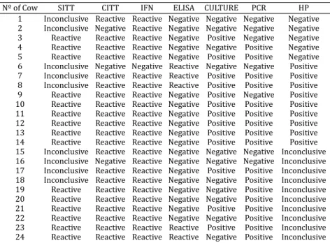
The Comparative Intradermal Tuberculin Test (CITT) con-irmed the irst 15 cows that were previously reactive, plus six out of nine that had been inconclusive at the irst SITT testing, totaling 21 skin-test reactive cows. The IFN conir-med all the 21 CITT reactive cows, and identiied as reacti-ve plus two cows that were negatireacti-ve to both SITT and CITT. MPB70 and MPB83-ELISAs presented identical results and were studied as one. They could identify four animals whi-ch were reactive to CITT plus one cow that was negative to SITT, CITT and IFN. Therefore, the study group was compo-sed by 24 cows that were positive to any of the employed tests (21 CITT, 2 IFN and 1 ELISAs). Those cows were slau-ghtered and fragments of lungs submitted to culture and histopathology. M. bovis was recovered by culture from tis-sues of 13 cows, while its DNA was evidenced on tissue of 17 samples (Table 1).
According to HP, in nine cows (37.5%) macroscopic le-sions positive for bovine pulmonary tuberculosis were ob-served. At microscopy, lesions presented typical tubercular granuloma displaying central necrosis with mineralization surrounded by macrophages, lymphocytes, plasma cells, neutrophils, epithelioid cells, and Langerhan’s giant cells, and enclosed partly or completely by a thin capsule. From those cows, eigth (88.9%) were IFN-positive and three (33.3%) reactive to ELISAs. Mycobacterium bovis was reco-vered from tissues of eight of these cows, while its DNA was evidenced on seven samples.
Ten cows presented macroscopic lesions suggestive of bovine pulmonary tuberculosis but were considered as inconclusive at HP (45.8%). At microscopy three animals (27.3%) presented lesions with diffuse neutrophilic inil-trate, and seven animals (63.3%) presented lesions with
clusters of epithelioid macrophages, two of them with i-brosis. From those cows, all the ten (100%) were IFN-posi-tive and two (18.2%) were reacIFN-posi-tive to ELISAs. M. bovis was recovered from tissues of three cows, while its DNA was evidenced on eight tissue samples.
Five animals were negative at HP (20.8%), although one of them presented macroscopic lesions suggestive of bovine pulmonary tuberculosis. From those cows, anyone was reactive to ELISAs while all the ive (100%) were IFN--positive. M. bovis was recovered from tissues of two cows, while its DNA was also evidenced on two samples.
DISCUSSION
The present study assessed the histopathology, in conjunc-tion with IFN, ELISA, Bacteriology and PCR, depicting the different stages of infection in a naturally infected herd and measuring the cellular differences among recent and ad-vanced infections.
Status of the herd was well deined since not only the herd was positive at the irst intradermal tests (single tu-berculinization) but also was conirmed by CITT. Additio-nally, M. bovis was recovered from the lesions and its DNA was evidenced by PCR.
It is not surprising that more animals (21) were detec-ted by the CITT than in the irst SITT testing (15). There was an interval of 90 days between the two skin tests, and it is possible that during that interval some cows that had been recently infected developed cell-mediated response and could only be detected at the second testing. In relation to the accuracy of IFN, this test has been extensively descri-bed to be more sensitive than skin tests (Lilenbaum et al. 1999, Wood & Jones 2001), and therefore the identiication of one supplementary cow that was skin-test negative is
Table 1. Results of Intradermal Tuberculin Test (SITT), Comparative Tuberculin Test (CITT), Interferon-gama Assay (IFN), MPB70/MPB83- ELISA (ELISA), Culture, Polymerase Chain Reaction (PCR) and Histopathology (HP)
for the diagnosis of bovine tuberculosis in the naturally infected herd
Nº of Cow SITT CITT IFN ELISA CULTURE PCR HP
not surprising. Additionally, the fact that MPB70 and 83 are very similar between themselves and presented identical results when used as capture antigens in an ELISA has alre-ady been described (McNair et al. 2001), as well as the role of ELISA in the detection of anergic cows (Silva et al. 2001, Pollock & Neill 2002, Lilenbaum & Fonseca 2006).
The positive microscopy results (9/24) were in accor-dance with the suggestive macroscopic lesions found in the lungs. These animals were probably in a more advanced stage of the disease, presenting classic tubercular granulo-ma, which is frequently observed with the progress of the disease (Welsh et al. 2005). In those animals, one was iden-tiied exclusively by ELISA. Additionally, eight out of nine were positive at the culture, the greatest proportion among the groups, suggesting a higher bacillary burden.
According to current knowledge regarding both IFN and ELISA, in this herd these cows may have been in different stages of the disease, what could justify the high percenta-ge of inconclusive samples (45.8%), as well as the negative results at histopathology. Initial immune responses against mycobacterial infections are expected to be predominantly cellular, while humoral responses become detectable later (Wood & Jones 2001). In that regard, IFN signalizes recent infection, since it has been described that this test detects infected cattle in very early stages of infection (Lilenbaum et al. 1999), as soon as 14 days after inoculation (Buddle et al. 1995). Similarly, ELISA has been described to be more useful for detecting animals with classic, advanced lesions, when bacterial load is higher and humoral response is evi-dent (Silva et al. 2001, Pollock & Neill 2002, Lilenbaum & Fonseca 2006).
In relation to the animals that presented mild lesions, considered as inconclusive to pulmonary tuberculosis, we assume that they had a recent infection. Three animals presented lesions with diffuse neutrophilic iniltrate, and other seven animals presented lesions with clusters of epi-thelioid macrophages, what is consistent to initial stages of infection. Cassidy et al. (1998) identiied neutrophils as one of the earliest cells associated with the developing granulo-ma, and Pollock and Neill (2002) described the initial inter-play between macrophage and mycobacteria. Additionally, IFN identiied as reactive all of those animals, while only two of them were reactive to ELISA. In relation to the direct evidenciation of the agent on those samples, an interesting inding was observed. Although only three samples were positive by bacterial culturing, eight of them were positi-ve by PCR. Molecular evidence of Mycobacterium bovis has been described as more sensitive than bacteriology since it requires a low bacterial load (Parra et al. 2008). Therefo-re, all the histopathological, bacteriological, molecular and immunological indings support the hypothesis that those animals were recently infected.
Regarding the ive animals that were negative to HP, all of them were identiied by IFN while ELISA was negative (Table 1). Therefore, there are evidences to support that those animals had been recently infected and, for that rea-son, they did not present visible lesions at HP. Tuberculin--reactive cattle with no visible lesions (NVL) have been re-ported when cows are in a very early stage of the disease,
leading to small and/or only few lesions (Corner 1994), what agrees with our results.
Concluding, histopathology was employed in the pre-sent study as a tool to identify the different stages that are observed in the development of TB pathophisiology. Consi-dering a multidisciplinary approach, when histopathologi-cal, bacteriologihistopathologi-cal, molecular and immunological indings were analyzed altogether, it was possible to identify all the infected animals and to determine the stage of the infec-tion in different cows from the same herd. This approach is mandatory for the interpretation of the various tools that are frequently employed for the diagnosis of TB, since it has been demonstrated that not all the cows forming the same herd are in the same stage of the infection, and therefore a multidisciplinary approach is required for detecting all the infected animals.
Acknowledgements.- This research was supported by CNPq and FAPERJ (Brazil). WL and AMRF are research fellows of CNPq. The authors are thankful for the help of Jim McNair (Agri-Food and BioSciences Institute, Ireland) for providing ELISA antigens and John Kastelic (Lethbridge Rese-arch Centre, Canada) for technical assistance.
REFERENCES
Buddle B.M., de Lisle G.W., Pfeffer A. & Aldwell F.E. 1995. Immunological responses and protection against Mycobacterium bovis in calves vacci-nated with a low dose of BCG. Vaccine 13:1123-1130.
Cassidy J.P., Bryson D.G., Pollock J.M., Evans R.T., Forster F. & Neill S.D. 1998. Early lesion formation in cattle experimentally infected with
Mycobacte-rium bovis. J. Comp. Pathol. 119:27-44.
Corner L.A. 1994. Post mortem diagnosis of Mycobacterium bovis infection in cattle. Vet. Microbiol. 40:53-63.
De la Rua Domenech R., Goodchild A.T., Vordemeier H.M., Hewinson R.G., Christiansen K.H. & Clifton-Hadley R.S. 2006. Ante mortem diagnosis in cattle: A review of the tuberculin tests, gama-interferon assay and other ancillary diagnostic techniques. Res. Vet. Sci. 81:190-21.
Figueiredo E.E.S., Silvestre F.G., Campos W.N., Furlanetto L.V., Medeiros L., Lilenbaum W., Fonseca L.S., Silva J.T. & Paschoalin V.M.F. 2009. Identiica-tion of Mycobacterium bovis isolates by multiplex PCR. Braz. J. Microbiol. 40:231-233.
Lilenbaum W. & Fonseca L. 2006. The use of ELISA as a complementary tool for bovine tuberculosis control in Brazil. Braz. J. Vet. Res. Anim. Sci. 43:256-261.
Lilenbaum W., Schettinni J., Souza G.N., Ribeiro E.R. & Moreira E.C. 1999. Comparison between a gama- IFN assay kit and intradermal tuberculin test for the diagnosis of bovine tuberculosis on ield trials in Brazil. J. Vet. Med. B, Infect. Dis. Vet. Public Hlth 46:353-358.
Marassi C.D., Medeiros L. & Lilenbaum W. 2010. The use of a Gamma-Inter-feron assay to conirm a diagnosis of bovine tuberculosis in Brazil. Acta Trop. 113:199-201.
McNair J., Corbett D.M., Girvin R.M., Mackie D.P. & Pollock J.M. 2001. Charac-terization of the early antibody response in bovine tuberculosis: MPB 83 is an early target with diagnostic potential. Scand. J. Immunol. 53:365-371. Medeiros L., Marassi C.D., Figueiredo E.E.S. & Lilenbaum W. 2010.
Poten-tial application of new diagnostic methods for controlling bovine Tuber-culosis in Brazil. Braz. J. Microbiol. 41:1-4.
Monaghan M.L., Doherty M.L., Collins J.D., Kazda J.F. & Quinn P.J. 1994. The tuberculin test. Vet. Microbiol. 40:111-124.
Parra A., García N., García A., Lacombe A., Moreno F., Freire F., Moran J. & Hermoso de Mendoza J. 2008. Development of a molecular diagnostic test applied to experimental abattoir surveillance on bovine tuberculo-sis. Vet. Microbiol. 127:315-324.
Pollock J.M., Welsh M.D. & McNair J. 2005. Immune response in bovine tuberculosis: towards new strategies for the diagnosis and control of disease. Vet. Immunol. Immunopathol 108:37-43.
Silva E. 2001. Evaluation of an enzyme-linked immunosorbent assay in the diagnosis of bovine tuberculosis. Vet. Microbiol. 78:111-117.
Thoen C.O., Lobue P.A., Enarson D.A., Kaneene J.B. & Kantor I.N. 2009. Tuberculosis: a re-emerging disease in animals and humans. Vet. Ital. 45:135-181.
Varello K., Pezzolato M., Mascarino D., Ingravalle F., Caramelli M. & Bo-zzetta E. 2008. Comparison of histologic techniques for the diagnosis of bovine tuberculosis in the framework of eradication programs. J. Vet. Diagn. Invest. 20:164-169.
Webpage OIE 2010. Manual of Diagnostic Tests and Vaccines for Ter-restrial Animals. Available at <http://www.oie.int/eng/normes/ mmanual/2008/pdf/2.04.07_BOVINE_TB.pdf> Accessed on Sept.10, 2010)
Welsh M.D., Cunningham R.T., Corbett D.M., Girvin R.M., McNair J., Skuce R.A., Bryson D.G. & Pollock J.M. 2005. Inluence of pathological progres-sion on the balance between cellular and humoral immune responses in bovine tuberculosis. Immunology 114:101-111.
Wood P.R. & Jones, S.L. 2001. BOVIGAMTM: An in vitro cellular diagnostic test for bovine tuberculosis. Tuberculosis 81:147-155.