Food Science and Technology ISSN 0101-2061
DI:
D http://dx.doi.org/10.1590/1678-457X.0017
1 Introduction
Yellow mealworm beetles Tenebrio molitor Linnaeus (Tenebrionidae, Coleoptera) are considered scavengers and are typically found to be injurious to insects in warehouses for agricultural products (Weaver et al., 1990; Ye et al., 2001; Morales-Ramos & Rojas, 2015). It is known to be suitable experimental material for physiological and genetic research (Takiguchi et al., 1992). Traditionally, Yellow mealworm has a long history of use as food supplement. They are typically used as a food source for reptile, fish, and avian pets. They are also provided to wild birds in bird feeders, particularly during the nesting season, when birds are raising their young and appreciate a ready food supply (Stoops et al., 2016). Experiments on mice fed with the filtrate of larval powder solution showed that the insect sample was safe to the mice, and it was effective on antifatigue, delaying aging, decreasing the level of serum total cholesterol and micronuclear rate in polychromatic erythrocytes, and increasing the perilymphocyte transformative rate in tested mice or mice (Siemianowska et al., 2013). T. molitor larvae are generally regarded as a rich source of protein, vitamins, essential amino acids, minerals and essential fatty acids like linolenic acid. The contents of toxic heavy metals were lower than national standards (Bai & Cheng, 2003; Gerber & Sabourin, 1984; Gnacadja, 1975). The various healthful function of the insect sample is based on the integration of its effective nutrition components. It was reported that dried larvae could be used in place of nuts, raisins and chocolate chips in many cookies, bread or dessert recopies. Powdered larvae can also replace part of the flour in cakes or pie crust. Barely
thawed whole larvae can be added to sauces (Ghaly & Alkoaik, 2009). They can be purchased at many pet stores. The potential for annual mealworm production in China is estimated at more than 100 tons (Zhang et al., 2008). As a result they are known as “golden grubs” and make excellent fish bait and serve as food for animals in aquariums and zoological parks. The insect is worthy of exploration as the healthful food for human.
Supercritical carbon dioxide (CD2) extraction as a green technology is certainly alternative method to replace or to complement conventional industrial process such as pressing and solvent extraction. Supercritical fluid extraction technique has many advantages over traditional methods, especially in preservation of thermo sensitive compounds using low temperatures, which results reduced energy consumption (Matthaus & Bruhl, 2008; Moslavac et al., 2014). A number of insects used in food systems have been demonstrated to modulate immune responses (Nowak et al., 2016). Even though T. molitor larvae has been used extensively as the food supplement of human and animal, very little information is available on the effect of supercritical CD2 fluid
T. molitor larvae extraction on nonspecific, cellular and humoral immune responses. In this study, we report immunomodulatory activity of supercritical CD2 fluid T. molitor larvae extraction in normal mice. The aim of this study was to provide the scientific basis for the comprehensive utilization of the yellow mealworm as a potential human food, improve the insect utilization efficiency, and evaluate the potential for future applications.
Immunomodulatory effects of supercritical fluid CO
2extracts from
freeze-dried powder of
Tenebrio molitor
larvae (yellow mealworm)
QingFeng TANG1, Yin DAI2*
Received 29 Jan., 2016 Accepted 24 Mar., 2016
1 Department of Entomology, Institute of Insect Resources Development and Utilization, Anhui Agricultural University - AAU, Hefei, PR, China 2 Institute of Animal Husbandry & Veterinary Medicine, Anhui Academy of Agricultural Sciences - AHAAS, Hefei, PR, China
*Corresponding author: [email protected]
Abstract
In order to take full advantage of Tenebrio molitor larvae (yellow mealworm) resources, the supercritical CD2 fluid freeze-dried powder of T. molitor larvae (fdTML) extraction on the immune systems of mice was carried out. The results about the effects of supercritical CD2 fluid fdTML extraction on carbon expurgation and phagocytosis of peritoneal macrophages experiments of mice indicated that the fdTML extraction enhanced observably carbon expurgatory index, phagocytic rate and phagocytic index. The fdTML extraction could stimulate response of delayed hypersensitivity. The proliferation of ConA-induced mitogenic reponse for spleen lymphocyte was also increased. The amount of hemolytic antibody in mice serum increased compared with those of the control group mice. The half of hemolysis values in serum of treated mice increased compared to the control group. Furthermore, serum ND content in all treatment groups was higher than that of the control group whereas acid phosphatase and alkaline phosphatase activity was only significantly higher relative to the control group. Dur findings suggest that supercritical CD2 fluid the fdTML extraction has potential as a health food supplement.
Keywords: food supplement; immunological function; immunoregulation; insect utilization; supercritical CD2 fluid extraction.
2 Materials and methods
2.1 Preparation of the freeze-dried powder of T. molitor larvae (fdTML)
T. molitor larvae (7-10th instars) used were obtained from the Anhui Agricultural University laboratory stocks, freeze-dried and ground to a powder with flat-hammer grinding mill and sifted through a 60-mesh screen. Lastly, the powder was stored in sealed aluminum foil bag at 4 ºC before analysis.
2.2 Supercritical CO2 fluid extraction of the fdTML soluble organic compounds
Supercritical CD2 fluid system (SFE- CD2, HL-1/32-50Mpa) was used to extract the organic soluble compounds. The temperatures and pressures of the extraction and liberation vessels were 50 ºC and 30 Mpa, and 45 ºC and 8 Mpa, respectively. The linear velocity of CD2 was maintained at 8 kg/h.
2.3 Animals
The experiments were conducted on female KunMing mice weighing 20 ± 2 g maintained at 25 ± 2 °C with normal mouse chow and water ad libitum. The animals were housed, five per cage and maintained on 12 h day and night cycle. The animals were divided into four groups comprising ten rats each. Group I: Control animals (distilled water control). Group II: Animals fed with supercritical CD2 fluid extraction (low-dosage group 1.89 g · kg-1 · d-1). Group III: Animals fed with supercritical CD2 fluid extraction (moderate-dosage group 3.78 g · kg-1 · d-1). Group IV: Animals fed with supercritical CD2 fluid extraction (high-dosage group 7.56 g · kg-1 · d-1).
Mice were orally administered a dose of 0.025 mL · g-1 body weight of supercritical CD2 fluid extraction once daily for four consecutive weeks. The control mice were given distilled water only by the same method. After four weeks oral administration, the immunomodulatory effect of supercritical CD2 fluid fdTML extraction was obtained from each experimental group by the method described previously.
2.4 Carbon clearance test
After four consecutive weeks’ oral administration, each mouse was injected with Indian ink 0.1 mL/10 g.bw from mouse tail vein. In the second minute (t1) and tenth minute (t2) after injection, 20 μL blood was drawn from the orbital venous plexus of mice’ eye sockets with suction tube in which was moistened with heparin solution and diluted in 2 mL 0.1% sodium carbonate solution. Mice were sacrificed and the body, thymus and spleen were collected and weighed.
The optical density (DD) values of blood solution were assayed with a 721 spectrophotometer at a wavelength of 600 nm, and the K (representing the capability of carbon granule clearance from mouse blood) (Equation 1) and α (representing the phagocytosis activity of macrophage) (Equation 2) values were calculated according to the formula.
2 1
lgOD at 2 min-lgOD at 10 min t
=
-t
K (1)
3
Body weight
K Spleen weight + Thymus weig =
ht
a (2)
2.5 Phagocytosis of peritoneal macrophages
Chicken blood was put into a sterile flask containing crystal ball and shaken to remove the fiber. The solution was rinsed three times with saline before centrifugation at 2000 rpm for 10 min. The supernatant was abandoned and the chicken red blood cells (CRBC) were prepared.
Phagocytosis of mice was detected using the method described by Lin et al. (1995) with slight modification. Briefly, on the last administration day, 1 mL of 20% (v/v) CRBC was intraperitoneally injected into each mouse, and the mice were euthanized 30 min later. Two milliliters saline was injected into the abdominal cavity and 1 ml fluid was then collected to make a smear for each mouse. The smears were incubated at 37 °C for 30 min in a wet box, fixed with acetone–methanol (1/1, v/v) solution, and then stained by 4% (v/v) Giemsa-phosphoric acid dye. The number of macrophage ingesting CRBC out of a total of at least 100 cells was calculated by direct visual enumeration using a light microscope. The phagocytic rate (PR) (Equation 3) and phagocytic index (PI) (Equation 4) were calculated using the following formula:
( )% =Number of macrophage ingesting CRBC 100%
Number of total macrophage
PR (3)
=Number of total ingested CRBC
Number of total macrop PI
hage
(4)
2.6 Serum hemolysin assay
Sheep blood was put into a sterile flask containing crystal ball and shaken to remove the fiber. The solution was rinsed three times with saline before centrifugation at 2000 rpm for 10 min. The supernatant was abandoned and the sheep red blood cells (SRBC) were prepared.
After four consecutive weeks’ oral administration, 0.2 mL of 20% (v/v) SRBC was intraperitoneally injected into each mouse. Five days later, blood was drawn from the orbital venous plexus of mice’ eye sockets with suction tube and serum was isolated from normal mice’ blood as well. Serum samples were diluted with normal saline to 1/400. The value of absorbance at the 50% hemolytic dose (HC50) (Equation 5) was determined by colorimetric method with a 721 spectrophotometer at a wavelength of 540 nm.
50
Value of samples absorbance
Dilution of serum Value of absorbance at SRBC 50% hemolytic dose
=
HC (5)
2.7 Delayed-type hypersensitivity reaction to dinitrofluorobenzene
1% (w/v) DNFB solution was used to challenge the right ear of the mice. The antigen challenge was evaluated by the methods described with some modification. Twenty-four hours later, a piece of left and right ear with the diameter of 8 mm was obtained with a stiletto. The delayed-type hypersensitivity reaction (DTH) (Equation 6) was calculated using the following formula:
DTH = Weight of right ear (diameter 8 mm) - Weight of left ear (diameter 8 mm) (6)
2.8 Lymphocyte-proliferation assays
After four consecutive weeks’ oral administration, spleens were removed aseptically from mice and teased in cold (4 °C) phosphate-buffered saline (PBS, pH 7.2) and pressed through a nylon sieve to liberate the cells. The suspension was allowed to sediment for several minutes and the supernatant was centrifuged (400 g, 10 min, 4 °C). The pellet was suspended in 2 ml 0.17 M ammonium chloride to disintegrate erythrocytes (2 min). The remaining cells were washed three times with cold PBS. The last pellet was suspended in Roswell Park Memorial Institute Medium 1640 (RPMI 1640, Sigma USA). Under sterile conditions, the spleen cells were adjusted to a concentration of 3 × 10⁶ cells/mL in 2 mL RPMI 1640, dispensed into 24-well tissue-culture plates (1 mL/well). The cell viability was measured by using the trypan blue dye exclusion method. Spleen cells were incubated at 37 °C in an atmosphere comprising 5% CD2/95% air in 24-well tissue-culture plates with 50 μL Concanavalin A (Con A, Sigma USA) solution or pure medium for 72 h. Lymphocyte proliferation was determined using a Methylthiazoletetrazolium (MTT) assay. At 4 h before the end of the incubation period, the 0.7 mL of supernatant was removed and 0.7 mL of RPMI 1640 medium was added to all wells of an assay. Subsequently, 50 μL of MTT solution (Sigma USA) was added to all wells of an assay, and plates were incubated for another 4 h under the same conditions. 1 mL isopropanol was added to all wells and mixed thoroughly to dissolve the dark blue crystals. After a few minutes, the plates were read on a Universal Microplate Spectrophotometer, using a test wavelength of 570 nm. The results are presented as optical density (D.D.) ± SD.
2.9 Antibody producing cells
The number of plaque forming cells from the spleen was determined by the Jerne’s Plaque assay (Jerne & Nordin, 1963). After 24 days oral administration, 0.2 ml of 2% (v/v) sheep red blood cells (SRBC) was intraperitoneally injected into each mouse. Five days later, spleens were removed aseptically from mice and teased in cold (4 °C) PBS (pH 7.2) and pressed through a nylon sieve to liberate the cells. The suspension was allowed to sediment for several minutes and the supernatant was centrifuged (400 g, 10 min, 4 °C). The pellet was suspended in 2 mL 0.17 M ammonium chloride to disintegrate erythrocytes (2 min). The remaining cells were washed three times with cold PBS. The last pellet was suspended in RPMI 1640 medium. Under sterile conditions, the spleen cells were adjusted to a concentration of 5 × 10⁶ cells/mL in 5 mL RPMI 1640. 25 μL of spleen cell suspension and 50 μL of 10% SRBC (v/v) were mixed with equal volumes of Hank’s solution (pH 7.2), and incubated at the slides which were spread by molten agarose in an atmosphere
comprising 5% CD2 /95% air for 1.5 h. Subsequently, Number of plaques was counted using a colony counter.
2.10 Acid phosphatase and alkaline phosphatase assay
After four consecutive weeks’ oral administration, blood was drawn from the orbital venous plexus of mice’ eye sockets with suction tube. The centrifuge tubes remained relatively static for 10 min and were incubated at 4 °C in the refrigerator overnight, fixed with the blood from which serum precipitated. According to the method which Reagent Kit (Nanjing jiancheng Bioengineering Institute, China) provided, the activity of acid phosphatase and alkaline phosphatase was measured.
2.11 Serum NO assay
After four consecutive weeks’ oral administration, blood was drawn from the orbital venous plexus of mice’ eye sockets with suction tube. The centrifuge tubes remained relatively static for 10 min and were incubated at 4 °C in the refrigerator overnight, fixed with the blood from which serum precipitated. According to the method which Reagent Kit (Nanjing jiancheng Bioengineering Institute, China) provided, the content of ND was measured.
2.12 Statistical analysis
Analysis of data was carried out by ANDVA. Comparison of means in homogeneous subsets was performed using the Tukey multiple comparisons test at 95% confidence interval employing the JMP version 5.0, NCSS, SigmaPlot 10.0.
3 Results
3.1 Effect of supercritical CO2 fluid fdTML extraction on the carbon particles clearance of mice
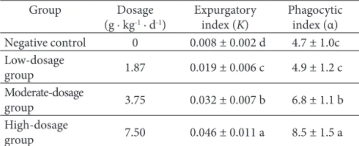
Effect of supercritical CD2 fluid fdTML extraction administration on the carbon particles clearance of mice is given in Table 1. Supercritical CD2 fluid fdTML extraction administration was found to enhance the Expurgatory index (K) and Phagocytic index (α). It was dose-dependent to potentiate the capability of carbon granule clearance and the phagocytosis activity of macrophage in mice. Expurgatory index was significant increased from 0.008 in negative control to 0.019, 0.032 and 0.046, respectively in group treated with 1.87 g · kg−1, 3.75 g · kg−1 and 7.50 g · kg−1 body
Table 1. Effect of supercritical CD2 fluid fdTML extraction on the
carbon particles clearance of mice.
Group Dosage
(g · kg-1 · d-1)
Expurgatory index (K)
Phagocytic index (α) Negative control 0 0.008 ± 0.002 d 4.7 ± 1.0c Low-dosage
group 1.87 0.019 ± 0.006 c 4.9 ± 1.2 c Moderate-dosage
group 3.75 0.032 ± 0.007 b 6.8 ± 1.1 b High-dosage
weight fdTML. The Phagocytic index was significant increased from 4.71 in negative control to 6.8 and 8.5, respectively in group treated with 3.75 g · kg−1 and 7.50 g · kg−1 body weight fdTML. No significant differences were found in dosage of 1.87 g · kg−1 body weight compared to negative control.
3.2 Effect of supercritical CO2 fluid fdTML extraction on phagocytosis of peritoneal macrophages
Effect of supercritical CD2 fluid fdTML extraction on phagocytosis of peritoneal macrophages is given in Table 2. It was dose-dependent to potentiate the activity of phagocytosis of peritoneal macrophages in mice. Phagocytic rate was significant increased from 17% in negative control to 25%, 26% and 27%, respectively in group treated with 1.87 g· kg−1, 3.75 g · kg−1and 7.50 g · kg−1 body weight fdTML (Table 2). The phagocytic index was 0.14 in negative control, which was significantly increased to 0.29, 0.31 and 0.58, respectively in groups treated with 1.87g· kg−1, 3.75 g· kg−1 and 7.50 g · kg−1 body weight fdTML.
3.3 Effect of supercritical CO2 fluid fdTML extraction on the hemolysin of mice
The effect of supercritical CD2 fluid fdTML extraction on humoral immune response in mice was tested. As shown in Table 3, difference in half of hemolysin values (HC50) was significant increased from 108 in negative control to 138, 133 and 142, in group treated with 1.87 g · kg−1, 3.75 g · kg−1 and 7.50 g · kg−1 body weight fdTML. No significant differences were found in low-dosage, moderate-dosage and high-dosage.
3.4 Evaluation of supercritical CO2 fluid fdTML extraction on cellular immune response
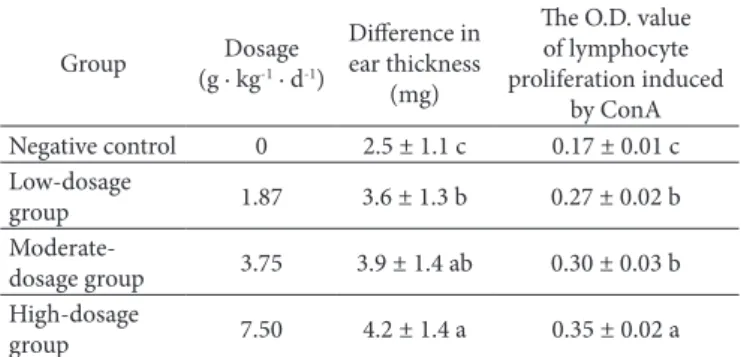
The effect of supercritical CD2 fluid fdTML extraction on the DTH reaction in mice was tested. As shown in Table 4, difference in ear thickness was significant increased from 2.5 mg in negative control to 3.6 mg, 3.9 mg and 4.2 mg, in group treated with 1.87 g · kg−1, 3.75 g · kg−1 and 7.50 g · kg−1 body weight fdTML. The D.D. value of lymphocyte proliferation induced by ConA was significant increased from 0.17 in negative control to 0.27, 0.30 and 0.35, respectively in group treated with 1.87 g · kg−1, 3.75 g · kg−1 and 7.50 g · kg−1 body weight fdTML (Table 4). Therefore, it was dose-dependent to potentiate the cellular immune response in mice.
3.5 Evaluation of supercritical CO2 fluid fdTML extraction on humoral immune response
Supercritical CD2 fluid fdTML extraction administration was found to significantly enhance the number of antibody producing cells in spleen. As shown in Table 5, the number of plaques was significant increased from 71 × 103 in negative control to 97 × 103 and 105 × 103, respectively in group treated with 3.75 g · kg−1 and 7.50 g · kg−1 body weight fdTML. No significant differences were found in dosage of 1.87 g · kg−1 body weight compared to negative control. However, it was dose-dependent to potentiate the humoral immune response in mice.
Table 4. Effect of supercritical CD2 fluid fdTML extraction on the
cellular immune response assessed by DTH reaction and lymphocyte proliferation.
Group Dosage (g · kg-1 · d-1)
Difference in ear thickness
(mg)
The D.D. value of lymphocyte proliferation induced
by ConA Negative control 0 2.5 ± 1.1 c 0.17 ± 0.01 c Low-dosage
group 1.87 3.6 ± 1.3 b 0.27 ± 0.02 b
Moderate-dosage group 3.75 3.9 ± 1.4 ab 0.30 ± 0.03 b High-dosage
group 7.50 4.2 ± 1.4 a 0.35 ± 0.02 a
Values in the same column with the same letters did not differ significantly at 0.05 level.
Table 2. Effect of supercritical CD2 fluid fdTML extraction on
the nonspecific immune response assessed by phagocytic rate and phagocytic index.
Group Dosage
(g · kg-1 · d-1)
Phagocytic rate (%)
Phagocytic index Negative control 0 17 ± 2 c 0.14 ± 0.02 c Low-dosage
group 1.87 25 ± 3 b 0.29 ± 0.08 b
Moderate-dosage group 3.75 26 ± 3 ab 0.31 ± 0.06 b High-dosage
group 7.50 27 ± 3 a 0.58 ± 0.09 a
Values in the same column with the same letters did not differ significantly at 0.05 level.
Table 3. Effect of supercritical CD2 fluid fdTML extraction on the
hemolysin of mice.
Group Dosage (g · kg-1 · d-1) Half of hemolysin
values (HC50)
Negative control 0 108 ± 11 b
Low-dosage group 1.87 138 ± 13 a
Moderate-dosage
group 3.75 133 ± 10 a
High-dosage group 7.50 142 ± 12 a
Values in the same column with the same letters did not differ significantly at 0.05 level.
Table 5. Effect of supercritical CD2 fluid fdTML extraction on the
humoral immune response assessed by antibody producing cells.
Group Dosage
(g · kg-1 · d-1)
Number of plaques (×103/spleen cells)
Negative control 0 71 ± 7 c
Low-dosage
group 1.87 74 ± 9 c
Moderate-dosage
group 3.75 97 ± 10 b
High-dosage
group 7.50 105 ± 9 a
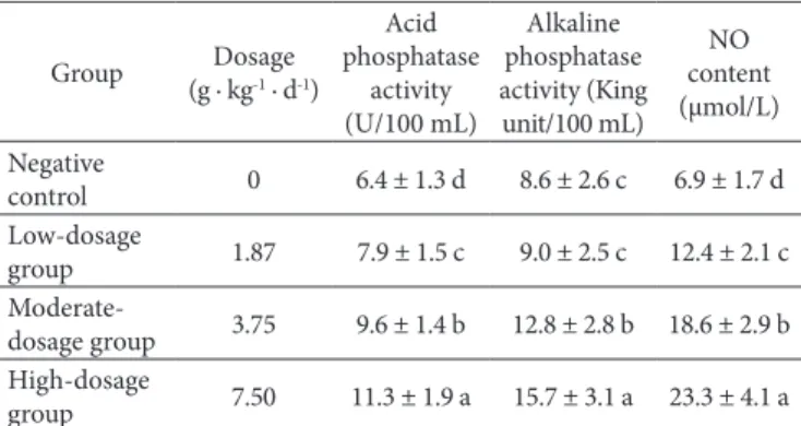
3.6 Effect of supercritical CO2 fluid fdTML extraction on the acid phosphatase, alkaline phosphatase activity and serum NO content of mice
Effect of supercritical CD2 fluid fdTML extraction administration on the acid phosphatase and alkaline phosphatase activity of mice is given in Table 6. The fdTML extraction administration was found to enhance the acid phosphatase and alkaline phosphatase activity. It was dose-dependent to potentiate the activity of the acid phosphatase and alkaline phosphatase in mice. The acid phosphatase activity was significant increased from 6.4 U/100 mL in negative control to 7.9 U/100 mL, 9.6 U/100 mL and 11.3 U/100 mL, in group treated with 1.87 g · kg−1, 3.75 g · kg−1 and 7.50 g · kg−1 body weight fdTML. The alkaline phosphatase activity was significant increased from 8.6 King unit /100mL in negative control to 12.8 King unit /100 mL and 15.7 King unit/100mL, in group treated with 3.75 g · kg−1 and 7.50 g · kg−1 body weight fdTML respectively. However, no significant differences were found in dosage of 1.87 g · kg−1 body weight compared to negative control.
The effect of supercritical CD2 fluid fdTML extraction on the serum ND content in mice was tested. As shown in Table 6, the serum ND content of mice was significant increased from 6.9 μmol/L in negative control to 12.4 μmol/L, 18.6 μmol/L and 23.3 μmol/L, in group treated with 1.87 g· kg−1, 3.75 g· kg−1 and 7.50 g · kg−1 body weight fdTML. It was dose-dependent to potentiate the the serum ND content in mice.
4 Discussion
The immune system is involved in the etiology as well as pathophysiologic mechanisms of various diseases and its function and efficiency is influenced by many exogenous and endogenous factors such as food, pharmaceuticals, physical and psychological stress, resulting in either immunosuppression or immunostimulation (Geetha et al., 2005). There is growing interest in identifying and characterizing insects with immunomodulatory activity. Immunoregulation is a complex balance between regulatory and effector cell and any imbalance in the immunological mechanism can lead to pathogenesis (Davis & Kuttan, 2000). Immunity has been shown to be suppressed in cancer. Chemotherapy and radiation therapy, useful in cancer treatment were found to deteriorate the immunity.
It is well known that macrophages are important cells in the immune response, especially in the antiinfection immunity. Administration of supercritical CD2 fluid fdTML extraction was found to increase the carbon expurgatory index and phagocytic index significantly indicating that the extract could stimulate the nonspecific phagocytic function. The supercritical CD2 fluid fdTML extraction could stimulate T cell-mediated immunity through delayed-type hypersensitivity. DNFB, played as the antigen, could induce the activating of the T cell, which would subsequently release different kinds of cytokine and attack the target cell (Yang et al., 2006).
The humoral immune response is the aspect of immunity that is mediated by secreted antibodies produced in the cells of the B lymphocyte lineage. Humoral immunity is so named because it involves substances found in the humours, or body fluids. Through the determination of hemolysin in serum content, we can know the immune response and its intensity (Zheng, 1991; Kielsen et al., 2016). The supercritical CD2 fluid fdTML extraction was found to enhance half of hemolysin values indicating that the extract could stimulate humoral immune response. The increase of the sensitivity in the T-dependent antibody response was assumed to be derived from the additive action of fdTML extraction to macrophages, helper T cells and B cells which are known to coordinate in immunization, activation and antibody production (Han et al., 1998).
Macrophages are cells produced by the differentiation of monocytes in tissues. Macrophages function in both non-specific defense (innate immunity) as well as help initiate specific defense mechanisms (adaptive immunity) of vertebrate animals (Netea et al., 2008). Their role is to phagocytose cellular debris and pathogens, either as stationary or as mobile cells. They also stimulate lymphocytes and other immune cells to respond to pathogens. In our paper, it was found phagocytosis of macrophage was greatly elevated in normal mice treated with fdTML, which may attribute to the nutritional components. Phosphatase is an important biological detoxification enzyme systems and its activity reflects the activated degree of macrophages. ND is the important medium and the initial factor that play an important cell functions, involved in a series of immune regulation effects (Ding et al., 2005). Supercritical CD2 fluid fdTML extraction administration was found to enhance the serum ND content, acid phosphatase and alkaline phosphatase activity, indicating that fdTML can effectively protect the biological immune system of mice, and enhance non-specific immune function.
5 Conclusions
In conclusion, nonspecific, cellular and humoral immune response are three of dominant indexes to evaluate the immunomodulatory activity of the test samples, which were determined to be efficient with the results positive. Therefore, supercritical CD2 fluid fdTML extraction enhanced all indexes compared with negative control. The results indicated that fdTML had a potency to potentiate the immune responses in mice.
Acknowledgements
The work was supported by fund from National Natural Science Foundation of China (31302044) and Key Project for University Excellent Young Talents by Anhui Province of China (gxyqZD2016035).
Table 6. Effect of supercritical CD2 fluid fdTML extraction on the acid
phosphatase, alkaline phosphatase activity and serum ND content of mice.
Group (g · kgDosage -1 · d-1)
Acid phosphatase
activity (U/100 mL)
Alkaline phosphatase activity (King unit/100 mL)
ND content (μmol/L) Negative
control 0 6.4 ± 1.3 d 8.6 ± 2.6 c 6.9 ± 1.7 d Low-dosage
group 1.87 7.9 ± 1.5 c 9.0 ± 2.5 c 12.4 ± 2.1 c
Moderate-dosage group 3.75 9.6 ± 1.4 b 12.8 ± 2.8 b 18.6 ± 2.9 b High-dosage
References
Bai, Y., & Cheng, J. (2003). Nutritive value and rearing methods of Tenebrio molitor in China. Entomological Knowledge, 40(4), 317-322. http://dx.doi.org/10.3969/j.issn.0452-8255.2003.04.007.
Davis, L., & Kuttan, G. (2000). Immunomodulatory activity of Withania somnifera.Journal of Ethnopharmacology, 71(1-2), 193-200. http:// dx.doi.org/10.1016/S0378-8741(99)00206-8. PMid:10904163. Ding, T. M., Chen, J. A., Zhang, Z. X., & Zhang, Y. H. (2005). The methods
for determination of nitric oxide in vivo and their applications. Progress in Pharmaceutical Sciences, 29(5), 221-226. http://dx.doi. org/10.3969/j.issn.1001-5094.2005.05.006.
Geetha, S., Singh, V., Ram, M. S., Ilavazhagan, G., Banerjee, P. K., & Sawhney, R. C. (2005). Immunomodulatory effects of seabuckthorn (Hippophae rhamnoides L.) against chromium (VI) induced immunosuppression. Molecular and Cellular Biochemistry, 278(1), 101-109. http://dx.doi.org/10.1007/s11010-005-7095-9. PMid:16180095. Gerber, G. H., & Sabourin, D. U. (1984). Dviposition site selection in
Tenebrio molitor (Coleoptera; Tenebrionidae). Canadian Entomologist, 116(1), 27-39. http://dx.doi.org/10.4039/Ent11627-1.
Ghaly, A. E., & Alkoaik, F. N. (2009). The Yellow Mealworm as a Novel Source of Protein. American Journal of Agricultural and Biological Sciences, 4(4), 319-331. http://dx.doi.org/10.3844/ajabssp.2009.319.331. Gnacadja, P. C. (1975). Value of alkane-produced protein and three
animal meals for growth and body nitrogen of meal worms (Tenebrio molitor L.). Annales de la Nutrition et de l’Alimentation, 29(1), 51-60. PMid:1227370.
Han, S. B., Kim, Y. H., Lee, C. W., Park, S. M., Lee, H. Y., Ahn, K. S., Kim, I. H., & Kim, H. M. (1998). Characteristic immunostimulation by angelan isolated from Angelica gigas Nakai. Immunopharmacology, 40(1), 39-48. http://dx.doi.org/10.1016/S0162-3109(98)00026-5. PMid:9776477.
Jerne, N. K., & Nordin, A. A. (1963). Plaque formation in agar by single antibody producing cells. Science, 140(3565), 405. http://dx.doi. org/10.1126/science.140.3565.405.
Kielsen, K., Shamim, Z., Ryder, L. P., Nielsen, F., Grandjean, P., Budtz-Jorgensen, E., & Heilmann, C. (2016). Antibody response to booster vaccination with tetanus and diphtheria in adults exposed to perfluorinated alkylates. Journal of Immunotoxicology, 13(2), 270-273. http://dx.doi.org/10.3109/1547691X.2015.1067259. PMid:26181512. Lin, P. Y., Feng, Z. M., Pan, J. Q., Zhang, D., & Xiao, L. Y. (1995). Effects
of artesunate on immune function in mice. Acta Pharmacologica Sinica, 16(5), 441-444. PMid:8701764.
Matthaus, B., & Bruhl, L. (2008). Virgin hemp seed oil: an interesting niche product. European Journal of Lipid Science and Technology, 110(7), 655-661. http://dx.doi.org/10.1002/ejlt.200700311. Morales-Ramos, J. A., & Rojas, M. G. (2015). Effect of larval density
on food utilization efficiency of Tenebrio molitor (Coleoptera: Tenebrionidae). Journal of Economic Entomology, 108(5), 2259-2267. http://dx.doi.org/10.1093/jee/tov208. PMid:26453714.
Moslavac, T., Jokić, S., Šubarić, D., Aladić, K., Vukoja, J., & Prce, N. (2014). Pressing and supercritical CD2 extraction of Camelina sativa oil. Industrial Crops and Products, 54(2), 122-129. http://dx.doi. org/10.1016/j.indcrop.2014.01.019.
Netea, M. G., Lewis, E. C., Azam, T., Joosten, L. A. B., Jaekal, J., Bae, S. Y., Dinarello, C. A., & Kim, S. H. (2008). Interleukin-32 induces the differentiation of monocytes into macrophage-like cells. Proceedings of the National Academy of Sciences of the United States of America, 105(9), 3515-3520. http://dx.doi.org/10.1073/pnas.0712381105. PMid:18296636.
Nowak, V., Persijn, D., Rittenschober, D., & Charrondiere, U. R. (2016). Review of food composition data for edible insects. Food Chemistry, 193, 39-46. http://dx.doi.org/10.1016/j.foodchem.2014.10.114. PMid:26433285.
Siemianowska, E., Kosewska, A., Aljewicz, M., Skibniewska, K., Polak-Juszczak, L., Jarocki, A., & Jędras, M. (2013). Larvae of mealworm (Tenebrio molitor L.) as European novel food. Agricultural Sciences, 4(6), 287-291. http://dx.doi.org/10.4236/as.2013.46041.
Stoops, J., Crauwels, S., Waud, M., Claes, J., Lievens, B., & Van Campenhout, L. (2016). Microbial community assessment of mealworm larvae (Tenebrio molitor) and grasshoppers (Locusta migratoria migratorioides) sold for human consumption. Food Microbiology, 53(Pt B), 122-127. http://dx.doi.org/10.1016/j.fm.2015.09.010. PMid:26678139. Takiguchi, M., Niimi, T., Su, Z. H., & Yaginuma, T. (1992). Trehalase
from male accessory gland of an insect, Tenebrio molitor, cDNA sequencing and developmental profile of the gene expression. The Biochemical Journal, 288(1), 19-22. http://dx.doi.org/10.1042/ bj2880019. PMid:1445264.
Weaver, D. K., Mcfarlane, J. E., & Alli, I. (1990). Repellency of volatile fatty acids present in frass of larval yellow mealworms, Tenebrio molitor L. (Coleoptera: Tenebrionidae), to larval conspecifics. Journal of Chemical Ecology, 16(2), 585-593. http://dx.doi.org/10.1007/ BF01021788. PMid:24263513.
Yang, F. M., Shi, Y., Sheng, J. C., & Hu, Q. H. (2006). In vivo immunomodulatory activity of polysaccharides derived from Chlorella pyrenoidosa.European Food Research and Technology, 224(2), 225-228. http://dx.doi.org/10.1007/s00217-006-0315-z. Ye, X. Q., Liu, D. H., & Hu, C. (2001). Some factors’ effects on the
solubility of protein from yellow mealworm (Tenebrio molitor L) larvae. Journal of Zhejiang University Science, 2(4), 436-438. http:// dx.doi.org/10.1631/jzus.2001.0436.
Zhang, C. X., Tang, X. D., & Cheng, J. A. (2008). The utilization and industrialization of insect resources in China. Entomological Research, 38(Suppl 1), S38-S47. http://dx.doi.org/10.1111/j.1748-5967.2008.00173.x.