ABSTRACT
BACKGROUND AND OBJECTIVES: Changes in motor control may contribute to muscle recruitment deficits. The ob-jective of this study was to evaluate strength, endurance, and re-cruitment of the torso muscles.
METHODS: We evaluated 35 women, divided into two groups: chronic low back pain (LBPG, n=20) and control (CG, n=15). The clinical conditions, incapacity, level of pain, strength, en-durance and the muscles recruitment: internal oblique (IO) and external oblique (EO), rectus abdominis (RA), lumbar iliocostal (LI) and lumbar multifidus (LM) were evaluated. MANOVA, MANCOVA and Kruskal-Wallis were used.
RESULTS: There was no significant difference between groups in the muscle strength test (p<0.172). The control group showed the greater capacity for muscle endurance in the muscle endur-ance test (p<0.001). In muscle recruitment, the chronic low back pain group presented greater muscle activation, which was evi-dent in the global stabilizing muscles EO and RA (p<0.05). CONCLUSION: Women with back pain had reduced muscular endurance and greater recruitment of the global muscles when compared to women with no back pain.
Keywords: Electromyography, Lumbar pain, Muscle strength, Physical resistance.
RESUMO
JUSTIFICATIVA E OBJETIVOS: Alterações no controle mo-tor podem contribuir para déficits no recrutamento muscular. O objetivo deste estudo foi avaliar a força, resistência e o recruta-mento dos músculos do tronco.
MÉTODOS: Foram avaliadas 35 mulheres, divididas em dois grupos: dor lombar crônica (GDL, n=20) e controle (GC, n=15). Avaliou-se as condições clínicas, incapacidade, nível da
Influence of pain in strength, resistance and recruitment of trunk muscles
Influência da dor na força, resistência e recrutamento dos músculos do tronco
Guilherme Thomaz de Aquino Nava1, Beatriz Mendes Tozim2, Mary Hellen Morcelli2, Marcelo Tavella Navega2
1. Universidade Estadual Paulista, Instituto de Biociências, Departamento de Educação Física, Rio Claro, SP, Brasil.
2. Universidade Estadual Paulista, Faculdade de Filosofia e Ciências, Departamento de Fi-sioterapia e Terapia Ocupacional, Marília, SP, Brasil.
Submitted on March 11, 2018.
Accepted for publication on August 20, 2018. Conflict of interests: none – Sponsoring sources: none.
Correspondence to:
Av. Hygino Muzzi Filho, 737 – Mirante 17525-000 Marília, SP, Brasil. E-mail: gtanava@gmail.com
© Sociedade Brasileira para o Estudo da Dor
dor, força, resistência e recrutamento dos músculos: oblíquo ex-terno (OE) e inex-terno (OI), reto abdominal (RA), iliocostal lom-bar (ICL) e multífido lomlom-bar (MUL). Utilizou-se MANOVA, MANCOVA e Kruskal-Wallis.
RESULTADOS: No teste de força muscular não houve diferença significativa entre os grupos (p=0,172). O grupo controle demons-trou maior capacidade de resistência no teste de resistência mus-cular (p<0,001). No recrutamento musmus-cular, o grupo dor lombar crônica apresentou ativação muscular maior, que ficou evidente nos músculos estabilizadores globais, OE e RA (p<0,05).
CONCLUSÃO: Mulheres com dor lombar apresentaram resis-tência muscular diminuída e maior recrutamento nos músculos globais, quando comparadas às mulheres sem dor lombar. Descritores: Dor lombar, Eletromiografia, Força muscular, Re-sistência física.
INTRODUCTION
Low back pain (LBP) is defined as pain or discomfort on the lumbar spine, lumbosacral or sacroiliac regions, being consid-ered one of the main musculoskeletal dysfunctions of the current time1-3. Approximately 80% of the population will have some LBP episode in its lifetime, and among these, approximate-ly 62% will have a recurrence after one year2-6. The etiology of chronic low back pain (CLBP) and its natural course are import-ant issues not yet well understood, and approximately 90% of the CLBP cases do not have a defined or identifiable etiology, making treatment more difficult1,5. Studies were conducted to evaluate the relationship between the strength and endurance of the torso extensors and CLBP, which is a phenomenon related to long-term disability1.6.
One of the hypotheses for the changes in muscle strength and en-durance are the alterations in the neural adaptation and conse-quent modification in the motor control7,8. This theory suggests that changes at different levels of the nervous system can result in the redistribution of muscle activity and change in the mechani-cal behavior, which can lead to deficits in the postural control of the torso muscles6,8,9. However, this relationship between strength, endurance, and muscle recruitment of the stabilizer torso muscles was poorly addressed. Therefore, it is evident how important it is to assess the recruitment of the torso muscles simultaneously to the assessment of the muscle strength and endurance parameters. Thus, to assess the muscular system, it is important to divide it into local and global muscles according to their functions8,10. The literature describes surface electromyography as an instru-ment with characteristics that allow the evaluation of the torso muscles function of healthy and injured individuals5,11,12.
The objective of this study was to evaluate the strength, endur-ance, and recruitment of the torso muscles in women with CLBP.
METHODS
Observational, cross-sectional study conducted with 35 wom-en aged betwewom-en 30 and 59 years, of which 20 were in the chronic low back pain group (LBPG, average age 46.6±8.34 years), based on personal report and pain location. The control group (CG) had 15 participants who did not have LBP (CG, average age 39.8±8 years). The eligibility criteria for the LBPG were no practice of physical activity and having recurrent idio-pathic CLBP for at least three months prior to the study. For the CG was no practice of any physical activity and no previous report of LBP. Non-eligibility criteria for both groups were: nerve compression on lower limbs (LL), pregnancy, history of severe cardiorespiratory problems, LL discrepancy, rheumatic, neurological or vestibular diseases, lumbar spine surgery, un-corrected visual alteration or being unable to understand the task. The exclusion criteria were: unable to perform any step of the data collection or any complication related to the collection procedure (n=2).
The sample calculation was obtained by the G*Power software,
performed from a pilot study (data of five participants in each group). The time values of the muscle endurance test were used for the sample calculation. It was used the power of 0.95, α error probability of 0.05, the effect size of 1.614, and it was estimated the need for 8 participants per group.
The data collection procedure consisted of clinical assessment, dorsal muscles strength test (MST) using a dorsal dynamome-ter, muscle endurance test (MET) by the Biering Sorensen test associated with surface electromyography of the torso stabilizer muscles. The Roland Morris Disability Questionnaire (RMDQ) was also used. The clinical evaluation consisted in collecting data on the dominance of LL used to place the electrodes in the elec-tromyographic assessment, and data about the pain, such as its location, duration, intensity, the presence of paresthesias and use of pain relief drugs. The intensity was quantified by the visual an-alog scale (VAS). The VAS is a reliable and easy-to-apply meth-od that quantifies the pain13. The scale has a horizontal line of 100mm. On the left end, there are the words “no pain/discom-fort,” and on the right, “worst pain/discomfort imaginable”13. To measure the pain, the participant was asked to inform the pain felt at the time of the evaluation and the intensity of the referred pain on a daily basis14. When finished, the measurement was per-formed with a ruler from the end “without pain/discomfort” to quantify the test13,15.
The RMDQ is an instrument that aims to determine the degree of disability due to the presence of LBP. It was translated and validated for the Portuguese language and consists of 24 self-re-spond questions16. The RMDQ is straightforward. The partici-pants must check “yes” or “no” in the options that best represent their current state regarding the presence of LBP, varying from zero to 24. Zero means no complaints, and as the result increas-es, the greater the incapacity to perform activities. The maximum result of 24 indicates participants with severe limitations16.
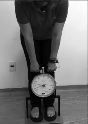
After the clinical evaluation, the MST of the torso extensors was performed using a dorsal dynamometer (Oswaldo Filizona), (Figure 1). For the MST, the participants were instructed to po-sition their feet on a given location and exert a tensile force in the direction of the torso extension, and not to exert force with the upper limbs (UL) nor bend the LL17. The test was performed twice at the beginning, and twice at the end of the collection, for familiarization with the task18. The highest value of the second attempt was used for analysis18. At each attempt, the participants were stimulated verbally to keep the contraction for 4 seconds, with a 1-minute rest1,11.
Figure 1. Muscle strength test
Fatigue was considered as the moment when the participants could no longer maintain enough muscle contraction to keep the horizontal position of the body19. During the MET, elec-tromyographic signals were collected from the local and global muscles on the dominant side of each participant. We used the dominance test that consisted of three tests, going up and down the stairs, kick a ball on target, and anterior displacement test20. For the electromyography, the selected local muscles were the internal oblique muscle (IO) and the lumbar multifidus (LM). And the selected global muscles were the external oblique (EO), the rectus abdominis (RA) and the iliocostalis lumborum (ICL). The participants were positioned in the supine position for the location and placement of the electrodes:
• RA: ½ of the distance between the xiphoid process and the
umbilical, approximately 3cm lateral to the median line15.
• IO: 2cm medially and below the ASIS21.
• EO: 50% of the distance between the lower rib cage and ASIS21. Then, the participants were positioned in the prone position for the location and placement of the electrodes:
• ICL: 6cm laterally to the space between the spinous process of L2-L321.
• LM: Positioned on the line connecting the posterior superior iliac spine and the space between L1-L2 at the level of L515. The muscles were located, and the electrodes were positioned unilaterally and longitudinally oriented with the muscle fiber on the dominant side20. The area of placement of the electrodes was shaved, and the skin abrasion with gauze was performed to de-crease the impedance, and the skin was cleaned with alcohol21. The electrodes used were the Ag/AgCl surface electrodes, active, with pre-amplification with 20 times gain. They were positioned in a bipolar configuration, with a 1cm diameter pickup area and distance of 2cm between them. The reference electrode was po-sitioned in the ulnar styloid process on the dominant side12. To capture the electromyographic signals, it was used a biological signal acquisition module, 8-channel EMG830c (EMG Sys-tem®), a EMGLab data collection, processing and storage soft-ware, calibrated with sampling frequency of 2000 Hz, total gain of 2000 times (20 times on the sensor and 100 times on the
equipment) and system impedance of 109Ω, mutual rejection module>100 dB and signal-to-noise ratio<3μV RMS.
The maximum voluntary isometric contraction (MVIC) was subsequently collected for the standardization of the electro-myographic signal. The RA muscle was assessed with the par-ticipants sitting on the stretcher, with knees flexed at 90°, feet
on a support base and LL united11. The UL were positioned
crossed in front of the chest, and the torso slightly inclined backwards11. The feet and trunk were stabilized by the evalua-tors, while the participants applied the maximum force to flex the torso anteriorly11,15.
The IO and EO muscles were assessed in lateral decubitus with the side to be assessed upwards11. The knees remained flexed at 90°, and the UL united and crossed in front of the torso, while the evaluators stabilized the knees, hip, shoulders, and elbows11. The participants applied force to the torso lateral flexion direction11,15. The ICL and LM muscles were assessed with the participants in the prone position, with the LL positioned on the box, from the ASIS region until the feet, and the torso outside the box11,19. The participants were stabilized in the regions of the ankles, knees, and hips with Velcro staps19. Another strap was positioned n the region of the shoulder blades to limit the torso extension, and the participants were encouraged to apply the maximum force in the direction of the torso extension11,15.
All the MVIC evaluations were performed twice with muscle contraction sustained for four seconds and with one minute of rest4. Oral motivation was given at all times, as well as in-structions at the beginning of the evaluations, on which muscle would be assessed to its better recruitment. The highest value was used for data analysis11.
The electromyographic analysis was done with the software Mat-lab® during the MET. A 4th order Butterworth high-pass filter with a cutoff frequency of 20Hz and a 4th order Butterworth low-pass filter with a cutoff frequency of 500Hz were used. The Root Mean Square (RMS) of the muscles were normalized by the highest value obtained on MVIC.
port number 1.054.270). All the participants were informed about the research and signed a Free and Informed Consent Term (FICT).
Statistical analysis
The statistical analysis was performed using PASW 18.0 (SPSS Inc., Chicago, USA). The parametric electromyographic data were presented as the mean and standard deviation, and the nonparametric as median, minimum and maximum. The Sha-piro-Wilk test was used to check data normality. After checking, the multivariate analysis of variance (MANOVA) was used to compare the characteristics of the subjects. Age and body mass index (BMI) of the participants were used as covariates for para-metric data analysis, using multivariate analysis of covariance (MANCOVA). The Kruskal-Wallis test was used for nonpara-metric data analysis. The significant value adopted was p<0.05.
RESULTS
The average of the age of the CG was 6 years lesser when compared with the LBPG, apart from presenting lower BMI. Therefore, the age and BMI were considered as covariates in the statistical analysis of the electromyographic activity for the comparison of groups (Table 1). Regarding the data analysis of the ICL and LM muscles electromy-ography, no significant differences were observed in MET in the comparison between the groups (p=0.331 and F=1.148) (Table 2). The RA and EO muscles showed a significant difference, where-as the IO muscle showed no difference between the groups in MET, as can be seen in table 3.
MST and MET values are shown in figure 3. Muscle strength showed no significant difference. A significant difference was ob-served in muscle endurance when comparing the groups. In the LBPG the difference was lower.
LBPG LBPG
(b) (a)
CG CG
1.6 1.4 1.2 1 0.8 0.6 0.4 0.2 0 250
200
150
100
50
0
Normalized muscle
str
ength
Seconds
Table 1. Demographic and pain characteristics of the participants (Mean± SD)
LBPG (n=20) CG (n=15) MANOVA
p-value
MANOVA F-value
Age (years) 45.90±8.45 39.80 ±8.01 0,038 4.661*
Body mass index (kg/m) 28.42±5.52 24.19±3.44 0,014 6.799*
Duration of low back pain (years) 8.55±8.86 - -
-Daily pain (VAS) 3.97±2.54 - -
-Pain on evaluation (VAS)
Roland Morris disability questionnaire
0.85±1.64 4.57±5.30
-LBPG = lower back pain group; CG = control group; VAS = visual analog scale; MANOVA = multivariate analysis of variance. * significant difference p<0.05.
Figure 3. Box plot showing the median, interquartile and minimum and maximum intervals in muscle endurance and muscular strength tests for both groups
(a) MET = muscle endurance test (p=0.001); (b) MST = muscle strength test (p=0.172); LBPG = low back pain group; CG = control group; *Significant difference (p<0.05).
Table 3. Root Mean Square normalized data of the internal and external oblique muscles, and rectus abdominis of the nonparametric data
LBPG CG p-value
Median Minimum Maximum Median Minimum Maximum
IO 0.98 0.3 6.23 0.52 0.12 2.5 0.053
EO 0.51 0.01 1.32 0.14 0.01 2.5 0.008*
RA 0.31 0.1 2.99 0.2 0.08 4.05 0.009*
IO = internal oblique muscle; EO = external oblique muscle; RA = rectus abdominal muscle; LBPG= low back pain group; CC = control group. * significant difference p<0.05. Table 2. Root Mean Square normalized data of the iliocostalis muscles and lumbar multifidus (mean±SD of parametric data)
LBPG CG p-value F-value
Mean±SD Mean±SD
ICL 0.77±0.15 0.64±0.17 0,175 1,928
LM 0.83±0.16 0.75±0.12 0,618 0,253
ICL = liliocostalis lumborum muscle; LM = lumbar multifidus muscle; LBPG = low back pain group; CG = control group.
DISCUSSION
There was no significant difference between groups (p=0.172) in MST. However, the CG had higher values on the torso ex-tensors strength. It is known that pain reduces muscle activi-ty6. The explanation for the non-appearance of significant dif-ference in this study may be related to the fact that the LBPG had high values of daily pain associated with the presence of limitations showed by the RMDQ5, leading to the restriction of movement in the region with pain, as well as of the whole body. Therefore, pain improvement may explain the
elevat-ed values of muscle activation5,6. Avoiding movements that
use the area of the lumbar spine favors sedentarism, and as a result, the overweight shown by the high BMI of the partici-pants with LBCP2,22,23. The overweight and the presence of a greater amount of fat facilitate fat infiltration in the muscle area of the ICL and LM muscles, and that increase is indica-tive of muscle atrophy that leads to a reduction in the torso extensors strength22.
The MET results show a greater muscle endurance in the CG, with time values higher than the LBPG (p<0.001). Previous studies have shown that MET is related with pain and the prognosis of LBP development, and individuals who present less than 58 seconds are three times more like-ly to have LBP than those who maintained more than 104
seconds19,23,24. This difference in MET may be due to the
predominance and prevalence of type II muscle fibers and reduction of type I muscle fibers of the ICL and LM mus-cles in the LBPG, which makes it difficult to maintain a horizontal posture during MET and in the postures during daily activities23,25.
The electromyography results give information regarding
neuromuscular activity6, and surface electrodes are
recom-mended, primarily because this is a volunteer activity, and therefore, it is preferable to use surface electrodes26. In ad-dition, the use of needle electrodes in a sustained contrac-tion could cause discomfort to participants. Due to the use of surface electrodes in the bipolar configuration, there is a high rejection rate of the common-mode, which purpose is to eliminate external noise, which results in a better quality of
the electromyographic signal26. When analyzing the
electro-myography data with the MET data, it was observed that the CG showed less muscle recruitment of all evaluated muscles. Greater results in muscle activity in the LBPG may have a relationship with the number of traction units selected for the task5,26. Thus, lower values of muscle activation in the CG may be related to the greater number of motor units recruited at the time of the test, with a lower amplitude value of mus-cle recruitment4-6,26,27. The higher levels of muscle activation in the LBPG were evident in the EO and RA muscles, con-sidered global stabilizers muscles10, which showed statistically significant results (p<0.05). The results of this study corrob-orate the studies that showed higher activation of the global muscles. However, this strategy that aims at increasing the torso stiffness by increasing the agonist and antagonist activa-tion, make the global muscles act as local, and in situations of
higher stability requirement it may have a harmful potential and increase pain7-9.
The CG showed better results in muscle endurance and re-cruitment in the tests conducted, and endurance is an aspect of performance and functional assessment. Women who were in the LBPG showed deficits in the neuromuscular capacity. The central nervous system uses this information to develop and correct movement patterns in our daily activities, i.e., pain produces a favorable environment for the onset and de-velopment of lesions and pain.
CONCLUSION
Women with chronic low back pain had lower muscle en-durance and more muscle requirement of the global muscles when compared with women without pain. Thus, it is import-ant to note that rehab training should also include muscle en-durance training of the torso extensor and focus on the motor control with the purpose of reorganizing muscle recruitment with emphasis on the local system.
REFERENCES
1. Cho KH, Beom JW, Lee TS, Lim JH, Lee TH, Yuk JH. Trunk muscles strength as a risk factor for nonspecific low back pain: A pilot study. Ann Rehabil Med. 2014;38(2):234-40.
2. Conway R, Behennah J, Fisher J, Osborne N, Steele J. Associations between trunk extension endurance and isolated lumbar extension strength in both asymptomatic participants and those with chronic low back pain. Healthcare. 2016;4(3). pii: E70. 3. Ono R, Higashi T, Takahashi O, Tokuda Y, Shimbo T, Endo H, et al. Sex differences
in the change in health-related quality of life associated with low back pain. Qual Life Res. 2012;21(10):1705-11.
4. Nelson-Wong E, Alex B, Csepe D, Lancaster D, Callaghan JP. Altered muscle recruit-ment during extension from trunk flexion in low back pain developers. Clin Biomech. 2012;27(10):994-8.
5. Butler HL, Hubley-Kozey CL, Kozey JW. Changes in electromyographic activity of trunk muscles within the sub-acute phase for individuals deemed recovered from a low back injury. J Electromyogr Kinesiol. 2013;23(2):369-77.
6. Schmit EF, Brito JD, Nóbrega SR, Araújo-Neto SA, Andrade PR, Ferreira JJ, et al. Efeitos da fisioterapia na força, atividade mioelétrica e dor, em lombálgicos crônicos. ConScientiae Saúde. 2016;15(2):183-90.
7. Hodges PW, Moseley GL, Gabrielsson A, Gandevia SC. Experimental muscle pain changes feedforward postural responses of the trunk muscles. Exp Brain Res. 2003;151(2):262-71.
8. D’hooge R, Hodges P, Tsao H, Hall L, Macdonald D, Danneels L. Altered trunk muscle coordination during rapid trunk flexion in people in remission of recurrent low back pain. J Electromyogr Kinesiol. 2013;23(1):173-81.
9. Tsao H, Galea MP, Hodges PW. Driving plasticity in the motor cortex in recurrent low back pain. Eur J Pain. 2010;14(8):832-9.
10. Bergmark A. Stability of the lumbar spine. A study in mechanical engineering. Acta Orthop Scand Suppl. 1989;230:1-54.
11. Vera-Garcia FJ, Moreside JM, McGill SM. MVC techniques to normalize trunk mus-cle EMG in healthy women. J Electromyogr Kinesiol. 2010;20(1):10-6.
12. Rossi DM, Morcelli MH, Marques NR, Hallal CZ, Gonçalves M, Laroche DP, et al. Antagonist coactivation of trunk stabilizer muscles during Pilates exercises. J Bodyw Mov Ther. 2014;18(1):34-41.
13. Astfalck RG, O’Sullivan PB, Straker LM, Smith AJ, Burnett A, Caneiro JP, et al. Sitting postures and trunk muscle activity in adolescents with and without nonspecific chron-ic low back pain: an analysis based on subclassifchron-ication. Spine. 2010;35(14):1387-95. 14. Morcelli MH, Faganello FR, Navega MT. Avaliação da flexibilidade e dor de idosos
fisicamente ativos e sedentários. Ter Man. 2010;8(38):298-304.
15. Schinkel-Ivy A, Nairn BC, Drake JD. Investigation of trunk muscle co-contraction and its association with low back pain development during prolonged sitting. J Elec-tromyogr Kinesiol. 2013;23(4):778-86.
16. Nusbaum L, Natour J, Ferraz MB, Goldenberg J. Translation, adaptation and valida-tion of the Roland-Morris quesvalida-tionnaire--Brazil Roland-Morris. Braz J Med Biol Res. 2001;34(2):203-10.
18. Gruther W, Wick F, Paul B, Leitner C, Posch M, Matzner M, et al. Diagnostic accu-racy and reliability of muscle strength and endurance measurements in patients with chronic low back pain. J Rehabil Med. 2009;41(8):613-9.
19. Alaranta H, Luoto S, Heliövaara M, Hurri H. Static back endurance and the risk of low-back pain. Clin Biomech. 1995;10(6):323-4.
20. Hoffman M, Schrader J, Applegate T, Koceja D. Unilateral postural control of the functionally dominant and nondominant extremities of healthy subjects. J Athl Train. 1998;33(4):319-22.
21. Marques NR, Hallal CZ, Gonçalves M. Padrão de co-ativação dos músculos do tronco durante exercícios com haste oscilatória. Motriz Rev Educ Fis. 2012;18(2):245-52. 22. Teichtahl AJ, Urquhart DM, Wang Y, Wluka AE, Wijethilake P, O’Sullivan R,
et al. Fat infiltration of paraspinal muscles is associated with low back pain, disability, and structural abnormalities in community-based adults. Spine J. 2015;15(7):1593-601.
23. Davarian S, Maroufi N, Ebrahimi I, Farahmand F, Parnianpour M. Trunk muscles
strength and endurance in chronic low back pain patients with and without clinical instability. J Back Musculoskelet Rehabil. 2012;25(2):123-9.
24. Mbada CE, Ayanniyi O, Ogunlade SO, Orimolade EA, Oladiran AB, Ogundele AO, et al. Rehabilitation of back extensor muscles’ inhibition in patients with long-term mechanical low-back pain. ISRN Rehabil. 2013;1-11.
25. Mazis N, Papachristou DJ, Zouboulis P, Tyllianakis M, Scopa CD, Megas P. The effect of different physical activity levels on muscle fiber size and type distribution of lumbar multifidus. A biopsy study on low back pain patient groups and healthy control sub-jects. Eur J Phys Rehabil Med. 2009;45(4):459-67.
26. De Luca CJ. The use of surface electromyography in biomechanics. J Appl Biomech. 1997;13(2):135-63.