Beatriz da Cunha Batista
Licenciatura em Bioquímica
Characterization and Biofilm Assessment of
Haemophilus influenzae
Isolates from
Patients with Chronic Obstructive
Pulmonary Disease and Otitis Media
Dissertação para obtenção do Grau de Mestre em Bioquímica
Orientador: Maria Paula Bajanca Lavado, Investigadora
auxiliar, Instituto Nacional de Saúde Doutor Ricardo Jorge
Co-orientador: Maria Luísa Forte Marques Jordão,
Investigadora auxiliar, Instituto Nacional de Saúde Doutor
Ricardo Jorge
Júri:
Presidente: Prof. Doutor José Ricardo Ramos Franco Tavares Arguente: Prof. Doutora Raquel Sá Leão Domingues da Silva
Vogal: Prof. Doutora Maria Paula Bajanca Lavado
Beatriz da Cunha Batista
Licenciatura em Bioquímica
Characterization and Biofilm Assessment
of
Haemophilus influenzae
Isolates from
Patients with Chronic Obstructive
Pulmonary Disease and Otitis Media
Dissertação para obtenção do Grau de Mestre em Bioquímica
Orientador: Maria Paula Bajanca Lavado, Investigadora
auxiliar, Instituto Nacional de Saúde Doutor Ricardo Jorge
Co-orientador: Maria Luísa Forte Marques Jordão,
Investigadora auxiliar, Instituto Nacional de Saúde Doutor
Ricardo Jorge
v Copyright
“Characterization of Haemophilus influenzae Isolates from Patients with Chronic Obstructive Pulmonary Disease and Otitis Media”
Copyright © Beatriz da Cunha Batista, Faculdade de Ciências e Tecnologia, Universidade Nova de Lisboa
vi
vii
Agradecimentos
Durante o período de realização da minha Tese, várias pessoas contribuíram, tanto directa como indirectamente para a concretização deste trabalho. A todas elas, gostaria de apresentar o meu maior agradecimento.
Começo por agradecer ao Instituto Nacional de Saúde Doutor Ricardo Jorge (INSA), sediado em Lisboa, na pessoa do seu Director Dr. Fernando de Almeida, por ter proporcionado e possibilitado a realização deste estágio no Laboratório Nacional de Referência de Infeções Respiratórias, no Departamento de Doenças Infeciosas.
De uma forma mais directa, gostaria de expressar um grande agradecimento à minha orientadora, Doutora Maria Paula Bajanca Lavado, Investigadora do Departamento de Doenças Infeciosas e Responsável pelo Laboratório Nacional de Referência de Haemophilus influenzae, que desde o primeiro dia se disponibilizou para me aceitar. Obrigada por todos os esclarecimentos, ajuda, disponibilidade, compreensão e dedicação. Em segundo lugar, gostaria de mostrar a minha gratidão à minha co-orientadora, Doutora Maria Luísa Forte Marques Jordão, Investigadora do Departamento de Saúde Ambiental e Responsável pela Microscopia Eletrónica, por todo o auxílio, conselhos e esclarecimentos que disponibilizou durante este período. A ambas, um muito obrigada por todo o apoio e paciência, durante o período de estágio e escrita desta Dissertação! Não podia deixar de agradecer à Célia Bettencourt por toda a disponibilidade e boa-disposição para esclarecer qualquer dúvida que pudesse ter. Obrigada e boa sorte para a continuação da Tese de Doutoramento.
De uma forma mais indirecta, muito obrigada aos meus amigos de Abrantes e da FCT-UNL por estarem sempre presentes depois de tantos anos de amizade! Um especial agradecimento a todos os estagiários do INSA com quem tive a oportunidade de conviver e que fizeram parte do meu dia-a-dia durante estes meses; aos que revi e aos que conheci, muito obrigada!
viii
ix
Resumo
Haemophilus influenzae é um microrganismo comensal da nasofaringe humana, responsável por doenças invasivas e não-invasivas. Este trabalho focou-se no estudo de 93 estirpes de H. influenzae, isolados entre 2013 e 2018, de indivíduos com duas doenças não-invasivas epidemiologicamente relevantes: Doença Pulmonar Obstrutiva Crónica (DPOC) e Otite Média (OM). As estirpes foram caraterizadas, fenotípica e molecularmente, relativamente à presença de cápsula, produção de β-lactamase, suscetibilidade a antibióticos, diversidade genética por MLST, presença/ausência de fatores de virulência (pilA, hifA, hmw1A, hmw2A, hia e ompP5) e a capacidade para formar biofilmes.
A DPOC incidiu numa população adulta (100%), enquanto a OM foi predominante em crianças (98,2%). Verificou-se que o H. influenzae não capsulado (HiNC) foi o agente etiológico responsável pela DPOC (97,4%) e OM (100%). Relativamente à susceptibilidade aos antibióticos, há a destacar a produção de β-lactamase em 15,1% das estirpes. O MLST revelou uma elevada diversidade genética, tanto na DPOC como na OM, com a caraterização de 31 STs em 41 estirpes analisadas. Os genes pilA e ompP5 foram identificados em mais de 50% dos isolados de DPOC e OM. Os genes hifA
e hia foram encontrados em menos de metade dos isolados, tendo-se verificado uma maior prevalência na OM. Os genes hmw1A e hmw2A foram identificados, respetivamente, em 25,5% e 32,7% das estirpes de OM, estando ambos presentes em 76,3% das estirpes de DPOC. A formação de biofilmes foi observada em 14,0% e 29,0% das estirpes após 24h e 48h, respetivamente. Não foi possível estabelecer uma relação entre formação de biofilmes e origem clínica, nem com a presença de fatores de virulência (pilA, hmw1A e hmw2A) envolvidos na produção de biofilmes.
DPOC e OM são doenças frequentemente associadas a HiNC. Não existindo uma vacina disponível, é importante a sua monitorização, pelo impacto social e económico que representam em Saúde Pública.
x
xi
Abstract
Haemophilus influenzae is a commensal microorganism of the human nasopharynx, responsible for both invasive and non-invasive diseases. This work focused on the study of 93 H. influenzae isolates, collected between 2013 and 2018, from patients with two epidemiologically relevant non-invasive diseases: Chronic Obstructive Pulmonary Disease (COPD) and Otitis Media (OM). Phenotypical and molecular characterizations were performed for the isolates, regarding capsular typing, β-lactamase production, antibiotic susceptibility, genetic diversity by MLST, presence/absence of virulence factors (pilA, hifA, hmw1A, hmw2A, hia and ompP5) and ability to produce biofilms.
COPD isolates were collected from adults (100%), while 98.2% of OM isolates were collected from children. Non-typeable H. influenzae (NTHi) was the aetiological agent in COPD (97.4%) and OM (100%). Regarding antibiotic susceptibility, it should be noticed that 15.1% of the isolates were β-lactamase producers. MLST revealed a high genetic diversity among COPD and OM isolates, with 31 STs in 41 analysed isolates. pilA and ompP5 genes were present in more than 50% of COPD and OM isolates. hifA and hia genes were identified in less than half of the isolates, with a higher prevalence of these among OM isolates. hmw1A and hmw2A genes were respectively identified in 25.5% and 32.7% of OM isolates, while both hmw genes were present in 76.3% of COPD isolates. Biofilm production was observed for 14.0% and 29.0% of all isolates after 24h and 48h, respectively. No relationship between biofilm production and clinical source could be established, as well as with the presence of virulence factors (pilA, hmw1A e hmw2A) involved in biofilm production.
COPD and OM are frequently associated with NTHi. Since no vaccines are available, monitoring of these diseases is highly recommended, as these constitute a Public Health threat associated with a high economic and social burden.
xii
xiii
Index
Index of Figures ... xvii
Index of Tables ... xix
List of Abbreviations and Acronyms ... xxi
1. Introduction ... 1
1.1. Haemophilus influenzae ... 1
1.1.1. Pasteurellaceae family, Haemophilus genus and Haemophilus influenzae ... 1
1.1.2. Haemophilus influenzae serotypes ... 3
1.1.3. Molecular typing ... 3
1.1.3.1. Capsular typing ... 3
1.1.3.2 Multilocus Sequence Typing ... 4
1.2. Haemophilus influenzae infections... 6
1.2.1. Invasive disease ... 6
1.2.2. Non-invasive disease ... 6
1.2.2.1. Chronic Obstructive Pulmonary Disease ... 6
1.2.2.2. Otitis Media ... 7
1.3. Epidemiology ... 9
1.4. Antibiotics in Haemophilus influenzae ... 11
1.4.1. β-lactams resistance mechanisms in H. influenzae ... 12
1.4.1.1. β-Lactamase-Positive Ampicillin-Resistant (BLPAR) isolates ... 12
1.4.1.2. β-Lactamase-Negative Ampicillin-Resistant (BLNAR) isolates ... 12
1.4.1.3. β-Lactamase-Positive Amoxicillin-Clavulanate-Resistant (BLPACR) isolates ... 13
1.5. Virulence factors ... 13
1.5.1. Adhesion ... 13
1.5.1.1. pilA ... 14
1.5.1.2. hifA ... 15
1.5.1.3. hmw1A and hmw2A ... 15
xiv
1.5.1.5. ompP5 ... 16
1.5.2. Persistence and invasion ... 16
1.6. Biofilms ... 18
1.6.1. H. influenzae biofilms ... 19
1.6.2. H. influenzae biofilms in patients with COPD and OM infections ... 21
1.7. Aim of the work ... 21
2. Methods ... 23
2.1. Clinical isolates ... 23
2.2. Bacterial growth and conservation ... 25
2.3. DNA extraction ... 25
2.4. Antimicrobial susceptibility testing ... 25
2.4.1. β-lactamase production ... 25
2.4.2. Minimum Inhibitory Concentration ... 25
2.5. Capsular typing... 26
2.5.1. Amplification... 26
2.5.2. Analysis of fragments by gel electrophoresis ... 27
2.6. Multilocus Sequence Typing ... 28
2.6.1. Amplification... 28
2.6.2. Purification ... 28
2.6.3. Sequencing ... 29
2.6.4. Multilocus Sequence Typing analysis ... 29
2.7. Virulence factors ... 30
2.8. Biofilms ... 32
3. Results ... 35
3.1. Clinical isolates ... 35
3.2. Capsular typing... 36
3.3. Antibiotic susceptibility ... 36
3.3.1. β-lactamase production ... 37
xv
3.4. Multilocus Sequence Typing ... 40
3.5. Virulence genes ... 43
3.6. Biofilms ... 44
3.6.1. Biofilm production ... 44
3.6.2. Virulence factors and biofilm production ... 45
4. Discussion ... 47
4.1. Haemophilus influenzae and non-invasive diseases ... 47
4.1.2. Chronic Obstructive Pulmonary Disease ... 47
4.1.3. Otitis Media ... 48
4.2. Capsular typing... 48
4.3. Antibiotic susceptibility ... 48
4.3.1. β-lactamase production ... 49
4.3.2. β-Lactamase-Negative Ampicillin-Resistance (BLNAR) mechanism ... 50
4.4. Multilocus Sequence Typing analysis ... 51
4.5. Virulence genes ... 51
4.6. Biofilms ... 53
4.6.1. Comparison between different classification methods ... 53
4.6.2. Production of biofilm ... 54
4.6.3. Correlation between production of biofilm and an underlying disease ... 55
4.6.4. Virulence factors and biofilm production ... 56
Conclusions and Future Perspectives ... 57
References ... 59
5. Appendix ... 67
Appendix 5.1. ... 67
Appendix 5.2. ... 69
Appendix 5.3. ... 72
Appendix 5.4. ... 73
Appendix 5.5. ... 74
xvi
xvii
Index of Figures
Figure 1.1 | (A) NTHi colonies and (B) encapsulated H. influenzae colonies in chocolate agar plates supplemented with polivitex. ... 2
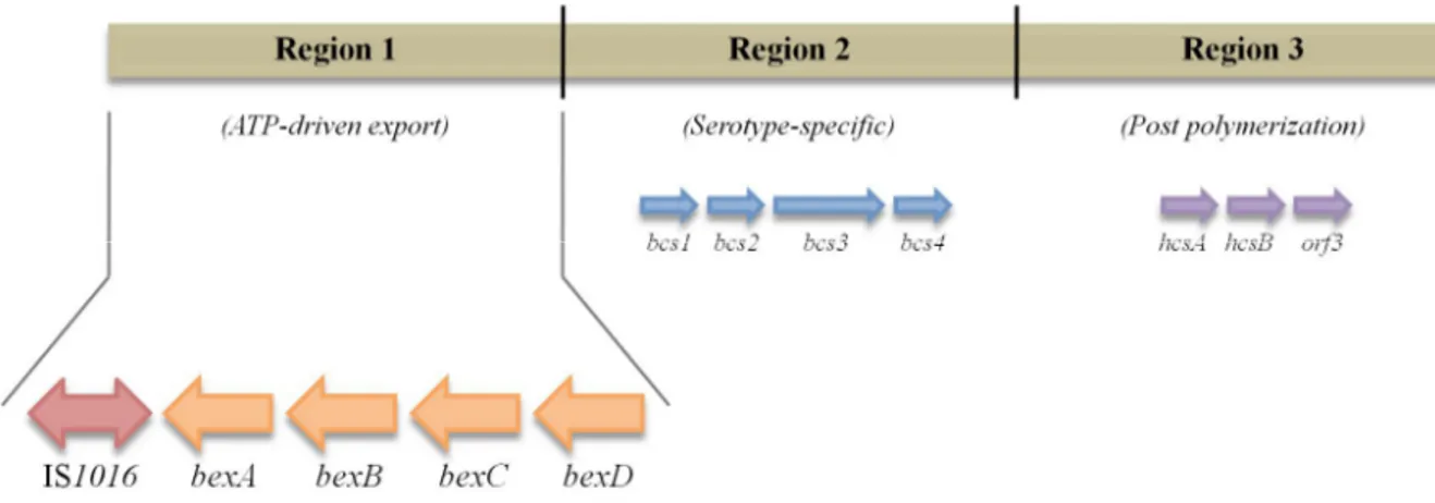
Figure 1.2 | Schematic presentation of the cap b locus. ... 4
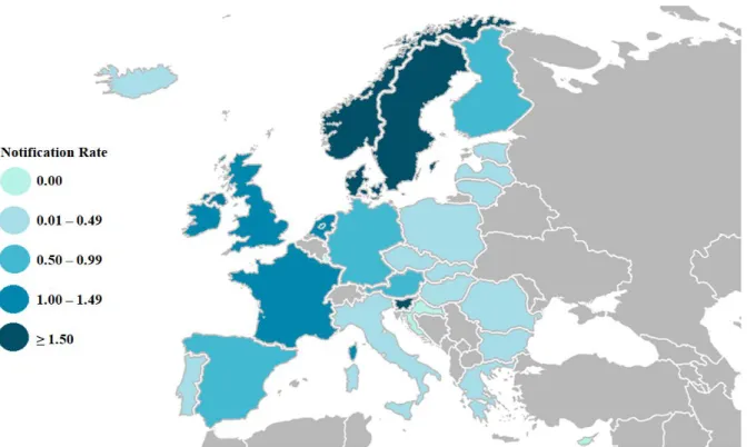
Figure 1.3 | Distribution of invasive H. influenzae disease cases per 100,000 population, in each EU/EEA country, in 2015. ... 10
Figure 1.4 | Penicillin inactivation through β-lactamase production. ... 12
Figure 1.5 | Schematic presentation of the three main stages of biofilm production: adhesion, maturation and dispersal ... 19
Figure 3.1 | Distribution of COPD isolates according to the age of patients. ... 35
Figure 3.2 | Distribution of OM isolates according to the age of patients. ... 36
Figure 3.3 | Schematic presentation of β-lactamase producer (Positive) and non-producer (Negative) COPD and OM isolates. ... 37
Figure 3.4 | Schematic presentation of genetic diversity among H. influenzae isolates (PHYLOViZ 2.0©, with goeBURST algorithm). ... 42
Figure 3.5 | Biofilm production after 24h and 48h. ... 46
xviii
xix
Index of Tables
Table 1.1 | Distribution of virulence genes among NTHi isolates. ... 17
Table 2.1 | Sample characterization. ... 24
Table 2.2 | Antibiotic concentrations on the microdilution panels. ... 26
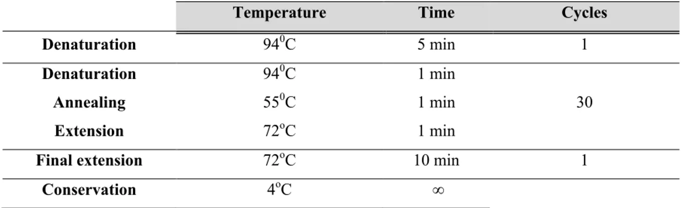
Table 2.3 | Capsular typing PCR protocol. ... 27
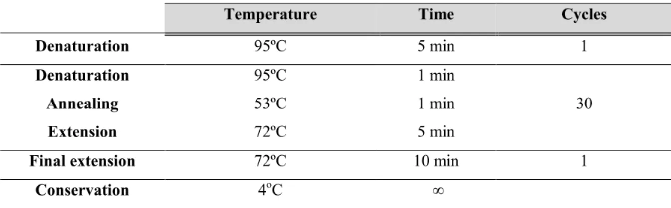
Table 2.4 | MLST amplification protocol. ... 28
Table 2.5 | Purification PCR protocol. ... 29
Table 2.6 | Sequencing PCR protocol. ... 29
Table 2.7 |pilA PCR protocol. ... 31
Table 2.8 | hifA PCR protocol. ... 31
Table 2.9 | hmw1A/2A PCR protocol. ... 31
Table 2.10 | hia PCR protocol. ... 32
Table 2.11 | ompP5 PCR protocol. ... 32
Table 2.12 | Criteria for classification of H. influenzae isolates ability to assemble biofilms. ... 33
Table 3.1 | Antibiotic breakpoints for H. influenzae. ... 38
Table 3.2 | Antibiotic susceptibility of COPD isolates. ... 39
Table 3.3 | Antibiotic susceptibility of OM isolates. ... 40
xx
Table 3.5 | Distribution of virulence genes in COPD and OM H. influenzae isolates. ... 43
Table 3.6 | Relation between hmw1A/2A and hia genes in COPD and OM H. influenzae isolates. ... 44
Table 3.7 | Biofilm production at 24h and 48h and classification of isolates as weak and moderate producers, according to each clinical group. ... 46
Table 5.1 | COPD isolates data and characterization. ... 67
Table 5.2 | OM isolates data and characterization. ... 69
Table 5.3 | Primers used for capsular typing. ... 72
Table 5.4 | Primers used for MLST. ... 73
xxi
List of Abbreviations and Acronyms
adK – Adenylate Kinase gene
Am – Ampicillin
AOM – Acute Otitis Media
atpG – ATP Synthase F1 Subunit Gamma gene
Aug – Amoxicillin-K clavulanate
Azi – Azithromycin
BHI – Brain Heart Infusion
BLNAR – β-Lactamase-Negative Ampicillin-Resistant
BLNAS – β-Lactamase-Negative Ampicillin-Susceptible
BLPACR – β-Lactamase-Positive Amoxicillin-Clavulanate-Resistant
BLPAR – β-Lactamase-Positive Ampicillin-Resistant
bp – Base-Pairs
C – Chloramphenicol
CAP – Community Acquired Pneumonia
CEACAM1 – Carcinoembryonic Antigen-Related Cell Adhesion Molecule-1
Cft – Cefotaxime
Cl- – Chloride Ion
COPD – Chronic Obstructive Pulmonary Disease
Cp – Ciprofloxacin
Cpe – Cefepime
Crm – Cefuroxime
CSOM – Chronic Suppurative Otitis Media
DGS – Direcção-Geral da Saúde
DLV – Double Locus Variant
DNA –Deoxyribonucleic Acid dsDNA – Double Stranded DNA
ECDC – European Centre for Disease Prevention and Control
EPS – Extracellular Polymeric Substance
EU/EAA – European Union and European Economic Area
EUCAST – European Committee on Antimicrobial Susceptibility Testing
FASTA format – Text format for representation of nucleotide, or peptide, sequences
Fe3+ – Ferric Ion
frdB – Fumarate Reductase Iron-Sulfur Protein gene
xxii Hap –Haemophilus Adhesion Protein
Hgp – Hemoglobulin:Haptoglobolin Binding Proteins
Hia –Haemophilus influenzae Adhesin
Hia – Encapsulated Haemophilus influenzae, serotype a
Hib – Encapsulated Haemophilus influenzae, serotype b
Hic – Encapsulated Haemophilus influenzae, serotype c
Hid – Encapsulated Haemophilus influenzae, serotype d
Hie – Encapsulated Haemophilus influenzae, serotype e
Hif – Encapsulated Haemophilus influenzae, serotype f
HifA – Major structural subunit of the fimbria gene cluster hifABCDE HMW – High Molecular Weight
Hsf –Haemophilus Surface Fibril
HxuC – Heme-Hemopexin
IgA – Immunoglobulin A
INSA – Instituto Nacional de Saúde Doutor Ricardo Jorge
LOS – Lipooligosaccharide
mdh – Malate Dehydrogenase gene
Mer – Meropenem
MIC – Minimum Inhibitory Concentration
MIC50 – Minimum Inhibitory Concentration for 50% of isolates
MIC90 – Minimum Inhibitory Concentration for 90% of isolates
MLEE – Multilocus Enzyme Electrophoresis
MLST – Multilocus Sequence Typing
MTS – Minimum Spanning Tree
NAD – Nicotidamide Adenine Dinucleotide
NTHi – Nontypeable Haemophilus influenzae OD – Optical Density
ODcut-off – Optical Density defined as the Cut-off
ODisolate – Optical Density defined as mean OD570 nm for each isolate ODnc – Optical Density of the Negative Control
OM – Otitis Media
OME – Otitis Media with Effusion
OMP – Outer Membrane Protein
PBP – Penicillin-Binding Protein
PCR – Polymerase Chain Reaction
xxiii
PHiD-CV – 10-valent Pneumococcal Nontypeable Haemophilus influenzae Protein-D Conjugate Vaccine
PilA – Major structural subunit of the type IV pilus pilABCD PRP – Polyribosylribitol Phosphate
recA – RecA protein gene
Rif – Rifampicin
SD – Standard Deviation
SDnc – Standard Deviation of the Negative Control
SLV – Single Locus Variant
ST – Sequence Type
T/S – Trimethoprim/Sulfamethoxazole
TBE – Tris-Borate-EDTA
Tbp – Transferrin Binding Protein
TBS – Tris-Buffered Saline
Te – Tetracycline
Tfp – Type IV Pilin
TLV – Triple Locus Variant
TSB – Trypic Soy Broth
USA – United States of America
UK – United Kingdom
WGS – Whole Genome Sequencing
xxiv
1
1. Introduction
1.1.
Haemophilus influenzae
1.1.1. Pasteurellaceae family, Haemophilus genus and Haemophilus influenzae
Pasteurellaceae family belongs to the Pasteurellales order and Gammaproteobacteria class.1
It is composed of a wide group of gram-negative opportunistic pathogens and commensal organisms, which have an important impact on human and animal health.2
Currently, the family is composed of 26 genera, including Actinobacillus, Aggregatibacter,
Avibacterium, Avibacteriumendocarditidis, Basfia, Bibersteinia, Bisgaardia, Chelonobacter, Cricetibacter, Frederiksenia, Gallibacterium, Haemophilus, Histophilus, Lonepinella, Mannheimia,
Mesocricetibacter, Muribacter, Necropsobacter, Nicoletella, Otariodibacter, Pasteurella,
Phoconobacter, Testudinibacter, Ursidibacter, Vespertiliibacterpulmonis, and Volucribacter.3 Haemophilus, Actinobacillus and Pasteurella are considered the three main classic genera.2
Of the Haemophilus genus, four species possess host specificity for animals (H. parasuis, H. felis, H. paracuniculus and H. haemoglobinophilus) and nine for humans (H. influenzae, H. aegyptius,
H. haemolyticus, H. parainfluenzae, H. ducreyi, H. pittmaniae, H. sputorum, H. paraphrohaemolyticus
and H. parahaemolyticus).2
H. influenzae was first described by physician Richard Pfeiffer in 1892. The bacilli were detected during the epidemic of the influenza virus, in sputum samples of patients suffering from this infection and was then named as “The influenza Bacillus”.4 In 1931, Margaret Pittman detected two
different types of colonies grown on agar plates.5 These two types differentiated in size and
opaqueness. One type of colonies was described as large, smooth, mucoid, slightly opaque and iridescent when observed in obliquely transmitted light and were denominated as S (Smooth) isolates; the second type of colonies were smaller, translucent, non-iridescent with transmitted light, with a rough surface, being denominated as R (Rough) isolates. S isolates appeared to be more virulent for laboratory animals and, in all of them, it was detected the presence of a capsule. This finding led to a differentiation of H. influenzae into isolates with a capsule, or typeable, and isolates lacking a capsule, also referred to as Nontypeable isolates (NTHi) (Fig. 1.1).
2
as haemin and Nicotinamide Adenine Dinucleotide (NAD), respectively.6 The X-factor consists of a
Ferric Ion (Fe3+) inserted in the centre of a porphyrin ring (protoporphyrin IX), with Chloride Ion (Cl-)
as a ligand.7 It can be used, optionally, for anaerobic growth. The V-factor is a pyridine nucleotide that
possesses important roles in metabolic conversions, such as signal transducers, plays a major role as electron carrier in oxidoreductase reactions and is essential for growth.8,9
H. influenzae is an important human-restricted commensal of the nasopharynx. A longitudinal study conducted in Portugal by Sá-Leão and colleagues10 collected 414 nasopharyngeal samples of
children from a day care center, during one year. The authors stated that H. influenzae was present in 87% of the nasopharyngeal samples. In addition, all children were colonized with this bacterium, at some point of the study and 34% were persistently colonised, highlighting the importance of this bacterium as a colonizer of the respiratory tract.
Similar to all gram-negative microorganisms, the cell wall of H. influenzae is composed of a layer of peptidoglycan – cross-linked by Penicillin-Binding Proteins (PBPs) – that confers strength, resistance against antibiotics and protects bacteria against lysis due to variations in osmotic pressure. Outer and inner membranes also protect bacteria against antibiotics, by hampering them from reaching the cytoplasm, where these could interfere with protein synthesis. These membranes serve as barriers of outer, as well as inner permeability.11 Lipooligosaccharides (LOS) are endotoxins present on the
bacterial cell wall and are fundamental for bacterial adhesion.12 Although H. influenzae lacks flagella,
Figure 1.1 | (A) NTHi colonies and (B) encapsulated H. influenzae colonies in chocolate agar plates
supplemented with polivitex. Both photographs were taken during this Master’s Thesis, with an Otitis Media
3
NTHi isolates commonly possess pili structures, which have been associated with twitching motility (non-flagellar based type IV pilin-dependent motility).13
1.1.2. Haemophilus influenzae serotypes
H. influenzae can be classified as encapsulated, or non-encapsulated based on the production of a capsular polysaccharide.
Encapsulated H. influenzae isolates are divided into six distinct serotypes, a, b, c, d, e, f – also referred to as Hia, Hib, Hic, Hid, Hie and Hif – based on the production of different capsular polysaccharides. Capsular polysaccharides confer virulence by avoiding complement system lysis.14 H. influenzae capsular serotypes may be organised into three groups, each composed of two serotypes, based on both the structures of the polysaccharides and an association with their resistance to antibody-free complement lysis effect.14 Types b and a were considered the most virulent, being
composed of a phosphodiester, a neutral sugar and an alcohol (ribitol). Types c, d, e and f were much less virulent, with types c and f being considered of intermediate complement resistance and composed of a phosphodiester, a N-acetylated amino sugar and a monosaccharide. Finally, types d and e possessed the least resistance to complement action and have a repeat unit of a N-acetylmannosamine uronic acid and N-acetylglucosamine.14 Encapsulated isolates are thus more
virulent and tend to be associated with invasive disease, such as septicaemia and meningitis.6
Although NTHi isolates lack a polysaccharide capsule, they can be differentiated by Outer Membrane Proteins (OMPs), LOS and High Molecular Weight (HMW) protein profiles, among other virulence factors, such as Hif and Hap adhesins, for instance.15 NTHi isolates are usually associated
with non-invasive diseases, of the upper (e.g.: Otitis Media [OM] and sinusitis) and lower respiratory tract (e.g.: chronic obstructive pulmonary disease [COPD] and cystic fibrosis).16
1.1.3. Molecular typing
1.1.3.1. Capsular typing
Capsular typing by molecular methods is considered the most accurate technique to differentiate NTHi from encapsulated isolates and for identification of H. influenzae serotypes.17 The
region of the chromosome of H. influenzae responsible for capsule expression is named the “cap
4
of genes, essential for processing and exporting capsular material to the cell surface. These two regions flank a serotype-specific region, region 2, which appears to be unique to each capsular serotype and contains genes involved in the synthesis and polymerization of the capsular polysaccharide (Fig. 1.2).18,19
Figure 1.2 | Schematic presentation of the cap b locus. bex genes are located in region 1 of the cap locus,
downstream from insertion element IS1016. Region 2 possesses capsule-specific genes and region 3 is responsible for post polymerization. (adapted from19)
Most Hib isolates usually possess a partial duplication of a DNA fragment. This duplication includes two copies of regions 2 and 3, one copy of region 1 and a truncated copy of region 1 with a deletion between the insertion element IS1016 and bexA sequences.19 However, these
isolates may undergo a recombination event that leads to the loss of the complete copy. The truncated copy would remain, with a loss of part of the coding sequence located downstream of the start of functional bexA gene, which is essential for the exportation of the capsular polysaccharide to the cell surface. This results in a capsule-deficient mutant, referred to as b- mutant, or Hib-minus.19 Falla and
colleagues have developed a Polymerase Chain Reaction (PCR) capsular serotyping method that has been used as a standard protocol for typing H. influenzae.20 This method distinguishes NTHi isolates
from encapsulated ones, by amplification of bexA gene, which is exclusively present in encapsulated isolates. The authors were able to obtain an amplification product of 343 base-pairs (bp), confirming the presence of this gene in encapsulated isolates. In addition, the authors further designed type-specific primers to all six capsular serotypes enabling a differentiation of each capsular serotype.
1.1.3.2 Multilocus Sequence Typing
5
number of clusters according to each serotype, which suggested that isolates belonging to a certain serotype show limited genetic diversity. The authors hypothesised that since H. influenzae is a naturally competent microorganism, the capsule may serve as a barrier for the uptake of extracellular DNA, which probably contributes to the clonal structure of encapsulated isolates. In fact, genetic diversity in NTHi isolates is mostly due to horizontal genetic exchange.12
NTHi isolates have been shown to be electrophoretically distant from serotype b and even Hib-minus isolates, presenting a very heterogeneous population, unlike encapsulated population. Considering NTHi heterogeneous population structure, Chiara and colleagues15 were able to organize
NTHi population into six different clades (I-VI) according to the presence/absence of six virulence factors, supporting a capacity of clonal evolution in the NTHi population.
Initial studies were mostly conducted by Multilocus Enzyme Electrophoresis (MLEE), with a main focus on the electrophoretic mobility patterns of major OMPs. However, despite of the elucidations provided by MLEE regarding the clonal structure of the encapsulated population and great diversity of NTHi isolates, an alternative method – Multilocus Sequence Typing (MLST) – was developed as a standard for studies of genetic diversity in H. influenzae species.
MLST aimed to address two major issues related to epidemiological surveillance: first, to understand if the isolates collected from a certain outbreak were equal, or different from the one that started the outbreak. Second, to know if a possible correlation between isolates causing a disease in a specific geographic area and worldwide collected isolates could be established.22 Therefore, a focus
was turned into identifying alleles in the nucleotide sequences of housekeeping genes. Considering that MLST provided analysis of nucleotide sequences instead of enzymatic electrophoretic patterns, it enabled the identification of more genetic variations, than MLEE. Furthermore, this technique permitted comparisons of sequences data between laboratories, due to the existence of worldwide databases.23 MLST analyses internal fragments of 450–500 bp from seven selected housekeeping
genes. Each gene has its sequence assigned to an allele number and the alleles at the seven loci provide an allelic profile, which in turn defines the Sequence Type (ST) for each isolate. Since the accumulation of nucleotidic changes in housekeeping genes is considered a slow process, the allelic profile of a certain bacterial isolate is stable enough in time, allowing this method to be regarded as ideal for epidemiologic studies.22 The STs are then displayed in a dendrogram that enables to establish
a correlation between identical, or highly similar, allelic profiles of isolates. Thus, a phylogenetic relation may be inferred.24
In the specific case of H. influenzae, Meats and colleagues23 selected Adenylate
6
Haemophilus influenzae MLST Database (https://pubmlst.org/hinfluenzae/), where a number is assigned to each allele and an ST to each isolate profile.23
1.2. Haemophilus influenzae infections
H. influenzae is a human-restricted commensal microorganism found in the nasopharynx of the respiratory tract, which makes it an ongoing source of potential infections in the upper and lower respiratory tract, as it is the case of OM, COPD, pneumonia, cystic fibrosis and bronchitis, in addition to invasive diseases.25
1.2.1. Invasive disease
In 2018, the European Commission26 updated the definition of invasive disease caused by H. influenzae as the isolation of this bacterium, or its nucleic acids from a clinical sample of a biological fluid considered sterile, such as blood, or cerebrospinal fluid, for instance. In Europe, the Surveillance Report of the European Centre for Disease Prevention and Control (ECDC) showed that the distribution of invasive H. influenzae disease tends to follow a seasonal pattern and the highest number of reported cases seems to occur during winter months, with a great decrease in August. From this month on, the number of cases increases until the end of the year.25,27
1.2.2. Non-invasive disease
1.2.2.1. Chronic Obstructive Pulmonary Disease
World Health Organization (WHO) has defined COPD as a disease that can be characterized by an airflow limitation, which is usually progressive and associated with alveolar or airway abnormalities of the lungs to certain noxious gases and particles. Patients with this disease usually present a chronic inflammation of the airways and parenchymal destruction.28 Inflammation,
7
and alteration of the airways and vasculature. Airway obstruction and tissue destruction ultimately reduce the capacity of lungs for gas exchange.30
COPD is currently the fourth leading cause of worldwide death and it is estimated to rank as third in 2020.28 According to WHO, there was a prevalence of 251 million worldwide cases of
COPD in 2016 and 3.17 million deaths were caused by this disease in 2015.31 Additionally, this
organization stated that despite of COPD being previously more common in men than women, due to increased tobacco smoking among women (in high-income countries) and a higher risk of exposure to indoor pollution (in low-income countries), this disease now equally affects both genders.32
Exacerbations are changes in the baseline dyspnea, sputum and/or cough of the patient that goes beyond the usual variability.33 Interestingly, it is estimated that approximately 50% of all
COPD exacerbations are actually caused by bacterial infections, being NTHi, along with
Streptococcus pneumoniae and Moraxella catarrhalis, the most frequent aetiological agents.34,35 NTHi
may be present in both stable and exacerbated states and it has been shown that patients colonised with this bacterium in the stable state tend to present more symptoms and sputum purulence during an exacerbation than non-colonised patients. Additionally, these patients present more cough, may take a longer period of time to recover from a peak flow at an exacerbation and present a higher exacerbation frequency.36 Furthermore, a patient can be colonised by more than one isolate of NTHi and acquisition
of a new isolate is related to occurrence of an exacerbation. Sethi and colleagues37 have demonstrated
that, in the majority of cases, after an exacerbation related to NTHi, serum antibodies are produced specifically to target that newly acquired isolate. However, this immunological response is only efficient against homologous isolates, and has no effect in newly acquired heterologous isolates. The authors postulate that these results may elucidate a mechanism that explains recurrent exacerbations, in COPD patients, in the presence of H. influenzae.
Given the significant role of bacteria in exacerbations, most of the patients are treated with antibiotics. However, since clinical features do not distinguish between bacterial and non-bacterial exacerbations, the benefits of the treatment would not be as successful in non-non-bacterial exacerbations.38 In Portugal, empirical treatment recommended is amoxicillin, with or without
clavulanic acid, macrolides, or doxycycline. However, it is recommended that antibiotics against COPD should only be taken by a patient previously confirmed to have bacterial exacerbations and not by a patient in a stable-state.39
1.2.2.2. Otitis Media
8
OM may be manifested either by acute, or chronic episodes. Three of its most common conditions are Acute Otitis Media (AOM), Otitis Media with Effusion (OME) and Chronic Suppurative Otitis Media (CSOM). AOM may be caused by either viruses or bacteria and common symptoms include otalgia and fever.41 OME, on the other hand, is a chronic inflammation.41 Common
symptoms are glue ear fluid (presence of fluid in the middle ear space behind the eardrum) with absence of acute signs, and hearing loss is a possible consequence.42 CSOM is a chronic inflammation
characterized by suppurative middle ear infection established for a long period of time and with perforation in the tympanic membrane, in most of the cases.41
In 2012, Monasta and colleagues43 conducted a review with data from 1980 to 2008
with relevant worldwide information regarding AOM and CSOM. From the information they gathered, the authors observed that the global AOM incidence rate is estimated as 709 million cases per year, with 51% of the cases being registered in children under the age of five. In Central Europe, 40% of the cases occurred in children with ages ranging from less than one to five years old. CSOM, on the other hand, registered 31 million cases each year, with 22.6% of these cases occurring in children under the age of five years old. As for mortality rates, the authors estimated that approximately 21,000 people die, each year, due to complications related with OM. The authors stated that, although the mortality rates associated with OM are relatively low, when considering the overall combination of AOM and CSOM, plus respective sequels, the numbers should be considered relevant, more specifically in the first five years of life.43
The most common treatment against AOM, in Portugal, is antibiotic administration. Amoxicillin, or amoxicillin-clavulanate are the elected choices for treatment of OM infections. Second, or third, generation cephalosporins may be an alternative in the case of ineffectiveness of the first-line antibiotic, or for penicillin allergic children.44
A 10-valent pneumococcal nontypeable Haemophilus influenzae protein-D conjugate vaccine – PHiD-CV (SynflorixTM) – was developed against H. influenzae and 10 serotypes of S. pneumoniae.45 Using protein D as carrier relates to it being a surface protein mostly conserved in both
9
1.3. Epidemiology
Due to the severity of invasive diseases, most epidemiologic studies available in literature are focused on these. Limited information is available regarding non-invasive diseases caused by H. influenzae. Therefore, the surveillance data here presented are related to invasive disease reports.
Conjugated Hib vaccine induces bactericidal antibodies to respond against Polyribosylribitol Phosphate (PRP), a capsular polysaccharide, regarded as a major virulence factor.47
In the pre-vaccine era, serotype b was one of the major causes of meningitis in children, particularly under five years old.48 In the United Kingdom (UK), a survey conducted in this period49
revealed that from all H. influenzae reported cases, 82% were caused by Hib, 88% were registered in children under five years old, and meningitis was the most common infection. In the United States of America (USA), 3–6% of infected children would die and in 20–30% of survivors, permanent mental retardation, or mild hearing loss were common sequels.47
Hib vaccines were first available in the European Union and European Economic Area (EU/EEA) in 1989.48 WHO reported that Hib vaccine is currently used as part of the routine
immunization programme in 192 countries,50 including all EU/EEA member states.25
In the post-vaccine era, although a decrease of infections caused by Hib was observed, invasive disease caused by NTHi increased. Among encapsulated isolates, a major incidence has been reported for serotype f, in Europe.25
An epidemiologic study comprising the years of 2007 and 2014 in 12 European countries51
showed that, overall, 78% of invasive cases were caused by NTHi and low notification rates for Hib supported the efficacy of Hib vaccine. The authors highlighted that these trends may be a result of improved surveillance programs, physicians awareness and even better serotyping techniques that were not available in the pre-vaccine era.
In 2015, ECDC reported 3,162 cases of invasive disease, caused by H. influenzae, confirmed in 30 European countries (Fig.1.3). Invasive cases were reported, mostly in children under the age of one and in elderly people over 65 years old.25 It was further reported that 82% of all cases were caused
by NTHi and that these were the most common cause of infection in all ages. Regarding encapsulated isolates, Hif, Hib and Hie accounted for 9%, 4% and 3% of all cases, respectively and the remaining cases were caused by Hia, Hic and Hid. Hie and Hif serotypes seemed to mostly affect people over 45 years of age.
Despite of worldwide surveillance data, comparisons of H. influenzae disease incidence between countries and overtime trends of infection should be carefully evaluated, since these are dependent on policies and surveillance systems of each country, along with reporting processes and case detection methods.48
10
studies testing protein-D and protein-E have been conducted, but the development of an effective vaccine against NTHi is still an ongoing subject of research.52,53
In Portugal, epidemiological data covers three distinct periods: 1989–2001,54 2002–201055 and
2010–2014,56 with the last period being specific to paediatric cases. All these studies were related to H. influenzae invasive disease and the results were in agreement with studies described above.25,51
Hib vaccine was available in Portugal, in 1994, being then recommended for children under five years old. It was implemented as part of the National Vaccination Programme in the year 2000. The first study period, 1989–2001, includes the pre-vaccine era. During this period, a majority of Hib isolates was characterized (60.5%) followed by NTHi (38.6%) and the first case of a Hif isolate was reported. Along with a decrease in the number of Hib infections, a decrease in multidrug resistance was also reported, which may be explained due to most of the resistant isolates being, then, of serotype b.54
Between the years 2002 and 201055 prevalence of Hib invasive disease was much lower
(13.2%), NTHi isolates accounted for the majority of cases (77.1%) and non-b serotypes appeared to be emerging, with reports of serotypes a (2.1%) and f (6.9%). Serotype d is a rare serotype that was characterized in Portugal for the first time in 2009 and since then, no other serotype d isolate was found among Portuguese disease.57
Figure 1.3 |Distribution of invasive H. influenzae disease cases per 100,000 population, in each EU/EEA
11
The paediatric study in the period 2010–201456 reported NTHi isolates as being responsible
for the majority of the cases (65.7%). Hib was characterized in nine children (23.7%), six of which were considered vaccine failures.
1.4. Antibiotics in Haemophilus influenzae
Antibiotics are biological, or synthetical, substances that either possess the ability to inhibit bacterial growth, or kill bacteria, which characterizes them as bacteriostatic, or bactericidal, respectively.58 Nowadays, it is recognized that infectious diseases are still a leading cause of
worldwide death and antimicrobial resistance is a topic of great concern. Antibiotics with a similar structure present identical patterns of toxicity, effectiveness and potential side effects.58
β-lactams usually possess a highly reactive ring – as a common structure – and include penicillins (e.g.: ampicillin, amoxicillin), cephalosporins (e.g.: cefepime, cefotaxime, cefuroxime) and carbapenems (e.g.: meropenem).58
These antibiotics usually target PBPs, which are fundamental for synthesis of the bacterial cell wall, as they cross link peptide units in peptidoglycan production. Covalent bonding with antibiotic disrupts the process by weakening peptidoglycan and the cell eventually bursts, due to osmotic pressure.58-60
Macrolides (e.g.: azithromycin) and tetracyclines bind to bacterial 50S and 30S ribosome subunit, respectively, which stops addition of amino-acids to polypeptide chains, ultimately resulting in inhibition of protein synthesis. Quinolones (e.g.: ciprofloxacin) interfere with DNA replication, by inhibiting the action of DNA gyrase, an enzyme that introduces negative supercoils in DNA during replication. Other agents, such as chloramphenicol, rifampicin and trimethoprim-sulfamethoxazole, may act as inhibitors of the 50S ribosome subunit, DNA replication and folic acid metabolism, respectively.58,59,61
For treatment of H. influenzae infections, β-lactams are commonly used, since the outer bacterial membrane presents little resistance to penetration of this class of antibiotics.60 However,
12
1.4.1. β-lactams resistance mechanisms in H. influenzae
In H. influenzae, both enzymatic and non-enzymatic mechanisms have been reported and production of β-lactamase constitutes the most common resistance mechanism against β-lactams.62
1.4.1.1. β-Lactamase-Positive Ampicillin-Resistant (BLPAR) isolates
In 1972, the first report of ampicillin resistance in H. influenzae was registered and was related to the production of TEM-1 β-lactamase, which is encoded by blaTEM-1 gene. H. influenzae
rarely produces a second β-lactamase, ROB-1, encoded by the blaROB gene.62 β-lactamases hydrolyse
the ester-amide bond in β-lactams, which interferes with the structure of the antibiotic and, ultimately, its effectiveness (Fig. 1.4).59,63
β-lactamases may be divided into four classes, from A to D, according to similarities in their structure. Classes A, C and D possess a serine residue in the active site, whereas class B are metalloproteins that require zinc as a cofactor.64 TEM-1 and ROB-1 are class A β-lactamases.62
As a solution for β-lactamase producing H. influenzae isolates, alternative treatments that combine amoxicillin and clavulanic acid have been administered to patients.65,66 In the presence of
clavulanic acid, β-lactamase irreversibly binds to its β-lactam ring, which inactivates the enzyme and prevents it from binding to β-lactams.67
1.4.1.2. β-Lactamase-Negative Ampicillin-Resistant (BLNAR) isolates
Ampicillin-resistant isolates that do not produce β-lactamase have been reported and designated as BLNAR. Usually, these isolates possess altered PBPs with reduced affinity for β-lactams, due to amino-acid substitutions in this protein.68 In addition to ampicillin, BLNAR isolates
also present reduced susceptibility for other β-lactams, such as cephalosporins.62 In opposite to
13
lactamase producing isolates, clavulanic acid has no effect in BLNAR isolates, due to lack of the target enzyme.69
H. influenzae possesses eight PBPs, PBP1 – PBP8.62 Mutations in ftsI gene, which
encodes PBP3, have been reported and are useful to confirm phenotypically identified BLNAR isolates. More specifically, amino-acid substitutions close to conserved KTG (K512TG) and SSN (S379SN) motifs in the transpeptidase domain of PBP3 are believed to be mostly responsible for antibiotic resistance.68 However, there is still not a clear definition for BLNAR isolates, since some
authors define them based on resistance breakpoint, while others consider ampicillin-intermediate isolates.62 These isolates can only be accurately identified by sequencing the ftsI gene.68
1.4.1.3. β-Lactamase-Positive Amoxicillin-Clavulanate-Resistant (BLPACR) isolates
BLPACR isolates possess both the production of β-lactamase and modified PBPs, which means that these are resistant to β-lactams, such as ampicillin and amoxicillin-clavulanate as well. Most authors define these isolates considering the amoxicillin-clavulanate breakpoint, but mutations identified in the ftsI gene ultimately serve as confirmation.62,69,68 BLPACR isolates have
higher amoxicillin resistance levels than BLNAR isolates, due to the presence of β-lactamases.69
1.5. Virulence factors
Identifying and understanding how virulence factors influence pathogenic, or commensal behaviour is very important and this is, nowadays, a main focus in NTHi research.12H. influenzae has
developed several mechanisms to resist immune responses and to adhere, persist and, consequently, invade host cells.70,71
1.5.1. Adhesion
The very first step in the NTHi pathogenesis is adherence of bacteria to the mucosa, after initial interaction.70,71
14
is the most common immunoglobulin in nasal secretions. Both Hib and NTHi isolates may produce IgA protease, which cleaves IgA1 antibodies into Fc and Fab fragments.72
LOS are lipopolysaccharides, with shorter saccharide chains, considered to be essential for interaction of bacteria with host cells.12 These structures vary among bacterial cells of the same isolate
and also between different isolates, which naturally affects interaction and invasion of host cells. LOS are present in both Hib and NTHi isolates.71
For adherence to host mucosa, H. influenzae also counts on the expression of several adhesins, which are proteins that facilitate this action.73 Five important adhesins, HifA, HMW1, HMW2,
Hia/Hsf and Hap, have been identified in encapsulated (Hib) and NTHi isolates.71 Piliated adhesins
include PilA and HifA proteins. PilA was shown to be essential for NTHi adherence to epithelium and twitching motility, since experiments with pilA-mutant isolates demonstrate a significant reduced ability of isolates to properly accomplish these mechanisms.13 Kubiet and colleagues74 have also
shown that for both Hib and NTHi isolates, absence of HifA pili significantly decreases the ability of isolates to adhere to mucin and that pre-treatment with antibodies that specifically targeted HifA has a similar effect.
However, isolates that lack pili structures still have the ability to adhere to human epithelium, which suggests expression of non-piliated adhesins. For instance, non-piliated high molecular weight proteins, HMW1 and HMW2, are present in encapsulated non-type b and most NTHi isolates, in addition to Hia protein, which seems to be a substitute for HMW-deficient isolates.75-77
1.5.1.1. pilA
pilA is a highly conserved gene that is present in, approximately, 100% of NTHi isolates,13,78 (Table 1.1). This gene is one of the four-member gene cluster pilABCD, expressing Type
IV Pilin (Tfp). Bakaletz and colleagues13 have showed that expression of pilA is essential for pilus
structures on the bacterial surface. pilA expression has been shown to be necessary for proper adherence, biofilm formation and epithelium colonization. Jurcisek and colleagues79 demonstrated that pilA mutants showed diminished capacity for in vitro adherence, which translated into loss of ability to colonize in vivo and decreased stability of biofilms in chinchilla middle ear. Tfp structures additionally play a significant role in competence, as these help the uptake of extracellular DNA through bacterial membranes.80 Pili structures have also been suggested to mediate interbacterial interaction, since pilA
15 1.5.1.2. hifA
HifA, encoded by hifA gene, is a structural subunit of the pilus structure encoded by
hifABCDE cluster. This protein mediates adherence to eukaryotic cells, by specifically binding to sialyl-lactosylceramide ganglioside receptor on epithelial cells. Expression of hifA is essential for adherence, since inactivation of this gene highly hampers the ability of H. influenzae to adhere to epithelial cells.81 Analysis of amino-acid sequences revealed conserved and variable regions, in which
variable regions possibly result in different antigenic sites.82 Geluk and colleagues82 found that hifABCDE cluster is present in both Hib and NTHi isolates with 18% of NTHi isolates possessing
hifABCDE cluster (Table 1.1). The authors additionally suggested that H. influenzae probably loses the ability to express fimbriae structures inside tissues, as a defence mechanism to avoid clearance. Although HifA plays a role in adherence to mucosa, other factors such as HMW proteins are also important for establishment of binding to epithelium.74,82
1.5.1.3. hmw1A and hmw2A
hmw1A and hmw2A genes encode HMW1 and HMW2 proteins, respectively. These genes present 80% similarity and are 71% identical.83 Despite of highly similar, HMW1 and HMW2
proteins possess different binding specificities: while HMW1 is specific for sialylated glycoprotein receptor containing sialic acid, a recent study has shown a high affinity of HMW2 to 2-6 linked N-acetylneuraminic acid, suggesting that this may be the receptor.84 Both proteins mediate attachment to
Chang epithelial cells.76,85 van Schilfgaarde and colleagues86 conducted a study for comparison of
adherence, hmw presence/absence and expression of HMW proteins in NTHi isolates. The authors found that isolates presenting both hmw genes and HMW proteins represented 72% of all isolates capable of adherence. These results highlighted a correlation between HMW and capacity for adherence. Other studies also corroborate the importance of expression of hmw genes for adherence.76,78 A study conducted by Ecevit and colleagues87 found, in NTHi isolates, percentages of
51% and 23% for hmw1A and hmw2A, respectively, while others have found prevalence for both genes from 55 to 100% of isolates (Table 1.1).78,88 NTHi isolates that do not express HMW proteins
are still able to adhere, since other adhesins, such as Hia and OMP P5, may serve as alternative adherence mechanisms.78
1.5.1.4. hia
16
when these are absent, since hmw and hia genes are mutually excluded in both NTHi and non-type b encapsulated isolates.75,77 In fact, hia has been described to be present in over 80% of NTHi isolates
lacking both HMW1 and HMW2 proteins.90 However, a receptor for Hia still remains unidentified.91
In the chromosome, hia and hsf are located in the same region. Although these genes encode proteins with similar functions, hia is present in NTHi isolates, while hsf is the major non-pilus adhesin of Hib isolates. Non-type b encapsulated isolates also possess a gene homologous to hsf.75
1.5.1.5. ompP5
OMP P5 is a β-barrel outer membrane protein. It is encoded by ompP5 gene and is a member of the OmpA protein family,92,93 which has been identified from 52% to 100% of NTHi
isolates (Table 1.1).78,92 OMP P5 binds to mucin and surface-expressed Carcinoembryonic
Antigen-Related Cell Adhesion Molecule-1 (CEACAM1) receptor.93 Duim and colleagues analysed NTHi
isolates from patients with chronic bronchitis and verified that, regarding ompP5, all isolates had conserved sequences, in addition to diverse regions located on the cell surface. These diverse regions resulted in OMPs with different molecular weights. The authors explain that this diversity may have resulted from selective pressure, which becomes an advantage of these isolates in persistence during a chronic infection.94 Similar results have been found by other authors when analysing isolates from
patients with non-respiratory and respiratory diseases, as well as with exacerbations of chronic diseases (COPD isolates included).92 Vuong and colleagues described that 13% of isolates lacking hmw1A and hmw2A genes, had ompP5 gene and these were still capable of adherence. The authors stated that although these results were not statistically significant, these may elucidate the importance of OMP P5 in adherence.78
1.5.2. Persistence and invasion
Ability of H. influenzae to adhere and persist on the mucosa is especially favoured in patients with underlying diseases of the upper and lower respiratory tract, in which mucociliary clearance is hampered.74 After adherence has been established, persistence on the mucosa is the following step. In
order to persist, H. influenzae depends upon a constant supply of iron and heme, among additional nutrients from the surrounding environment. Uptake of nutrients not only promotes survival, but also bacterial replication. Furthermore, bacteria must develop further mechanisms to resist host immune system responses.70,71
17
isolates count on Transferrin-Binding Proteins (Tbp), Tbp1 and Tbp2, for direct iron uptake from transferrins.95 For heme uptake, H. influenzae uses hemoglobulin and Hemoglobulin:Haptoglobolin
binding Proteins (Hgp), encoded by hgp genes and Heme-Hemopexin (HxuC), the most important complexes for heme binding and transfer to cytoplasm.96
Table 1.1 | Distribution of virulence genes among NTHi isolates.
Gene Prevalence Reference
pilA ~ 100 % Bakaletz et al., 200513 and Vuong et al., 201378
hifA ~ 18 % Geluk et al., 199882
hmw 1A 51%
55–100 % Ecevit et al., 2004
87 Vuong et al., 2013, 78 and Busher et al., 200488
hmw 2A 23% Ecevit et al., 200487
hia 32% ; 55.6% Ecevit et al., 200487 and Cardines et al., 200789
ompP5 52% ; 100 % Vuong et al., 201378 and Martí-Lliteras et al., 201192
In addition to genetic diversity, H. influenzae has the ability to express phase variable virulence factors. Phase variation is an adaptation mechanism developed when bacteria are faced with changes in the surrounding environment, or must resist certain immune responses. It involves a variation of a structure and is usually associated with the number of nucleotides in a gene sequence.71
This means that each isolate may independently express some genes and switch-off different ones, considering the surrounding environment. The result is hundreds of different H. influenzae virulence phenotypes in a single culture, which can be translated to different structures of LOS, or expression of fimbriae surface antigens. Ultimately, the phenotype that better fits the environment requirements is the one that persists. Therefore, there is a very high probability that phenotypes of the same isolate observed in vivo and in vitro may quite differ.12,91 Key targets of phase variation are structures
18
Hap protein was shown to be a major virulence factor for establishment of interaction between Hib, or NTHi isolates and host cells and as an adhesin, by binding to laminin, fibronectin and collagen IV, which are components present on the respiratory epithelium. Additionally, considering the opportunistic behaviour of H. influenzae, Hap is also important for interaction and consequent invasion of damaged epithelium.97 Naturally, it is expected that other factors besides Hap, such as
proteins D and E, may also contribute to invasion of the epithelium.70,71
1.6. Biofilms
Biofilms are assemblies of microorganisms enclosed in a self-produced Extracellular Polymeric Substance (EPS) matrix, adhered to a surface.98 Changing from a planktonic to a biofilm
state enables a multicellular behaviour that favours bacterial survival in unfavourable conditions.99
Cell signalling (e.g.: quorum sensing) is a fundamental mechanism for bacterial communication and, ultimately, biofilm production.100
Biofilm production is a dynamic process divided in three main stages. The first is adhesion and involves attachment to a surface, plus formation of microcolonies. The second phase is maturation of biofilm, which involves an adaptation to conditions of the surrounding environment and the production of an EPS matrix. The final stage is biofilm dispersion that can be triggered by different factors, such as nutrient availability, or antibiotic action, among others (Fig.1.5).99
Attachment may occur on any surface – whether biotic, or abiotic – such as host tissues, or medical devices. Biofilms that may be a source of infection for humans pose a major concern of public health, since the presence of biofilms in humans constitutes a major virulence behaviour, being mostly associated with chronic infections (e.g.: recurrent OM, CSOM, pneumonia, cystic fibrosis, endocarditis and osteomyelitis). Biofilms serve as a protection mechanism for bacteria and can be repeatedly produced.100,101 Bacteria commonly associated with biofilms include Escherichia coli, Pseudomonas aeruginosa, Staphylococcus aureus, Streptococcus epidermidis, Enterobacter cloacae
and Klebsiella pneumoniae. Biofilms formed by these bacteria are related to several human infections of the urinary tract, respiratory tract and soft tissue, for instance. Most of these bacteria are associated with hospital-acquired infections.101
Challenges related to biofilm production include spreading of bacteria to bloodstream, causing secondary infections, resistance to antibiotics in the presence of a mature biofilm and, ultimately, recurrence of infection.101,102 Furthermore, routine bacterial cultures are designed to detect planktonic
19
As referred, bacteria in biofilms usually present increased resistance to drug treatment. Resistance may come from hampering the penetration of biofilm, or from inhibition mechanisms through the action of enzymes (e.g.: β-lactamases), for instance, that inactivate antibiotics and protect bacteria.101,102 Therefore, the conventional use of antibiotics may not be efficient in these cases and
surgical interventions, or alternative therapies that directly target the EPS matrix, or inhibition of quorum sensing signalling molecules, for example, have to be considered.100
1.6.1. H. influenzae biofilms
Until recently, H. influenzae biofilms were a topic of controversy, as to whether isolates were able to produce an EPS matrix and its significance for virulent behaviour.12,104 However, studies have
shown a capacity of H. influenzae to aggregate in biofilms, which affect bacterial virulence.105
Considering that attachment is the first step towards biofilm production, various studies have focused on inhibiting bacterial adhesion, thus preventing biofilm production.99
Several virulence factors expressed by H. influenzae were suggested to contribute to adhesion and to be constituents of the EPS matrix, including LOS, PilA, OMPs, HMW1 and HMW2 proteins. Jurcisek and Bakaletz106 have described the presence of dsDNA, LOS, OMPs and type IV pilin
structures in the EPS matrix of NTHi biofilm. They hypothesised that pilin proteins may have a fundamental role in the structural stabilization of the biofilm, as interbacterial bridges. The fact that a PilA-mutant isolate could not form a biofilm as robust as isolates expressing this protein and that it presented a decreased adherence to the middle ear of a chinchilla host, further supported this statement. Similar to what has been reported for other bacterial biofilms,99 these authors106 described a
compartmentalization of the EPS matrix constituents with dsDNA being mostly located in the outer edges of the biofilm. This possibly served for stabilization of the structure. A more recent study further
Figure 1.5 | Schematic presentation of the three main stages of biofilm production: adhesion, maturation
20
supported such results, since antisera treatment targeting PilA protein translated into a significant inhibition of biofilm production by NTHi isolates.107
Furthermore, HMW1 and HMW2 are thought to be major constituents of H. influenzae
biofilms, as well, since antisera specific for these proteins has been shown to cover most of NTHi biofilm surface, suggesting a wide distribution of the adhesins on the EPS matrix.108
Biofilms are known to provide protection against antibiotics, as previously referred. In the specific case of H. influenzae, in vitro studies have demonstrated a diminished effect of antibiotics against bacteria inside a biofilm. In fact, for the same bacterial isolate, the required antibiotic concentration to inhibit bacterial growth in a biofilm may be 100 folds higher than the dose required to inhibit the planktonic form.109 These results demonstrate that, in a biofilm associated infection, a
patient may receive an empirical treatment that may, ultimately, have no effect, leading to bacterial survival and persistence of infection. The fact that routine culture methods may not detect bacteria inside biofilms and that there are no standard methods of antibiotic susceptibility tests for bacteria in biofilms, further hampers a proper treatment of infection.102,103 Moreover, it has been suggested that
sub-inhibitory concentrations of β-lactams – the antibiotics most commonly used to treat H. influenzae
infections – may actually induce biofilm production of NTHi isolates.110
An additional challenge related to biofilm production is the development of polymicrobial biofilms in infected tissues, resulting from interplay of different bacterial species. H. influenzae, for example, has been shown to form polymicrobial biofilms with Moraxella catarrhalis and
Streptococcus pneumoniae.111,112