Selin Pars
BScCHARACTERIZATION OF A DAND5
P.R152H CONTROL-DERIVED IPS CELL
LINE TO BE USED AS A TOOL FOR
HEART DISEASE MODELING
Dissertation to obtain the Master degree in Biochemistry for Health
Advisor: José A. Belo, PhD, CEDOC Co-advisor: Graça Rosas, PhD, CEDOC
Selin Pars
BScCHARACTERIZATION OF DAND5
P.R152H CONTROL-DERIVED IPS CELL
LINE TO BE USED AS A TOOL FOR
HEART DISEASE MODELING
Dissertation to obtain the Master degree in Biochemistry for Health
Advisor: José A. Belo, PhD, CEDOC Co-advisor: Graça Rosas, PhD, CEDOC
Jury:
President: Prof. Doutor António Sebastião Rodrigues Main discussant: Prof. Doutora José Eduardo Marques Bragança Other members: Prof. Doutor José António Henriques de Conde Belo
Profª. Doutora Maria Teresa Nunes Mangas Catarino
CEDOC – Chronic Diseases Research Center - Nova Medical School Universidade Nova de Lisboa
Funding: FCT (PTDC/BIM-MED/3363/2014)
StemCellResearch29(2018)202–206
Lab Resource: Stem Cell Line
Generation and characterization of a human iPS cell line from a patient-
related control to study disease mechanisms associated with DAND5
p.R152H alteration
Selin Parsa,1, Fernando Cristoa,1, José M. Inácioa,1, Graça Rosasa, Isabel Marques
Carreirab,c,d, Joana Barbosa Melob,c,d, Patrícia Mendese, Duarte Saraiva Martinsf, Luís Pereira de Almeidag, José Maioe, Rui Anjosf, José A. Beloa,*
a
Stem Cells and Development Laboratory, CEDOC, NOVA Medical School/Faculdade de Ciências Médicas, Universidade Nova de Lisboa, Lisbon, Portugal
b
Cytogenetics and Genomics Laboratory, Faculty of Medicine, University of Coimbra, Coimbra, Portugal
c
CNC.IBILI Consortium, University of Coimbra, Coimbra, Portugal
d
CIMAGO - Center of Investigation on Environment Genetics and Oncobiology, Faculty of Medicine, University of Coimbra, Portugal
e
Departamento Materno-Infantil, Centro Hospital do Algarve, EPE, Faro, Portugal
f
Hospital de Santa Cruz, Centro Hospitalar Lisboa Ocidental, Lisbon, Portugal
g
CNC - Center for Neurosciences & Cell Biology, University of Coimbra, Coimbra, Portugal
A B S T R A C T
A DAND5-control human iPSC line was generated from the urinary cells of a phenotypically normal donor. Exfoliated renal epithelial (RE) cells were collected and reprogrammed into iPSCs using Sendai virus repro- gramming system. The pluripotency, in vitro differentiation potential, karyotype stability, and the transgene-free status of generated iPSC line were analyzed and confirmed. This cell line can be exploited as a control iPSC line to better understand the mechanisms involved in DAND5-associated cardiac disease.
Resource table.
Unique stem cell line NMSUNLi002
Type of Modification N/A
Associated disease N/A
Gene/locus NM_152654.2:c.455G; DAND5 c.G455G; p.R152R
identifier
Alternative name(s) of stem cell line
iUC-DAND5_455/control Method of modification
Name of transgene or
N/A N/A
Institution CEDOC, NOVA Medical School
Contact information of José A. Belo, [email protected]
distributor
resistance Inducible/constitutive
system
N/A
Type of cell line iPSC
Origin Human
Additional origin info Sex: male
Ethnicity: Caucasian
Date archived/stock date
Cell line repository/ bank
January 2018 N/A
Cell Source Exfoliated renal epithelial cells isolated from urine
Clonality Clonal
Method of Transgene-free (Sendai virus vector)
reprogramming
Ethical approval Approved by the Ethics Committee of NOVA Medical School (Protocol N° 13/2016/ CEFCM) and by the National Committee for Data Protection (CNPD, Permit N° 8694/ 2016).
S. Pars et al. Stem Cell Research 29 (2018) 202–206
*
Corresponding author.
E-mail address: [email protected] (J.A. Belo). 1
Equal authors.
https://doi.org/10.1016/j.scr.2018.04.015
Received 6 April 2018; Received in revised form 19 April 2018; Accepted 26 April 2018 Available online 28 April 2018
S. Pars et al. Stem Cell Research 29 (2018) 202–206
Resource utility
This DAND5-control iPSC line is essential in studying the disease related impairment in heart formation of DAND5 found in a previous study. This control cell line will allow to uncover the role of DAND5
c.455 G > A variant in the molecular mechanisms of cardiomyocyte proliferation along with the previously established line.
Resource details
DAND5 is a Nodal antagonist that is involved in the correct left/ right
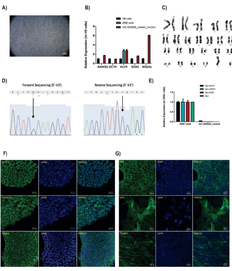
body axis establishment during gastrulation (Belo et al., 2017). The heart is the first organ to be formed and the first organ to be af- fected by improper levels of Nodal signaling throughout embryo de- velopment. Several variants of different genes involved in this signaling pathway have been associated with Congenital Heart Disease (Deng et al., 2015). Recently, we have identified and functionally character- ized a c.455G > A DAND5-variant in a patient diagnosed with ven- tricular septal defect with overriding aorta, right ventricular hyper- trophy, and pulmonary atresia (a case of extreme tetralogy of Fallot phenotype) (Cristo et al., 2017a). The variant was inherited from the apparently healthy mother. To further study the impact of altered DAND5 proteins in molecular pathways that are involved in cardiac development and on cardiomyocyte behavior, we have generated and characterized the NMSUNLi001-A variant cell line, which has a het- erozygous non-synonymous variant in exon 2 of DAND5 gene (c.455G > A), causing an amino acid change of p.R152H in the func- tional domain of the DAND5 protein (Cristo et al., 2017b). However, to unveil the precise mechanism of how this variant affects early heart development, the generation of control iPSC lines is essential. Here, we generated and characterized a DAND5 patient-related (father) control iPSC line that does not carry any alteration in the locus of the variant described in the NMSUNLi001-A cell line. This cell line will be utilized for disease modeling purpose. Hence, exfoliated renal epithelial (RE) cells were collected from urine sample collected from a healthy donor. After being grown during 7 days in culture, the RE cells were repro- grammed using CytoTune™-iPS 2.0 Reprogramming Kit (Life Technol- ogies, Invitrogen). The kit utilizes non-transmissible, non-integrating form of Sendai Virus (SeV) vectors that deliver four key transcription factors (SOX2, OCT3/4, c-MYC and KFL-4) to reprogram the somatic cells into a pluripotent state, and the iPSC colonies appeared 17 days after the delivery of the reprogramming factors. At this time, we ob- served that cells assumed typical stem cell morphology. To obtain homogeneous and clonal iPSCs lines, we manually picked and expanded several single cell-derived iPSCs colonies. Among those, one sub-clone that best displayed the ESC-like morphology (Fig. 1A) was chosen for further characterization. Firstly, after 27 passages in culture, DNA Sanger sequencing and karyotype analysis proved the genotype 455G in DAND5 exon 2 (Fig. 1D), and the number (46, XY) and arrangement of chromosomes (Fig. 1C). We assessed the transgene-free status of the iPSC line by qPCR (Fig. 1E), confirming the clearance of the viral vectors. Since the cytoplasmic nature of SeV only allows the exogenous reprogramming vectors to be cleared after several passages, we used an early passage of iPSCs as a positive control. The pluripotency of the cells was analyzed by both fluorescence immunocytochemistry (Fig. 1F) and qPCR (Fig. 1B). We confirmed the expression of the key plur- ipotency factors OCT4, NANOG, and SSEA4 both at protein and mRNA level. At mRNA level, we additionally contfihremexpression of pluripotency markers NODAL and SOX2. Embryoid body (EB) forma- tion assay was performed to assess the spontaneous differentiation potential of the iPSCs in vitro. From this assay, we assessed that the EBs cultured for 19 days expressed markers of the three germ layers: en- doderm, mesoderm, ectoderm, i.e., alpha-fetoprotein (AFP), smooth muscle actin (SMA), tubulin beta-3 chain (TUBB3), respectively (Fig. 1G). Finally, STR
analysis was performed showing that all the 16 loci tested matched (Table 1).
Materials and methods
Reprogramming of RE cells
RE cells were collected and expanded in culture. After ~4 days in culture, cells started to become evident and the medium was changed to REBM™ supplemented with REGM™ BulletKit (Lonza). When cells reached ~80% confluency, they were seeded on a 6-well plate and reprogrammed using CytoTune™-iPS 2.0 Sendai Reprogramming Kit (Life Technologies). At day 8 post-transduction, cells were passaged onto a 100 mm culture dish coated with Geltrex (Gibco, Thermo Fisher Scientific) and the next day medium was changed to Essential 8™ (E8) Flex medium, replaced until the iPSCs have emerged. 17 days post- transduction, colonies that best display an ESC-like morphology were picked and expanded with daily renewal of the E8 Flex medium.
Sequencing
To confirm the absence of the c.455G nucleotide in the established
DAND5-control cell line, genomic DNA was extracted using ISOLATE II
Genomic DNA kit (Bioline). Then, using the primers indicated in Table 2, exon 2 of DAND5 was amplified by PCR and purified using NZYGelpure kit (NZYTech). Sequencing was conducted by STAB VIDA
(http://www.stabvida.com/).
RNA extraction and real time qRT-PCR
The clearance of SeV transgenes and the expression of pluripotency markers OCT4, NANOG, SSEA4 (primers listed in Table 2) were carried out using Direct-zol™ RNA MiniPrep (Zymo Research). Subsequently, reverse transcription and qRT-PCR were performed.
Embryoid body formation assay
Embryoid bodies (EBs) consisting of approximately 2000 iPS cells/ 20 μl drop in Essential 8™ (E8) medium with 4 mg/ml polyvinylalcohol and RevitaCell™ Supplement (Thermo Fisher Scientific) were generated using hanging drop method. After 2 days, EBs were suspended in 50% E8 medium and 50% differentiation medium (DMEM with 20% FBS, Pen/Strep, NEAA, 2 mM L-glutamine, and 0,1 mM β-mercaptoethanol) and grown 3 more days. At day 5, EBs were placed on 24-well-plate with differentiation medium changes every other day. By day 19, EBs were fixed with 4% formaldehyde and the immunocytochemistry as- sayed for the three germ layers AFP, SMA and TUBB3.
Fluorescent immunocytochemistry (ICC)
Cells were fixed with 4% paraformaldehyde, permeabilized, blocked and incubated with primary and secondary antibodies (listed in Table 2) overnight at 4 °C. Prior to image acquisition, DAPI was used to stain DNA. All fluorescent images were acquired with confocal microscopy.
Karyotyping
Chromosome analysis was performed using GTG high resolution banding technique, according to standard procedures with a minimum of 10 metaphase spreads analyzed. Analysis of GTG-banded chromo- somes was performed at a resolution of 400 bands per haploid genome and karyotypes were established according to the International System for Human Cytogenetic Nomenclature (ISCN 2016).
S. Pars et al. Stem Cell Research 29 (2018) 202–206
204
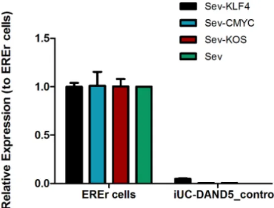
Fig. 1. Characterization of the iUC-DAND5_455/control iPSC line. A. Morphology of the iUC-DAND5_455/control line. B. mRNA expression levels of endogenous pluripotency markers in H9 cells (Black - positive control), ERE cells (Blue) and iUC-DAND5_455/control line (Red). CT-values were normalized to the geometric mean of the two housekeeping genes GAPDH and β-actin and with H9 human embryonic stem cell line as reference (set to 1). C. Karyotype of representative metaphase showing normal 46 chromosomes (XY). D. DNA sequence confirming the normal homozygous c.455G genotype in the iUC-DAND5_455/control line. E. Absolute quantitative real-time PCR showing absence of the vectors and the exogenous reprogramming factor in iPSCs (right) and presence of the reprogramming factors in the EREr control cells (left). F. Immunodetection of pluripotency markers of iUC-DAND5_455/control line. G. Immunofluorescence analyses of in vitro differentiation of EBs using specific antibodies against the endodermal marker α-fetoprotein (AFP), ectodermal marker βIII-tubulin (TUBB3) and mesodermal markers α-smooth muscle actin (SMA). Nuclei were stained with DAPI (scale bars = 20µm).
S. Pars et al. Stem Cell Research 29 (2018) 202–206
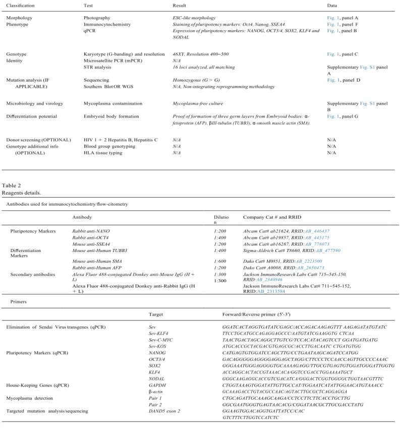
Table 1
Characterization and validation.
Classification Test Result Data
Morphology Photography ESC-like morphology Fig. 1, panel A
Phenotype Immunocytochemistry Staining of pluripotency markers: Oct4, Nanog, SSEA4 Fig. 1, panel F qPCR Expression of pluripotency markers: NANOG, OCT3/4, SOX2, KLF4 and
NODAL
Fig. 1, panel B
Genotype Karyotype (G-banding) and resolution 46XY, Resolution 400–500 Fig. 1, panel C
Identity Microsatellite PCR (mPCR) N/A
STR analysis 16 loci analyzed, all matching Supplementary Fig. S1 panel
A Mutation analysis (IF
APPLICABLE)
Sequencing Homozygous (G > G) Fig. 1, panel D
Southern Blot OR WGS N/A, Non-integrating reprogramming methodology
Microbiology and virology Mycoplasma contamination Mycoplasma-free culture Supplementary Fig. S1 panel
B Differentiation potential Embryoid body formation Proof of formation of three germ layers from Embryoid bodies: α-
fetoprotein (AFP), βIII-tubulin (TUBB3), α-smooth muscle actin (SMA).
Fig. 1, panel G
Donor screening (OPTIONAL) HIV 1 + 2 Hepatitis B, Hepatitis C N/A N/A
Genotype additional info (OPTIONAL)
Blood group genotyping N/A N/A
HLA tissue typing N/A N/A
Table 2 Reagents details.
Antibodies used for immunocytochemistry/flow-citometry
Antibody Dilutio
n Company Cat # and RRID
Pluripotency Markers Rabbit anti-NANO 1:200 Abcam Cat# ab21624, RRID:AB_446437
Rabbit anti-OCT4 1:400 Abcam Cat# ab19857, RRID:AB_445175
Mouse anti-SSEA4 1:200 Abcam Cat# ab16287, RRID:AB_778073
Differentiation
Markers Mouse anti-Human TUBB3 1:400 Sigma-Aldrich Cat# T8660, RRID:AB_477590
Mouse anti-Human SMA 1:600 Dako Cat# M0851, RRID:AB_2223500
Rabbit anti-Human AFP 1:200 Dako Cat# A0008, RRID:AB_2650473
Secondary antibodies Alexa Fluor 488-conjugated Donkey anti-Mouse IgG (H + L)
Alexa Fluor 488-conjugated Donkey anti-Rabbit IgG (H + L)
1:300
1:300
Jackson ImmunoResearch Labs Cat# 715–545-150, RRID:AB_2340846
Jackson ImmunoResearch Labs Cat# 711–545-152, RRID:AB_2313584
Primers
Target Forward/Reverse primer (5′-3′)
Elimination of Sendai Virus transgenes (qPCR) Sev GGATCACTAGGTGATATCGAGC/ACCAGACAAGAGTTT AAGAGATATGTATC
Sev-KLF4 TTCCTGCATGCCAGAGGAGCCC/AATGTATCGAAGGTG CTCAA Sev-C-MYC TAACTGACTAGCAGGCTTGTCG/TCCACATACAGTCCT GGATGATGATG Sev-KOS ATGCACCGCTACGACGTGAGCGC/ACCTTGACAATC CTGATGTGG
Pluripotency Markers (qPCR) NANOG CATGAGTGTGGATCCAGCTTG/CCTGAATAAGCAGATCCATGG
OCT3/4 GACAGGGGGAGGGGAGGAGCTAGG/CTTCCCTCCAACCAGTTGCCCCAAAC SOX2 GGGAAATGGGAGGGGTGCAAAAGAGG/TTGCGTGAGTGTGGATGGGATTGGTG KLF4 ACCAGGCACTACCGTAAACACA/GGTCCGACCTGGAAAATGCT
NODAL GGGCAAGAGGCACCGTCGACATCA/GGGACTCGGTGGGGCTGGTAACGTTTC
House-Keeping Genes (qPCR) GAPDH CTGGTAAAGTGGATATTGTTGCCAT/TGGAATCATATTGGAACATGTAAACC
β-actin GCAAAGACCTGTACGCCAAC/AGTACTTGCGCTCAGGAGGA
Mycoplasma detection Pair 1 CTGCAGATTGCAAAGCAAGA/CCTCCTTCTTCACCTGCTTG
Pair 2 GGCGAATGGGTGAGTAACACG/CGGATAACGCTTGCGACCTATG
Targeted mutation analysis/sequencing DAND5 exon 2 GGAAGTGGACAGGTGATTATCC/CAC
GTCTTTCTTGGTCCATCTC
Mycoplasma contamination test
The sterility of the iPSC culture from mycoplasma was verified by PCR using the primers in Table 2.
STR analysis
authenticated by STR analysis performed by STAB VIDA (http://www. stabvida.com/).
Supplementary data to this article can be found online at https://
doi.org/10.1016/j.scr.2018.04.015.
S. Pars et al. Stem Cell Research 29 (2018) 202–206
206
clinical data collection: FC, JMI, PM, JM, RA, DM; Analyzed the data: FC, JMI, GR, JB; Performed the experiments: SP, FC, JMI, GR;
Karyotype experiment and analysis: IMC, JBM, LPA; Contributed to writing the manuscript: SP, FC, JMI and JB. All authors read and ap- proved the final manuscript.
Ethical statement
All the experimental protocols were approved by the Ethics Committee of the NOVA Medical School (Protocol N.°13/2016/CEFCM) and by the National Committee for Data Protection (CNPD, Permit N.° 8694/2016), according to European Union legislation. Written in- formed consent was obtained from patient guardian prior to sample collection.
Acknowledgements
We would like to thank the patient and their guardians for their generous donation of the urine sample used in this study. We also would like to thank Ana Jardim for technical support in karyotype analysis. This work was supported by Fundação para a Ciência e a Tecnologia (PTDC/BIM-MED/3363/2014). iNOVA4Health - UID/ Multi/04462/2013, a program financially supported by Fundação para a Ciência e Tecnologia/Ministério da Educação e Ciência, through na- tional funds and co-funded by FEDER under the PT2020 Partnership Agreement is acknowledged.
References
Belo, J., Marques, S., Inácio, J., 2017. The role of Cerl2 in the establishment of left-right
asymmetries during axis formation and heart development. J. Cardiovasc. Dev. Dis. 4, 13.
Cristo, F., Inácio, J.M., de, Almeida S., Mendes, P., Martins, D.S., Maio, J., Anjos, R., Belo, J.A., 2017a. Functional study of DAND5 variant in patients with congenital heart disease and laterality defects. BMC Med. Genet. 18.
Cristo, F., Inácio, J.M., Rosas, G., Carreira, I.M., Melo, J.B., de, Almeida L.P., Mendes, P., Martins, D.S., Maio, J., Anjos, R., Belo, J.A., 2017b. Generation of human iPSC line from a patient with laterality defects and associated congenital heart anomalies carrying a DAND5 missense alteration. Stem Cell Res. 25, 152–156.
Deng, H., Xia, H., Deng, S., 2015. Genetic basis of human left-right asymmetry disorders.
ACKNOWLEDGEMENTS
I would firstly like to thank my thesis advisor Prof. José António Belo for the opportunity he provided for me to work in Stem Cells and Development Laboratory, CEDOC. He was always reachable and supportive during the time I was working on my thesis in his lab. I am also profoundly grateful to Dr. Fernando Cristo for his support and expertise in the lab, as well as his valuable recommendations and input in my thesis. Dr. Graça Rosas has helped me from the beginning even in the busiest working days, and I am very thankful for the time she spent to teach me laboratory techniques and her handy advices in the lab. I would also like to thank to Dr. José Manuel Inácio for his support and helpfulness. The rest of the Stem Cells and Development Lab, and the whole CEDOC family were also very welcoming and I appreciate all their help.
Lastly, I would like to express my gratitude to my parents and my partner, for their endless encouragement and support. I could not have completed this work without them. Many thanks!
ii
ABSTRACT
Congenital heart diseases (CHDs) and associated laterality defects are a major health concern and 1,35 million people are diagnosed with CHDs each year worldwide. The complexity of heart development and the hurdles to investigate the disease phenotypes make it challenging to identify the underlying causes of CHDs.
DAND5 is the human homologue of mouse Cerl-2 gene that codes for a protein involved
in regulating the Nodal signalling pathway by antagonizing the Nodal protein in the node and inhibiting the Nodal signaling in the right lateral plate mesoderm (R-LPM). In a previous study, a new DAND5 heterozygous nonsynonymous variant c.455G>A was identified and linked to the risk of disease in two patients with CHDs arising from laterality defects. In order to model the phenotype of the patients with the variant c.455G>A, a human iPSC line was previously generated and characterized (Cristo et al., 2017).
In this work, we characterized a DAND5-control iPSC line from a healthy male donor (without the variant) to serve as control for the DAND5 c.455G>A line.
The characterization was based on the detection of pluripotency markers at gene- and protein level, and in vitro differential potential. Short tandem repeats (STR) analysis has used to prove the genetic identity of the ERE cells and the reprogrammed iPSCs. Karyotyping of the iPSCs after ≥20 passages has shown the stability of the highly proliferating iPSCs. Additionally, Mycoplasma detection test showed the sterility of the cell culture.
This control cell line will be compared to the DAND5 variant c.455G>A cell line to further exploit our knowledge on the consequences of the variant in the phenotypes of the patients (disease modelling) and more precisely in the modulation of cardiomyocyte proliferation (possible therapy).
RESUMO
As doenças cardíacas congênitas (DCC) e os defeitos de lateralidade associados são uma das principais preocupações de saúde e 1,35 milhões de pessoas são diagnosticadas com DCC a cada ano em todo o mundo. A complexidade do desenvolvimento do coração e os obstáculos para investigar os fenótipos da doença tornam difícil identificar as causas subjacentes das DCC.
O gene DAND5 é o homólogo humano do Cerl-2 de camundongo que codifica uma proteína envolvida na regulação da via de sinalização Nodal antagonizando a proteína Nodal no nódulo e inibindo a sinalização Nodal na mesoderme da placa lateral direita (R-LPM). Em um estudo anterior, uma nova variante não-sinônima c.455G> A da DAND5 heterozigótica foi identificada e vinculada ao risco de doença em dois pacientes com DCC decorrentes de defeitos de lateralidade. Para modelar o fenótipo dos pacientes com a variante c.455G> A, uma linha iPSC humana foi previamente gerada e caracterizada (Cristo et al., 2017).
Neste trabalho, caracterizamos uma linha iPSC de controle DAND5 de um doador do sexo masculino sadio (sem a variante) para servir de controle para a linha DAND5 c.455G> A.
A caracterização foi baseada na detecção de marcadores de pluripotência a nível de genes e proteínas, e potencial diferencial in vitro. A análise de repetições curtas em tandem (STR) usou para provar a identidade genética das células ERE e das iPSCs reprogramadas. A cariotipagem das iPSCs após ≥ 20 passagens mostrou a estabilidade das iPSCs altamente proliferativas. Além disso, o teste de detecção de Mycoplasma mostrou a esterilidade da cultura de células.
Esta linha celular de controlo será comparada com a linhagem celular c.455G> A da variante DAND5, para explorar ainda mais o nosso conhecimento sobre as consequências da variante nos fenótipos dos doentes (modelação da doença) e, mais precisamente, na proliferação de cardiomiócitos (terapia possível).
iv
ABBREVIATION LIST
ActRII: activin A receptor type 2 AFP: alpha fetoprotein
ALK4: activin A receptor type 1B AoS: aortic stenosis
A/P: anterior/posterior AP: arterial pole
APC: adenomatous polyposis coli ARC105: mediator complex subunit 15 AS: aortic sac
ASD: atrial septal defect AV: atrioventricular
AVE: anterior visceral endoderm AVR: atrioventricular ring
AVSD: atrioventricular septal defects A-P: anterior-posterior
BL-2: Biosafety Level 2 BSA: bovine serum albumin Ccnd1: cyclin D1
cDNA: complementary deoxyribonucleic acid Cerl-2: cerberus-like 2
CFC1: cripto, FRL-1, cryptic family 1 CHDs: Congenital Heart Diseases
CITED2: Cbp/p300 interacting transactivator with Glu/Asp rich carboxy-terminal domain 2
c-Myc: MYC proto-oncogene, bHLH transcription factor CM: cardiomyocyte
CNS: central nervous system CK1: casein kinase 1
Coarc: coarctation
Cripto: teratocarcinoma-derived growth factor 1 DAPI: 4′,6-diamidino-2-phenylindole
DAND5: Dan domain family member 5 DL: dextral looping
DMEM: Dulbecco's Modified Eagle Medium DMSO: dimethyl sulfoxide
DNA: deoxyribonucleic acid
dNTP: deoxyribonucleotide triphosphate DORV: double outlet right ventricle Drp1: dynamin 1 like
DV: dorsoventral DVL: dishevelled EBs: embryoid bodies
EGF-FGF: epidermal growth factor-fibroblast growth factor ERE: exfoliated renal epithelial
FBS: fetal bovine serum FZL: Frizzled
GAPDH: glyceraldehyde 3-phosphate dehydrogenase GATA4: GATA binding protein 4
GSK3ß: glycogen synthase kinase 3 beta hESC: human embryonic stem cell
hiPSC: human induced pluripotent stem cell ICC: immunocytochemistry
ICM: inner cell mass
iPSC: induced pluripotent stem cell kDa: kilodalton
Klf-4: kruppel-like factor 4 KO: knock-out
KOS: Klf4–Oct3/4–Sox2
LDEV: lactose dehydrogenase elevating virus Lefty1: left-right determination factor 1 Lefty2: left-right determination factor 2 LPM: lateral plate mesoderm
LR: left/right
LRP5/6: LDL receptor related protein 5 Nanog: Nanog homeobox
NEAA: non-essential amino acids NF: nuclease-free
NKX2-4: NK2 homeobox 4 NKX2-5: NK2 homeobox 5
Nodal: nodal growth differentiation factor NOTCH1: notch 1
NOTCH2: notch 2
Oct4: POU class 5 homeobox 1 PA: primitive atrium
PAHs: polycyclic aromatic hydrocarbons PBS: phosphate-buffered saline
PCR: polymerase chain reaction PDA: patent ductus arteriosus PenStrep: penisilin/streptomycin Pitx2: paired-like homeodomain 2 PLV: primitive left ventricle
Ppm1a: protein phosphatase, Mg2+/Mn2+ dependent 1A PR: primary ring
PRV: primitive right ventricle PS: pulmonary stenosis PVA: polyvinyl alcohol
qPCR: quantitative polymerase chain reaction RNA: ribonucleic acid
R-LPM: right lateral plate mesoderm ROCK: rho-associated protein kinase
vi
RT-qPCR: real time quantitative polymerase chain reaction SAR: sinoatrial ring
SeV: Sendai virus
SMA: smooth muscle actin Smad2: SMAD family member 2 Smad3: SMAD family member 3 Smad4: SMAD family member 4 Sox2: sex determining region Y-box 2 SSEA4: stage-specific embryonic antigen-4 SSCs: somatic stem cells
STR: short tandem repeats SV: sinus venosus
TBX1: T-box 1 TBX20: T-box 20
TCL/LEF: T cell factor/lymphoid enhancer factor TGA: transposition of the great arteries
TGF-ß: transforming growth factor beta TOF: Tetralogy of Fallot
TUBB3: class III ß-tubulin VAR: ventriculoarterial ring VP: venous pole
VSD: Ventricular Septal Defect Wnt1: Wnt family member 1
INDEX
ACKNOWLEDGEMENTS ... i
ABSTRACT ... ii
RESUMO ... iii
ABBREVIATION LIST ... iv
INDEX ... vii
INDEX OF FIGURES ... ix
1. INTRODUCTION ... 1
1.1. Congenital Heart Diseases ... 1
1.2. An Overview of Heart Development ... 3
1.2.1. Embryonic Origins of the Heart ... 4
1.2.2. Lateral Plate Mesoderm (LPM) Formation ... 6
1.2.3. Two Crucial Steps of the Heart Formation: Fusion of the Endocardial Tubes and Heart Looping ... 7
1.2.4. Maturation of the Heart ... 9
1.3. Cardiac Left-Right Asymmetry ... 11
1.3.1. Regulation of the Left-Right Axis Formation ... 11 1.3.2. Nodal Signalling and the Role of DAND5 ... 13 1.4. iPSC Technology ... 18 1.4.1. Reprogramming of iPSCs ... 18 1.4.2. iPSCs: Generation and Characterization ... 20 1.4.3. Patient-derived hiPSC-CMs ... 22 1.5. Objectives ... 23
2. MATERIALS AND METHODS ... 25
2.1. Ethical Approval ... 25
2.2. iPSC Source and Maintenance ... 25
2.3. Cell Culture Conditions of Human iPSCs ... 25
2.4. Characterization of Human DAND5-control iPSC Line ... 26
2.4.1. Embryonic Stem Cell (ESC)-like Morphology ... 26 2.4.2. DNA Sequencing ... 26 2.4.3. Short Tandem Repeats (STR) Analysis ... 27 2.4.4. Transgene-free Status ... 27 2.4.5. Mycoplasma Contamination Detection ... 27 2.4.6. RNA Extraction, cDNA Synthesis and qRT-PCR ... 27 2.4.7. Detection of Pluripotency Markers - Immunocytochemistry ... 28 2.4.8. Detection of in vitro Differentiation Potential ... 28 2.4.9. Karyotype Analysis ... 29
3. RESULTS ... 30
3.1. Morphology of DAND5-control iPSCs ... 30
viii
3.4. Clearance of the Transgenes – Lack of Transgene Expression (qPCR) ... 32
3.5. Mycoplasma-free Culture ... 33
3.6. Expression of Pluripotency Factors – Gene Level ... 34
3.7. Expression of Pluripotency Factors – Protein Level ... 35
3.8. Differentiation Potential of iPSCs in vitro ... 36
3.9. Karyotype Analysis ... 37
4. DISCUSSION AND CONCLUSIONS ... 39
5. BIBLIOGRAPHICAL REFERENCES ... 41
6. ATTACHMENTS ... 49
INDEX OF FIGURES
Figure 1.1. Prevelance of Congenital Heart Diseases (CHDs)
Figure 1.2. The developmental stages of the human heart formation. Figure 1.3. From the endocardial tubes to the chamber specification.
Figure 1.4. Nodal signalling pathway components in left-right symmetry breaking in vertebrates.
Figure 1.5. The components of Nodal signalling pathway.
Figure 1.6. The delivery of the pluripotency genes into the target cell using non-integrative methods.
Figure 1.7. Processing of iPSC: from the patient to the therapeutics. Figure 3.1. Generated DAND5-control iPSCs in culture.
Figure 3.2. Forward and Reverse sequencing of DAND5 exon2.
Figure 3.3. STR analysis of the iPSC in comparison with the ERE cells. Figure 3.4. Transgene-free status of the established iPSC line.
Figure 3.5. Mycoplasma contamination test.
Figure 3.6. Relative expression of pluripotency genes NANOG, OCT4, KLF4, SOX2, and NODAL.
Figure 3.7. Pluripotency of the DAND5-control cell line displayed at protein level. Figure 3.8. Spontaneous differentiation potential of iPSC in vitro.
Figure 3.9. Karyotype analysis of the iPSCs.
1
1. INTRODUCTION
1.1. Congenital Heart Diseases
Congenital heart diseases (CHDs) represent the abnormalities of the heart or the great vessels at birth, which accounts for 9 of 1,000 live births globally (Linde, Van Der et al., 2011). This translates into an annual number of 1.35 million live births with CHDs out of 150 million births in the world. The symptoms of CHDs include shortness of breath, fatigue, heart murmur (Sun et al., 2015). CHDs represent a wide range of heart defects that include ventricular septal defect, atrial septal defect, pulmonary stenosis, patent ductus arteriosus, tetralogy of Fallot, coarctation of aorta, and atrioventricular septal defect (Kumar V, Abbas AK, 2012). The most common subtype of CHDs is ventricular septal defect (VSD) on a global scale (Abdulkadir & Abdulkadir, 2016; Chelo et al., 2016), followed by atrial septal defect (ASD) (Linde, Van Der et al., 2011) (Fig. 1.1). Laterality defects, another type of CHD, display a prevalence of 1.1 in 10.000 live births (Lin et al., 2014) and are caused by the improper left-right axis determination of the internal organs during the developmental stage. The disruption of the normal left-right axis establishment culminates in a variety of complex cardiac and extracardiac aberrations (Versacci et al., 2018). Among these malformations, situs inversus totalis occurs when the internal organs develop as their complete mirror images, whereas in situs
ambiguous (or heterotaxy) the internal thoracic or abdominal organs are assembled
aberrantly (Vetrini et al., 2016). However, whereas in situs inversus the prevalence of CHD is 3%, almost all the patients (90%) with heterotaxy also have complex congenital heart defects like pulmonary atresia/stenosis, transposition of the great arteries (TGA), double outlet right ventricle (DORV), ventricular septal defects (VSDs), atrial septal defects (ASDs), atrioventricular septal defects (AVSD), hypoplastic left heart, anomalous venous return and coarctation of the aorta (Mohapatra et al., 2009).
The aetiology of CHDs is not well understood and characterized in most of the cases, nevertheless environmental, genetic and epigenetic factors together are known to play a role in the development of the disease. Environmental factors that of maternal origin include pregestational diabetes, pollakiuria, febrile illnesses, viral infections such as rubella and/or rubeola, influenza, alcohol consumption, cigarette smoking, use of medications, and teratogens (Chaix, 2016; Sun et al., 2015). Moreover, recently, maternal
hypertension (Ramakrishnan et al., 2015) and maternal exposure to polycyclic aromatic hydrocarbons (PAHs) were also been associated with the increased risk of CHDs (Li et
al., 2018). Among the genetic factors, chromosomal aberrations, single gene mutations
or deletions are to be listed. Moreover, the patients can be categorized as syndromic and non-syndromic phenotypes. In Down, Edward, Patau, DiGeorge syndromes, chromosomal aneuploidies are responsible for the CHDs, whereas single gene deletions or mutations are the causative of Holt-Oram, Nooan, Allagille syndromes (Chaix, 2016). In the, case of non-syndromic patients, mutations in several genes, inherited according to Mendelian inheritance, have been shown as the cause of CHDs. Some of them include
NKX2-4, NKX2-5, CITED2, CFC1, GATA4, TBX1, TBX20, NOTCH1, NOTCH2 whose
mutations are linked to CHDs (Deng et al., 2014; Djordjevic et al., 2015).
Additionally, since the major cause of mortality in laterality defects is related to complex CHDs, we can postulate that these defects can result from the disturbances of the left-right axis patterning. Therefore, variants in the genes involved in the establishment of the LR axis might also function as a risk factor or as a direct cause of CHDs (Ramsdell, 2005).
Due to the advances in early diagnostic methods and the availability of pediatric cardiac surgery, the mortality rate of the children born with CHDs decreased significantly over the last years. Between 1987-2005, the mortality of children with complex CHDs was reduced 67%, resulting in an increase of the median age of patients death by 15 years. For the first time, the number of adults living with congenital heart diseases is higher than children. There are now an estimated 1.8 million adults in Europe with CHDs, and this number is expected to increase rapidly in the next years (Ávila et al., 2014; Moons et al., 2010). This brings several consequences, and adults with congenital heart disease, particularly those with moderate or complex disease, presented longer-term sequelae and face significant cardiac and noncardiac comorbidities and a shorted life expectancy that need be managed with special attention, care and differently than the pediatric patients. Moreover, these patients are an important adulthood problem with a high healthcare resource utilization and a significant source of global economic burden.
3
Figure 1.1. Prevelance of Congenital Heart Diseases (CHDs). The results include global data of birth prevelance over the period shown in the graph. AoS=aortic stenosis, ASD: atrial septal defect, Coarc: coarctation, PDA: patent ductus arteriosus, PS: pulmonary stenosis, TGA: transposition of the great arteries, TOF: tetralogy of Fallot, VSD: ventricular septal defect. (Obtained from Lin et al., 2014).
In order to discover therapeutic solutions for the CHDs and other cardiac diseases, an elaborate knowledge of the heart development is necessary. Moreover, since the heart have a limited regeneration capacity and heart transplantation is not always possible due to the inadequate organ supplies and immune rejection risk, the need for better understanding the cardiac disease mechanisms and the generation of desired amounts of cardiomyocytes is urgent.
Therefore, discovery of patient-specific iPSC raised the hopes that these cells could be used in multiple areas of research, disease modelling, drug screening, and translational therapies to serve as a solution to those patients. In the future, with the improvements of iPSC technology, functional cardiomyocyte generation may be scaled up and the the solution for cardiac diseases may be widely available.
1.2. An Overview of Heart Development
A functional heart requires the adequate pumping of the blood to all body parts, which is achieved by the coherence of the great vessels and heart chambers. The development of the human heart includes numerous steps that begins with the formation of mesoderm at
day 12 of gestation and is completed by the 8th week (Kumar V, Abbas AK, 2012). Initially, cardiac precursor cells that are located symmetrically in the splanchnic layer of the lateral plate mesoderm fuse in the midline and give rise to a primitive heart tube at day 17-19 of development in human and day 7 in the mouse embryo (Sylva, Hoff, Van den & Moorman, 2014). Afterwards, the heart tube elongates, bends and undergoes several looping steps, in order to provide the correct positioning of the atria and the ventricles. At the later stages with the addition of ectoderm-derived neural crest cells, specification and septation of the heart chambers is achieved, so that, the oxygenated and deoxygenated blood will precisely be present in separate chambers, allowing the proper functioning of the heart (Sieber-Blum, 2004).
All of these steps are strictly regulated and involving many genetic pathways that are highly conserved along the species (Sun & Kontaridis, 2018). Therefore, any impairment of these molecular pathways might disrupt the correct formation of the heart, resulting in congenital heart diseases (CHDs), the major cause of both infant morbidity and mortality in the first year of life (Fixler et al., 2014; Gilboa et al., 2016).
1.2.1. Embryonic Origins of the Heart
The journey of a human embryo begins with the fertilization of an egg cell with a spermatozoid to form a newly specialized cell called the zygote. The zygote undergoes repetitive cell divisions without gaining any size, being called morula. As the cell divisions of the sphere-shaped morula continue, the cells in the middle unequally take up nutrients due to the geometric nature of a sphere and therefore will need a wider surface area. At this embryonic stage, called blastocyst, a cavity in the middle part of the diving cells is formed, presenting an internal mass of cells called inner cell mass (ICM) or embryoblast and an outer cell layer called trophoblast (Fig. 1.2A) (Moore, Persaud & Torchia, 2016). Upon the implantation of the embryo into the uterine wall, trophoblast cells will undergo proliferation and differentiation and their lineages will mainly give rise to the extraembryonic tissues such as placenta (Silva & Serakides, 2016). The cells that make up the ICM will then differentiate into hypoblast (or primitive endoderm) and epiblast, forming a structure called bilaminar embryonic disc (Fig. 1.2B). The hypoblast will contribute to the extraembryonic tissues, whereas the epiblast contains pluripotent
5
cells that will form the three germ layers (i.e. endoderm, mesoderm, and ectoderm) and the germ cell lineage (Roode et al., 2012).
At this stage, the epiblast and hypoblast cells of the bilaminar embryonic disc appear as a flat disc (Fig. 1.2B). Later on, at the midline of the embryonic disc, a structure called primitive streak will form, marking the first morphological sign of the gastrulation process.
The primitive streak also determines the anterior-posterior (A/P) axis of the embryo, a process that begins by the implantation of the blastocyst into the uterus. Upon implantation, in mice, the epiblast cells and the extraembryonic lineages (trophectoderm and primitive endoderm) undergo a series of proliferation and form an egg cylinder (Beddington e Robertson, 1999; Morris et al., 2012). Once the egg cylinder is formed, the epiblast is positioned at the distal part of the embryo, whereas the extraembryonic ectoderm, derived from trophectoderm, is located at the proximal part. Around E5.5, primitive endoderm-derived anterior visceral endoderm (AVE) cells at the distal part of the embryo move unilaterally towards the anterior of the embryo and defines the point where the head will form (Srinivas, 2004). At the same time, the dorsoventral (DV) axis is specified according to the proximal-distal axis of the implantation site (Ferguson, 1996; Meinhardt, 2015). So, after implantation, the future A/P axis is marked anteriorly by the polarization of the head primordium and posteriorly (tail) by the appearance of the primitive streak, while the prospective DV axis is defined by the formation of the three germ layers, the endoderm marking its ventral side and the ectoderm being dorsal (Hamada & Tam, 2014; Hirokawa, Tanaka & Okada, 2009) Once the A/P and DV axes are established, it is argued that the left-right (LR) axis is established consistently oriented relative to the dorsoventral and anterior.
The patterning of LR axis starts at gastrulation in a process involving the Nodal signalling pathway and a leftward flow of secreted molecules in the node that culminates in the creation of a LR-biased signal in this embryonic structure. Afterwards, this signal is transferred to the lateral plate mesoderm (LPM), and subsequently to internal organs for their asymmetric morphogenesis (Raya & Izpisúa Belmonte, 2006). The regulation of this process will be further elucidated in more detail in Section 1.2.
Figure 1.2. The developmental stages of the human heart formation. (Adapted from Moore, Persaud, Torchia, The Developing Human: Clinically Oriented Embryology, 2016).
1.2.2. Lateral Plate Mesoderm (LPM) Formation
At day 16 of development in humans and E6.5 in mouse, around the midline of the epiblast, the primitive streak forms in the posterior region of the embryo (Fig. 1.2B). This structure will continue from the midline of the bilaminar embryo to the caudal part. The cells around the primitive streak continuously divide laterally and cranially until they make up a lining above the hypoblast, forming two bilateral heart-forming regions (Fig.
1.2C). When the cells in the primitive streak stop dividing, we are left with three different
layers that are solely derived from the epiblast; ectoderm, mesoderm, and endoderm (Schleich et al., 2013). These three germ layers will give rise to the particular parts of the developing embryo. Ectoderm mainly will form the epidermis as well as the central nervous system (CNS), endoderm will line the gut and respiratory tract and mesoderm
7
The mesoderm can be divided into somatic mesoderm, intermediate mesoderm, and the
lateral plate mesoderm (LPM). The somatic mesoderm cells will give rise to somites (Fig.
1.2C), which will form axial structures like skeleton and skeleton muscles and the intermediate mesoderm will form the kidneys and the gonads (Moorman, 2003; Musumeci et al., 2015). The lateral plate mesoderm will differentiate into two distinct parts, the somatic LPM and the visceral LPM, which will originate the heart and some parts of the circulatory system like blood cells and blood vessels (Moorman, 2003).
1.2.3. Two Crucial Steps of the Heart Formation: Fusion of the
Endocardial Tubes and Heart Looping
As the development of human foetus proceeds, in the visceral LPM, dorsal to the developing gut tube, the left and right dorsal aortae will appear, whereas the ventral
endocardial tubes (or cardiogenic plate, Fig. 1.3A) will be formed ventral to the gut tube.
The ventral endocardial tubes were initially thought to be symmetrical, but now we know that they are not completely symmetrical (Gittenberger-De Groot et al., 2005). They are composed of crescent-shaped group of cells, where the first and second heart fields can be distinguished. The transition from mesodermal to cardiac progenitor cells requires the induction and specification of the splanchnic mesoderm into cardiac mesoderm, which is mediated by intrinsic signals from the primitive streak and from the surrounding tissues. Upon the fusion of the left and the right endocardial tubes, an arterial pole (AP) at the anterior and venous pole (VP) at the posterior will come into existence (Fig. 1.3A). During this time, the left and right parts of the ectoderm at the most outer layer of the embryo will band together and along with the somatic LPM, originating the body wall.
A. B.
Figure 1.3. From the endocardial tubes to the chamber specification. A. Endocardial tubes emerge at the left and the right side of the embryo. At about day 23 of development they fuse and create one single endocardial tube. Venous pole (VP) at the posterior take up the blood and carry it to the arterial pole. The rightward (dextral) looping (DL) of the endocardial tube locates the heart chambers in their final positions, along with the subsequent septations. B. The spatial locations of sinus venosus (SV), primitive atrium (PA), primitive left- and right ventricles (PLV, PRV), and aortic sac (AS). Atrioventricular- and ventriculoarterial rings (AVR, VAR) will form the valves with the migration of endocardial cushions. (PR: primary ring, SAR: sinoatrial ring). (Adapted from Gittenberger-de Groot et al., 2005).
Around day 21 of development, the two endocardial tubes at the left and right side of the embryo will start to bind to each other and will fuse completely around day 23. Likewise, the two dorsal aortae will also fuse to be in contact with the endocardial tubes via the 1st and the 2nd aortic arches.
At this stage, the heart is still a long tube divided in the sinus venosus (SV), primitive
atrium (PA), primitive ventricle (PV), bulbus cordis, and the aortic sac (AS) (Fig. 1.3B).
SV will give rise to the right atrium, vena cavae, and the coronary sinus. PA and PV will give rise to the atria and the ventricles of the heart. Bulbus cordis will mostly form the right ventricle, whereas the aortic sac constructs the aortic arches.
The four-chambered heart will form after the looping of the heart. Before the looping, the aortic sac is at the superior part of the elongated tube and the sinus venosus in the inferior part. During the heart looping, the primordial atrium will end up being superior to the primordial ventricle, and at the same time, the bulbus cordis loops to the right side of the body (Fig 1.3B), forming the left and right ventricles together with the primordial ventricle.
9
Rightward cardiac looping is the first morphological indication of asymmetry in the vertebrate embryo and the mechanisms that drive this process are intrinsically related with the correct establishment of the LR axis during gastrulation. In turn, this correct left or right identity depends on a complex network of signals and molecules that are strictly regulated and maintained on the LPM in the form of Pitx2 expression, which will be described in Section 1.2 (Patel, Isaac & Cooke, 1999).
The incorrect looping of the endocardial tube at this stage is associated with some of the CHDs categorized as laterality defects such as sinus inversus totalis, heterotaxia (Schleich
et al., 2013).
1.2.4. Maturation of the Heart
Until this point, the positioning of the heart chambers is partially established. However, the septation and the rearrangement of the arteries need to occur for the heart to become a fully functional organ that pumps the sufficient blood to nourish all the body. Therefore, to achieve this maturation, firstly, the atrioventricular (AV) canal needs to be closed by
dorsal and ventral endocardial cushions, followed by left and right lateral cushions and
form the left and right AV canal (Fig 1.3B) (Sieber-Blum, 2004).
As mentioned before, primordial ventricle and the bulbus cordis will give rise to the left and the right ventricles. The looped parts of the endocardial tube, sinus venosus and primordial atria will form the right and the left atria. In addition to that, part of bulbus cordis will become the proximal aorta and the pulmonary trunk.
Once the four-chambered heart structure is established, the differentiating myocardium will be responsible for the conduction system instead of the peristaltic construction of the heart tube (Ya et al., 1997).
The normal blood circulation will start with the entry of the venous blood from the body parts to the right atrium. Via the tricuspid valve, the venous blood will flow to the right ventricle and subsequently will be pumped to the lunges by pulmonary arteries. Simultaneously, the left atrium takes up the oxygenated blood from the pulmonary veins and pumps it to the left ventricle through the mitral valve. The oxygenated blood from left ventricle is then pumped to different body parts by aorta.
The conduction of the electrical impulses throughout the heart is achieved by nodes. Sinoatrial node, atrioventricular node, bundle of His and Purkinje fibers together orchestrate the conduction in the heart (Sánchez-Quintana & Yen Ho, 2003).
At later stages, coronary vascular formation of the heart will be accomplished by epicardium. The endothelial network that is derived from the epicardium will provide nutrition and oxygen for the heart itself (Tomanek, 2005).
Each and every step of the heart development should be correctly established, since their disturbance might result in congenital heart diseases.
11
1.3. Cardiac Left-Right Asymmetry
Vertebrates have an external bilateral symmetry along the right axis but present a left-right asymmetry of the internal organs (Levin, 2005). Although the first sign of left-left-right asymmetry occurs during the morphogenesis of the internal organs at the time of cardiac looping, the specification of the left-right axis takes place much earlier at gastrulation. At this time, cells that migrate from the primitive streak to the left and right parts of the embryonic midline are already exposed to different left and right patterning signals and will contribute to the distinct regions of the heart. These patterning signals, in turn, result from a number of events that are mainly controlled by the TGF-ß/Nodal signalling pathway and DAND5, a 20-kDa secreted growth factor with the ability to directly bind to Nodal and to inhibit its signalling pathways, play a critical role in this process (Hamada
et al., 2002; Zhou et al., 1993).
1.3.1. Regulation of the Left-Right Axis Formation
The establishment of the left-right axis can be divided into three main steps; initial symmetry breaking at the node, the transfer of the LR-biased signal from the node to the LPM, and the left-right asymmetric positioning and morphogenesis of the visceral organs (Shiratori & Hamada, 2014).
The initial left-right symmetry breaking seems to originate in a structure called left-right organizer (node in amniotes; Kupffer’s vesicle in Zebrafish). According to the widely accepted fluid flow model in symmetry breaking, the cells that are aligned along the LR organizer create a leftward flow by the motile cilia. This flow, namely nodal flow has been shown to be dependent on the motile cilia which reside in the mouse node (Nonaka
et al., 1998).
In Figure 1.4, the symmetry breaking mechanism in the vertebrate embryo is depicted. In the centre of the LR organizer, motile cilia are responsible to create the leftward nodal flow by rotating in a clockwise direction, whereas at the periphery, the immotile sensory cilia sense this flow in a mechanosensory way (Zinski, Tajer & Mullins, 2017). As a result of this leftward flow, the cells on the left part of the LR organizer degrade RNA of the
Cerl2 (DAND5), which results in the biased Cerl2 expression on the right side of the LR
embryo. The second step in the establishment of the LR axis involves the transference of the this LR biased signal to the left-LPM. The mechanism of the transduction of this signal is currently not known, but it is suggested that the peripheral cells of the LR organizer secrete Nodal ligands and the upon receptor binding, Nodal signalling will start in the left part of the LPM, where Nodal regulates its own expression by a positive-feedback mechanism. On the right side of the LPM, this signalling is disrupted by Nodal inhibitors, Lefty1 and Lefty2. Lefty1 functions as a midline barrier that prevents Nodal to cross the midline and to induce bilateral expression of left-specific genes, whereas Lefty2 regulates the expression of Nodal in left LPM, restricting the intensity and area of Nodal signalling in a highly precise manner (Smith et al., 2011).
After the LR biased signalling in the node has been accomplished and subsequently propagated through the left LPM, the signal must reach to the internal organs and be interpreted in order to initiate the asymmetrical morphogenesis of visceral organs. The key gene responsible for this 3rd step of the LR axis formation is called Pitx2. As it can be seen in Fig. 1.4., Pitx2 is initially expressed in the left LPM, and its expression is expanded throughout the whole left LPM.
The signalling of the asymmetry from the left LPM through to internal organs are achieved by Pitx2 expression, which retains longer than other components of Nodal signalling pathway. During the next steps of cardiac development, when the primitive heart tube undergoes dextral looping to form the four-chambered heart, Pitx2 expression is essential. Among the different isoforms of Pitx2, the cardiac-specific Pitx2 (Pitx2c) is responsible for this cardiac looping, whose ectopic expression causes laterality defects of the heart such as situs inversus and cardiac isomerism (Liu et al., 2001). Moreover, in the formation of atrioventricular canal by endocardial cushions, left-sided expression of Pitx2 in the myocardium is detected. Additionally, Pitx2c expression was also shown in left atrium, left outflow tract, and interventricular myocardium (Ai et al., 2006). The abnormal expression of the Pitx2 during the outflow tract rotation, as well as the leftward shifting of the heart tube might contribute to a number of congenital heart defects (CHDs) such as double outlet right ventricle (DORV), transposition of the great arteries (TGA), and ventricular septal defects (VSD). These findings suggest the multifunctional role of this gene in the cardiac left-right asymmetry.
13
Figure 1.4. Nodal signalling pathway components in left-right symmetry breaking in vertebrates. (Adapted from Zinski, Tajer & Mullins, 2017).
1.3.2. Nodal Signalling and the Role of DAND5
The molecular cascade of the Nodal signalling pathway and its components are depicted in Fig. 1.5.
Nodal signalling is activated when the Nodal protein binds to a target cell through interaction to the ALK4 threonine kinase type I receptor) and ActRII (serin-threonine kinase type I receptor). Upon the receptor-ligand interaction and co-receptor involvement (Cripto or Cryptic), ALK4 phosphorylation at the cell membrane occurs. This leads to the subsequent phosphorylation of Smad2/Smad3, their association with Smad4 and the translocation of this complex to the nucleus. Here, the complex interacts with the transcription factor FoxH1 leading to activation of downstream target genes like
Nodal itself (auto regulation), Lefty2 and Pitx2, which is the main responsible for
transducing the Nodal signaling afterwards (Hill, 2018; Shen, 2007).
The Nodal signalling pathway needs to be strictly regulated and this is achieved by the inhibitor Lefty1 and Lefty2 in the LPM and by the Cerl2/DAND5 protein at the node.
Figure 1.5. The components of Nodal signalling pathway. (Obtained from Hill, 2018)
Cerberus like 2 (Cerl-2) or DAND5 gene in humans encodes a protein that belongs to the
TGF-ß (transforming growth factor-ß) antagonists.
As a Nodal antagonist, the secreted Cerl-2 protein plays a crucial role in the determination of left-right asymmetry in mice (Marques et al., 2004). At the mouse node, the asymmetric expression pattern of Cerl-2 represents the most immediate molecular response to the flow.
Prior to the appearance of the LR-biased signalling, the levels of Cerl-2 in peripheral crown cells are equal on the left and right side of the node. At this stage, the Nodal flow achieved by the motile cilia is very weak. As the leftward Nodal flow accelerates (E7.75),
Cerl-2 expression on the left side is downregulated, which breaks the symmetry of the
15
The resulting Nodal signalling is therefore increased on the left side of the node (L>R), due to the relatively higher Nodal-inhibitory effect of Cerl-2 on the right side.
At the 2/3 somite stage in mice, once the expression of Cerl-2 in the right side of the node starts to become higher than the left side, Cerl-2 protein accumulates in the right side of the node, preventing the activation of Nodal cascade on the R-LPM (Saijoh et al., 2003). When the velocity of the nodal flow becomes intense, it drives the translocation of the inhibitory Cerl-2 protein from the right to the left side of the node. This assumption is in agreement with the small size (20kDa) of this protein, which may render it easily transported (Inácio et al., 2013). Until the 6-somite stage, the Cerl-2 protein remains in the left side of the node. By then, the signal starts to decrease and terminate.
As a summary, Cerl-2 is initially present in the right side of the mouse embryo, where it starts its Nodal inhibiting activity. Afterwards, the translocation of the Cerl-2 protein to the left LPM turns the Nodal signalling off (Oki et al., 2009).
Cerl-2 is initially present in the right side of the mouse embryo, where it starts its Nodal inhibiting activity. Afterwards, the translocation of the protein to the left side of the embryo turns the Nodal signalling off (Inácio et al., 2013).
The crucial role of Cerl-2 in the establishment of LR axis and consequent heart development is supported in the significant mortality rate due to the cardiac defects (including laterality defects) seen in Cerl-2 knock-out (KO) mice. Moreover, the absence of Cerl-2, the only member of the Cerberus/DAN family expressed in the heart, observed in Cerl-2 knock-out mice also results in a massive increase of the ventricular cardiac walls, without any laterality defect, suggesting that Cerl2 has a role in heart development other than establishing the left-right axis in mice (Arauȷ́o, Marques & Belo, 2014).
In fact, the hypertrophy of the ventricular walls in Cerl-2 KO mice were shown to be caused by increased levels of phospo-Smad2 that culminates in prolonged TGF-ß/Nodal signalling. Moreover, the ventricles of these mice also presented an increased mitotic index associated with an increased expression of Ccnd1, which functions as a cell cycle regulator. Considering that this signalling pathways regulates cardiac proliferation (in
addition to its roles in early cardiac specification, and differentiation), the presence of Cerl-2 seems to be specifically required in the heart to control the proliferation of cardiomyocytes through the control of TGF-B/Nodal signalling.
In addition to these findings, nuclear ß-catenin levels in the Cerl-2 KO mice was also shown to be increased. Wnt/ß-catenin signalling is also an important pathway that regulates proliferation and differentiation. Therefore, its upregulation highly affects the cardiomyocyte proliferation.
Overall, the lack of Cerl-2 in mice is associated with the dysregulation of both TGF-ß/Nodal, and Wnt/ß-catenin pathways, suggesting a possible role of Cerl-2 in cardiomyocyte proliferation.
Recently, the human homologue of Cerl-2, DAND5 (Dan domain family member 5) was also studied and associated to the risk of developing CHDs in a cohort of CHD patients arising from defects during the establishment of the LR axis. In this study, conducted in our lab, a heterozygous nonsynonymous nucleotide variant c.455G>A, leading to a p.R152H a.a change in the functional domain of the DAND5 protein, has been found in two Caucasian patients with CHDs (Cristo et al., 2017). Phenotypically, Proband 1 presents left isomerism, VSD with overriding aorta, and pulmonary atresia, whereas tetralogy of Fallot and pulmonary atresia was observed in Proband 2one of the patients (Table 1.1). To get further insight in the consequences of the variant, a functional luciferase assay was performing and the results showed that the c.455G>A variant leads to a decreased activity of the DAND5 protein and consequently an increase in Nodal signalling (Cristo et al., 2017). This means that during the embryonic development of the patients, NODAL and DAND5 protein binding might have been less efficient than normal, resulting in a reduced inhibitory effect of DAND5 in the node. Equally, NODAL could be expressed higher than its normal levels on the node and then in the LPM. It remains unknown if the excess level of NODAL was located on the left or right side of the LPM, however, from the patients’ phenotypes, it can be concluded that an ectopic expression of NODAL (and PITX2) on the right-LPM might have occurred during the embryonic stage of the patients.
17
Altogether, these results link for the first time the reduced activity of a DAND5 variant with the human vulnerability for CHDs.
1.4. iPSC Technology
Due to the limitations of accessibility of some tissues in the body, such as the heart and the brain, there are diseases that cannot be easily studied. The use of stem cells represents a good solution to overcome this problem.
An important milestone in 1998 was achieved by the isolation and the cultivation of embryonic stem cells (ESCs) for the first time (Thomson, 1998). ESCs are pluripotent stem cells obtained at a very early stage of development that are capable of differentiating into all the cell types in an adult organism, and to proliferate indefinitely. Since ESCs are derived from oocytes and embryos their use in research and therapeutic approaches brings about ethical concerns with disputes about the onset of human personhood. To overcome these issues, the reprogramming of somatic cells to produce induced pluripotent stem cells has become, in the last years, the “gold standard” of stem cell research.
In 2006, for the first time, iPSCs from mouse fibroblast cells were generated in vitro by the groundbreaking work reported by Yamanaka (Takahashi & Yamanaka, 2006). A year after, the same approach was used to generate iPSC from adult human cells (Takahashi
et al., 2007). The researchers who developed this technique called reprogramming have
used different combinations of 24 candidate genes to induce pluripotency in differentiated cells (Takahashi & Yamanaka, 2006) and after several attempts, they discovered that the combination of only four transcription factors (Oct4, Sox2, Klf4, c-Myc) were needed.
1.4.1. Reprogramming of iPSCs
Nowadays, it is possible to revert differentiated cells into an undifferentiated state by a technology called cell reprogramming (Yamanaka & Blau, 2010).
There is a vast amount of reprogramming strategies that have been developed in the last years. They can be distinguished as the integrative (retroviral and lentiviral vectors) and non-integrative methods (adenoviruses, Sendai virus vector, self-replicating episomal vectors), which depends on the integration of genetic material of the vector into the host genome. Integrative methods are more efficient, however, non-integrative methods are more advantageous because transgene integration bring safety concerns for therapeutic approaches. (Abou-Saleh et al., 2018).
19
Retroviral vectors are using RNA as genetic material and integrate to the host genome. Target cells that are transduced with retroviral vectors mostly depend on the exogenous expression of pluripotency genes and the persistence of the transgene expression in the host cells may cause tumour formation (Shao & Wu, 2010). Lentiviral vectors are another type of RNA based vectors, which are also integrative. They are more efficient than the retroviral vectors in the reprogramming of the differentiated cells and have a wider scale of the cell types that they can infect.
To overcome the drawbacks of the integrative methods, new delivery systems have been developed. They include the use of adenoviruses, Sendai virus vectors, and the self-replicating episomal vectors.
Adenoviruses are DNA viruses whose genetic material retains in the host cell as extrachromosomal element. Episomal vectors are advantageous due to their long expression time, however they have a low efficiency of reprogramming. Another type of non-integrative vector, Sendai virus (SeV), which is a negative-sense single stranded RNA virus, was shown to have a good efficiency in the generation of iPSC and allows exogenous expression of the pluripotency genes (Fusaki et al., 2009). Although the genetic material of SeV does not integrate into the host genome, the transgenes are retained in the cytoplasm of the iPSCs and should be cleared after several passages. SeV vector penetrates to the host cell via the sialic acid receptors that are present on the cell membrane of a variety of animal species. As infection, replication and subsequent transcription occurs, (Figure 1.6.) the protein synthesis of the pluripotency factors begins.
Figure 1.6. The delivery of the pluripotency genes into the target cell using non-integrative methods. (Abou-Saleh et al., 2018).
1.4.2. iPSCs: Generation and Characterization
IPSCs can be generated from different origins, using distinct reprogramming methods and initial cell sources.
Firstly, somatic cells from a patient are collected and cultured following specific protocols and under appropriate conditions (Figure 1.7).
The cell source can be from different origins such as fibroblasts, hepatocytes, B and T lymphocytes, keratinocytes, blood cells (cord blood cells, peripheral blood cells), and urine cells (Raab et al., 2014; Singh et al., 2015; Zhao et al., 2013). The choice of the cell type will depend on several factors such as the availability of the cells, the reprogramming efficiency and non-invasive collection.
The accessibility and the reprogramming efficiency are some of the advantages of using fibroblasts, however, the risk of infection and chromosomal aberrations whilst taking biopsies are the drawbacks of using them. Peripheral blood cells are another type of cell sources, which is also easily accessible. When compared to fibroblasts, they require less effort in maintenance in the cell culture before the reprogramming experiments. Urine cells are readily available and accessible to be collected without the need of medical