R E S E A R C H
Open Access
Triiodothyronine modulates neuronal
plasticity mechanisms to enhance
functional outcome after stroke
Daniela Talhada
1,2, Joana Feiteiro
2, Ana Raquel Costa
2, Tiago Talhada
1, Elisa Cairrão
2, Tadeusz Wieloch
1,
Elisabet Englund
3, Cecília Reis Santos
2, Isabel Gonçalves
2and Karsten Ruscher
1,4*Abstract
The development of new therapeutic approaches for stroke patients requires a detailed understanding of the mechanisms that enhance recovery of lost neurological functions. The efficacy to enhance homeostatic mechanisms during the first weeks after stroke will influence functional outcome. Thyroid hormones (TH) are essential regulators of neuronal plasticity, however, their role in recovery related mechanisms of neuronal plasticity after stroke remains unknown. This study addresses important findings of 3,5,3′-triiodo-L-thyronine (T3) in the regulation of homeostatic mechanisms that adjust excitability– inhibition ratio in the post-ischemic brain. This is valid during the first 2 weeks after experimental stroke induced by photothrombosis (PT) and in cultured neurons subjected to an in vitro model of acute cerebral ischemia. In the human post-stroke brain, we assessed the expression pattern of TH receptors (TR) protein levels, important for mediating T3actions. Our results show that T3modulates several plasticity mechanisms that may operate on different temporal and spatial scales as compensatory mechanisms to assure appropriate synaptic neurotransmission. We have shown in vivo that long-term administration of T3after PT significantly (1) enhances lost sensorimotor function; (2) increases levels of synaptotagmin 1&2 and levels of the post-synaptic GluR2 subunit in AMPA receptors in the peri-infarct area; (3) increases dendritic spine density in the peri-infarct and contralateral region and (4) decreases tonic GABAergic signaling in the peri-infarct area by a reduced number of parvalbumin+/ c-fos+neurons and glutamic acid decarboxylase 65/67 levels. In addition, we have shown that T3 modulates in vitro neuron membrane properties with the balance of inward glutamate ligand-gated channels currents and decreases synaptotagmin levels in conditions of deprived oxygen and glucose. Interestingly, we found increased levels of TRβ1 in the infarct core of post-mortem human stroke patients, which mediate T3actions. Summarizing, our data identify T3 as a potential key therapeutic agent to enhance recovery of lost neurological functions after ischemic stroke.
Keywords: Ischemia, Photothrombosis, Recovery, Stroke, Thyroid hormones, Thyroid hormone receptors, 3,5,3 ′-triiodo-L-thyronine (T3), 3,5,3′,5′-tetraiodo-L-thyronine (T4)
Introduction
Loss of motor function following ischemic stroke is
the most enduring and disabling consequence [4, 26].
Despite the attempt to find neuroprotective treat-ments that mitigate tissue damage and loss of motor function, their translation into clinical practice has
been disappointing. So far, thrombectomy and
thrombolysis in the acute phase after stroke are the only effective treatments to restore blood flow and minimize brain damage. However, acute therapies are limited to the first 4.5 h for thrombolysis or up to 24 h for thrombectomy after stroke onset and are access-ible to less than 10% of stroke patients [36, 49]. Be-yond the acute phase constant and consistent specific rehabilitation programs are instrumental to partially regain brain function, dependent on size and brain
re-gions affected by stroke [37]. Therefore, the options
to minimize the damage after ischemic stroke remains
© The Author(s). 2019 Open Access This article is distributed under the terms of the Creative Commons Attribution 4.0 International License (http://creativecommons.org/licenses/by/4.0/), which permits unrestricted use, distribution, and reproduction in any medium, provided you give appropriate credit to the original author(s) and the source, provide a link to the Creative Commons license, and indicate if changes were made. The Creative Commons Public Domain Dedication waiver (http://creativecommons.org/publicdomain/zero/1.0/) applies to the data made available in this article, unless otherwise stated.
* Correspondence:[email protected]
1Laboratory for Experimental Brain Research, Division of Neurosurgery, Department of Clinical Sciences, Lund University, BMC A13, S-22184 Lund, Sweden
4LUBIN Lab - Lunds Laboratorium för Neurokirurgisk Hjärnskadeforskning, Division of Neurosurgery, Department of Clinical Sciences, Lund University, Lund, Sweden
sub-optimal and there is need for new therapeutic ap-proaches that target restorative processes.
In response to loss of input from the infarct core, surviving neurons adopt self-repair and self-organizing homeostatic mechanisms in order to stabilize the ra-tio between excitatory and inhibitory circuits and
maintain adequate synaptic input [21]. First, blood
flow is restored and there is a temporary resolution
in neuronal activity and metabolism in regions
surrounding and connected to the infarct [33, 82].
Concomitantly, in response to cell death in the infarct core, there is a change in neuronal pathways and
reorganization of neuronal connectivity, namely
axonal growth, spine remodeling and dendritic
arborization [29, 82]. A wide variety of homeostatic
mechanisms contribute to the maintenance of overall excitability, involving the regulation of neuronal in-trinsic excitability and synaptic transmission. These mechanisms include changes in receptor expression at the post-synaptic level, neurotransmitter release at the pre-synaptic level, ion channel function and synapse number or synaptic strength [52, 76, 77].
Processes of neuronal reorganization and cellular re-sponses to the infarct occur during the first weeks
after stroke in mice [7] and up to months and years
in humans [23, 24]. During this period the brain is
highly plastic, and distinct overlapping events promot-ing recovery of neurological function can be
modu-lated by external interventions [82]. Therefore,
current research is focused to understand mechanisms of post-injury plasticity that occur spontaneously after stroke [50, 82].
Current epidemiological human studies suggest that thyroid hormones (TH) signaling is related to a better outcome after stroke although the mechanisms involved are poorly investigated [71]. Several studies have pointed out that TH contribute to neuroprotection when admin-istered before [46, 61] or during the first hours after stroke or traumatic brain injury [20, 22,27,40, 55]. TH also protect cortical neurons against glutamate-induced neuronal damage [42].
However, long-term effects of TH during the
recov-ery phase after stroke remain largely unknown [71].
Here we hypothesized that 3,5,3′,5′-tetraiodo-L-thyro-nine (T4) and 3,5,3′-triiodo-L-thyronine (T3) might be
actively involved to enhance post-stroke recovery, since they are essential in several mechanisms for
brain development [3, 5] and normal function of the
adult brain [47]. Summarizing, TH are involved in
mechanisms of neuronal proliferation, migration and differentiation, neurite outgrowth, synaptic plasticity, dendritic branching and myelination during brain
de-velopment [5, 28]. In the adult brain, several
pro-cesses of neurorepair are particularly dependent on
T3 action, namely neuronal plasticity and
neurogen-esis [35, 56].
To study the role of TH in mechanisms of neuronal repair, we analyzed post-ischemic brains of mice sub-jected to intraperitoneal (i.p.) administration of T4and
T3 at 5 or 50μg/kg starting at day two after
photo-thrombosis (PT) and every second day, in a total of six
administrations; we assessed T3 effects in ionotropic
glutamate receptors (iGluRs) in cultured glutamatergic neurons; and we analyzed expression pattern of TH re-ceptors (TR) alpha 1 (TRα1) and beta 1 (TRβ1) in post-ischemic brains of mice and human patients. In the
present investigation we demonstrate that T3modulates
pathways during critical periods of recovery after stroke involved in reorganization of neuronal circuits and syn-aptic plasticity, functional connectivity and motor
re-covery. Summarizing, we demonstrate that (1) T3
enhanced recovery of lost motor function in an experi-mental model of stroke, (2) T3 increased levels of
syn-aptotagmin 1&2 and levels of post-synaptic glutamate receptor 2 (GluR2) subunit in alpha-amino-3-hydroxy-5-methyl-4-isoxazolepropionic acid (AMPA) receptors in the peri-infarct area, (3) T3increased dendritic spine
density in the ipsilateral and contralateral regions and
(4) T3 decreased tonic GABAergic signaling in the
peri-infarct area by a reduced number of
parvalbumin-positive (PV+) / c-fos+ neurons and
glu-tamic acid decarboxylase 65/67 (GAD 65/67) protein
levels. In cultured neurons (5) T3 modulates
mem-brane properties with the balance of inward glutamate
ligand-gated channels currents and (6) T3 modulates
synaptotagmin levels in an in vitro model of ischemia. In the human post-ischemic brain (7) TRβ1 has a
spatial expression pattern, which may drive T3
tran-scriptional activity. Materials and methods
Ethical considerations
Mice were bred and genotyped at the conventional facil-ity of the Biomedical Centre, (BMC, Lund, Sweden). All animal experiments (Studies I and II) were carried out in accordance with the international guidelines on ex-perimental animal research, with the approval of the Malmö-Lund Ethical Committee (ethical permit no. M50/2015) and followed the ARRIVE guidelines. All in vitro experiments (Study III) were carried out in com-pliance with directives on animal experimentation (Decreto-Lei 113/2013 and 2010/63/EU) in Portugal and European Union and with approval of the committee of Animal Research at Universidade da Beira Interior (CICS-UBI, Covilhã, Portugal). Human brain tissue used in this study was used with the approval of the Lund Ethical Review Board for research involving humans (Dnr 2011/80).
Thyroid hormones effects after experimental stroke (study I)
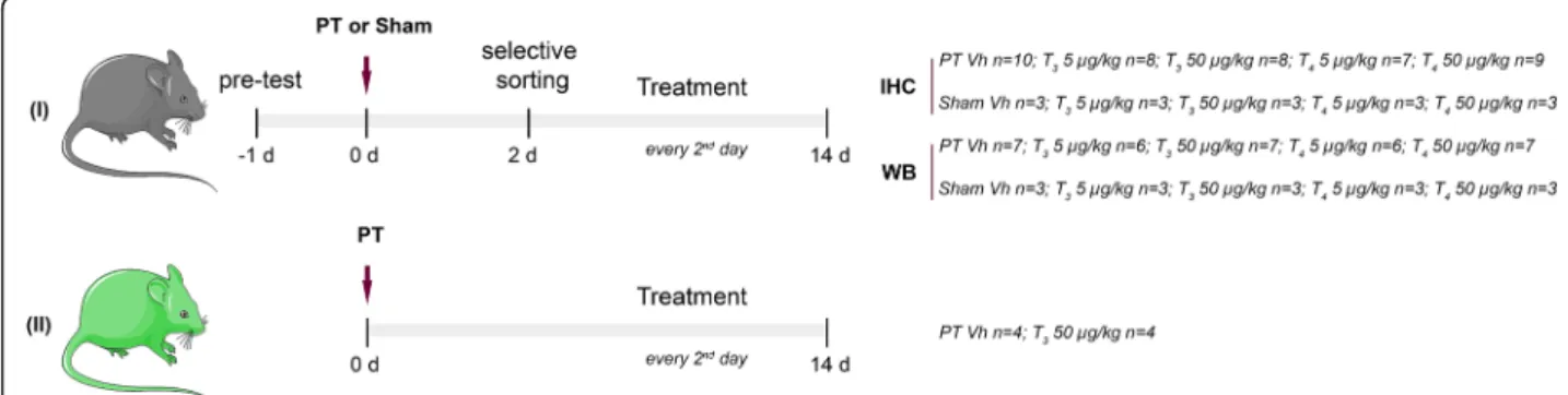
For this study, 117 C57BL/6 male mice (20 to 26 g, aged 9 to 10 weeks, purchased from Charles River) were used. Out of 117 animals, 12 were excluded due to problems during surgery and mortality before entering the treat-ment phase and 105 animals were randomly assigned into the treatment groups (Fig. 1). Treatment was initi-ated on day two after PT and every other day until the endpoint of the study. Vehicle (Vh, NaCl 0.9%), T3(5 or
50μg/kg) or T4(5 or 50μg/kg) were administered by i.p.
injection in a total of six administrations. On days two, seven and 14 after stroke onset or sham surgery, animals were evaluated for motor function.
Photothrombosis
Focal ischemic stroke was induced by PT, as described previously [65, 81]. Ischemic stroke was induced in the right hemisphere through illumination of a squared aperture measuring 4.0 to 2.0 mm (equal to an area of
8.0 mm2). The light position related to bregma (+ 1.5
mm lateral and + 0.5 mm anterior) affected the mouse primary motor cortex of forelimb-responsive sites, in the
left body side [72]. The same procedure was performed
in sham operated animals, with saline injection instead of photosensitizing dye.
Behavior analysis
Motor function and exploratory behavior after TH treat-ment was assessed using a neuroscore consisting of the rotating pole test (RPT) and the open field test, respect-ively [60, 79]. These assessments were performed in a blinded fashion to the investigator that performed the surgeries and treatments.
The RPT was used to assess postural and locomotor asymmetry that results from an unilateral brain lesion
[57]. After stroke or sham surgery, animals were evalu-ated on day two for randomization into treatment groups. Each trial was video recorded, and videos were used to assess motor dysfunction by using a zero to six scoring system (Table 1). Animals that did not fulfill in-clusion criteria were excluded from behavior analysis (see Additional file1: Supplementary methods).
The open field test was performed 14 days after stroke to assess both spontaneous post-ischemic locomotor ac-tivity and post-ischemic exploration behavior [78].
Immunohistochemistry and immunofluorescence
Tissue collection for immunostainings was performed as described before [53, 63]. Primary antibodies used for immunofluorescence were rabbit TRβ1 (Millipore, 1: 1000), rabbit TRα1 (Abcam, 1:1000), goat parvalbumin (PV235, Swant, 1:5000), mouse neuronal nuclei (NeuN, Millipore, 1:1000), glial fibrillary acidic protein (GFAP)-Cy3 (Sigma, 1:5000), rat cluster of differentiation (CD) 68 (Abd Serotec, 1:300), and mouse glutathione S-transferase (GST)-pi isoform (BD Transduction Labora-tories, 1:1000).
Infarct size measurement
Coronal brain sections from the start until the end of the infarct and spaced one millimeter were collected and stained for NeuN (rabbit NeuN, Millipore, 1:5000). The non-injured portion of the ipsilateral and contralateral hemisphere were encircled and the indirect infarct vol-ume was calculated by integration of areas from serial sections of each brain as described previously [70] using Fiji software [64].
Counting of parvalbumin positive cells
For each animal one coronal section (− 2.0 mm relative to
bregma) was stained for PV+neurons using a monoclonal
Fig. 1 Experimental design. In Study I C57BL/6 mice were pre-tested before photothrombosis (PT) or sham operations to assess limb placement. Selective sorting was assessed 2 days after surgeries. Animals were randomized into the treatment groups: Vehicle (Vh, NaCl 0.9%); T35 or 50μg/ kg; T45 or 50μg/kg. Treatment was administrated via intraperitoneal injection every second day after PT or sham operations. Neurological outcome was assessed by the rotating pole test, seven and 14 days after surgeries and brains were perfusion fixed or frozen, for
immunohistochemistry (IHC) or Western blot (WB), respectively. Study II was performed for dendritic spine analysis and Thy1-YFP transgenic mice were used. Treatment with Vh or T350μg/kg was administered as described for Study I
goat primary antibody (PV235, Swant, 1:5000), and visualization accessed using a VECTOR NovaRED Perox-idase (HRP) Substrate Kit (Vector Laboratories, CA, USA). Rabbit c-fos (Santa Cruz, 1:500) positive immuno-reactivity (c-fos+) was accessed using the avidin–biotin– HRP system.
Immunoblotting
Brains from mice were collected as previously described [63] and the tissue correspondent to the infarct core and peri-infarct was collected. Tissue from human brains were dissected out by a pathologist following autopsy. Primary antibodies used for Western blots were rabbit TRα1 (Abcam, 1:1000), rabbit TRβ1 (Millipore, 1:20000), mouse postsynaptic protein 95 (PSD95; BD Transduc-tion Laboratories, 1:1000), rabbit synaptophysin (Ther-moscientific, 1:15000), rabbit GluR1 (Millipore, 1:2000), mouse GluR2 (Millipore, 1:1000), mouse N-methyl-D-as-partate receptor 1 (NMDAR1) (BD Transduction La-boratories, 1:1000), rabbit synaptotagmin 1&2 (Abcam, 1:1000) and rabbit GAD 65/67 (Millipore, 1:2000).
Mem-branes were reprobed with antiβ-actin HRP conjugated
(1:150000, Sigma-Aldrich). Levels were calculated as a
percentage of β-actin expression, after densitometric
analysis using Fiji software.
Dynamics of dendritic spines after administration with T3
(study II)
To study the effects of T3on dendritic spine dynamics
in mouse neocortical neurons after experimental stroke, eight Thy1-yellow fluorescent protein (YFP) transgenic mice (25 to 40 g, aged 1 year, own breeding), that ex-press YFP in neuronal population were used. Mice were randomly assigned in the following treatment groups: PT/Vh, n = 4; PT/T350μg/kg, n = 4 (Fig.1). Treatment
was administered as described above for Study I. Four-teen days after the surgery, mice were sacrificed, perfu-sion fixed with paraformaldehyde 4% and brains were
collected for further infarct volume assessment and den-dritic spine analysis.
Photothrombosis
To induce PT in animals for dendritic spine analysis (Study II) the surgical procedure was performed as in Study I, and the left hemisphere was illuminated with a cold light source through a round aperture measuring 1.5 mm in diameter (equal to an area of 1.767 mm2) for 20 min. This approach induced smaller infarct sizes so that dendritic spines could be analyzed in different re-gions in the peri-infarct area. The same procedure was performed in Sham operated animals, with saline injec-tion instead of photosensitizing dye.
Detection and classification of dendritic spines from fluorescence laser scanning microscopy
Three coronal sections per animal were collected at dif-ferent levels: + 2.0 mm, + 1.0 mm and 0 mm relatively to bregma, corresponding to the rostral pole, center and caudal pole of the infarct, respectively. For each animal, we analyzed layers II/III correspondent to the apical pyr-amidal neurons in the ipsilateral motor cortex (Region 1, R1), ipsilateral somatosensory cortex (Region 2, R2), contralateral motor cortex (Region 3, R3) and contralat-eral somatosensory cortex (Region 4, R4).
Dendritic spine density and shape classification was accurately quantified and characterized using a three-dimensional computational approach as previously de-scribed, after image deconvolution [58].
For each region, three to five dendritic branches were randomly selected. Dendrites were manually selected, and spines were automatically detected using Neuron-Studio software. Dendritic spines were classified accord-ing to the head to neck ratio and head diameter as stubby, mushroom or thin [30,58], using default param-eters from NeuronStudio. Dendritic spine density was calculated with the ratio number of spines / dendrite length.
In vitro modulation of T3in glutamatergic neurons (study
III)
An in vitro model of cerebral ischemia and electrophysi-ology studies were performed to study immediate effects
of T3in homeostatic plastic mechanisms, namely
modu-lation of synaptic proteins crucial for neurotransmission and NMDA and AMPA evoked currents.
Cell cultures
Cultured cortical neurons were used after 7–8 days in vitro (DIV). Primary cortical neuronal cultures were prepared as described before [59]. Cells were obtained from the cerebral cortex from Wistar rats on embryonic day 16–18. Briefly, meninges were removed, and the
Table 1 Motor function assessed by the rotating pole test before and after photothrombosis at days 2, 7 and 14
Score Criteria
0 animal falls off immediately upon entry onto the pole 1 animal remains embraced to the pole unable to cross and
eventually falls off the pole
2 animal falls off during crossing or if the hindlimbs do not contribute to forward movement
3 animal crosses the pole while continuously slipping with the forelimbs or hindlimbs
4 animal crosses pole with > 3 ft slips 5 animal traverses the pole with 1–3 ft slips 6 animal crosses the pole without any foot slips
cortex dissected and subjected to enzymatic dissociation, using 0.05 / 0.02% w/v in phosphate buffered saline (PBS) trypsin / EDTA (#15400054, Thermofisher) for 15 min at 37 °C. The homogenized was rinsed with Dulbecco’s Modified Eagle’s medium (#11880036, DMEM, GIBCO) with 10% fetal bovine serum (#10500–064, GIBCO), 100 U penicillin and streptomycin/ml (#15140122, Thermo-fisher), 2 mM L-glutamine (#G5792, Sigma-Aldrich),
dis-sociated with a Pasteur pipette, centrifuged and
redissociated in starter medium (#21103049, Neurobasal medium, GIBCO) supplemented with B27 (#17504044, GIBCO), 100 U penicillin and streptomycin/ml, 2 mM
L-glutamine (#G5792, Sigma-Aldrich) and 25μM glutamate
(#49621, Sigma-Aldrich). The cells were plated onto poly-L-lysine (#P4707, Sigma-Aldrich) pre-coated multiwells at 1.5 × 10⁵cells/cm2and grown in starter medium at 37 °C
and 5% CO2. One-half of the medium was replaced with
cultivating medium (starter medium without glutamate) from 4 DIV. Cells were used after 7–8 DIV for in vitro assays.
In vitro ischemic model and experimental treatments
After 7 DIV neurobasal medium was collected and stored to be replaced after the experiments. Neuron cul-tures were washed with PBS, and oxygen and glucose deprivation (OGD) was induced with a deoxygenated aglycemic solution. OGD was generated in a hypoxia in-cubator chamber (StemCell Technologies), flushed with gas: 5% CO2, 95% N2. In control cultures, medium was
replaced by basic salt solution (BSS) after washing with PBS and cells were incubated in a normoxic atmosphere
containing 5% CO2. Cultures were in OGD or BSS
solu-tions for 120 min and after replaced by the previous col-lected medium. After OGD / BSS conditions, cells were
incubated with Vh (DMSO in PBS, 0.01%) or T3 1 μM
for 48 h. Subsequently, cells were washed with cold PBS to remove excess of culture medium and cells collected and frozen at− 80 °C until protein extraction.
Immunocytochemistry
For immunocytochemistry, neurons were plated on glass coverslips and fixed after 7 DIV. Antibodies used for im-munofluorescence were rabbit TRα1 (Thermoscientific, 1:500) or rabbit TRβ1 (Millipore, 1.500). The next day,
neurons were stained with Hoechst-33,342 (4μg/ml, Life
Technologies).
Immunobloting
Protein extraction was performed as previously
de-scribed [38, 63]. Western blot was performed to
evaluate levels of mouse synaptotagmin (BD Transduction Laboratories, 1:2000).
Electrophysiological recording of membrane currents
To study ligand-gated channels AMPA and NMDA, we adopted the voltage-ramp method [85].
Individual currents were recorded after incubation with T31 μM (n = 4) or Vh (n = 3) during the 48 h
pre-ceding the experiments. A sequence of voltage ramps at a rate of 0.23 mV/millisecond were applied at a holding
potential of − 80 mV. To obtain the agonist induced
current-voltage (I-V) relation, ramps I-V curves were constructed applying a 500 milliseconds voltage ramp
ranging from − 110 mV to + 20 mV elicited every 8 s.
Voltage ramps were applied in the absence and in the presence of AMPA and NMDA agonist glutamate at
50μM and co-agonist of NMDA channels glycine at
3μM, to enable subtraction of leak currents. The antag-onists of AMPA and NMDA channels,
6-cyano-7-nitro-quinoxaline-2,3-dione (CNQX; Sigma-Aldrich) and
dizocilpinehydrogen maleate (MK-801; Sigma-Aldrich), respectively, were used both at 10μM.
Cell currents were recorded sequentially in the
presence of specific K+- channel blockers
tetraethy-lammonium sodium salt (5 mM) and 4-Aminopyridine (1 mM), that were applied in the perfusion system
to-gether with the other drugs. Voltage-gated K+
chan-nels needed to be blocked, since those chanchan-nels were contributing to the conductance as well to the rever-sal potential obtained.
Statistical analysis
Data are expressed as means ± standard error of the mean (SEM) for parametric data or as medians for non-parametric data. P values < 0.05 were considered as sta-tistically significant. Statistical analysis was performed using IBM SPSS statistics 24 software for dendritic spine analysis or GraphPad Prism 6.0 software (GraphPad, San Diego, CA, USA), using one-way analysis of variance (ANOVA) followed by Bonferroni’s multiple comparison test when three or more groups were present or two-tailed unpaired Student’s t-test when comparing two groups. For non-parametric data, Kruskal Wallis test was employed for more than two groups followed by the Dunn’s multiple comparisons test and the Mann-Whitney U-test for comparison of two groups. Graphs were designed using GraphPad Prism 6.0 software.
For additional details about techniques and analysis per-formed, please refer to the Additional file1: Supplementary Methods.
Results
Treatment with T3improves functional recovery after PT
without affecting infarct size
We first assessed if treatment with T3or T4at 5 or 50μg/kg
enhances motor function in mice subjected to unilateral PT. Motor function was assessed by RPT on day 7 and 14 after
stroke onset. We observed some degree of spontaneous re-covery in mice of all groups subjected to PT. T3-treated mice
at 50μg/kg could traverse the pole with a score higher than three at 10 rpm, to the right and left sides, showing that all animals crossed the pole without falling (Additional file 2: Video S1, Additional file 3: Video S2, Additional file 4: Video S3 and Additional file 5: Video S4). However, a significantly enhanced functional recovery was only observed when the pole rotated at 10 rpm to the left, in animals treated with T3
at 50μg/kg, when compared to Vh-treated animals (Fig.2a). Fourteen days after stroke, 73% (eight out of 11) and 64% (seven out of 11) of mice treated with T350 μg/kg had a
score higher or equal to four points, at 3 rpm and 10 rpm to the left, respectively. In contrast, only 9% (one out of 11) of mice subjected to PT and treated with saline had a score of four points and not higher, at 3 and 10 rpm to the left (Add-itional file1: Figure S1).
Infarct size influences the severity of neurological defi-cits and differences of infarct size among treatment groups may influence behavior assessment to evaluate motor recovery over time. Overall the infarct volume did not differ between animals assigned to treatment groups
(2.5 ± 0.78 mm3 Vh, 3.2 ± 0.97 mm3 T3 5 μg/kg, 1.6 ±
0.47 mm3T3 50μg/kg, 3.1 ± 1.5 mm3 T45 μg/kg, 4.0 ±
1.3 mm3T450μg/kg; mean ± SEM) as shown in Fig. 2b.
All treatments had no influence on the behavior of sham-operated mice (data not shown).
The doses used in the present studies have been de-termined in preliminary studies (data not shown). No adverse effects related to hyperthyroidism were seen following any of the given doses. In addition, no dif-ferences were observed in body weight or temperature in animals from all groups throughout the studies
(Additional file 1: Table S1). In all experimental
groups, plasma levels of T3 and T4 were in
physio-logical range at the endpoint of the study (Additional file 1: Figure S2).
We performed the open field test to ascertain that TH administration was not associated with anxiety or depression-like behavior. Treatment with TH did not affect open field scores, indicative that the treatment did not induce anxiety (Additional file 1: Figure S3).
Treatment with T3did not affect the expression of TH
receptors after PT
To characterize if functional improvement after T3
ad-ministration was mediated by its binding to respective TR, we assessed their expression in the post-ischemic brain. We found that both isoforms, TRα1 and TRβ1, were ubiquitously expressed in the brain. TR were
expressed in the cytoplasm of NeuN and PV+neurons in
the peri-infarct region and in GFAP positive reactive as-trocytes in the glial scar surrounding the infarct (Fig.
2c). In contrast, CD68 positive monocytic phagocytes
and GST-pi positive oligodendrocytes were not immu-noreactive for TR (Additional file1: Figure S4).
Importantly, treatment with T3or T4at 5 or 50μg/kg
did not change the levels of TRα1 (Fig.2d), despite there was a nonsignificant elevation of TRα1 protein levels found in protein extracts obtained from the peri-infarct area (0.67 ± 0.22 Vh, 0.15 ± 0.05 T3, 1.15 ± 0.12 T4;
arbi-trary units, mean ± SEM). Likewise, no changes have been found in TRβ1 levels (1.59 ± 0.51 Vh, 1.61 ± 0.20 T3, 1.74 ± 0.42 T4; arbitrary units, mean ± SEM) (Fig.2e).
Thyroid hormone receptor pattern expression in human stroke patients
Both receptor isoforms were also found in post-mortem brain tissues. The levels for both isoforms did not differ between the peri-infarct area from stroke patients and cortex samples from non-stroke patients. However, dif-ferences were observed in the infarct core. Here, TRβ1 protein levels increased (0.37 ± 0.02 Ctrl, 0.32 ± 0.03 PI, 0.63 ± 0.07 IC; arbitrary units, mean ± SEM) while levels of TRα1 decreased (0.85 ± 0.13 Ctrl, 0.91 ± 0.16 PI, 0.38 ± 0.15 IC; arbitrary units, mean ± SEM) (Fig.2f, g).
Treatment with T3increases dendritic spine density in
principal neurons and modulates synaptic neurotransmission
Using Thy1-YFP transgenic mice, we performed a sec-ond study to evaluate if T3 at 50μg/kg was involved in
modulation of dendritic spine density and morphology as an estimate of structural plasticity in the postischemic brain. The study design including surgeries and
treat-ment with T3 at 50μg/kg or Vh were adopted from
Study I. To determine the possibility of formation of
new synaptic connections 14 days after T3
administra-tion, we evaluated dendritic spine density and morpho-logic classification in four regions corresponding to the peri-infarct area and remote areas to stroke (Fig.3a).
Infarct volumes did not differ between the treatments
(1.0 ± 0.45 mm3 Vh, 1.32 ± 0.41 mm3 T3; mean ± SEM)
and did not affect dendritic spines in regions of interest. Representative dendritic branches from mice treated
ei-ther with T3 at 50μg/kg or Vh are shown in Fig. 3b.
Each dendritic spine was classified as mushroom, thin or stubby using the NeuronStudio software (Fig.3c).
Throughout all three levels covering the anterior, mid-dle and posterior peri-infarct area and homotypic re-gions of the contralateral hemisphere, the overall number of dendritic spines was increased in T3-treated
animals compared to Vh-treated animals. In particular, a significant increase in mushroom type spines was ob-served in R1, level 1, thin spines in R3 and R4 from level 1 and R1 and R2 from level 2 and stubby spines in R2 and R4 of levels 2 and 3 (p < 0.001, all regions). To-gether, we found an increment of dendritic spine density
in T3-treated animals, in all regions and sections
ana-lyzed, particularly in the region correspondent of ipsilat-eral somatosensory cortex (Fig.3d).
These findings prompted us to investigate if treatment
with T3at 50μg/kg modulates pre- and/or postsynaptic
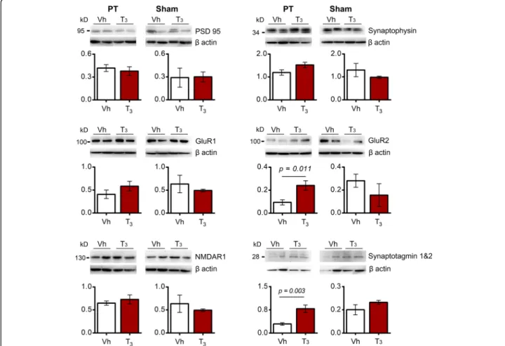
proteins, which reflects structural changes in dendritic spines and the number of functional synapses relevant for synaptic neurotransmission in the peri-infarct area. We observed no differences in the level of the
presynap-tic synaptophysin and the PSD95 (Fig. 4). Likewise, no
differences were detected in NMDAR1. Interestingly, we found that glutamate receptor 2 (GluR2), one of the AMPA receptor subunits, was significantly increased in mice subjected to PT and treated with T3while levels of
GluR1 remained stable (Fig. 4). Accompanied we found
increased levels of synaptotagmin 1&2. In sham operated control experiments, treatment with T3had no effect on
all studied proteins.
Synaptotagmins are downregulated by T3in an in vitro
model of ischemia and are downregulated in the infarct core of human stroke
The finding that T3at 50μg/kg modulates levels of
syn-aptotagmin 1&2 in vivo prompted us to evaluate its ex-pression in OGD-treated neuronal cultures pre-treated with T31μM for 48 h. Levels of synaptotagmin were
sig-nificantly decreased in neuronal cultures in the presence
of T3 (Fig. 5a). This pre-synaptic protein was also
expressed in the ischemic territory of stroke patients, be-ing significantly reduced in the infarct core (Fig.5b).
T3inhibits glutamate evoked currents in glutamatergic
cortical neurons
To study the relevance of T3 for neuron function we
used the method of voltage ramp to establish informa-tion about the I-V relainforma-tions of calcium permeable NMDA and AMPA post-synaptic receptors in the pres-ence and abspres-ence of T3. For each cell tested, membrane
current amplitudes were normalized in order to obtain current density (pA/pF).
Glutamatergic neurons responsiveness to T3
stimula-tion was consistent with the positive immunoreactivity for TRα1 and TRβ1 (Fig.5c). Application of agonist
glu-tamate at 50μM and NMDA co-agonist glycine at 3 μM
elicited an inward component at negative potentials. Glycine together with glutamate potentiated the glutam-ate induced current, even in the presence of Mg2+in the extracellular bath. We also examined the possibility of glycine to induce currents by itself. Application of gly-cine at 3μM did not induce a current in any of the neu-rons tested (Additional file1: Figure S8). We also tested if response was mediated by postsynaptic iGluRs NMDA and AMPA, by application of non-competitive
antago-nists MK-801 and CNQX at 10μM, respectively. After
application, currents were almost reversed (Fig. 5d).
Similarly, to the application of the antagonists, currents are also almost reversed after washout with extracellular bath (data not shown).
Compared with cells in control conditions, the presence of T3(1μM) in cell cultures for 48 h before the
experi-ments significantly decreased glutamate / glycine response in the neurons analyzed (Fig.5e).
T3downregulates GABA synthesis and activity of cortical
Parvalbumin immunoreactive cells
To determine whether functional recovery mediated by
i.p. injection of T3 at 50μg/kg modulates GABAergic
signaling, we evaluated GAD 65/67 expression in stroke
mice treated with T3compared with Vh. Longterm
ad-ministration of T3 50 μg/kg for 14 days after ischemic
stroke significantly reduced GAD 65/67 expression in the ischemic territory. In sham operated animals, admin-istration of T3did not alter the expression of GAD 65/
67 (Fig.6a).
To understand the significance of lower GAD 65/67 expression in animals treated with T3, we assessed the
(See figure on previous page.)
Fig. 2 Treatment with T350μg/kg improves functional recovery 14 days after photothrombosis (PT) without affecting infarct size. a Difference between the rotating pole test (rpt) scores from day 2 (selective sorting) and 14 (Δd2-d14) at 10 rotations per minute to the right and to the left sides, from mice subjected to PT (right hemisphere). Scores are shown as individual data and group median. Statistical analysis was performed by Kruskal-Wallis test followed by Mann-Whitney test (p = 0.0021 in T350μg/kg versus Vehicle treatment). Vehicle (n = 11), T35μg/kg (n = 10), T350 μg/kg (n = 11), T45μg/kg (n = 10), T450μg/kg (n = 9). b Representative coronal brain sections from stroke mice treated with Vehicle, T350μg/ kg or T450μg/kg. Staining with NeuN was performed to measure cortical infarcts. Infarct volumes are displayed as means ± SEM. On day 14 after PT in mice treated with Vehicle (n = 10), T35μg/kg (n = 8), T350μg/kg (n = 8), T45μg/kg (n = 7) or T450μg/kg (n = 7). Statistical analysis was performed by one-way ANOVA and Bonferroni’s multiple comparisons test. c Thyroid hormone receptors (TR) α1 and TRβ1 (AF488, green) expression in mouse brain cell populations. Both TR isoforms are expressed in NeuN (Cy5, blue) positive neurons and Parvalbumin (Cy3, red) positive neurons. GFAP (Cy3, red) immunoreactive astrocytes express TRβ1 in the ischemic territory, 14 days after PT. Scale bars 50 μm and 10 μm for insets at higher magnification. d Levels of TRα1 and e TRβ1 in the infarct core and peri-infarct area were analyzed 14 days after PT and after treatment with Vehicle (n = 3), T350μg/kg (n = 3) or T450μg/kg (n = 3). No difference was observed in levels of TRα1 and TRβ1. f Levels of TRα1 and g TRβ1 in the grey matter of human brain in non-stroke (Ctrl), and stroke cases, including the peri-infarct (PI) and infarct core (IC). For uncropped images of western blots see Additional file1: Figure S5. Levels of TRβ1 are increased in the IC. Statistical analysis was performed by One-way ANOVA and Bonferroni’s multiple comparisons test. Two-tailed unpaired Student’s t test was employed to determine p values. Data are expressed as mean ± SEM
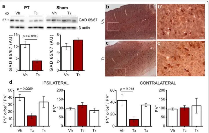
activity of cortical PV neurons, a class of interneurons that regulate GABA neurotransmission. PV immunoreactivity was co-localized with the activity-dependent marker c-fos, through NovaRED Peroxidase (PV) and DAB (c-fos)
im-munohistochemistry (Fig. 6b, c). As shown in Fig. 6d,
there is a significant reduction in PV+ / c-fos+ ratio
be-tween Vh and T3 50 μg/kg-treated animals in the
peri-infarct region (40.46 ± 4.26 Vh; 14.62 ± 3.4 T3 50 μg/kg;
mean ± SEM) and the homotypic region in the contralat-eral hemisphere (43.61 ± 9.43 Vh, 11.54 ± 4.12 T3 50 μg/
kg; mean ± SEM). In contrast, treatment with T450μg/kg
did not change the activity of PV+ cells in the same re-gions (Ipsilateral 33.97 ± 7.59; Contralateral 35.91 ± 2.65; mean ± SEM). Importantly, treatment with TH did not in-fluence the total number of PV immunoreactive cells in the ipsilateral and contralateral hemispheres (Fig.6d).
(See figure on previous page.)
Fig. 3 Treatment with T350μg/kg increases dendritic spine density 14 days after photothrombosis (PT). a Dendritic spine analysis 14 days after PT at different distances from bregma correspondent to the rostral pole (level 1), center (level 2) and caudal pole (level 3) of the cortical infarct. The regions analyzed correspond to the ipsilateral (R1) and contralateral (R3) motor cortex; and ipsilateral (R2) and contralateral (R4) somatosensory cortex. b Representative dendritic segments from animals treated with T350μg/kg (n = 4) and Vehicle (Vh; n = 4). c Apical dendritic spines from cortex layers II/III were automatically detected by NeuronStudio software and classified as mushroom, thin or stubby. Three to five dendritic segments were analyzed per animal. d Dendritic spine density (number of total spines / dendritic length) per region and classification of dendritic spines as mushroom, thin or stubby and their density per region, at each level analyzed. Results are displayed as means ± SEM. Statistical analysis was performed with two-tailed unpaired Student t-test, *p < 0.05, **p < 0.01, ***p < 0.001
Fig. 4 Levels of synaptic proteins in the infarct core and peri-infarct area 14 days after photothrombosis (PT) or in the homotypic area in sham operated mice have been analyzed after treatment with Vehicle (Vh; n = 6 for PT and n = 3 for sham) or T350μg/kg (n = 6 for PT and n = 3 for sham). There are no significant differences between levels of postsynaptic density protein 95 (PSD95), synaptophysin, glutamate receptor 1 (GluR1) and NMDA receptor 1 in the infarct core and peri-infarct in T3-treated mice compared with Vh. Levels of AMPA receptor subunit GluR2 and synaptotagmin 1&2 are increased in the infarct core and peri-infarct in T3-treated mice compared with Vh. Synaptotagmins are vesicle-associated synaptic proteins involved in neurotransmitter release. For uncropped images of western blots see Additional file1: Figure S6. No differences were observed in sham operated mice. Results are displayed as means ± SEM. Statistical analysis was performed with two-tailed unpaired Student’s t test
Discussion
After an ischemic stroke, there is a disruption of normal neuron function i.e. synaptic activity due to cell death occur-ring in the infarct core and therefore, disruption in the
nor-mal neuronal circuity [87]. As consequence, surviving
neurons adjacent to the infarct spontaneously adopt homeo-static mechanisms that contribute to maintain overall excit-ability, although to a limited extent [18, 21, 48]. The molecular mechanisms of homeostatic processes characterize the recovery phase of ischemic stroke and enhancing those with adjuvant interventions might be a key therapeutic
strategy [19]. This may create a wider therapeutic window to optimize and restore lost neurological function.
TH have been recently proposed as a key modulator in stroke [71] and brain injury recovery [41]. The lacking evi-dence of the underlying mechanisms of TH promoting functional recovery after stroke prompted us to evaluate the role of TH in the post-ischemic brain. Summarizing,
our work demonstrates for the first time that T3
modu-lates key homeostatic regulatory mechanisms that are cru-cial to maintain appropriate levels of excitation and mechanisms that stabilize neuronal activity in the
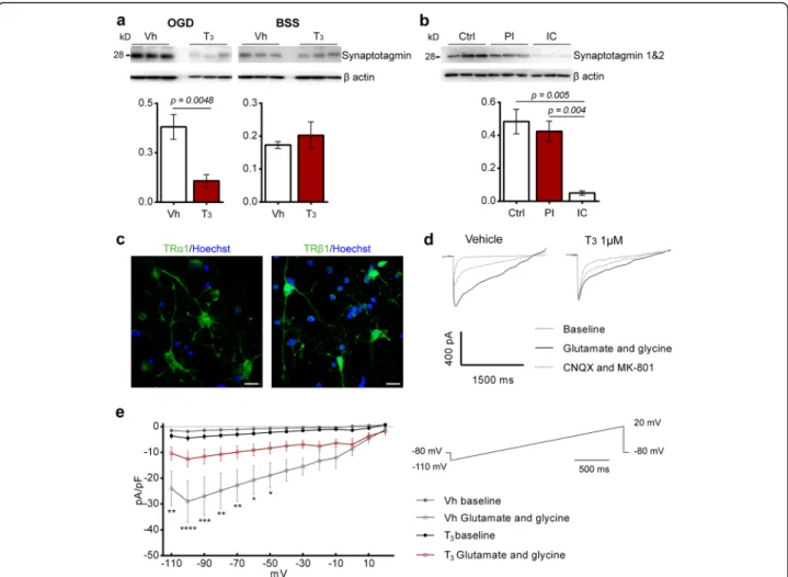
post-Fig. 5 Treatment with T31μM for 48 h inhibits iGluRs evoked currents in cultured cortical glutamatergic neurons and downregulates
synaptotagmin levels after oxygen and glucose deprivation (OGD). a In an in vitro model of acute cerebral ischemia, levels of synaptotagmin are decreased in cells pre-treated with T31μM for 48 h but no difference was observed in the control conditions (Vh, n = 5; T31μM, n = 5). b Human brains of stroke and non-stroke control cases have been analyzed for levels of synaptotagmin 1&2. Levels of synaptotagmin are decreased in the infarct core (IC) in comparison with control (Ctrl) and peri-infarct (PI) regions (n = 3 for each brain region). For uncropped images of western blots see Additional file1: Figure S7 c Representative images of expression of Thyroid hormone receptors TRα1 and TRβ1 (AF488, green) in cultured cortical glutamatergic neurons. TRα1 was mainly localized in the cytoplasm and TRβ1 was expressed in the cytoplasm and nucleus. Scale bar 20μm. d Representative traces obtained during voltage ramps from − 110 to + 20 mV after application of glutamate 50 μM and glycine 3μM, held at − 80 mV. After application of AMPA and NMDA antagonists, CNQX and MK-801 respectively, currents were almost fully reverted. e I-V relationship of glutamate 50μM and glycine 3 μM induced current in cortical neurons under voltage clamp condition under the membrane potential of− 80 mV. Each trace is the result of the average of three ramps for each 10 mV (Vh, n = 3; T31μM, n = 4). Results are displayed as means ± SEM. Statistical analysis was performed with two-tailed unpaired Student’s t test to compare glutamate induced currents in cells pre-treated with T31μM for 48 h, *p < 0.05, ** p < 0.01, *** p < 0.001
ischemic brain, contributing to cortical reorganization and to functional recovery.
Given that TH signaling could be related to better out-come, we first assessed behavioral recovery after
experi-mental stroke in mice treated with T4 or T3 at 5 or
50μg/kg. The photothrombotic model adopted for our
study induced a well-defined ischemic damage in the primary motor cortex that produced consistent hemipar-esis 2 days after stroke [53], allowing behavioral assess-ment of motor function following ischemia. As expected, all mice spontaneously recovered some of lost motor function over time in analogy to spontaneous recovery in humans [23,24]. Interestingly, the group treated with
T3 50 μg/kg had significant higher neurological scores
14 days after PT, with no difference in the infarct size compared to control group. However no significant stat-istical differences were observed in the T4-treated mice
groups. T4 is the prohormone and it needs to be
con-verted to T3before it can exert any biological effect [47].
In the rodent, half of T3 levels in the brain is provided
from its free fraction in blood circulation and cerebro-spinal fluid and the other half relies in local deiodination of T4in astrocytes and tanycytes, which concentration is
regulated by deiodinases activity [44, 74]. Although we did not verify deiodinase expression in the post ischemic brain, the possible scenario is that administration of T4
is less effective to exert action in the brain, since it still needs to be converted to the active form T3.
Next, we investigated the key T3-mechanisms that
might contribute for stroke recovery. Taking into ac-count that genomic actions of T3in the brain are mainly
mediated by binding to TRα1 and TRβ1 [71], we
assessed their levels and expression pattern in the post-ischemic mouse brain. TR levels were not altered after
Fig. 6 Treatment with T350μg/kg is associated with downregulation of GABA synthesis in the infarct core and peri-infarct area 14 days after photothrombosis (PT) and the activity of cortical parvalbumin immunoreactive cells (PV+) in the ipsilateral and contralateral areas. a and b Immunohistochemistry NiDAB (c-fos) counterstained with NovaRed (PV) was performed to count PV+c-fos+(white arrows) and PV+(black arrows) immunoreactive cells, in the ipsilateral and contralateral motor and somatosensory areas of mice treated with Vehicle (Vh) or T350μg/kg. Scale bars 100μm (a and b) and 20 μm (a’ and b’). c Levels of glutamate decarboxylase (GAD) 65/67 was downregulated in mice treated with T350 μg/kg, in the peri-infarct area 14 days after PT. Vh (n = 6 for PT and n = 3 for sham), T350μg/kg (n = 6 for PT and n = 3 for sham). For uncropped images of western blots see Additional file1: Figure S9. Results are displayed as means ± SEM. Statistical analysis was performed with two-tailed unpaired Student’s t test. d Functional recovery after T350μg/kg treatment may be related to a decrease in the ratio between PV+c-fos+/ PV+ immunoreactive cells observed in the ipsilateral area and the correspondent region in the contralateral hemisphere. Vh (n = 7), T350μg/kg (n = 6), T450μg/kg (n = 3). Results are displayed as means ± SEM. Statistical analysis was performed by One-way ANOVA and Bonferroni’s multiple comparisons test. Two-tailed unpaired Student’s t test was employed to determine p values
administration of T4 or T3 at 50μg/kg, suggesting that
recovery induced by T3 was mediated by other
mecha-nisms. However, our results do not exclude the possibil-ity that genomic actions in the brain have an impact on stroke recovery also at different temporal and spatial scales, in other animal models or in humans. Indeed, one study reported a reduction of TRβ1 expression in the infarct core compared with unaffected peri-infarct cortex and contralateral hemisphere 14 days after
per-manent middle cerebral occlusion (MCAO) [43]. We
also found that TRβ1 was significantly increased in the infarct core in the human brain, when compared to non-stroke patients. Taken together, we show that cerebral ischemia induces heterogenic changes in human brain TR expression, which may imply an important role for T3signaling.
Although TR are mainly nuclear, TRα1 and TRβ1 have been also found in the cytoplasm, which may
in-crease T3 nuclear import [2]. Interestingly, we
ob-served that TRα1 and TRβ1 was heterogeneously expressed in the cytoplasm of neurons and in reactive astrocytes from the glial scar, in accordance with a
previous study performed 14 days after MCAO [43].
However, none of TR isoforms were found in positive
GFAP astrocytes in the naïve rodent brain [14]. If TR
expression has implications in the formation and function of the glial scar should be the subject for subsequent studies.
Besides genomic actions, other TH-mediated
non-genomic mechanisms may contribute for stroke [71] and
brain injury [41] recovery. After ischemic stroke, there is an extensive and rapid loss of neurons and degeneration of their axons and dendritic spines in remote areas [87], in both ipsilateral and contralateral cortex [31], leading to a disruption in normal function of neuronal circuits and loss of brain function. In analogy to brain develop-ment and learning/plasticity mechanisms, surviving neu-rons after stroke attempt to stabilize the ratio between excitatory – inhibitory circuits, in order to adjust brain excitability [21]. A wide variety of homeostatic mecha-nisms might contribute to the maintenance of overall excitability, involving the regulation of neuronal intrinsic excitability and synaptic transmission [52,76,77]. Here, we have identified for the first time T3-modulated
mech-anisms of homeostatic plasticity that were related to motor recovery after experimental stroke. In particular,
we have shown that T3modulates plasticity mechanisms
that may operate on different temporal and spatial scales as compensatory mechanisms to assure appropriate syn-aptic neurotransmission.
Dendritic spines are highly dynamic [6,84] and espe-cially after stroke it occurs an extensive reorganization in dendritic arbors, which includes an increase in spine density and spine turnover [12, 13, 25], particularly in
apical cortical pyramidal neurons within the first 2
weeks [11]. In Study II we observed overall enhanced
cortical reorganization in T3-treated Thy1-YFP mice
reflected in increased spine density in cortical layers II/ III, especially in the peri-infarct area, which may con-tribute for spontaneous recovery. The process of spine formation or spinogenesis includes the formation of thin and long dendritic filopodia that are highly dy-namic and establish contact with presynaptic axons. The presence of appropriate signals would result in stabilization of the contact and maturation of filopodia into functional dendritic spines [6]. Interestingly, we
found increased density of thin protrusions in T3
-treated animals, especially in the peri-infarct area, al-though in a temporal scale we could not distinguish newly formed protrusions from the pre-existing ones. We also observed an increased number of mushroom-like spines in the peri-infarct region in all sections
ana-lyzed from T3-treated mice. Although we could not
as-sure that all protrusions are or will be transformed in more stable thin or mushroom-like spines over time,
this was a direct finding that T3 modulated the
reorganization of spines in numbers and structure 2 weeks after stroke onset.
Based on these findings, we further evaluated synaptic efficacy. To address this question, we studied levels of pre-synaptic proteins synaptophysin and synaptotagmin, important to regulate endocytosis and exocytosis of syn-aptic vesicles, respectively [39, 68, 69] and therefore neurotransmitter release. In particular, synaptotagmins are crucial for the docking of synaptic vesicles and
fu-sion with neuron membrane [69]. We demonstrate that
in the human ischemic infarct core, levels of synaptotag-min 1&2 were very low due to cell death and loss of syn-aptic neurotransmission. Nevertheless, their levels in the peri-infarct remained as the same as non-stroke brain tissue, which makes synaptotagmin a molecular target. The increase in synaptotagmin 1&2 levels in the post-ischemic brain of T3-treated mice supports an increase
of neurotransmitter release probability, which in turn may increase synaptic efficacy [9]. In contrast, we ob-served that synaptotagmin is reduced in OGD T3-treated
cultured glutamatergic neurons, which demonstrated
homeostatic regulation by T3 in order to reduce
neuro-transmitter release and hyperexcitability in an in vitro model of acute brain ischemia. Synaptotagmin related gene 1 is a TH responsive gene during brain develop-ment, regulating synaptic activity and structure [73] and
T4 has been reported to restore synaptotagmin 1 levels
to normal in hypothyroid rats [83]. However, how T3
tivates / inhibits synaptic vesicles for synaptotagmin ac-tion remains to be elucidated.
Besides neurotransmitter release, efficacy of neuro-transmission is dependent on post-synaptic response to
glutamate in neuron terminals, that can be modulated by changing the number or function of iGluRs AMPA and NMDA [10,51,75,76]. Indeed, stroke-induced
glu-tamate release activates AMPA receptors [17] and
NMDA receptors [54], changes that are related with ex-citatory synaptic transmission and motor recovery. Here we show an increase in levels of AMPA receptor subunit GluR2 in the peri-infarct area of mice treated with T3.
The AMPA receptor subunit GluR2 regulates critical as-pects of AMPA receptor function, neurotransmission
and synaptic plasticity [32, 66] which ultimately
contributes to increased excitability in the post-ischemic brain and recovery [67].
We characterized AMPA and NMDA excitatory post-synaptic currents with a voltage-clamp method in cultured glutamatergic neurons pre-treated for 48 h with T31 μM.
Interestingly, we found that glutamate evoked currents were significantly lower in neurons previously incubated with T3.
Similarly, in a previous study, T3at 10μM has been
impli-cated in the reduction of miniature excitatory post-synaptic currents frequency and glutamate induced toxicity in hippo-campal neurons [42]. Interestingly, we found that T3recruits
Fig. 7 Proposed mechanisms of homeostatic regulation of neurotransmission by T3. (A) After photothrombosis (PT), administration of T350μg/kg for 14 days modulates pathways during the recovery period after stroke involved in reorganization of neuronal circuits and synaptic plasticity, to balance excitation and inhibition ratio. a T3increases levels of post-synaptic glutamate receptor 2 (GluR2) subunit in AMPA receptors in the peri-infarct area and b increases levels of synaptotagmin 1&2, increasing the probability of neurotransmitter release. c T3increases dendritic spines density in the ipsilateral and contralateral regions. d T3decreases tonic GABAergic signaling in the peri-infarct area by a decrease in the levels of GAD 65/67 and e reduced parvalbumin (PV) activity. (B) In an acute model of cerebral ischemia and hyperexcitability, f in glutamatergic neurons pre-treated with T3at 1μM for 48 h there is a decrease in levels of synaptotagmin and g T3modulates neuron membrane properties with the balance of inward glutamate ligand-gated channels currents
divergent mechanisms to achieve homeostasis in two differ-ent systems regarding synaptic network organization, i.e., in vitro and in vivo and dependent on the activation status of neurons and brain tissues, respectively. Important for stroke recovery, T3could modulate synaptic neurotransmission to
an optimal firing rate.
After an ischemic insult, synaptic glutamate signaling is depressed also due to tonic inhibition of neuronal circuits, which ultimately restricts the process of recovery [8, 15,
80]. Modulation to shift the excitation - inhibition ratio by stimulation of glutamate signaling [15, 17] and reducing GABA inhibition [1,15,16] in the motor and somatosen-sory cortex accelerates motor recovery in mice. GABAergic neurotransmission is mediated by cortical interneurons, a group of cells expressing calcium-binding proteins, includ-ing PV. In fact, a correlation between reduction of PV/ GABA cells and functional recovery in rodents subjected to stroke has been shown [86]. Also, different therapeutic ap-proaches such as environment enrichment [29], benzodi-azepine inverse agonist [1], but also intravenous infusion of human bone marrow mesenchymal stromal cells after tran-sient MCAO [62] decreased cortical PV immunoreactivity or activity and were associated with enhanced recovery of lost neurological function. Treatment with T3reduced the
activity of PV immunoreactive cells in the peri-infarct area and in the contralateral hemisphere, without affecting the total number of PV+cells.
Concomitantly, in the peri-infarct area of animals treated with T350μg/kg, expression levels of GAD 65/67
was significantly reduced, and directly GABA production. Our results are in accordance with studies describing an increased GAD activity and GABA uptake in neurons in hypothyroid state [34] and the finding that T3
administra-tion inhibits GABA-induced Cl− currents [45]. Thus, the decrease in PV cortical activity may facilitate experience dependent plasticity and decrease GABA availability and tonic inhibition, and therefore contribute to restoration of neuronal networks.
Together, our findings reveal important implications of T3-mediated mechanisms in stroke recovery (Fig. 7). At
the cellular and structural level, we demonstrated that T3
is involved in mechanisms of neuronal plasticity that col-lectively contributed to functional recovery following ex-perimental stroke. Based on our findings it will be possible
to develop specific approaches targeting T3-mediated
mechanisms in the post-ischemic brain. Those may result in specific treatments to be tested in clinical trials.
Supplementary information
Supplementary information accompanies this paper athttps://doi.org/10. 1186/s40478-019-0866-4.
Additional file 1. Supplementary Methods and Results.
Additional file 2. Rotating pole test mouse 1 selective sorting after photothrombosis.
Additional file 3. Rotating pole test mouse 1 after vehicle treatment at 14 days.
Additional file 4. Rotating pole test mouse 2 selective sorting after photothrombosis.
Additional file 5. Rotating pole test mouse 2 after T3treatment (50 µg/kg) at 14 days.
Acknowledgements
This work was supported by funds from the Health Sciences Research Center (CICS-UBI) through National Funds by FCT - Foundation for Science and Technology UID / Multi / 00709/2019 (CRS and IG); by a doctoral fellowship (SFRH/BD/104679/2014) from FCT (DT); partially supported by“Programa Operacional do Centro, Centro 2020” through the funding of the ICON project (Interdisciplinary Challenges on Neurodegeneration; CENTRO-01-0145-FEDER-000013) (ARC, CRS and IG)”; the Swedish Brain Fund; the Cra-foord Foundation, Sveriges Stroke Riksförbundet; the Hans-Gabriel and Alice Trolle-Wachtmeister Foundation, The Alborada Research Fund; Sparbanksstif-telsen Färs & Frosta and a regional grant - ALF-Skåne (TW and KR). Image ac-quisition for dendritic spine analysis was performed in the Microscopy facility of CICS-UBI, a node of PPBI (Portuguese Platform of BioImaging): POCI-01-0145-FEDER-022122.
Authors’ contributions
DT and KR designed the research; DT, JF, ARC, TT, EC and KR performed research studies. EE provided human post-mortem brain tissues. EC, TW, CRS, IG and KR provided funding and material for all the experiments. EC, CRS, IG and KR supervised the studies. DT wrote the paper with input from all authors. All authors read and approved the final manuscript. Competing interests
The authors declare that they have no competing interests. Author details
1Laboratory for Experimental Brain Research, Division of Neurosurgery, Department of Clinical Sciences, Lund University, BMC A13, S-22184 Lund, Sweden.2CICS-UBI-Health Sciences Research Centre, Faculdade de Ciências da Saúde, Universidade da Beira Interior, Covilhã, Portugal.3Division of Oncology and Pathology, Lund University Hospital, Lund, Sweden.4LUBIN Lab - Lunds Laboratorium för Neurokirurgisk Hjärnskadeforskning, Division of Neurosurgery, Department of Clinical Sciences, Lund University, Lund, Sweden.
Received: 29 October 2019 Accepted: 8 December 2019
References
1. Alia C, Spalletti C, Lai S, Panarese A, Micera S, Caleo M (2016) Reducing GABA A-mediated inhibition improves forelimb motor function after focal cortical stroke in mice. Sci Rep 29:37823.https://doi.org/10.1038/srep37823
2. Anyetei-Anum CS, Roggero VR, Allison LA (2018) Thyroid hormone receptor localization in target tissues. J Endocrinol 237:R19–R34.https://doi.org/10. 1530/JOE-17-0708
3. Bárez-López S, Guadaño-Ferraz A (2017) Thyroid hormone availability and action during brain development in rodents. Front Cell Neurosci 11:1–7.
https://doi.org/10.3389/fncel.2017.00240
4. Benjamin EJ, Blaha MJ, Chiuve SE, Cushman M, Das SR, Deo R et al (2017) Heart disease and stroke Statistics’2017 update: a report from the American Heart Association. Circulation. 135(10):e146–e603.https://doi.org/10.1161/ CIR.0000000000000485.
5. Bernal J (2000) Thyroid hormones in brain development and function. [updated 2015 Sep 2]. In: Feingold KR, Anawalt B, Boyce A et al (eds) Endotext. MDText.com, Inc., South Dartmouth, pp 1–136
6. Bhatt DH, Zhang S, Gan W-B (2009) Dendritic spine dynamics. Annu Rev Physiol 71:261–282.https://doi.org/10.1146/annurev.physiol.010908.163140
7. Biernaskie J (2004) Efficacy of rehabilitative experience declines with time after focal ischemic brain injury. J Neurosci 24:1245–1254.https://doi.org/10. 1523/JNEUROSCI.3834-03.2004
8. Boddington LJ, Reynolds JNJ (2017) Targeting interhemispheric inhibition with neuromodulation to enhance stroke rehabilitation. Brain Stimul 10: 214–222.https://doi.org/10.1016/j.brs.2017.01.006
9. Branco T, Staras K (2009) The probability of neurotransmitter release: variability and feedback control at single synapses. Nat Rev Neurosci 10: 373–383.https://doi.org/10.1038/nrn2634
10. Bredt DS, Nicoll RA (2003) AMPA receptor trafficking at excitatory synapses. Neuron 40:361–379.https://doi.org/10.1016/S0896-6273(03)00640-8
11. Brown CE, Boyd JD, Murphy TH (2010) Longitudinal in vivo imaging reveals balanced and branch-specific remodeling of mature cortical pyramidal dendritic arbors after stroke. J Cereb Blood Flow Metab 30:783–791.https:// doi.org/10.1038/jcbfm.2009.241
12. Brown CE, Li P, Boyd JD, Delaney KR, Murphy TH (2007) Extensive turnover of dendritic spines and vascular remodeling in cortical tissues recovering from stroke. J Neurosci 27:4101–4109.https://doi.org/10.1523/JNEUROSCI. 4295-06.2007
13. Brown CE, Wong C, Murphy TH (2008) Rapid morphologic plasticity of peri-infarct dendritic spines after focal ischemic stroke. Stroke 39:1286–1291.
https://doi.org/10.1161/STROKEAHA.107.498238
14. Carlson DJ, Strait KA, Schwartz HL, Oppenheimer JH (1994)
Immunofluorescent localization of thyroid hormone receptor isoforms in glial cells of rat brain. Endocrinology 135:1831–1836.https://doi.org/10. 1210/endo.135.5.7525253
15. Carmichael ST (2012) Brain excitability in stroke: the yin and yang of stroke progression. Arch Neurol 69:161–167.https://doi.org/10.1001/archneurol. 2011.1175
16. Clarkson AN, Huang BS, Macisaac SE, Mody I, Carmichael ST (2010) Reducing excessive GABA-mediated tonic inhibition promotes functional recovery after stroke. Nature 468:305–309.https://doi.org/10.1038/nature09511
17. Clarkson AN, Overman JJ, Zhong S, Mueller R, Lynch G, Carmichael ST (2011) AMPA receptor-induced local brain-derived Neurotrophic factor signaling mediates motor recovery after stroke. J Neurosci.https://doi.org/ 10.1523/JNEUROSCI.5780-10.2011
18. Cramer SC (2008) Repairing the human brain after stroke: I. mechanisms of spontaneous recovery. Ann Neurol 63:272–287.https://doi.org/10.1002/ana. 21393
19. Cramer SC (2018) Treatments to promote neural repair after stroke. J Stroke 20:57–60.https://doi.org/10.5853/jos.2017.02796
20. Crupi R, Paterniti I, Campolo M, Di Paola R, Cuzzocrea S, Esposito E (2013) Exogenous T3 administration provides neuroprotection in a murine model of traumatic brain injury. Pharmacol Res 70:80–89.https://doi.org/10.1016/j. phrs.2012.12.009
21. Dalise S, Ambrosio F, Modo M (2014) Adaptive plasticity and recovery in preclinical models of stroke. Arch Ital Biol 152:190–215.https://doi.org/10. 12871/00039829201442
22. Doyle KP, Suchland KL, Ciesielski TMP, Lessov NS, Grandy DK, Scanlan TS et al (2007) Novel thyroxine derivatives, thyronamine and
3-iodothyronamine, induce transient hypothermia and marked
neuroprotection against stroke injury. Stroke 38:2569–2576.https://doi.org/ 10.1161/STROKEAHA.106.480277
23. Dromerick AW, Edwardson MA, Edwards DF, Giannetti ML, Barth J, Brady KP et al (2015) Critical periods after stroke study: translating animal stroke recovery experiments into a clinical trial. Front Hum Neurosci 9:231.https:// doi.org/10.3389/fnhum.2015.00231
24. Duncan PW, Min Lai S, Keighley J (2000) Defining post-stroke recovery: implications for design and interpretation of drug trials.
Neuropharmacology 39:835–841. https://doi.org/10.1016/S0028-3908(00)00003-4
25. Enright LE, Zhang S, Murphy TH (2007) Fine mapping of the spatial relationship between acute ischemia and dendritic structure indicates selective vulnerability of layer V neuron dendritic tufts within single neurons in vivo. J Cereb Blood Flow Metab 27:1185–1200.https://doi.org/10.1038/sj. jcbfm.9600428
26. Feigin VL, Norrving B, Mensah GA (2017) Global burden of stroke. Circ Res 120:439–448.https://doi.org/10.1161/CIRCRESAHA.116.308413
27. Genovese T, Impellizzeri D, Ahmad A, Cornelius C, Campolo M, Cuzzocrea S et al (2013) Post-ischaemic thyroid hormone treatment in a rat model of acute stroke. Brain Res 1513:92–102.https://doi.org/10.1016/j.brainres.2013.03.001
28. Gothié JD, Demeneix B, Remaud S (2017) Comparative approaches to understanding thyroid hormone regulation of neurogenesis. Mol Cell Endocrinol 459:104–115.https://doi.org/10.1016/j.mce.2017.05.020
29. Hakon J, Quattromani MJ, Sjölund C, Tomasevic G, Carey L, Lee JM et al (2018) Multisensory stimulation improves functional recovery and resting-state functional connectivity in the mouse brain after stroke. NeuroImage Clin 17:717–730.https://doi.org/10.1016/j.nicl.2017.11.022
30. Harris KM, Jensen FE, Tsao B (1992) Three-dimensional structure of dendritic spines and synapses in rat hippocampus (CA1) at postnatal day 15 and adult ages: implications for the maturation of synaptic physiology and long-term potentiation. J Neurosci 12:2685–2705
31. Huang SY, Chang CH, Hung HY, Lin YW, Lee EJ (2018) Neuroanatomical and electrophysiological recovery in the contralateral intact cortex following transient focal cerebral ischemia in rats. Neurol Res 40:130–138.https://doi. org/10.1080/01616412.2017.1411454
32. Isaac JTR, Ashby M, McBain CJ (2007) The role of the GluR2 subunit in AMPA receptor function and synaptic plasticity. Neuron 54:859–871.https:// doi.org/10.1016/j.neuron.2007.06.001
33. Jones TA, Adkins DL (2015) Motor system reorganization after stroke: stimulating and training toward perfection. Physiology 30:358–370.https:// doi.org/10.1152/physiol.00014.2015
34. Kalaria RN, Prince AK (1985) Effects of thyroid deficiency on the development of cholinergic, GABA, dopaminergic and glutamate neuron markers and DNA concentrations in the rat corpus striatum. Int J Dev Neurosci 3:655–666.https://doi.org/10.1016/0736-5748(85)90056-5
35. Kapoor R, Fanibunda SE, Desouza LA, Guha SK, Vaidya VA (2015) Perspectives on thyroid hormone action in adult neurogenesis. J Neurochem 133:599–616.https://doi.org/10.1111/jnc.13093
36. Kim JT, Fonarow GC, Smith EE, Reeves MJ, Navalkele DD, Grotta JC et al (2017) Treatment with tissue plasminogen activator in the Golden hour and the shape of the 4.5-hour time-benefit curve in the National United States get with the Guidelines-Stroke Population. Circulation 135:128–139.https:// doi.org/10.1161/CIRCULATIONAHA.116.023336
37. Knecht S, Hesse S, Oster P (2011) Rehabilitation after stroke. Dtsch Arztebl Int 108:600–606.https://doi.org/10.3238/arztebl.2011.0600
38. Kuric E, Ruscher K (2014) Reduction of rat brain CD8+ T-cells by levodopa/ benserazide treatment after experimental stroke. Eur J Neurosci 40:2463– 2470.https://doi.org/10.1111/ejn.12598
39. Kwon SE, Chapman ER (2011) Synaptophysin regulates the kinetics of synaptic vesicle endocytosis in central neurons. Neuron 70:847–854.https:// doi.org/10.1016/j.neuron.2011.04.001
40. Li J, Donangelo I, Abe K, Scremin O, Ke S, Li F et al (2017) Thyroid hormone treatment activates protective pathways in both in vivo and in vitro models of neuronal injury. Mol Cell Endocrinol 452:120–130.https://doi.org/10.1016/ j.mce.2017.05.023
41. Liu YY, Brent GA (2018) Thyroid hormone and the brain: mechanisms of action in development and role in protection and promotion of recovery after brain injury. Pharmacol Ther 186:176–185.https://doi.org/10.1016/j. pharmthera.2018.01.007
42. Losi G, Garzon G, Puia G (2008) Nongenomic regulation of glutamatergic neurotransmission in hippocampus by thyroid hormones. Neuroscience 151: 155–163.https://doi.org/10.1016/j.neuroscience.2007.09.064
43. Lourbopoulos A, Mourouzis I, Karapanayiotides T, Nousiopoulou E, Chatzigeorgiou S, Mavridis T et al (2014) Changes in thyroid hormone receptors after permanent cerebral ischemia in male rats. J Mol Neurosci 54: 78–91.https://doi.org/10.1007/s12031-014-0253-3
44. Maia AL, Kim BW, Huang SA, Harney JW, Larsen PR (2005) Type 2 iodothyronine deiodinase is the major source of plasma T3 in euthyroid humans. J Clin Invest 115:2524–2533.https://doi.org/10.1172/JCI25083
45. Martin JV, Williams DB, Fitzgerald RM, Im HK, Vonvoigtlander PF (1996) Thyroid hormonal modulation of the binding and activity of the GABA(a) receptor complex of brain. Neuroscience 73:705–713.https://doi.org/10. 1016/0306-4522(96)00052-8
46. Mdzinarishvili A, Sutariya V, Talasila PK, Geldenhuys WJ, Sadana P (2013) Engineering triiodothyronine (T3) nanoparticle for use in ischemic brain stroke. Drug Deliv Transl Res 3:309–317. https://doi.org/10.1007/s13346-012-0117-8
47. Mullur R, Liu YY, Brent GA (2014) Thyroid hormone regulation of metabolism. Physiol Rev 94:355–382.https://doi.org/10.1152/physrev.00030.2013
48. Overman JJ, Carmichael ST (2014) Plasticity in the injured brain: more than molecules matter. Neuroscientist 20:15–28.https://doi.org/10.1177/ 1073858413491146
49. Peisker T, Koznar B, Stetkarova I, Widimsky P (2017) Acute stroke therapy: a review. Trends Cardiovasc Med 27:59–66.https://doi.org/10.1016/j.tcm.2016.06.009