Pathogenesis of Alzheimer’s Disease
Jun Wang1,3, Zhong Zhao1, Emi Lin2, Wei Zhao1, Xianjuan Qian1, Daniel Freire1, Amanda E. Bilski1, Alice Cheng1, Prashant Vempati1, Lap Ho1,3, Kenjiro Ono4, Masahito Yamada4, Giulio M. Pasinetti1,3* 1Department of Neurology, Mount Sinai School of Medicine, New York, New York, United States of America,2Department of Neurobiology, Harvard Medical School, Boston, Massachusetts, United States of America,3Geriatric Research Education and Clinical Center, James J. Peters Veterans Affairs Medical Center, Bronx, New York, New York, United States of America,4Department of Neurology and Neurobiology and Aging, Kanazawa University Graduate School of Medical Science, Kanazawa, Japan
Abstract
Alzheimer’s disease (AD) is rapidly becoming one of the leading causes of disability and mortality in the elderly. As life-expectancy increases, an increasing number of people will rely on modern medicines to treat age-associated disorders. Among these medications, some might benefit, while others might exacerbate, the pathogenesis of AD. We screened 1,600 FDA approved drugs forb-amyloid (Ab)-modifying activity and identified drugs that can potentially influence amyloid precursor protein processing. In this study, we focused on cardiovascular drugs and demonstrated that some hypertensive medication can differentially modulate Ab, bothin vitroandin vivo. Our study suggests that some commonly prescribed
drugs might exert unintended effects and modulate AD and provides the basis for continuing investigation of the role of individual drugs on a case-by-case basis. This line of investigation will lead to the identification of common medications that are potentially beneficial or detrimental to AD as a reference for physicians to consider when prescribing the most appropriate drugs for their patients, particularly for treating chronic disorders among the growing geriatric population.
Citation:Wang J, Zhao Z, Lin E, Zhao W, Qian X, et al. (2013) Unintended Effects of Cardiovascular Drugs on the Pathogenesis of Alzheimer’s Disease. PLoS ONE 8(6): e65232. doi:10.1371/journal.pone.0065232
Editor:Tsuneya Ikezu, Boston University School of Medicine, United States of America
ReceivedOctober 17, 2012;AcceptedApril 23, 2013;PublishedJune 6, 2013
Copyright:ß2013 Wang et al. This is an open-access article distributed under the terms of the Creative Commons Attribution License, which permits unrestricted use, distribution, and reproduction in any medium, provided the original author and source are credited.
Funding:This work was supported by grants from the National Institutes of Health UO1 (AG29310) and Department of Veterans Affairs to GMP, and in part by Altschul Foundation. The funders had no role in study design, data collection and analysis, decision to publish, or preparation of the manuscript.
Competing Interests:The authors have declared that no competing interests exist. * E-mail: [email protected]
Introduction
Alzheimer’s disease (AD) is one of the most persistent and devastating disorders of old age, often leading to severe memory loss and functional impairment [1]. Its prevalence increases dramatically with aging. It is estimated that up to ,5 million people in the US currently have AD and it is projected that up to 14 million people will be affected by AD by the middle of this century.
AD is characterized neuropathologically by the accumulation of extracellular neuritic plaques composed ofb-amyloid (Ab) protein, intracellular neurofibrillary tangles of hyperphosphorylated tau protein, and neuron loss [2]. A major hypothesis regarding the pathogenesis of AD is that abnormally elevated Abcontent in the brain of AD patients is critical for the development of AD dementia. This hypothesis, commonly referred to as the ‘‘amyloid hypothesis,’’ suggests that increasing accumulation of Abpeptides promotes assembly of Ab proteins into neurotoxic, extracellular soluble oligomeric Ab aggregates that are largely responsible for cognitive deterioration and neuronal loss in AD [3–9]. Continuing recruitment of Ab peptides to oligomeric Ab aggregates leads to the formation of larger, insoluble Abfibrils that contribute to the formation of AD type neuritic plaques in the brain [10]. The amyloid hypothesis is supported by substantial genetic [11] and preclinical evidence [12]. However, to date, clinical trials based on amyloid hypothesis with the target mechanism of reducing brain amyloid load have produced null results including the most
recently completed antibody therapies (bapineuzamab), suggesting that the amyloid hypothesis and the optimal molecular targets or the optimal timing for intervention remains to be elucidated. Nonetheless, Abremains to be one of the major disease modifying targets for AD drug discovery.
In addition to AD, the aged population is associated with higher risks for many chronic diseases, such as cardiovascular disorders, diabetes, arthritis, cancer or cognitive impairment. Many elderly individuals require one or more medications to treat and/or manage age-related chronic disease. A survey conducted by Boston University Epidemiology Center showed that over 80% of people 65 years and older take at least one medication and almost 50% take three or more - As life-expectancy increases with advances in medicine the number of elderly continues to increase. Thus, the incidence and prevalence of chronic disease is rising dramatically, together with the number of elderly requiring one or more medications.
cognition. For example, recent evidence strongly supports the possibility that the use of some antihypertensive drugs such asb -blockers or Ca++ channel receptor antagonists, and certain potassium-sparing antihypertensive diuretics, may decrease the incidence of AD [14–17]. More recently, Hajjar et. al found that patients with or without AD, treated with angiotensin receptor blockers (ARBs), showed much lower amyloid burden compared to patients treated with other anti-hypertensisive medication [18]. Despite evidence associating some drugs with unintended activities on cognitive dysfunction, we do not know the potential role of how specific medications promote or inhibit the generation of Ab.
Since Abis one of the major contributory factors responsible for AD-type dementia, we surveyed 1600 FDA approved drugs that are commonly used by the general population for their ability to modulate Ab accumulation. Our intention is to identify medica-tions that might be potentially beneficial or harmful for Ab -mediated cognitive dysfunction. Outcomes from our studies will provide important information for physicians to consider when making decisions on prescribing the most appropriate drugs for their patients, particularly for treating chronic disorders among the growing geriatric population. Moreover, our observations also provide the impetus for future studies to systematically evaluate FDA- approved medications for potential repurposing as novel reagents to prevent or treat AD dementia.
Materials and Methods
Drug Screening Procedure
Embryonic-day (E)16 cortico-hippocampal neuronal cultures were prepared from heterozygous Tg2576 transgenic mice (Tg2576 neurons) [19;20]. Neurons were seeded onto poly-D-lysine–coated 96-well plates at 1.06105cells per well and cultured in Neurobasal medium supplemented with 2% B27, 0.5 mM L-glutamine and 1% penicillin-streptomycin (Gibco-BRL) in the tissue culture incubator at 37uC with 5% CO2. The absence of
astrocytes (,2%) was confirmed by the virtual absence of glial fibrillary acidic (GFAP) protein immunostaining (data not shown). For primary screening, cultured neurons were treated with 100mM of drug in duplicates; all drugs were obtained in stock from MicroSource Discovery Systems Inc (Gaylordsville, CT). Conditioned medium was collected for Ab detection. For
secondary screening, primary neurons prepared in 96-well plates were treated with 0.1mM, 1mM, 10mM, 50mM, and 100mM of each drug in duplicate for,16 hours and conditioned medium was tested for Ab content. Cell viability was assessed using a commercial available LDH assay kit according to the manufac-ture’s instruction (Promega) and by MTT (3-(4,5-Dimethylthiazol-2-yl)-2,5-diphenyltetrazolium bromide) assay.
Animals and Treatment
Female Tg2576 mice transgenic mice carrying a human amyloid precursor protein (APP) containing the familial Swedish KM670/671NL double mutation [21] were purchased from Taconic. Tg2576 mouse is a well characterized rodent of model with Ab-mediated neuropathology and cognitive impairment and were previously used in our studies on valsartan and carvedilol [20;22].
Testing drugs were prepared to desired concentrations and delivered to mice by diluting them directly into the drinking water. Typically, a 25,30 g mouse drinks ,4–5 ml per day. The amount of testing drugs to be diluted into the drinking water was based on 1) average daily water consumption and 2) the targeted dose of testing drugs to be delivered to animals. For example, a 25 g mouse would need to take 0.375 mg propranolol per day to achieve 15 mg/kg body weight/day as in the instance of chronic propranolol treatment. Typically a 25 g mouse drinks 4.5 ml liquid per day, therefore, propranolol was diluted in the drinking water to a concentration of 0.083 mg/ml or 83 mg/L. The average liquid consumption was monitored and doses were adjusted accordingly. We acknowledge that there is variability in the amount of liquid each animal consumed. Animals under treatment were providedad libitumaccess to testing drug-infused water as the sole source of drinking fluid.
For short-term treatment, Tg2576 mice were treated with individual compounds starting at 6 months of age and the treatment lasted one month.
For chronic treatment, Tg2576 mice were treated with 17.2 mg/kg/day nicardipine (equivalent to human 100 mg/day) or 15 mg/kg/day propranolol (equivalent to human 90 mg/day) delivered through their drinking water for 6 months, starting at 8 months of age.
Figure 1. Schematic diagram of primary screening of 1600 FDA approved drugs.
Table 1.115 commonly prescribed cardiovascular drugs used in screening for potential Ab-modifying activity.
Drug Name Ab Drug Name Ab Drug Name Ab
ANTIHYPERTENSIVE
A.b-Adrenergic blocker D. Vasodilator G. Angiotensin converting enzyme inhibitor
RESERPINE – PAPAVERINE HYDROCHLORIDE – PERINDOPRIL ERBUMINE 83.8
CARVEDILOL 38.3 HYDRALAZINE 66.1 FOSINOPRIL SODIUM 94.7
PROPRANOLOL HYDROCHLORIDE (2) 55.0 DIPYRIDAMOLE 72.2 ENALAPRIL MALEATE 100.7
PROPRANOLOL HYDROCHLORIDE 65.2 ISOXSUPRINE HYDROCHLORIDE 78.3 CAPTOPRIL 104.5
NYLIDRIN HDROCHLORIDE 70.5 NICOTINYL TARTRATE 80.0 RAMIPRIL 107.4
LABETALOL HYDROCHLORIDE 84.9 ISOSORBIDE DINITRATE 80.5 BENAZEPRIL HYDROCHLORIDE 116.3
ATENOLOL 90.4 PROTOVERATRINE A 91.7 QUINAPRIL HYDROCHLORIDE 119.3
METOPROLOL TARTRATE 91.3 MINOXIDIL 94.2 TRANDOLAPRIL 136.7
ALPRENOLOL 93.8 VINCAMINE 99.1 H. Diuretic
NADOLOL 94.2 METHYLDOPA 104.0 BUMETANIDE –
PINDOLOL 99.0 MOLSIDOMINE 118.3 ETHACRYNIC ACID –
PRACTOLOL 99.7 DIAZOXIDE 113.5 TRIAMTERENE –
TIMOLOL MALEATE 101.3 ADENOSINE PHOSPHATE 121.8 AMILORIDE 60.4
GUANETHIDINE SULFATE 125.5 E. Ganglionic blocking agent CYCLOTHIAZIDE 71.4
ACEBUTOLOL HYDROCHLORIDE 133.6 PEMPIDINE TARTRATE 80.9 ALTHIAZIDE 74.4
B. a-Adrenergic Blocker HEXAMETHONIUM BROMIDE 84.8 BENDROFUMETHIAZIDE 76.2
PRAZOSIN HYDROCHLORIDE – PENTOLINIUM TARTRATE 121.0 SPIRONOLACTONE 77.5
PHENTOLAMINE HYDROCHLORIDE 83.4 MECAMYLAMINE HYDROCHLORIDE 124.4 TRICHLORMETHIAZIDE 89.3 PHENOXYBENZAMINE HYDROCHLORIDE 87.5 F. Ca channel blocker HYDROCHLOROTHIAZIDE 89.5
TOLAZOLINE HYDROCHLORIDE 90.8 AMLODIPINE BESYLATE – METOLAZONE 91.4
URAPIDIL 96.4 BEPRIDIL HYDROCHLORIDE – CLOPAMIDE 92.0
TAMSULOSIN HYDROCHLORIDE 106.5 FENDILINE HYDROCHLORIDE – HYDROFLUMETHIAZIDE 92.4
GUANABENZ ACETATE 131.2 FLUNARIZINE HYDROCHLORIDE – UREA 92.8
C. Angiotensin receptor blocker TETRANDRINE – CANRENOIC ACID, POTASSIUM SALT 100.6
CANDESARTAN CILEXTIL – NICARDIPINE 10.7 THEOBROMINE 101.3
VALSARTAN 51.6 NITRENDIPINE 33.0 BENZTHIAZIDE 105.7
LOSARTAN 70.0 VERAPAMIL HYDROCHLORIDE 70.9 INDAPAMIDE 107.0
OLMESARTAN CILEXTIL 83.9 NIMODIPINE 79.1 CHLOROTHIAZIDE 107.3
IRBESARTAN 89.7 NIFEDIPINE 83.4 TORSEMIDE 107.4
TELMISARTAN 91.1 DILTIAZEM HYDROCHLORIDE 107.8 CHLORTHALIDONE 108.5
BERBAMINE HYDROCHLORIDE 119.1 FUROSEMIDE 138.5
ANTIARRHYTHMIC
PROPAFENONE HYDROCHLORIDE – DISOPYRAMIDE PHOSPHATE 83.5 AJMALINE 114.0
AMIODARONE HYDROCHLORIDE – MEXILETINE HYDROCHLORIDE 88.8 PROCAINAMIDE HYDROCHLORIDE 118.1
QUINIDINE GLUCONATE 63.3 HYDROQUINIDINE 96.5
ANTITHROMBOTIC and FIBRINOLYTIC
A. Coagulant B. Antifibrinolytic C. Antihyperlipidemic
SULOCTIDIL – TRANEXAMIC ACID 84.9 FENOFIBRATE –
DICUMAROL 51.3 AMINOCAPROIC ACID 124.4 SIMVASTATIN –
SCOPOLETIN 71.9 ATORVASTATIN CALCIUM 66.0
WARFARIN 89.4 ROSUVASTATIN 83.6
ANISINDIONE 90.1 PROBUCOL 86.8
PENTOXIFYLLINE 97.1 CLOFIBRATE 90.9
PHENINDIONE 104.0 NIACIN 101.1
BEZAFIBRATE 107.2
CONGESTIVE HEART FAILURE
LANATOSIDE C – DOPAMINE HYDROCHLORIDE 85.0 PERUVOSIDE 115.3
All mice were housed with foodad libitumand maintained on a 12:12-h light/dark cycle with lights on at 07:00 h in a temperature-controlled (2062uC) room prior to experimental manipulation. Mice were group housed with 3–5 mice per cage and average food and water intake was measured weekly. All procedures and protocols were approved by the Mount Sinai School of Medicine’s Institutional Animal Care and Use Committee (IACUC) through the Center for Comparative Medicine and Surgery.
Blood Pressure Measurements
Blood pressure and heart rate were measured using a non-invasive commercial blood pressure analysis system designed specifically for small rodents (Hatteras Instruments, NC) as previously described [20;23]. Mice were temporarily immobilized in a restraining chamber with the tail inserted through the tail cuff, laid down into the tail slot and secured with a piece of tape. Every mouse underwent 5 preliminary cycles for acclamation and the following 10 measurements of systolic, diastolic, and mean arterial pressure (MAP) and heart rate was recorded.
Behavioral Assessment of Cognitive Functions by the Morris Water Maze (MWM) Test
Spatial learning memory was assessed by the Morris water maze behavioral test, as previously described [20;24]. Briefly, mice were tested in a circular pool filled with water mixed with non-toxic white paint (Dick Blick Art Materials, IL). The water temperature was kept between 70 and 74uF. Mice were first tested in a visible trial for 3 consecutive days where the escape platform was clearly marked with a white sail. Following the visible trial, the white sail was removed and replaced by local visual cues. Mice were trained to mount the submerged escape platform in a restricted region of the pool using the visual cues. Mice were given 4 trials per day with 60 seconds per trial. Each day, the mice would start at different quadrant and if the testing mouse failed to reach the platform in 60 seconds, it would be gently led to the platform and let stay on the platform for 15 seconds before returning to the home cage. Spatial memory was assessed by recording the latency time for the animal to escape from the water onto a submerged escape platform as a function of the number of learning trials during the learning phase. Twenty-four hours after the last learning session, mice were subjected to a 45 second probe trial wherein the escape platform is removed. The water maze activity was monitored with the San Diego Instrument Poly-Track video tracking system (San Diego, CA). The cued-platform learning curve was used as control for the non-spatial factors on MWM performance, e.g., sensory-motor performance, motivation, anx-iety etc., which can be influenced by the potential effect of the testing drug. The mentstrual cycle was not controlled for the behavior testing.
Assessment of AD-type Amyloid Neuropathology Total Ab1-40 or Ab1-42 in the brain and in plasma were quantified by sandwich ELISA, as previously described [25]. Specifically, frozen pulverized tissue was homogenized in 5.0 M guanidine buffer, diluted (1:10) in phosphate-buffered saline containing 0.05% (v/v) Tween-20 and 1 mM Pefabloc protease inhibitors (Roche Biochemicals, Indianapolis, IN) and centrifuged for 20 min at 4uC. Supernatant was subjected to Ab1–40or Ab1–42
quantification by sandwich ELISA (BioSource, Camarillo, CA).
Statistical Analysis
Differences between means were analyzed using two-tailed Student t-test. For behavior testing, data were analyzed using two-way repeated measures ANOVA followed by Newman-Keuls post-hoc analysis. In all analyses, the null hypothesis was rejected at the 0.05 level. All values are expressed as mean and standard error of the mean (SEM). All statistical analyses were performed using the prism Stat program (GraphPad Software, Inc.).
Results
Identification of Cardiovascular Drugs with AD-modifying Activity
Our high throughput screening study assessed 1600 FDA approved drugs for their ability to modulate Abactivity. We found 559 drugs of the 1600 had no effect on APP processing or were toxic to neurons at the testing concentration, while 800 drugs could reduce Ab content over 10% in primary neurons derived from Tg2576 mice, among which, 184 drugs were able to reduce Abcontent greater than 30% compared to vehicle. We also found 241 drugs could potentially promote Ab accumulation including 26 drugs that could increase the level of Ab greater than 30% compared to vehicle treatment (Figure 1).
Cardiovascular medications are one of the most commonly prescribed drugs, especially in the elderly. Among the 1600 drugs we tested, 115 are cardiovascular drugs representing all pharma-cological classes of antihypertensive, antithrombotic and fibrino-lytic, antianginal, antiarrhythmic and congestive heart failure medications (Table 1). Primary screening of these drugs identified 13 cardiovascular drugs that could reduce$30% Ab accumula-tion in the condiaccumula-tioning medium from Tg2576 neurons following 16 hours treatment at 100mM concentration without toxicity as tested by MTT assay and LDH release assay (Table 1). We continued our secondary screening for dose response on these candidates and found that among the 13 candidates, carvedilol, propranolol, valsartan, losartan, hydralazine, nicardipine and amiloride demonstrated a concentration-dependent reduction of Ab1–40 and Ab1–42 in primary embryonic cortico-hippocampal
neuron cultures. All 7 drugs are antihypertensive agents of different pharmacological subclasses. We also found that 2 drugs,
The 115 cardiovascular agents were obtained as part of the Spectrum Collection from MicroSource Discovery Systems Inc. and listed in 4 pharmacological categories (source: Physician’s Desk Reference and Martindale Complete Drug Reference). Potential Ab-modifying activity was assessed using primary cortico-hippocampal neuron cultures derived from embryonic Tg2576 AD mice and the levels of Abin the conditioned medium were measured and expressed as percentage of vehicle-treated control (Abcolumn). In the high-throughput screening studies, 13 drugs lowered the Abcontent greater than 30% and two drugs (in bold and Italic) increased Ab content greater than 30% compared to the vehicle controls. The 13 Ab-lowering drugs were proceeded to a follow-up dose dependent test and the 8 drugs in bold were found to exert significant dose dependent Ab-lowering activity in the absence of cellular toxicity. ‘‘–’’ indicates that the drug is toxic for primary neurons at 100mM.
namely trandolapril and furosemide, could promote $30% Ab accumulation compared to vehicle control.
Short-termin vivoEfficacy Study using Tg2576 Mouse Model of AD
Based on thein vitrodose-dependent results, we tested 8 drugs (6 with Ab-lowering activity and 2 with Ab-promoting activity, Table 2) to explore whether these drugs can exert similar Ab modification activity in an experimental model of AD.
We performed a short-term feasibility study using the Tg2576 mice model of AD. We used the doses equivalent to the recommended dose for treating cardiovascular disease in humans (Table 2). All dosage ranges were obtained from online Physicians’ Desk Reference (http://www.pdr.net) and the conversions of drug equivalent dosages across species were derived using FDA criteria based on body surface area [26]. The detailed dosage for the treatment is listed in Table 2.
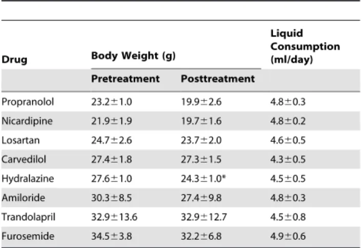
We found that short-term drug treatment for one month in Tg2576 mice, delivered in the drinking water at clinical dosage, did not significantly influence animal body weight, except for hydralazine treatment (Table 3), which showed a significant body weight drop following 1 month treatment. However, propranolol and losartan treatment resulted in a significantly influenced blood pressure, ,20% drop in systolic, diastolic and mean arterial pressure (MAP) measurements was observed, while other drugs showed no effect on the blood pressure measurements in the normotensive mice (Figure 2, A-F). The lack of hypotensive effect of the other drugs can be due to: 1) some of the drugs, such as nicardipine, significantly reduce the blood pressure in hypertensive subjects, but have minimal effect on blood pressure in normoten-sive subjects. 2) the drug dose conversion between species we used in the study is based on the body surface area. It is possible that the absorption and metabolism of certain drug might be different in mice compared to humans.
Consistent with the in vitro data, treatment of Tg2576 mice with propranolol, nicardipine or carvedilol resulted in a ,40% reduction in total guanidine-extractable Ab1-42 peptide and Ab 1-40 peptide in the brain (Figure 2G, 2H, and 2J), while treatment with hydralazine and amiloride had no effect on brain amyloid peptides levels. Losartan treated mice showed a significant reduction of Ab1-42 in the brain while no change in Ab1-40 level. We also found that propranolol, nicardipine and losartan treatment resulted in significant reductions of plasma level of Ab (Fig 2M, 2N and 2O).
To our surprise, the drugs that increased Ab in vitro, at the dosage equivalent to human prescription dosage (14.8 mg/kg/ day for furosemide and 0.74 mg/kg/day for trandolapril), did not increase total Ab1-40 or Ab1-42 in the brain following one-month treatment. On the contrary, both furosemide and trandolapril treatment significantly reduced the levels of total brain amyloid content in the Tg2576 mice following one-months short-term treatment (Figure 3B and 3E) without significantly changing of blood pressure. The reduction of brain Ab content in both treatments was associated with significant increases of plasma levels of Ab (Figure 3C and 3F). In parallel studies, we confirmed that the drug treatments did not alter the expression of APP in the brains of the mice using western blot analysis (data not shown).
Table 2.Short-term in vivo treatment dosage conversion in Tg2576 mice.
Equivalent human dose Recommended dose for
Tg2576 treatment Prescribed clinical dose hypertension treatment in human
(mg/kg/day) (mg/day) (mg/day)
propranolol 41.3 240 120,240
carvedilol 8.6 50 25,50
nicardipine 17 100 50,100
losartan 20.6 120 60,120
amiloride 3.4 20 5,20
hydralazine 51.7 300 100,300
furosemide 14.8 80 20,80
trandolopril 0.74 2 1,2
The dosage recommended for hypertension treatment in humans is listed as prescribed clinical dose. The equivalent dosage in animals is calculated using FDA criteria for converting drug equivalent dosages across species, based on body surface area [39]. The mice equivalent dosage was used in the short-term treatment. doi:10.1371/journal.pone.0065232.t002
Table 3.Effect of short-term in vivo on body weight and liquid consumption in Tg2576 mice.
Drug Body Weight (g)
Liquid Consumption (ml/day)
Pretreatment Posttreatment
Propranolol 23.261.0 19.962.6 4.860.3 Nicardipine 21.961.9 19.761.6 4.860.2 Losartan 24.762.6 23.762.0 4.660.5 Carvedilol 27.461.8 27.361.5 4.360.5 Hydralazine 27.661.0 24.361.0* 4.560.5 Amiloride 30.368.5 27.469.8 4.860.3 Trandolapril 32.9613.6 32.9612.7 4.560.8 Furosemide 34.563.8 32.266.8 4.960.6
Animals were treated with the anti-hypertensive drugs for four weeks and body weight and liquid consumption were monitored weekly. Data presented here are the end point body weight and the average liquid consumption throughout the study.
Chronic Administration of Nicardipine and Propranolol on Cognitive Function and Brain Neuropathology
The validation of the effect of any medication on AD pathology is cognitive function.
Based on the results from the short-term study (Figure 2), we chose nicardipine and propranolol, both of which significantly changed the levels of total Ab1-40 and Ab1-42 in the brain following one-month treatment for chronic studies, to evaluate their effect on cognitive function. Since short-term treatment with propranolol resulted in a significant hypotensive effect in the normatensive mice and significantly reduced heart rate (Figure 2A), we adjusted the dose to 15 mg/kg/day for chronic studies. We used the same dose for nicardipine as short-term treatment since it did not have any adverse effect.
Following 6 months treatment, we tested spatial memory function in the Tg2576 mice using the Morris water maze test.
First, we used the visible trial to confirm that propranolol treatment did not affect any non-spatial factors e.g., sensory-motor performance, motivation, anxiety etc. which might affect their water maze performance. Both groups were able to identify the target platform and both groups had similar swimming speed (Figure 4A and 4B). In the hidden platform training session, we found that 6 months of chronic propranolol treatment did not affect AD-type cognitive deterioration reflected by equally impaired spatial memory function between the treated and non-treated control Tg2576 mice. Neither group showed significant learning during the 7 day hidden platform testing (Figure 4C). This lack of learning was also evident during the probe trial, as neither group spent more than 25% chance time in the target quadrant (Figure 4D). Since propranolol is a non-selective beta adrenergic receptor blocker that has been used for memory relief [27–29], in parallel Figure 2. Effect of Ab-lowering drug treatment on blood pressure and amyloid neuropathology in Tg2576 mice.(A–F) Measurements of systolic, diastolic blood pressure, and mean arterial blood pressure (MAP) and heart beat (pulse) in response to,4 weeks of drug treatments. (G-L)
Assessment of Ab1-42 and Ab1-40 peptide concentrations in the brain of drug treated micevs.the control mice. (M-R) Assessment of Ab1-42 and
Ab1-40 in peripheral blood of drug treated micevs.the control mice. Blood pressure determination for each animal was calculated as the mean of 10
individual measurements. Values represents group mean values (+SEM); n = 3–5 mice per group. **P,0.01, *P,0.05, 2-tailed student t-test. doi:10.1371/journal.pone.0065232.g002
Figure 3. Effect of Ab-promoting drugs treatment on blood pressure and amyloid neuropathology in Tg2576 mice.(A and D) Measurements of systolic, diastolic blood pressure, and mean arterial blood pressure (MAP) in response to,4 weeks of drug treatments. (B and E)
Assessment of Ab1-42 and Ab1-40 peptide concentrations in the brain of drug treated micevs.the control mice. (C and F) Assessment of Ab1-42 and Ab1-40 in peripheral blood of drug treated micevs. the control mice. Values represents group mean values (6SEM); n = 3–5 mice per group.
studies, we tested whether it will interfere with spatial memory in wild type (WT) mice. WT mice were treated with the same treatment regime for 6 months and subjected to MWM test. We found propranolol at 15 mg/kg/day dose did not affect spatial memory in WT mice, as reflected by their normal learning progress during the 7 day training (Figure 4E). The probe trial also confirms that both propranolol-treated and non-treated control WT mice spent significantly more time in the target quadrant (,40% of time, much more than the 25% chance level, Figure 4F), indicating that propranolol at the treatment dose did not affect spatial memory function.
We next measured the brain neuropathology and found that, similar to the short-term treatment, 6 months of chronic propranolol treatment significantly reduced the level of both Ab1-40 and Ab1-42 in the brains compared to the control non-treated mice (Figure 4G). However, measurements of plasma levels of Abshowed that there was no change in Ab1-40 and Ab1-42 (Figure 4H) which is different from the short-term treatment results (Figure 2M).
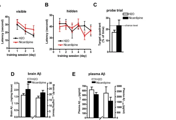
Chronic nicardipine treatment in Tg2576 showed similar behavior results. No benefits of cognitive improvement were observed following 6 months treatment by MWM test (Figures 5A– 5C). Surprisingly, contrary to the short-term results that nicardipine treatment significantly lowered the brain levels of
Ab1-40 and Ab1-42 (Figure 2H), chronic nicardipine treatment did not affect the levels of Abin the brain (Figure 5D). There was no difference in plasma levels of Ab1-40 and Ab1-42 following chronic treatment, which is also different from the short-term result that there was a significant reduction in plasma Abfollowing 1 month treatment (Figure 2N).
Discussion
AD is rapidly becoming one of the leading causes of disability and mortality, and it is expected that the prevalence of AD in the US will quadruple over the next 50 years [30;31]. With baby boomers reaching retirement age and life expectancy continuing to increase, an increasing number of elderly people will be taking one or more medications. Whether current commonly prescribed medications can worsen or improve AD dementia, and to what extent they might influence dementia from normal aging or from other neurodegenerative conditions, are issues with enormous public health implications.
In this study, we surveyed 1600 FDA approved drugs for their ability to modulate aberrant generation of AD-type Ab peptides from the amyloid precursor protein (APP) process in primary cortico-hippocampal cell cultures generated from the Tg2576 mouse AD model. We found a subset of medication that Figure 4. Effect of chronic propranolol treatment on spatial memory and neuropathology in Tg2576 mice.(A-D) The influence of chronic propranolol treatment on Abrelated spatial memory in Tg2576 mice by Morris water maze test (A) Visible trial (B) Swimming speed (C) Hidden platform learning acquisition, latency score represents time taken to escape to the platform from the water. (D) Probe trial. Percent of time in quadrant is calculated as the ratio of time spent in the target quadrant area relative to the time spent in the rest of the pool. (E-F) The influence of chronic propranolol treatment wild type mice by Morris water maze test: (E) Hidden platform learning acquisition (F) Probe trial. (G-H) Brain and plasma levels of total Ab1-40 and Ab1-42 peptides following chronic propranolol treatment. Values represent group mean6SEM, n = 7–8 per group. doi:10.1371/journal.pone.0065232.g004
Figure 5. Effect of chronic nicardipine treatment on spatial memory and neuropathology in Tg2576 mice.(A-C) The influence of chronic nicardipine treatment on Abrelated spatial memory in Tg2576 mice by MWM test: (A) Visible trial (B) Hidden platform learning acquisition (C) Probe trial. (D-E) Brain and plasma levels of total Ab1-40 and Ab1-42 peptides following chronic nicardipine treatment. Values represent group mean
6SEM, n = 9–11 per group.
significantly increased and a second subset of medications that significantly reduced generation of Abpeptides in primary neuron cultures.
Since cardiovascular disorder is one of the most prevalent diseases among the elderly, we focused on the potential role of cardiovascular drugs in regulating Ab generation. We found that most of the FDA-approved cardiovascular drugs have no detectable activity with regard to the generation of Ab peptides from our in vitro primary cortico-hippocampal screening system. However, we identified select cardiovascular drugs, such as propranolol and nicardipine, that exert in vitro Ab-lowering activity, and other cardiovascular drugs, such as furosemide, that significantly promote the generation of Abpeptidesin vitro.
Among the subset of cardiovascular drugs we found to promote Ab generation in vitro, several (e.g., trandolapril, quinapril) are angiotensin converting enzymes (ACE) inhibitors. This observa-tion is consistent with recent evidence suggesting ACE may be involved in the degradation of Ab peptides [32;33]. Indeed, the ACE inhibitor, captopril, has previously been shown to increase Ab accumulation in vitro [32;33]. However, in our primary screening, we found captopril barely increased Ab levels in the conditioned medium of primary neurons following treatment. This discrepancy might have been caused by the usage of different in vitro systems. Zou et. al used COS cells for their study [33] and Hemming et. al. used CHO and HEK293 cells [32]. In both studies, ACE was over expressed in the cell lines. While in our study, we used primary neurons derived from Tg2576 mice and the inhibition was towards endogenous ACE. It is possible that ACE is not a major contributor in Ab catabolism in primary neuronsin vitro.Another observation is that the majority of ACE inhibiting cardiovascular drugs we screened (5 of 8 ACE inhibitors screened) did not exhibit Ab-modification activity. Thus it is also likely that not all ACE inhibitors are equally effective in preventing Abdegradation.
Based on evidence that trandolapril is capable of promoting Ab generation in vitro, we explored the physiological impact of trandolapril on Ab neuropathology in vivo. In contrast to our in vitroevidence, results from our short-term study in Tg2576 mice treated for 1 month with trandolapril at a dose equivalent to the clinical dosage showed that trandolapril significantly reduced Ab contents in the brain while it significantly increased Abcontents in the plasma. Interestingly, the other drug furosemide, which showed Ab promoting activityin vitro, also reduced brain levels of Abaccompanied by increased levels of Abin plasma following short-term treatment in Tg2576 mice. Based on these observa-tions, we suggest that some of the cardiovascular drug, when administered in short-term, might promote generation of Abin the brain. However, the treatment might temporarily alter the dynamic balance of Abbetween brain and plasma, e.g. increase the Ab efflux or reduce the Ab influx which may result in the promotion of transport of Abfrom the brain to the periphery. It would be interesting to measure the levels of transport proteins such as receptor for advanced glycation end products (RAGE) and Lipoprotein receptor-related protein (LRP) following the short-term treatments.
Since most cardiovascular diseases are chronic conditions, the medications are generally prescribed for a prolonged period of time. Therefore, in our study, we also tested the effect of chronic drug application in cognitive function and brain neuropathology. Previously, we have shown that valsartan, one of the angiotensin receptor blocker medications identified in the secondary screening
(Table 1), could reduce Abneuropathology and improve spatial memory function in Tg2576 mice following chronic treatment [20]. We also showed in another study that carvedilol, a nonselectiveb-adrenergic receptor blocker, can prevent cognitive deterioration and reduce brain neuropathology by interfering with Aboligomerization and improving basal synaptic transmission in the TgCRND8 mouse model of AD [22;34].
In this study, we found that chronic treatment with propranolol could significantly reduce brain amyloid neuropathology but had no effect on cognitive function. It is possible that reducing amyloid alone might not be sufficient to improve cognition. It is also possible that propranolol is able to reduce total amyloid but is not able to influence the level of soluble oligomeric Abspecies, which are increasingly regarded as neurotoxic and largely responsible for synaptic failure in AD models [6–8;35–37]. Brain neuropathology results from nicardipine, again suggesting that short-term treat-ment might temporarily alter the hemodynamic between the brain and blood resulting in increased clearance while prolong treatment may not have any effects. Future studies will focus on mechanistic investigation of how certain drugs might influence APP processing or Ab catabolism and their possible role in Ab oligomerization which may explain the lack of behavior improvements in propranolol treated mice.
It is not surprising that in vitro and in vivo studies showed differences in biological efficacies. First of all, brain bioavailability is one of the major obstacles and some of the drugs might not be able to pass the blood brain barrier to influence brain amyloid processing and, therefore, are not able to exert their activities in the brain. Secondly, some of the drugs might have gone through extensive metabolism and the metabolites might not behave as the original drugs. Thirdly, some of the drugs might also impact other systems (such as vessel permeability, periphery protein degradation pathway, etc.) that will eventually affect the net outcome of the drug treatment. More importantly, drugs modifying amyloid may or may not have significant impacts in modulating AD-type cognitive function.
Therefore, in order to identify drugs that might have beneficial effects, such as reducing Ab neuropathology and improving cognitive function, for the consideration of physicians over drugs that might be potentially detrimental to cognition, individual drugs must be investigated on a case by case basis and these results should be supplemented with data obtained from clinical studies with patients following long-term drug treatment. A good example is that our preclinical study demonstrated that chronic application of ARB valsartan can significantly reduce the levels of soluble Ab as well as total amyloid plaque load in the brain in a mouse model of AD [20]. Data collected from the National Alzheimer Coordinating Center (including 29 centers across the US) confirms that patients receiving ARB medication have less amyloid deposition compared to untreated patients or patients with non-ARB antihypertensive medications [38]. Collectively, these studies may provide useful information for physicians when prescribing antihypertensive drugs.
Author Contributions
References
1. Cummings JL (2004) Alzheimer’s disease. N.Engl.J Med, 351: 56–67. 2. Selkoe DJ (2001) Alzheimer’s Disease: Genes, Proteins, and Therapy.
Physiol.Rev., 81: 741–766.
3. Cleary JP, Walsh DM, Hofmeister JJ, Shankar GM, Kuskowski MA, et al. (2005) Natural oligomers of the amyloid-[beta] protein specifically disrupt cognitive function. Nat Neurosci, 8: 79–84.
4. Klyubin I, Walsh DM, Lemere CA, Cullen WK, Shankar GM, et al. (2005) Amyloid beta protein immunotherapy neutralizes A beta oligomers that disrupt synaptic plasticity in vivo. Nature Medicine, 11: 556–561.
5. Kotilinek LA, Bacskai B, Westerman M, Kawarabayashi T, Younkin L, et al. (2002) Reversible memory loss in a mouse transgenic model of Alzheimer’s disease. J.Neurosci., 22: 6331–6335.
6. Lesne S, Koh MT, Kotilinek L, Kayed R, Glabe CG, et al. (2006) A specific amyloid-beta protein assembly in the brain impairs memory. Nature, 440: 352– 7.
7. Shankar GM, Li S, Mehta TH, Garcia-Munoz A, Shepardson NE, et al. (2008) Amyloid-[beta] protein dimers isolated directly from Alzheimer’s brains impair synaptic plasticity and memory. Nat Med, 14: 837–842.
8. Lambert MP, Barlow AK, Chromy BA, Edwards C, Freed R, et al. (1998) Diffusible, nonfibrillar ligands derived from Abeta 1–42 are potent central nervous system neurotoxins. PNAS, 95: 6448–6453.
9. Gylys KH, Fein JA, Tan AM, Cole GM (2003) Apolipoprotein E enhances uptake of soluble but not aggregated amyloid-beta protein into synaptic terminals. J Neurochem, 84: 1442–1451.
10. Braak H, Braak E (1997) Staging of Alzheimer-related cortical destruction. Int.Psychogeriatr., 9 Suppl 1: 257–261.
11. Citron M, Oltersdorf T, Haass C, McConlogue L, Hung AY,et al. (1992) Mutation of the beta-amyloid precursor protein in familial Alzheimer’s disease increases beta-protein production. Nature, 360: 672–674.
12. Emilien G, Maloteaux JM, Beyreuther K, Masters CL (2000) Alzheimer disease: mouse models pave the way for therapeutic opportunities. Arch Neurol, 57: 176–181.
13. Fox C, Richardson K, Maidment ID, Savva GM, Matthews FE, et al. (2011) Anticholinergic medication use and cognitive impairment in the older population: the medical research council cognitive function and ageing study. J Am Geriatr Soc, 59: 1477–1483.
14. Forette F, Seux ML, Staessen JA, Thijs L, Babarskiene MR, et al. (2002) The Prevention of Dementia With Antihypertensive Treatment: New Evidence From the Systolic Hypertension in Europe (Syst-Eur) Study. Arch Intern Med, 162: 2046–2052.
15. Guo ZC, Fratiglioni L, Zhu L, Fastbom J, Winbald B, Viitanen M (1999) Occurrence and progression of dementia in a community population aged 75 years and older - Relationship of antihypertensive medication use. Arch Neurol, 56: 991–996.
16. Khachaturian AS, Zandi PP, Lyketsos CG, Hayden KM, Skoog I, et al. (2006) Antihypertensive Medication Use and Incident Alzheimer Disease: The Cache County Study. Arch Neurol, 63: 686–692.
17. Lopez-Arrieta JM, Birks J (2002) Nimodipine for primary degenerative, mixed and vascular dementia. Cochrane Database Syst Rev, 3: CD000147. 18. Hajjar I, Brown L, Mack WJ, Chui H (2012) Impact of Angiotensin Receptor
Blockers on Alzheimer Disease Neuropathology in a Large Brain Autopsy Series 1. Arch Neurol: 1–7.
19. Anonymous.
20. Wang J, Ho L, Chen L, Zhao Z, Zhao W, et al. (2007) Valsartan lowers brain beta-amyloid protein levels and improves spatial learning in a mouse model of Alzheimer disease. J Clin.Invest, 117, 3393–3402.
21. Hsiao K, Chapman P, Nilsen S, Eckman C, Harigaya Y, et al. (1996) Correlative Memory Deficits, Abeta Elevation, and Amyloid Plaques in Transgenic Mice. Science, 274: 99–103.
22. Wang J, Ono K, Dickstein DL, rrieta-Cruz I, Zhao W, et al (2010) Carvedilol as a potential novel agent for the treatment of Alzheimer’s disease. Neurobiol.A-ging.
23. Diaz-Ruiz C, Wang J, Ksiezak-Reding H, Ho L, Qian X, et al. (2009) Role of Hypertension in Aggravating Abeta Neuropathology of AD Type and Tau-Mediated Motor Impairment. Cardiovasc.Psychiatry Neurol, 2009: 107286. 24. Morris R (1984) Developments of a water-maze procedure for studying
spatial-learning in the rat. Journal of Neuroscience Methods, 11: 47–60.
25. Wang J, Ho L, Qin W, Rocher AB, Seror I, et al. (2005) Caloric restriction attenuates beta-amyloid neuropathology in a mouse model of Alzheimer’s disease. FASEB J, 19: 659–661.
26. Reagan-Shaw S, Nihal M, Ahmad N (2008) Dose translation from animal to human studies revisited 2. FASEB J, 22: 659–661.
27. Schwabe L, Nader K, Wolf OT, Beaudry T, Pruessner JC (2012) Neural signature of reconsolidation impairments by propranolol in humans 2. Biol Psychiatry, 71: 380–386.
28. Schwabe L, Nader K, Pruessner JC (2013) beta-Adrenergic blockade during reactivation reduces the subjective feeling of remembering associated with emotional episodic memories 1. Biol Psychol., 99: 227–32.
29. Lonergan MH, Olivera-Figueroa LA, Pitman RK, Brunet A (2012) Propran-olol’s effects on the consolidation and reconsolidation of long-term emotional memory in healthy participants: a meta-analysis 1. J Psychiatry Neurosci, 37: 120111.
30. Sano M (2003) Current concepts in the prevention of Alzheimer’s disease. Cns Spectrums, 8: 846–53.
31. Cummings JL (2004) Treatment of Alzheimer’s disease: current and future therapeutic approaches. Rev Neurol Dis., 1: 60–9.
32. Hemming ML, Selkoe DJ (2005) Amyloid beta-protein is degraded by cellular angiotensin-converting enzyme (ACE) and elevated by an ACE inhibitor 5. J Biol Chem, 280: 37644–37650.
33. Zou K, Yamaguchi H, Akatsu H, Sakamoto T, Ko M, Mizoguchi K, et al. (2007) Angiotensin-converting enzyme converts amyloid beta-protein 1–42 (Abeta(1–42)) to Abeta(1–40), and its inhibition enhances brain Abeta deposition 1. J Neurosci, 27: 8628–8635.
34. Arrieta-Cruz I, Wang J, Pavlides C, Pasinetti GM (2010) Carvedilol Reestablishes Long-Term Potentiation in a Mouse Model of Alzheimer’s Disease. J Alzheimers.Dis, 21: 649–54.
35. Lacor PN, Bunie MC, Chang L, Fernandez SJ, Gong Y, et al. (2004) Synaptic Targeting by Alzheimer’s-Related Amyloid {beta} Oligomers. J.Neurosci., 24: 10191–10200.
36. Lacor PN, Buniel MC, Furlow PW, Sanz Clemente A, Velasco PT, et al. (2007) A{beta} Oligomer-Induced Aberrations in Synapse Composition, Shape, and Density Provide a Molecular Basis for Loss of Connectivity in Alzheimer’s Disease. J.Neurosci., 27: 796–807.
37. Shankar GM, Bloodgood BL, Townsend M, Walsh DM, Selkoe DJ, et al. (2007) Natural Oligomers of the Alzheimer Amyloid-{beta} Protein Induce Reversible Synapse Loss by Modulating an NMDA-Type Glutamate Receptor-Dependent Signaling Pathway. J.Neurosci., 27: 2866–2875.
38. Hajjar I, Brown L, Mack WJ, Chui H (2012) Impact of Angiotensin Receptor Blockers on Alzheimer Disease Neuropathology in a Large Brain Autopsy Series 1. Arch Neurol: 1–7.