CO1 – PROGREssIOn OF stRUCtURal DamaGE On mRI OF thE sPInE anD saCROIlIaC JOInts In PatIEnts WIth axIal sPOnDylOaRthRItIs Is lImItED: thE 5–yEaR REsUlts In thE DEsIR COhORt
Alexandre Sepriano1, 2, Sofia Ramiro1, Robert Landewé3, Dougados M4, Désirée van der Heijde1
1. Rheumatology, Leiden University Medical Center, Leiden, Netherlands
2. CEDOC, NOVA Medical School, Nova University, Lisboa, Portugal
3. Amsterdam Rheumatology and Immunology Center (ARC), Amsterdam, Netherlands
4. Department of Rheumatology, Cochin Hospital, Paris, France
Background: Reliably detecting radiographic structural
change in patients with axial spondyloarthritis (axSpA), especially in the sacroiliac joints (SIJ), is notorious ly difficult. Magnetic resonance imaging (MRI) is an alter -native for radiographs to assess structu ral damage. However, so far the utility of MRI in capturing change in structural damachange over time has been poorly stu -died.
Objectives: We aimed to evaluate the change over time
of structural lesions on MRI of the SIJ and spine in pa-tients with axSpA.
Methods: Patients with recent onset (≤3 years)
axSpA (according to the treating rheumatologist) from the DESIR cohort were included. MRI of the SIJ (MRI--SIJ) and spine (MRI-spine) were obtained at baseline and 5 years and scored by 3 trained central readers una -ware of the chronology. Structural damage in the SIJ (MRI-SIJ-STR) and in the spine (MRI-spine-STR) was defined according to 3 binary rules (A1: ≥5 fatty lesions and/or erosions; B1: ≥3 erosions; and C1: ≥3 fatty sions) and 3 continuous scores (A2: number of fatty le-sions /erole-sions; B2: number of erole-sions; and C2: num-ber of fatty lesions). For binary outcomes, structural damage was defined by the agreement of at least 2 out of 3 readers and the % of net progression by
subtract-Comunicações orais
acta reumatol port. 2018:43:25-45 (sup)
ing the number of patients that ‘improved’ from those that ‘worsened’ divided by the total number of patients with complete baseline and 5-year data. For contin uous outcomes, the mean of the 3 readers was used and the difference between year 5 and baseline was calculated.
Results: In total, 151 and 145 patients had complete
MRI-SIJ and MRI-spine data available from 3 readers, respectively. The percentages of net progression at SIJ level are summarized in the Figure 1. These were 6.6%, 0.7% and 7.9% for the binary outcomes A1, B1 and C1 respectively. Notably, the percentage of ‘improvement’ (4.6%) was almost as high as the percentage of ‘worse -ning’ (5.3%) for definition B1 (≥3 erosions); while no ‘improvements’ were seen by the 3 readers for defini-tion C1 (≥3 fatty lesions). Similar differences were seen for the mean (standard deviation) change of the 3 MRI--SIJ-STR continuous outcomes (A2: 1.02 (2.60); B2: 0.20 (1.39); and C2: 0.83 (2.20); p<0.01 for all). MRI-spine-STR net change over time was almost absent (A1: -0.7%; B1: 0.0%; C1: 0.7%) considering the binary outcomes, and small (though statistically signi ficant) considering definition A2 (0.18 (0.52); p<0.01) and C2 (0.14 (0.48); p<0.01) but absent for
7.9% 1.3% 6.6% 5.3% 4.6% 0.7% 7.9% 0.0% 7.9% A1
(≥5 fatty lesions/erosions) (≥3 erosions)B1 (≥3 fatty lesion)C1
% P ro gr es si on
% patients who worsened after 5 years of follow-up % patients who improved after 5 years of follow-up Net % progression (worsened minus improved)
FIGURE 1.Changes in different binary MRI-SIJ-STR outcome measures. All outcomes are assessed according to the ‘2 out of 3' definition in the completers population (N=151).
MRI-SIJ-STR, structural damage on magnetic resonance imaging of the sacroiliac joints.
Background: Several strategies have been proposed to
promote early referral of patients with axial spondy-loarthritis (axSpA), but consensus on the ‘best’ strate-gy is yet to be achieved. Moreover, few studies com-pared referral strategies (RS) head-to-head and, up to now, none has neither evaluated these in a ‘nationwide’ setting (external validity) nor assessed the entire spec-trum of SpA (i.e. axSpA and peripheral SpA).
Objectives: To evaluate the performance of the
screen-ing strategy for SpA of a nationwide epidemiological study (EpiReumaPt), as compared to previously pro-posed RS.
Methods: EpiReumaPt was a three-stage national
health survey (2011-2013) where, in the first phase, 10,661 adult participants were randomly selected and interviewed using a structured face-to-face question-naire that included screening for rheumatic diseases (RD), such as SpA. In the second phase, positive screen-ings for ≥1 rheumatic complaint plus 20% nega tive screenings were invited for an assessment by the rheumatologist. Finally, 3 rheumatologists revised all the information and defined the final diagnosis by con-sensus. All participants of the second phase were in-cluded (N=3,877). Each RS (Table I) was tested against the SpA revised diagnosis using the following metrics: definition B2 (0.03 (0.24); p=0.109).
Conclusion: These results suggest that patients with
early axSpA only show modest structural progression in the MRI of the SIJ and that fatty lesions are more sen-sitive to change compared to erosions. In this early axSpA population, MRI-detected structural progres-sion in the spine is very limited/absent.
CO8 – PERFORmanCE OF REFERRal stRatEGIEs FOR sPOnDylOaRthRItIs: a POPUlatIOn-basED natIOnWIDE stUDy
Alexandre Sepriano1,2, Sofia Ramiro1,2,
Filipe Araújo1,3, Pedro MAchado4, Rodrigues AM1, 5, Nélia Gouveia 1,5, Mónica Eusébio5, Helena Canhão5,6, Jaime C. Branco1, 5
1. CEDOC, Nova Medical School, Lisboa, Portugal 2. Rheumatology, Leiden University Medical Center, Leiden, Netherlands
3. Rheumatology Unit, Hospital Ortopedico de SantAna, SCML, Cascais, Portugal
4. University College London, London, United Kingdom 5. EpiReumaPt Study Group, Lisboa, Portugal
6. EpiDoC Unit, CEDOC, NMS & National School of Public Health, UNL, Lisboa, Portugal
ÓrgÃo oficial da sociedade portuguesa de reumatologia
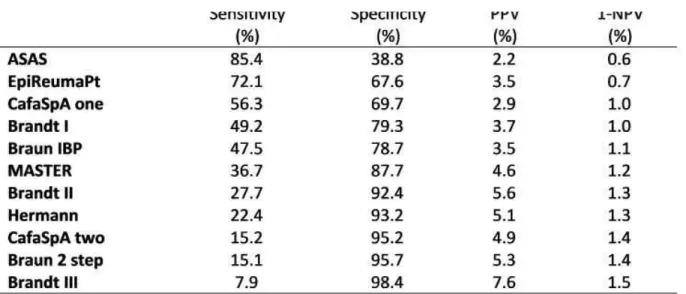
tablE I. PERFORmanCE OF thE REFERRal stRatEGIEs aGaInst thE RhEUmatOlOGIst ClInICal DIaGnOsIs (n=3,877; PRE-tEst PRObabIlIty: 1.6% – WEIGhtED natIOnal sPa PREvalEnCE)
Sensitivity Specificity PPV 1-NPV (%) (%) (%) (%) ASAS 85.4 38.8 2.2 0.6 EpiReumaPt 72.1 67.6 3.5 0.7 CafaSpA one 56.3 69.7 2.9 1.0 Brandt I 49.2 79.3 3.7 1.0 Braun IBP 47.5 78.7 3.5 1.1 MASTER 36.7 87.7 4.6 1.2 Brandt II 27.7 92.4 5.6 1.3 Hermann 22.4 93.2 5.1 1.3 CafaSpA two 15.2 95.2 4.9 1.4 Braun 2 step 15.1 95.7 5.3 1.4 Brandt III 7.9 98.4 7.6 1.5
ASAS (≥1/5+): IBP (ASAS definition), good response to NSAIDs, family history of SpA, peripheral manifestations (arthritis, enthesitis and/or dactylitis), extra-articular manifestations (uveitis, psoriasis and/or IBD); EpiReumaPt (≥1/5+): previous SpA/PsA diagnosis, IBP (≥3/8 features), CBP (≥3 months) starting <45 years and ≥1/6 SpA features, dactylitis, enthesitis; CafaSpA one (≥1/3+): IBP (ASAS definition), good response to NSAIDs, family history of SpA;
CafaSpA two (≥2/3+): see CafaSpA one; Brandt I (≥1/1+): IBP (morning stiffness >30 min, pain at night/early morning, improvement by exercise; ≥1/3); Brandt II (≥1/1+): ≥3/3 IBP features (see Brandt I); Brandt III (≥1/1+): ≥3/3 IBP features (see Brandt I); Braun IBP (≥2/5+): start BP ≤35 years, waking second half of the night, alternating buttock pain, improvement by movement, not rest; MASTER (≥2/3+): IBP (morning stiffness >30 min, improvement exercise, not rest, awakening in the night because of BP), good response to NSAIDs, family history of AS; Hermann (≥1/1): IBP (Calin's criteria): Braun 2 step (≥ 2/3): psoriasis, alternating buttock pain, improvement BP by exercise. HLA-B27 excluded from ASAS, Brandt I-III and Braun 2 step; elevated CRP/ESR excluded from ASAS. PPV: positive predictive value; 1-NPV 1:negative predictive value
CHLO, Lisboa, Portugal
2. CEDOC, NOVA Medical School. Faculdade de Ciências Médicas da Universidade NOVA de Lisboa., Lisboa, Portugal
3. Rheumatology, Leiden University Medical Center, Leiden, Netherlands
4. Rheumatology Department, Hospital Garcia de Orta, Almada, Portugal
5. Rheumatology Department, Centro Hospitalar e Universitário de Coimbra, Coimbra, Portugal
6. Rheumatology Department, Unidade Local de Saúde do Alto Minho, Ponte de Lima, Portugal
7. Rheumatology Department, Instituto Português de Reumatologia, Lisboa, Portugal
8. Rheumatology Department, Hospital de Santa Maria (CHLN), Lisbon Medical and Academic Centre, Lisboa, Portugal
9. Rheumatology Research Unit, Instituto de Medicina Molecular, Lisboa, Portugal
10. Rheumatology Department, Centro Hospitalar de São João, Porto, Portugal
Background: A BASDAI ≥4 has been often required to
start TNFi therapy in patients with axSpA. However, this cut-off of high disease activity (HDA) is largely ar-bitrary. Unlike BASDAI, ASDAS incorporates objective measures (e.g. CRP) and has a validated definition of HDA (≥2.1). It has thus been suggested that ASDAS could also be used to guide treatment decisions, but evidence to support this is still scarce.
Objective: To compare the impact of applying the
ASDAS and BASDAI definitions of HDA in selecting patients for TNFi-treatment in daily clinical practice.
Methods: Patients from Reuma.pt (Rheumatic Diseases
Portuguese Register), with diagnosis of axSpA accord-ing to their rheumatologists (both treated and not treat-ed with their first TNFi), with complete baseline BASDAI and ASDAS data, and complete 6-month of follow-up (i.e. baseline, 3 and 6 months visits avai lable) were included. Four subgroups [cross-tabulation be-tween ASDAS (≥2.1) and BASDAI (≥4) definitions of HDA], were compared according to baseline demographic and clinical characteristics in the ‘eligible po -pulation’ (i.e. irrespective of TNFi-treatment). In addi-tion, for patients starting TNFi and with complete fol-low-up BASDAI/ASDAS data (‘efficacy population’), the subgroups were also compared according to different response criteria (see Table I), at 3 and 6 months.
Results: In total, 466 patients were included (59%
males and 66% HLA-B27 positive). The large majority sensitivity, specificity, positive predictive value (PPV),
and post-test probability of disease given a negative test (1-negative predictive value). RS with an imaging (e.g. MRI) or laboratory component (e.g. CRP, HLA-B27) were modified (by excluding these components) given limited data obtained in the survey (Table I). A weight-ing factor was used to take the survey design into account.
Results: From the total 3,877 participants, 92 received
a SpA diagnosis [weighted prevalence: 1.6% (95%CI: 1.2; 2.1)], 3,107 other RD diagnosis [e.g. knee os-teoarthritis (31%)] and 678 no RD diagnosis. The ASAS RS was the most sensitive (85%) followed by the EpiReumaPt strategy (72%) (Table I). The ASAS and EpiReumaPt RS had the lowest post-test pro babilities of SpA in the presence of negative screening (0.6% and 0.7% respectively), thus, yielding a marked decrease in the probability of disease if negative [(1.60.6)/1.6*100=63%; (1.60.7)/1.6*100=56% respecti -vely). On the other hand, the likelihood of SpA in-creased by 38% (2.2-1.6)/1.6*100) and 119% (3.5--1.6)/1.6*100) in case of a positive ASAS and EpiReumaPt RS, respectively. Brandt III was the least sensitive strategy in this study and not contributive to excluding SpA (1-NPV: 1.5%; pre-test probability: 1.6%), but expectedly increased the likelihood of SpA by 3.8 times if positive. The performance of the re-maining RS is described in the Table I.
Conclusion: For the first time, a wide range of SpA RS
were tested head-to-head in a population-based setting where the ASAS and EpiReumaPt RS were shown to be the most sensitive. Our data suggest that these strate-gies can be effectively used as screening tools for SpA especially when laboratory and imaging data are not available.
CO15 - ElIGIbIlIty CRItERIa FOR tnFi thERaPy In axspa: GOInG bEyOnD basDaI
José Adriano Oliveira Marona1,2, Alexandre Sepriano2,3, Santiago
Rodrigues-Manica1,2, Fernando Pimentel-Santos1,2, Ana Filipa Mourão1,2, Nélia Gouveia2,
Jaime C. Branco1,2, Filipe Vinagre4, Raquel Roque4, João Rovisco5, Mary Lucy Marques5,
José Tavares-Costa6, Joana Leite Silva6,
Helena Santos7, Nathalie Madeira7, Elsa Vieira-Sousa8,9, Rita C Machado8, Miguel Bernardes10,
Raquel Miriam Ferreira10, Sofia Ramiro2,3
1. Rheumatology Department, Hospital Egas Moniz,
the use of ASDAS≥2.1 as a selection criterion for treat-ment decisions.
Disclosures: Supported in part by a research Grant
from Investigator-Initiated Studies program of MSD. CO35 – PattERn OF DRUG UsE In systEmIC lUPUs ERythEmatOsUs anD REasOns FOR DRUG DIsCOntInUatIOn In REal WORlD ClInICal PRaCtICE
Tiago Costa1, 2, Mónica Eusébio3, Sandra Falcao1, 2, Filipe Araújo4, Patrícia Nero5, Ines L6,
Marília Rodrigues6, Diogo Jesus6, Sofia Barreira7, 8, Carla Macieira7, Marta Cabral9, Graça Sequeira10, Jaime C. Branco1, 2, Maria José Santos8, 11
1. Rheumatology Department, Hospital Egas Moniz, CHLO, Lisboa, Portugal
2. CEDOC, NOVA Medical School, Nova University, Lisboa, Portugal
3. Sociedade Portuguesa de Reumatologia, Lisboa, Portugal
4. Rheumatology and Osteoporosis Unit, Hospital de Sant Ana, SCML, Cascais, Portugal
5. Rheumatology, Hospital CUF Descobertas, Lisboa, (n=382; 82%) fulfilled the definition of HDA accor ding
to both BASDAI and ASDAS at baseline (i.e. BASDAI≥4 and ASDAS≥2.1). The frequency of ASDAS≥2.1, if BASDAI<4, was much higher than the opposite condi-tion (i.e. ASDAS<2.1, if BASDAI≥4) (70% vs 0.5%). Compared to patients fulfilling both definitions, those who were ASDAS≥2.1 only, were more likely to be male (82.5% vs 54%), HLA-B27 positive (79% vs 54%), to show higher levels of CRP (2.6 ± 2.5 vs 2.2 ± 2.8mg/dL) and lower BASFI (3.1 ± 2.6 vs 5.6 ± 2.3). In the ‘effi-cacy population’ (n=296), better responses were observed among patients with ASDAS≥2.1 only, espe-cially for the most ’stringent’ outcomes [e.g. ASDAS in-active disease (ASDAS ID): 59% and 50%, at 3 and 6 months respectively], compared to patients fulfilling both definitions (ASDAS ID: 26% and 25% at 3 and 6 months respectively) (Table I).
Conclusion: Our results show that the ASDAS-HDA
definition (ASDAS≥2.1) is more inclusive than the BASDAI-HDA definition (≥4) in selecting axSpA pa-tients for TNFi treatment. Importantly, the additional-ly ‘captured’ patients respond better and have higher likelihood of predictors thereof. These results support
ÓrgÃo oficial da sociedade portuguesa de reumatologia
tablE I. tnFi REsPOnsE CRItERIa aCROss sUbGROUPs aCCORDInG tO basDaI/asDas CatEGORy ('EFFICaCy POPUlatIOn')
ASDAS ≥2.1 ASDAS <2.1
Overall BASDAI ≥4 BASDAI <4 BASDAI ≥4 BASDAI <4
Variables (N=296)* (N=256) (N=34) (N=1) (N=5) p-value** Outcomes – 3 months, n (%) ASAS20 159 (59) 142 (60) 15 (56) 0 (0) 2 (40) 0.48 ASAS40 127 (47) 111 (46) 14 (52) 0 (0) 2 (40) 0.74 ASAS PR 73 (26) 56 (22) 14 (56) 0 (0) 3 (60) <0.01 BASDAI50 184 (62) 160 (63) 21 (62) 0 (0) 3 (60) 0.64 ASDAS CII 207 (70) 179 (70) 26 (77) 0 (0) 2 (40) 0.16 ASDAS MI 123 (42) 111 (43) 12 (35) 0 (0) 0 (0) 0.16 ASDAS ID 90 (30) 66 (26) 20 (59) 0 (0) 4 (80) <0.01 Outcomes – 6 months, n (%) ASAS20 160 (61) 139 (61) 18 (62) 0 (0) 3 (60) 0.67 ASAS40 124 (46) 104 (45) 17 (57) 0 (0) 3 (60) 0.43 ASAS PR 74 (27) 57 (24) 13 (48) 0 (0) 4 (80) <0.01 BASDAI50 188 (64) 167 (65) 18 (53) 0 (0) 3 (60) 0.29 ASDAS CII 216 (73) 190 (74) 25 (74) 0 (0) 1 (20) 0.02 ASDAS MI 128 (43) 117 (46) 11 (32) 0 (0) 0 (0) 0.08 ASDAS ID 84 (28) 63 (25) 17 (50) 0 (0) 4 (80) <0.01 *axSpA patients treated with TNFi, with complete 6 months of follow-up and data for BASDAI/ASDAS at every time-point; **comparison between subgroups according to BASDAI/ASDAS category of disease activity (ANOVA for continuos variables and Chi2 for categorical variables).
Portugal
6. Rheumatology Department, Centro Hospitalar e Universitário de Coimbra, Coimbra, Portugal
7. Rheumatology Department, Hospital de Santa Maria (CHLN), Lisbon Medical and Academic Centre, Lisboa, Portugal
8. Rheumatology Research Unit, Instituto de Medicina Molecular, Faculdade de Medicina da Universidade de Lisboa, Lisboa, Portugal
9. Pediatric, Hospital Fernando Fonseca, Amadora, Portugal
10. Rheumatology Department, Centro Hospitalar Universitário do Algarve, Faro, Portugal
11. Rheumatology Department, Hospital Garcia de Orta, Almada, Portugal
Background: Pharmacological treatment for systemic
lupus erythematosus (SLE) is aimed at reducing disea
se activity, preventing flares and minimizing the dama -ge. The use of medication varies widely and therapeu-tic strategies are well defined only for certain organ mani festations. Hydroxychloroquine is the standard treatment for most SLE patients during the entire disea se course, while immunosuppressants are recommen ded for those with severe organ involvement. Beli -mumab is the only biological currently licensed for SLE, although others are used off-label in clinical practice.
Objectives: To describe the real-world patterns of drug
use in SLE patients, and their relationship with disease phenotype. To describe reasons for drug discontinua-tion and drug retendiscontinua-tion (DR) in SLE patients.
Methods: Observational study of adult SLE patients
registered in Reuma.pt, who have clinical diagnosis of SLE, followed for at least 1 year and with available data on medication. Sociodemographic and clinical chara -cteristics were compared among treatment groups
de-ÓrgÃo oficial da sociedade portuguesa de reumatologia
tablE I. sOCIODEmOGRaPhIC anD ClInICal ChaRaCtERIstICs aCCORDInG tO tREatmEnt GROUPs
CO40 – REUmahEaRt: CaRDIOvasCUlaR RIsk In InFlammatORy RhEUmatIC DIsEasE – a PORtUGUEsE POPUlatIOn basED stUDy
Vital Da Silva Domingues1,2,3, Rodrigues AM3,4,5,6, Sara Dias3,7, Jaime C. Branco3,4,5,8,
Helena Canhão3,4,5,9,10
1. Instituto Gulbenkian de Ciência, Oeiras, Portugal 2. Pediatrics, Centro Hospitalar do Porto, Hospital de Santo António, Porto, Portugal
3. EpiDoC Unit, CEDOC, NOVA Medical School, NOVA University, Lisboa, Portugal
4. CEDOC, NOVA Medical School. Faculdade de Ciências Médicas da Universidade NOVA de Lisboa., Lisboa, Portugal
5. Sociedade Portuguesa de Reumatologia, Lisboa, Portugal
6. Rheumatology Research Unit, Instituto de Medicina Molecular, Lisboa, Portugal
7. Unidade de Investigação em Saúda (UIS), Escola Superior de Saude do Insituto Politecnico de Leiria, UIS – ESSLei-IPLeiria, Leiria, Portugal
8. Rheumatology Department, Hospital Egas Moniz, CHLO, Lisboa, Portugal
9. Escola Nacional de Saúde Pública | Nova University of Lisbon, Lisboa, Portugal
10. Departamento Músculo-Esquelético, Centro Hospitalar Lisboa Central – Hospital Curry Cabral, Lisboa, Portugal Introduction: Individuals diagnosed with rheumatic
diseases have shown an increased risk of developing several comorbid conditions, of which cardiovascular (CV) comorbidities are the most common and have the greatest effect on mortality. Our global aim is to assess the impact of Inflammatory Rheumatic Diseases (IRD) in the development of cardiovascular diseases con -trolling for traditional CV risk factors in a Portuguese national-wide population-based cohort.
Methods (study design, setting, participants, expo-sure, outcome, analytics): This study used data from
a population-based longitudinal cohort study – the Epi-DOC cohort. IRD participants ere selected according to Rheumatoid Arthritis (RA), Systemic Lupus Erythe-matous (SLE), Ankylosing Spondylitis (SpA) and po-lymyalgia rheumatic (PMR) diagnosis criteria fulfil-ment. Outcome was defined as a composite of myo-cardial infraction or angor pectoris (ischemic heart di-sease), arrhythmias, valvular disease, stroke or transient ischemic attack and peripheral artery disease. Multi-variate logistic regression models were used to assess fined as: group 1 antimalarials ± glucocorticoids (GCs);
group 2 immunosuppressants (azathioprine (AZA)/my-cophenolate mofetil (MM)/methotrexate (MTX)) ± (an-timalarials ± GCs); group 3 biologics ± immunosup-pressants ± (antimalarials ± GCs). To assess possible differences between the groups, univariate regression analyses were made. DR was assessed by Kaplan-Meier survival analysis. In all analyses significance level was set at 0.05.
Results: A total of 824 SLE patients were included,
mean age of 47.3±14.4 years, 92.3% female. The mean age at first symptoms was 31.6±14.1 and at SLE dia -gnosis of 34.1±14.3 years. On their last assessment, 678 (82.3%) were being treated with antimalarials, 463 (56.2%) GCs, 343 (41.6%) immunosuppressants (149 AZA, 99 MM, 67 MTX, 14 cyclosporine, 11 cyclo phos -phamide (CP), 3 leflunomide), 53 (6.4%) biologics (32 rituximab, 21 belimumab) and 26 (3.2%) were off medication. The sociodemographic and clinical chara -cteristics according to treatment groups are shown in Table I. Gender distribution was similar across groups. A high prevalence of women, Caucasians, nonsmo -kers, acute cutaneous lupus and arthritis was found in all groups. Patients in group 1 had lower disease acti -vity measured by SLEDAI, less organ damage measured by SLICC and lower score on physician’s global assess-ment. In group 2 patients were younger and had high-er prevalence of renal involvement. Patients in group 3 had higher SLEDAI score and damage, higher preva-lence of mucocutaneous, articular, neurologic and hematologic involvement and more use of GCs. The main reported reasons for discontinuation of antima -larials and rituximab were adverse events (AE) in 19 (41.3%) and 4 (33.3%), respectively; AZA, MM, MTX and belimumab was loss of response in 25 (60.9%), 7 (38.9%), 8 (33.3%) and 4 (30.8%), res pectively; CP was remission in 5 (50%). DR was higher with antima -larials (9.3 years (mean)) and smaller with belimumab (1.9 years (mean)).
Conclusion: Almost all SLE patients with established
disease were chronically medicated, most with anti-malarials ± GCs. As expected, this group 1 had less se-vere disease. Patients under immunosuppressants had a higher frequency of renal involvement, which denotes a targeted therapeutic strategy. In routine clinical set-tings biologics are rarely used, being restricted to patients with very active SLE and multiple clinical mani -festations. Treatment persistence on antimalarials is high, and AE are the most frequent reason for its dis-continuation.
studied.
Objectives: To estimate the clinical predictors of NP in
RA patients adjusting for their radiographic damage.
Methods: Cross-sectional study was performed with
RA patients followed at our Rheumatology department. Patients with diagnosed neuropathy or non-RA risk factors for NP were excluded. Selected patients were eva luated in a medical visit. Demographic, clinical and la -boratorial data were collected and two questionnaires were applied to assess NP: the Leeds Assessment of Neuropathic Symptoms (LANSS) and the painDETECT (PDQ). Wrists, hands and feet radiographic studies from the previous 12 months were classified according to the modified van der Heijde Sharp s method by one trained reader, blinded for patient clinical variables and treatment allocation. Univariate and multivariate lo-gistic regression were performed adjusting for global radiographic score (GS). Signi ficance level was set as <0.05.
Results: Ninety one RA patients were included. Se
-venty (77%) were women, with a mean (SD) age of 55.6 (10.8) years and median disease duration of 12 years (range: 2–41); 84% patients were seropositive for Rheumatoid Factor and/or ACPA; 85 (93%) were treat-ed with DMARDs and 41% with a biological DMARD (bDMARDs). The mean (SD) DAS28 4V CRP was 3.15 (0.77). The median joint erosion score (JE) was 28 (range: 3-143) and the median joint space narrowing (JN) was 46 (range: 10-133). Forty-two (46%) patients had NP by the LANSS (≥12) and 29% had a possi-ble/likely NP in the PDQ (>12). JN was a significant negative predictor of LANSS NP (OR: 0.98, p=0.02). After adjusting for GS, gender was not associated with NP. Pain VAS, patient global activity and the tender joint count were positive predictors of NP by both tests. Swollen joint count, ESR or CRP levels were not signifi -cantly associated with NP. DAS 28 CRP was a signifi-cant posi tive predictor of NP by both tests (OR 1.89 for LANSS; OR: 2.06 for PDQ, p<0.05); as well as the HAQ score (OR: 2.68 and OR: 4.85, respectively, p<0.05). Positivity for ACPA was a negative predictor of LANSS NP once more (OR: 0.31, p=0.048). Current me -thotrexate had lower odds of LANSS NP (OR: 0.35, p=0.04) but did not remained significant after adjust-ment for DAS28 CRP. Previous/current Hydroxy-chloroquine (HCQ) treatment was again a negative pre-dictor for PDQ NP (OR: 0.11, p<0.04) and remained significant after adjustment for DAS28 CRP. Previ-ous/current leflunomide (LFN) was newly a positive predictor of NP in both tests (OR: 3.41 for LANSS and predictors of CV events in IRD participants. Calibration
and discrimination of a predictive model were assessed by goodness-of-fit and area under receiver operating characteristic curve.
Results: In a national cohort of 10 661 people, patients
with RA (n=61), SLE (n=13), SpA (n=92), PMR (n=8) were identified. Patients with IRD had similar age as non-IRD (mean age 55 vs 53-year-old; 72,1% female), with a predominance of dyslipidaemia diagnosis (40.7% vs 31.4%; p=0,033) and sedentary lifestyle (exercise practise 22.7% vs 33%; p=0,016). IRD parti-cipants were followed by a median follow-up of 2.6 years compared with 2.4 years in the non-IRD group (p<0,01). Cardiovascular events were proportional in both populations, leading ischemic heart disease on IRD group (34.6%) and arrhythmias in controls (29.4%). After adjustment for risk factors, the odd of cardiovascular event is high (OR 1.64, 95% CI: 1.04--2.58; p=0.03). A stepwise approach to find the best predictive model attained that gender, age, history of hypertension, body mass index, IRD and follow-up time are the most important predictive variables of CV event, with an area under ROC of 0,80.
Conclusions: We report an increase odd of major CV
events in inflammatory rheumatic disease in Portugal adjusting for potential modifiers. This study brings for-ward a contemporary awareness of physicians and pa-tients with IRD for a premature identification and con-trol of higher risk patients among this population. CO45 – DEtERmInants OF nOn-nOCICEPtIvE PaIn In RhEUmatOID aRthRItIs
Teresa Martins-Rocha1, Sofia Pimenta1, Miguel Bernardes1, Alexandra Bernardo1, Margarida Barbosa2, Raquel Lucas3, Lúcia Costa1
1. Serviço de Reumatologia, Centro Hospitalar de São João, Porto, Portugal
2. Unidade de Dor Crónica, Centro Hospitalar de São João (CHSJ), Porto, Portugal
3. Epidemiologia Clínica, Medicina Preditiva e Saúde Pública, Faculdade de Medicina da Universidade do Porto (FMUP), Porto, Portugal
Introduction: Neuropathic component (NP) of
Rheumatoid Arthritis (RA) pain was described in near-ly a third of the patients. Radiographic damage is a re-flection of cumulative disease activity and other patho-physiological processes. Some clinical predictors of RA NP were recently identified by our group, but associa-tion and adjustment for radiographic dama ge were not
ÓrgÃo oficial da sociedade portuguesa de reumatologia
OR: 2.95 for PDQ, p<0.05), persisting significant after disease activity adjustment. No other signifi cant asso-ciations were found.
Conclusions: Consistently with our previous data, this
study supports an association between NP and disease activity/functional scores but not with objective in-flammatory measures. Possible increased risk of NP in LFN treated patients was newly pointed and protective role of ACPA positivity and HCQ was reinforced. REFEREnCEs
1. AW Christensen et al. Scand J Rheumatol 2016;1–9 2. Martins-Rocha T, et al. Ann Rheum Dis, vol 76, suppl 2, 2017,
p 1177
CO53 – mEmbRanOUs vERsUs PROlIFERatIvE lUPUs nEPhRItIs: tWO DIFFEREnt DIsEasEs?
Filipa Farinha1, Brett Sydney Bernstein1,
Ruth Pepper2, David A Isenberg1, Anisur Rahman1
1. Centre for Rheumatology, University College of London, London, United Kingdom
2. Centre for Nephrology, University College London, London, United Kingdom
Background: Lupus nephritis (LN) is currently
classi-fied according to the 2003 International Society of Nephrology/Renal pathology Society (ISN/RPS) classi-fication system, which is based on histology.(1) Most patients have proliferative lupus nephritis (PLN), which has been the most studied type of LN. Membranous lupus nephritis (MLN) is less frequent, accoun -ting for 10-20% of the cases.(2) In some patients there is a combination of the two types.
Objectives: To compare MLN and PLN with respect to
demographic, clinical and laboratory characteristics.
Methods: Single-centre retrospective observational
study. All patients with biopsy-proven proliferative (class III and IV), membranous (class V) and mixed (class III or IV + V) LN (according to the 2003 ISN/RPS classification), followed at UCLH Rheumatology department from 1975 to 2017, were included. Indivi dual clinical files were reviewed to obtain demogra
-tablE I. COmPaRIsOn bEtWEEn thE thREE GROUPs OF PatIEnts
Class III and IV Class V III+V or IV+V p
Total, N 135 38 14 Sex F, N (%) 123 (91) 33 (87) 11 (79) 0.303 M, N (%) 12 (9) 5 (13) 3 (21) Ethnicity Caucasian, N (%) 67 (50) 12 (32) 3 (21) 0.044 Afro-Caribbean, N (%) 35 (26) 18 (47) 6 (43) Asian, N (%) 33 (24) 8 (21) 5 (36)
uPCR at LN diagnosis, median; IQR 261.5; 372 254.0; 276 143.0; 195 0.663 Creatinine at LN diagnosis, median; IQR 73.5; 40 54.5; 17 73; 58 0.106 Albumin at LN diagnosis, median; IQR 32.5; 13 31; 9 35; 4 0.624 C3 at LN diagnosis, median; IQR 0.61; 0.34 0.81; 0.57 0.64; 0.32 0.002 Anti-dsDNA at LN diagnosis, median; IQR 863.0; 1616.75 80; 149.5 296; 242 0.000
Ever Low C3, N (%) 107 (80) 35 (92) 11 (79) 0.203
Ever anti-dsDNA positive, N (%) 111 (83) 32 (84) 12 (86) 0.950
Ever anti-Sm positive, N (%) 25 (19) 16 (42) 6 (43) 0.004
Ever anti-RNP positive, N (%) 42 (31) 19 (50) 8 (57) 0.030
Ever anti-Ro positive, N (%) 54 (40) 16 (42) 10 (71) 0.081
Ever anti-La positive, N (%) 21 (16) 3 (8) 4 (29) 0.168
Use of antimalarials, N (%) 82 (66) 27 (73) 9 (69) 0.732
Use of immunosuppressants, N (%) 121 (95) 35 (95) 14 (100) 0.669
Use of corticosteroids, N (%) 125 (97) 36 (95) 13 (93) 0.668
ÓrgÃo oficial da sociedade portuguesa de reumatologia
phic, clinical, laboratory and pathological data. We also recorded data on treatment with corticosteroids, im-munosuppressants and antimalarials. We compared groups using Pearson’s chi-squared test for qualitative variables and MannWhitney test for quantitative varia -bles. Renal survival was analysed through the Kaplan--Meier method. Significance level was defined at 0.05.
Results: 187 patients were included (Table I). Age at
diagnosis was not significantly different between groups (p=0.474). The groups differ regarding ethnici -ty – higher proportion of Caucasians with PLN versus higher proportion of Afro-Caribbeans with MLN. Patients with MLN present with higher C3 levels and si -gnificantly lower anti-dsDNA levels than the ones with proliferative changes. Conversely, the proportion of positive anti-Sm and anti-RNP antibodies is lower in patients with pure PLN. Thirty-four patients with PLN, 3 with MLN and 2 with mixed nephritis, progressed to end-stage renal disease. Cumulative renal survival rates at 5, 10, 15 and 20 years were 91, 81, 75 and 66% for PLN and 100, 97, 92 and 84% for MLN, respectively (Figure 1).
Conclusions: In spite of presenting in the context of the
same autoimmune systemic disease, PLN and MLN appear to be very different entities, showing significant diffe -rences regar ding serologic profiles and renal survival. REFEREnCEs
1. Weening JJ, D’Agati VD, Schwartz MM, Seshan SV, Alpers CE,
Appel GB, et al. The classification of glomerulonephritis in sys-temic lupus erythematosus revisited. J Am Soc Nephrol. 2004;15(2):241-50.
2. Austin III HA, Illei GG, Balow JE. Lupus membranous nephro-pathy. In: Lewis EJ, Schwartz MM, Korbet SM, Chan DTM, edi-tors. Lupus Nephritis. second ed. Oxford: Oxford University Press; 2011. p. 169-98.
CO55 – COmPaRatIvE lOnG-tERm
EFFECtIvEnEss OF sWItChInG tO anOthER tUmOUR nECROsIs FaCtOR antaGOnIsts, tOCIlIzUmab OR RItUxImab In PatIEnts WIth RhEUmatOID aRthRItIs anD InaDEqUatE REsPOnsE tO a FIRst-lInE tnF InhIbItOR
Daniela Santos-Faria1, Mónica Eusébio2, Joana Leite Silva1, Joana Ramos Rodrigues1, Joana Sousa-Neves1, Ana Catarina Duarte3, Carina Lopes4, Joana Silva-Dinis5, Ana Valido5, João Freitas6, Mariana Santiago6,
Raquel Miriam Ferreira7, Sara Santos7, Luís Cunha-Miranda8, Daniela Peixoto1, Filipa Teixeira1, Sérgio Alcino 1, Carmo Afonso1, José Tavares-Costa1, Maria José Santos3
1. Rheumatology Department, Unidade Local de Saúde do Alto Minho, Ponte de Lima, Portugal
2. Sociedade Portuguesa de Reumatologia, Lisboa, Portugal 3. Rheumatology Department, Hospital Garcia de Orta, Almada, Portugal
4. Rheumatology Department, Hospital Egas Moniz, CHLO, Lisboa, Portugal
5. Rheumatology Department, Hospital Santa Maria, CHLN, Lisbon Academic Medical Center, Lisboa, Portugal 6. Rheumatology Department, Centro Hospitalar
Universitário de Coimbra, Coimbra, Portugal
7. Rheumatology Department, Centro Hospitalar de São João, Porto, Portugal
8. Instituto Português de Reumatologia, Lisboa, Portugal Background: Tumour necrosis factor inhibitors (TNFi)
are highly effective treatments for active Rheumatoid Arthritis (RA). However, up to 40% of patients either fail to respond adequately to TNFi or lose response over time. Options available for these patients include treat-ment with a second TNFi or switching to a biological therapy with a different target such as Tocilizumab (TCZ) or Rituximab (RTX).
Objectives: To compare the effectiveness of a 2nd TNFi,
TCZ or RTX, measured by stratified persistency rates, in RA patients with previous inadequate response to their 1st TNFi; to compare the response rates of TNFi, TCZ 1,0 Cu m u la ti ve r en al s u rv iv al 0,8 0,6 0,4 0,2 0,0 ,0 0,0 20,0 30,0 40,0 50,0 Time III and IV V
FIGURE 1.Kaplan-Meier curves showing renal survival for patients with membranous and proliferative lupus nephritis
ÓrgÃo oficial da sociedade portuguesa de reumatologia
or RTX at 6 months, 1 and 2 years; to clarify the fre-quency and reasons for treatment discontinuation.
Methods: Non-interventional prospective study of RA
patients exposed to a 2nd TNFi, TCZ or RTX treatment after previous TNFi descontinuation using real-world data from the Reuma.pt database. Patient baseline cha-racteristics (demographic data, disease chacha-racteristics and activity), disease activity at follow-up (6 and 12 months and every year thereafter), discontinuation date and reason were collected and compared according to biologic class. Persistency of RTX, TCZ and TNFi were estimated using Kaplan-Meier analysis, from initiation of each therapy until discontinuation/ switch and last follow-up visit. All analyses were performed with SPSS v23 and significance level was set at 0.05.
Results: 643 patients were included, 88.8% females,
with a mean age of 59.4 (±12.8) years and mean
di-sease duration until 1st biologic of 10.1 (±8.5) years. After 1st TNFi discontinuation, 390 (60.7%) patients initiated a 2nd TNFi, 147 (22.9%) TCZ and 106 (16.5%) RTX. There were no significant differences in patient and disease characteristics among the 3 groups, except for extra-articular manifestations, higher in RTX group (p=0.013), education (p=0.002) and current full-time employment (p<0.001), both lower in RTX patients. At baseline, TNFi group included more pa-tients treated with concomitant methotrexate (p=0.002) and a higher swollen joint count-28 (p=0.010). However, the disease activity according to DAS28, CDAI and SDAI were similar between the dif-ferent therapy groups at baseline. The persistency ra-tes according to Kaplan-Meier survival curve were sig-nificantly greater (log rank test, p<0.001) among pa-tients who initiated TCZ or RTX after TNFi failure, tablE I. PERCEntaGE OF PatIEnts WhO REmaInED On thERaPy, at 6 mOnths, 1, 2, 3, 4 anD 5 yEaRs, FOR EaCh thERaPEUtIC GROUP
Survival Rate n(%) TNF inhibitor n=390 Tocilizumab n=147 Rituximab n=106
6 Months 71% 81% 85% 1 Year 62% 78% 82% 2 Years 52% 77% 78% 3 Years 46% 73% 72% 4 Years 41% 70% 67% 5 Years 29% 64% 50% 1,0 Cu m u la ti ve R et en ti on R at e 0,8 0,6 0,4 0,2 0,0 0 20 40 60 80 100 120
Month from Biologic initiation
Therapetic groups TNFi Tocilizumab Rituximab TNFi-censored Tocilizumab-censored Rituximab-censored
ÓrgÃo oficial da sociedade portuguesa de reumatologia
compared with those who initiated a 2nd TNFi (Figu-re 1, Table I). The multivariate analysis showed a lower risk of discontinuation for TCZ (HR 0.39, 95% CI 0.23 to 0.64, p<0.001) and RTX (HR 0.42, 95% CI 0.25 to 0.72, p=0.001) and a significant risk for discontinua-tion for smoking (HR 2.43, 95% CI 1.50 to 3.95, p<0.001) and higher HAQ at baseline (HR 1.51, 95% CI 1.14 to 2.00, p=0.004), adjusted for gender, disea-se duration and comorbidities. The proportion of pa-tients with a EULAR good response at 6 months (p<0.001), 1 (p<0.001) and 2 years (p=0.021) were, respectively, 23.7%, 28.0%, 31.4% for TNFi, 51.7%, 54.4%, 55.9% for TCZ and 27.5%, 23.1%, 25.6% for RTX. The main reason for discontinuation was ineffi-cacy in the case of TNFi and RTX and adverse events in the case of TCZ (p<0.001).
Conclusions: Our findings showed a significantly hi
-gher drug retention for RTX and TCZ compared with 2nd TNFi and a similar persistence among RTX and TCZ, in patients with a previous TNFi discontinuation. These data corroborate the notion that switching to a biologic with a different mechanism of action might be more effective after prior TNFi discontinuation. CO87 – Fall DEtERmInants In thE aDUlt PORtUGUEsE POPUlatIOn
Ana João Marques1, Rodrigues AM2,3,4, Sara Dias2,4,5, Helena Canhão2,3,4, Jaime C. Branco2,6,7, Carlos Vaz8,9
1. Faculdade de Medicina da Universidade do Porto, Porto, Portugal
2. EpiDoC Unit, CEDOC, NOVA Medical School, NOVA University, Lisboa, Portugal
3. EpiDoC Unit, CEDOC, NMS & National School of Public Health, UNL, Lisboa, Portugal
4. Associação EpiSaúde, Évora, Portugal
5. Unidade de Investigação em Saúde (UIS), Escola Superior de Saude do Insituto Politecnico de Leiria, UIS-ESSLei-IPLeiria, Leiria, Portugal
6. Serviço de Reumatologia do Hospital Egas Moniz -Centro Hospitalar Lisboa Ocidental (CHLO- E.P.E.), Lisboa, Portugal
7. EpiReumaPt Study Group, Lisboa, Portugal 8. Faculdade de Medicina da Universidade do Porto, Departamento de Medicina e CINTESIS, Porto, Portugal 9. Serviço de Reumatologia, Centro Hospitalar de São João, Porto, Portugal
Introduction: Falls are a major public health issue, given
its prevalence and social impact. This study aimed to (1) cha racterize fallers in the adult Portuguese
popula-tion as well as (2) identify falls determinants.
Methods: Our data of 7403 adults (≥18 years) was re-trieved from phase 1 survey of EpiReumaPt, a repre-sentative sample of adult Portuguese population. We analyzed sociodemographic variables and the presence of chronic diseases, which was evaluated by self-report. Anxiety/depression symptoms were assessed using The Hospital Anxiety and Depression Scale (HADS). Fall was defined by the presence of a self-report fall in the previous 12 months to the interview. Univariate and Multivariable logistic regression were used to assess fall determinants. Analyses were conducted in Stata v13.
Results: The estimated prevalence of falls in the
Por-tuguese population is 24,1%. Women are at 2.12 times higher risk of fall than man (95% CI 1.79 – 2.51) and there’s also a progressive increasing association between age and falls, with people with 75+ years having greater odds of falling (OR = 1.86 95% CI 1.49 – 2.31). Dif-ferent chronic health conditions were identified as ma-jor determinants of falls in the Portuguese population. Neurologic (OR = 1.64 95% CI 1.17 – 2.32) and rheumatic (OR = 1.44 95% CI 1.18 – 1.74) disease were significantly and independently associated with falls. Similar results were found for presence of anxiety (OR = 1.33 95% CI 1.04 – 1.71) or depression (OR = 1.61 95% CI 1.20 – 2.15) symptoms.
Conclusion: Our results show a perspective of the
de-terminants of falls in the Portuguese population, al-lowing us to know that women and elders are at greater risk. We have showed that some chronic diseases are as-sociated with falls, in particular musculoskeletal and mental diseases. Implementing specific and adapted prevention strategies might reduce the number and complications of falls ultimately improving Portuguese overall health.
CO93 – REtEntIOn OF tOCIlIzUmab as mOnOthERaPy vERsUs tnF InhIbItORs WIth COnvEntIOnal synthEtIC DmaRDs In RhEUmatOID aRthRItIs PatIEnts WIth InaDEqUatE REsPOnsE tO tnF InhIbItORs: a stUDy FROm thE tOCERRa COllabORatIOn
Maria José Santos1, Kim Lauper2, 3, Dan Nordström4, Karel Pavelka5, M Victoria Hernández6, Žiga Rotar7, Florenzo Iannone8, C t lin Codreanu9,
Galina Lukina10, Sara Gale11, Khaled Sarsour11, Attila Pethoe-Schramm12, Delphine Courvoisier2, Cem Gabay2, 3
ÓrgÃo oficial da sociedade portuguesa de reumatologia
Almada, Portugal
2. Rhumatologie, University Hospital of Geneve, Geneve, Switzerland
3. SCQM Registry, Zurich, Switzerland
4. Rheumatology, ROB-FIN Helsinki University Central Hospital, Helsinki, Finland
5. Charles University, Prague, Czech Republic 6. Reumatologia, Hospital Clinic, Barcelona, Spain 7. BioRx.si Univ Med Center, Ljubljana, Slovenia 8. GISEA, University Hospital of Bari, Bari, Italy 9. University of Medicine and Pharmacy, Bucharest, Romania
10. ARBITER, Institute of Rheumatology, Moscow, Russian Federation
11. Genentech, San Francisco, United States 12. F. Hoffman-La Roche, Basel, Switzerland
Background: Tocilizumab (TCZ) as monotherapy has
been shown to be more efficacious than the TNF inhi-bitor (TNFi) adalimumab as monotherapy in patients with rheumatoid arthritis (RA). However, effectiveness data comparing TCZ as monotherapy versus TNF inhi -bitors in combination with csDMARDs are limited.
Objectives: To examine retention of TCZ administered
alone (TCZ mono) versus TNFi in combination with csDMARDs (TNFi combo) in patients with RA who had an inadequate response to ≥1 TNFi (TNFi-IR).
Methods: Patients with RA who were TNFi-IR and
treated with TCZ mono or TNFi combo with baseline (BL) data, not immediately lost to follow-up and star-ted treatment after TCZ was available across 9 Euro-pean registries in TOCERRA from 2009 to 2016 were included. The hazard for TCZ discontinuation was mo-deled using a country-stratified Cox proportional ha-zards model, adjusting for age, gender, disease dura-tion, seropositivity, HAQ and CDAI at BL, number of previous csDMARD and biologic DMARD (bDMARD), glucocorticosteroid and calendar year of treatment ini-tiation. Missing data on covariates were imputed using multiple imputation with chained equations.
Results: A total of 4748 patients were eligible,
inclu-ding 585 who received TCZ mono and 4163 who re-ceived TNFi combo. Patients who rere-ceived TCZ mono were older with a longer disease duration, more pre-vious bDMARDs and less glucocorticosteroids at base-line (Table I) compared with patients who received TNFi combo. The crude median retention for TCZ mono was 1.82 years (95% CI: 1.59–2.09) and 1.54 years (95% CI: 1.43–1.64) for TNFi combo, (P=0.65). Causes of discontinuation differed between TCZ mono and TNFi combo (P<0. 001): TCZ mono stopped more frequently for ineffectiveness (25.7% vs. 13.8%) and TNFi combo stopped more frequently for safety issues (18.3% vs. 12.8%). In a country-stratified, covariate--adjusted analysis, we found that hazards of disconti-nuation were significantly lower among patients who tablE I. basElInE ChaRaCtERIstICs
TCZ mono (N=585) TNFicombo (N=4163) p
Age, yr (median, IQR) 57.8 [44.2-65.5], n=585 54.3 [44.2-61.7], n=4161 <0.001 Female gender, N (%) 485 (82.9%), n=585 3333 (80.1%), n=4161 0.12 Disease duration, yr (median, IQR) 9.7 [4.5-16.7], n=569 7.8 [3.3-14.3], n=3575 <0.001 Seropositivity (RF and/or ACPA), N (%) 445 (83.6%), n=532 2573 (81.0%), n=3175 0.17
N previous bDMARDs, N (%) <0.001 1 250 (42.7%) 2882 (69.2%) 2 206 (35.2%) 526 (12.6%) ≥3 129 (22.1%) 755 (18.1%) Glucocorticoids 193 (33.0%), n=585 2487 (59.7%), n=4163 <0.001 Concomitant csDMARD – MTX – 1766 (42.4%) MTX + other – 1291 (31.0%) Other – 1106 (26.6%) CDAI (mean, SD) 23.2 (16.1), n=322 21.9 (14.7), n=3021 0.25 HAQ (mean, SD) 1.4 (0.7), n=226 1.1 (0.7), n=2429 <0.001
IQR: interquartil range; yr: years; N: number; RF: rheumatoid factor; ACPA: anti–citrullinated peptide antibodies; MTX: methotrexate; SD: standard deviation
ÓrgÃo oficial da sociedade portuguesa de reumatologia
received TCZ mono (HR: 0.71, P<0.001). More pre-vious treatment with bDMARDs and a greater HAQ and CDAI at BL were significantly associated with greater risk of discontinuation.
Conclusions: In routine care across 9 European
coun-tries, TCZ mono retention is better than TNFi combo in patients with RA who were TNFi-IR.
aCknOWlEDGEmEnts
Funding by F. Hoffmann-La Roche/ Genentech.
DIsClOsURE OF IntEREst
K. Lauper: None declared, D. Nordström Grant/research support from: AbbVie, BMS, MSD, Pfizer, Roche and UCB, Consultant for: AbbVie, BMS, MSD, Roche, UCB and Pfizer, K. Pavelka Grant/re-search support from: AbbVie, Roche, Medis, MSD and Pfizer, Con-sultant for: AbbVie, Roche, Amgen, MSD, BMS, UCB and Egis, V. Hernandez: None declared, M. J. Santos: None declared, Z. Ro-tar: None declared, F. Iannone: None declared, C. Codreanu: None declared, G. Lukina Consultant for: BMS, Roche, MSD, AbbVie and Pfizer, S. Gale Employee of: Genentech, K. Sarsour Employee of: Genentech, A. Pethoe-Schramm Employee of: F. Hoffmann-La Ro-che, D. Courvoisier: None declared, C. Gabay Grant/research support from: AB2 Bio, AbbVie, Actelion, BMS, Debiopharm, MSD, Novartis, Pfizer, Regeneron, Roche, Sanofi and UCB, Consultant for: AB2 Bio, AbbVie, Actelion, BMS, Debiopharm, MSD, Novartis, Pfi-zer, Regeneron, Roche, Sanofi and UCB.
CO104 – a PhasE 3 RanDOmIzED, PlaCEbO-COntROllED, DOUblE-blInD stUDy OF UPaDaCItInIb (abt-494), a sElECtIvE Jak-1 InhIbItOR, In PatIEnts WIth aCtIvE RhEUmatOID aRthRItIs WIth InaDEqUatE REsPOnsE tO COnvEntIOnal synthEtIC DmaRDs Gerd R. Burmester1, Joel Kremer2, Van den Bosch F3, Yihan Li4, Yijie Zhou4, Ahmed A Othman5,
Aileen L. Pangan4, Heidi S. Camp5
1. Department of Rheumatology and Clinical Immunology, Charité - University Medicine Berlin, Berlin, Germany 2. Research division at The Center for Rheumatology, Albany Medical College, Albany, New York, United States 3. Rheumatology, Ghent University Hospital, Ghent, Belgium
4. AbbVie Inc. North Chicago, Chicago, United States 5. Abbvie, North Chicago, Chicago, United States
Background/Purpose: Upadacitinib (UPA) is an oral,
selective JAK-1 inhibitor in development for the treat-ment of patients (pts) with moderate to severe rheuma-toid arthritis (RA) and other immune-mediated dis-eases.
Methods: This Phase 3 study in pts with inadequate
res ponse (IR) to csDMARDs included a double blind
placebo (PBO)-controlled period (Period 1, reported here), during which pts were randomized 1:1:1 to re-ceive once-daily (QD) extended-release formulation of UPA at 15 mg or 30 mg, or PBO for 12 weeks (wks). The primary efficacy endpoints were the proportion of pts who achieved an ACR20 response and the propor-tion who achieved DAS28-CRP low disease activity (LDA, ≤3.2) at Wk 12, using non-responder imputa-tion (NRI).
Results: Of 661 pts who were randomized, all received
study drug, and 618 (93.5%) completed Period 1. At baseline, demographics and disease characteristics were similar across arms. The study met all primary and key secondary endpoints with p values < 0.001 for both doses. At Wk 12, significantly more pts receiving UPA 15 mg and 30 mg QD vs PBO achieved an ACR20 response (63.8% and 66.2% vs 35.7%, p<.001), and DAS28-CRP LDA (48.4% and 47.9% vs 17.2%, p<.001) (Table I). Onset of action was rapid with significantly more pts in both UPA arms achieving ACR20 at Wk 1 vs PBO. At Wk 12, significantly more pts met ACR50 and ACR70 in the UPA 15 mg (38% and 20.8%) and 30 mg QD arms (43.4% and 26.5%) vs PBO (14.9% and 5.9%). Significantly more patients receiving UPA 15 mg and 30 mg QD vs PBO achieved DAS28-CRP <2.6 (30.8% and 28.3% vs 10%, p<.001)] and CDAI-LDA (40.3% and 42% vs 19%, p<.001), and pts receiving UPA at both doses experi-enced significantly greater improvements in DAS28--CRP, HAQ-DI, morning stiffness and FACIT-F vs PBO (p<.001).
Adverse events (AEs) and serious AEs were nume -rically higher with UPA than PBO (Table II). The over-all incidence of infection was higher for UPA 15 mg and 30 mg QD vs PBO, but few were serious infections. There were 4 cases of herpes zoster/Varicella Zoster Virus infection (1 on PBO). Asymptomatic CPK eleva-tions were only reported for patients on UPA. Two ma-lignancies and 3 adjudicated cardiovascular events were reported. There were no deaths, cases of TB or GI perforations. Types and frequency of laboratory ab-normalities were similar to findings in Phase 2 studies with UPA.
Conclusion: The efficacy of UPA at 15 mg and 30 mg
QD vs PBO was demonstrated in this csDMARD-IR study population. The most notable responses were ob-served in the more stringent endpoints of LDA (by ei-ther DAS28-CRP or CDAI) and ACR70. The safety and tolerability profile was consistent with observations in the Phase 2 studies with UPA.
ÓrgÃo oficial da sociedade portuguesa de reumatologia
tablE I. EFFICaCy EnDPOInts at WEEk 12#
Upadacitinib Upadacitinib Placebo 15 mg QD 30 mg QD Endpoint N=221 N=221 N=219 Primary Endpoints ACR20 (%) 35.7 63.8*** 66.2 *** DAS28-CRP LDA (%) 17.2 48.4*** 47.9 ***
Key Secondary Endpoints
ACR20 at Week 1 (%) 8.6 22.2*** 28.3*** ACR50 (%) 14.9 38.0*** 43.4*** ACR70 (%) 5.9 20.8*** 26.5*** DAS28-CRP <2.6 (%) 10.0 30.8*** 28.3*** CDAI LDA (%) 19.0 40.3*** 42.0*** Δ DAS28-CRP -1.02 -2.20*** -2.34*** Δ HAQ-DI -0.25 -0.59*** -0.54*** Δ SF-36 PCS 3.03 7.58*** 8.01***
Δ Morning Stiffness Duration (min.) -34.27 -85.28*** -85.13***
Δ FACIT-F 2.96 7.91*** 7.74***
Values are LS mean unless otherwise specified. Δ, Change from baseline; QD, once daily; ACR20/50/70, 20/50 or 70% improvement in ACR criteria; DAS28-CRP, 28-joint disease activity score using C-reactive protein; HAQ-DI, health assessment questionnaire disability index; SF-36 PCS, short form 36- physical component score; LDA, low disease activity; FACIT-F, functional assessment of chronic illness therapy-fatigue (FACIT-F)
#Results for binary endpoints are based on NRI analysis. Results for DAS28-CRP and HAQ-DI are based on Multiple Imputation analysis. Results for other continuous endpoints are based on MMRM (Mixed Effect Model Repeat Measurement) analysis. ***p< .001
tablE II. aDvERsE EvEnts sUmmaRy
Upadacitinib Upadacitinib
Placebo 15 mg QD 30 mg QD
n (%) N=221 N=221 N=219
Any Adverse Event (AE) 108 (48.9) 125 (56.6) 118 (53.9)
Serious AE 5 (2.3) 9 (4.1) 6 (2.7)
AE Leading To Discontinuation Of Study Drug 7 (3.2) 7 (3.2) 13 (5.9)
Severe AE 5 (2.3) 8 (3.6) 7 (3.2) AE of Special Interest Infection 47 (21.3) 64 (29.0) 69 (31.5) • Serious InfectionϮ 1 (0.5) 1 (0.5) 3 (1.4) • Opportunistic Infection ǂ 1 (0.5) 0 3 (1.4) Anemia 3 (1.4) 0 3 (1.4) Neutropenia 1 (0.5) 4 (1.8) 8 (3.7) Herpes Zoster 1 (0.5) 1 (0.5) 2 (0.9)¥ Hepatic disorder 5 (2.3) 4 (1.8) 6 (2.7) CPK elevation 0 5 (2.3) 6 (2.7) Malignancy (including NMSC) 0 0 2 (0.9)
Cardiovascular event (adjudicated)6 0 2 (0.9) 1 (0.5)
AE, adverse event; CPK, creatine phosphokinase; NMSC, non-melanoma skin cancer
Ϯ Serious Infection events: PBO: pneumonia; UPA 15 mg: enterocolitis infection; UPA 30 mg: 1 varicella zoster, 1 viral upper respiratory tract infection, 1 staphylococcal wound infection
ǂ Opportunistic infection events: PBO: oral candidiasis; UPA 30 mg: 2 oral candidiasis, 1 varicella zoster pneumonia ¥ 1 pt on UPA 30 mg was exposed to chicken pox and had primary varicella infection
γ Malignancies: UPA 30 mg: 1 case of basal cell carcinoma in pt with history of skin cancer, 1 case of chronic lymphocytic leukemia/small lymphocytic lymphoma. Both were deemed unrelated to study drug by the investigator
6. Cardiovascular events (adjudicated): UPA 15 mg: 1 congestive cardiac failure, 1 stent placed in pt with prior history of angina, coronary artery disease and transient ischemic attack; UPA 30 mg: ischemic stroke in pt with history of hypertension
ÓrgÃo oficial da sociedade portuguesa de reumatologia
DIsClOsURE
G. R. Burmester, AbbVie Inc., Eli Lilly, Gilead, Janssen, Merck, Ro-che, Pfizer, and UCB.,5,AbbVie Inc., Eli Lilly, Gilead, Janssen, Merck, Roche, Pfizer, and UCB., 8; J. Kremer, Corrona,1,Corrona, 3,Abb-Vie, 2,BMS, Genentech, Gilead, GSK, Eli Lilly and Pfizer, 5; F. van Den Bosch, AbbVie Inc., Celgene, Eli Lilly, Janssen, Merck, Novar-tis, Pfizer and UCB, 5,AbbVie Inc., Celgene, Eli Lilly, Janssen, Merck, Novartis, Pfizer and UCB, 8; Y. Li, AbbVie, 1,AbbVie, 3; Y. Zhou, Abbvie, 1,AbbVie, 3; A. Othman, AbbVie, 1,AbbVie, 3; A. L. Pan-gan, AbbVie, 1,AbbVie, 3; H. S. Camp, AbbVie, 1,AbbVie, 3.
CO134 – assOCIatIOn bEtWEEn mEmORy b-CElls anD PhEnOtyPIC FEatUREs OF PRImaRy sJÖGREns synDROmE
Filipe Barcelos1,2,3, Catarina Martins1, Ana Luísa Pa-poila1, 4, Carlos Geraldes1,4, Joana Cardigos5, Glória Nunes1,
Teresa Lopes1, Nuno Alves5, José Vaz Patto2, Jaime C. Branco1,3,6, L Miguel Borrego1,7
1. CEDOC, NOVA Medical School. Faculdade de Ciências Médicas da Universidade NOVA de Lisboa., Lisboa, Portugal
2. Instituto Português de Reumatologia, Lisboa, Portugal 3. Rheumatology, Hospital CUF Descobertas, Lisboa, Portugal
4. CEAUL, Centro de Estatística e Aplicações, Universidade de Lisboa, Lisboa, Portugal
5. Ophthalmology, Centro Hospitalar de Lisboa Central, Hospital de Santo António dos Capuchos, Lisboa, Portugal 6. Rheumatology Department, Hospital Egas Moniz, CHLO, Lisboa, Portugal
7. Immunoallergy, Hospital CUF Descobertas, Lisboa, Portugal
Background: B-cells play a pivotal role in primary
Sjö-gren’s syndrome (pSS) pathogenesis and evolution. In pSS, the distribution of peripheral B-cell subpopula-tions is reported to be altered, with an increase of the naive subset and the decrease of circulating memory cells. A decreased frequency of memory cells has also been identified in patients Sicca syndrome without cri-teria for pSS. The identification of distinct B-cell sub-set profiles that may have a role in the diagnosis of au-toimmune diseases is of great interest.
Objectives: Our study aims to evaluate the distribution
of B-cell subsets in patients with pSS and sicca syn-drome through flow cytometry and to establish cut-off points for pSS classification in relation to healthy con-trols. Moreover, we aim to evaluate the relation between lymphocyte subpopulations and phenotypic features in pSS patients.
Methods: Fifty-seven pSS patients, 68 non-Sjögren
Sic-ca and 24 healthy controls were included. Frequencies of circulating Bcells were determined by flow cytome -try. B-cells were characterized as naïve and memory (switched and unswitched) subsets, and classified from Bm1 to Bm5, based on surface marker expression of the following monoclonal antibodies: CD19, CD24, CD27, CD38, Anti-IgD and Anti-IgM.
KruskalWallis test was applied for groups’ compari -son, with multiple comparisons used whenever suit-able. Receiver Operating Characteristic (ROC) curves were used to establish cut-off points in the B-cells sub-set levels and to estimate corresponding sensitivity and specificity. Data analysis was performed using R-pro-gramme.
Results: Absolute numbers of lymphocytes in pSS
pa-tients were lower compared to controls, particularly the memory subset. pSS had higher percentage of naïve and lower percentage of memory B-cells than controls. Absolute numbers of memory B-cells in Sicca were in-termediate between pSS and controls.
Significant differences were found between pSS and controls in absolute counts of all memory subsets: total memory (TMem) (CD19+CD27+), switched memo ry (SwM) (CD19+IgDCD27+) and unswitched memo -ry (UnSwM) (p<0.001 for all). Comparing pSS with controls, we found a weak evidence of lower percenta -ges of TMem B-cells (p=0.078) in patients, and more significant differences in the UnSwM subset (p=0.043). Percentages of memory B-cells in Sicca were similar to pSS and lower than controls, but the differences were not statistical significantly. Absolute memory B-cells numbers in Sicca were intermediate between those of pSS and controls.
Through ROC curves, the B-cell subsets that better discriminate between pSS and controls were TMem and SwM. A cut-off of ≤58 TMem cells/µl yelded a speci-ficity of 0.88 and a sensitivity of 0.60 for pSS, and was met by 59.6% of pSS patients, 12.5% of controls and 38.8% of Sicca patients, and a cut-off of <23.5 SwM cells/µl yelded a specificity of 0.88 and a sensitivity of 0.54 and was met by 54.4% of pSS patients, 12.5% of controls and 37.3% of Sicca patients.
pSS patients with cell counts lower than the cut-off points had longer disease duration, higher disease ac-tivity (ESSDAI), and were more likely to present auto--antibodies and positive biopsy. Several Sicca also pre-sented memory B-cell subsets counts lower than the pSS cut-off.
Conclusions: Decreased numbers of memory B-cell
ÓrgÃo oficial da sociedade portuguesa de reumatologia
controls. Lower memory B-cells counts is associated with more active pSS di sease profile. It remains to be clarified whether decreased memory B-cells in Sicca represents pSS and if B-cell profiling could help in the diagnosis of pSS.
CO149 – thIRty mOnths OF
REUma.Pt/sClERODERma – sPECIal FOCUs On IntERstItIal lUnG DIsEasE
Ana Catarina Duarte1, Ana Cordeiro1,
Patrícia Martins2,3, Inês Cordeiro2,3, Tânia Santiago4,5, Maria João Salvador4,5, Raquel Miriam Ferreira6, Sara Santos6, Maria José Santos1,3
1. Rheumatology Department, Hospital Garcia de Orta, Almada, Portugal
2. Rheumatology Department, Hospital de Santa Maria (CHLN), Lisbon Medical and Academic Centre, Lisboa, Portugal
3. Unidade de Investigação em Reumatologia, Instituto de Medicina Molecular, Faculdade de Medicina, Universidade de Lisboa, Centro, Lisboa, Portugal
4. Rheumatology Department, Centro Hospitalar e Universitário de Coimbra, Coimbra, Portugal 5. Faculdade de Medicina, Universidade de Coimbra, Coimbra, Portugal
6. Rheumatology Department, Centro Hospitalar de São João, Porto, Portugal
Background: Systemic sclerosis (SSc) is a rare systemic
rheumatic disease (SRD) characterized by inflamma-tion and fibrosis. Lung involvement, particularly
in-terstitial lung disease (ILD), is a major cause of morbi-mortality. Reuma.pt allows the follow-up of patients (pts) with different SRD and is very useful in daily prac-tice. In September 2015 a new protocol for scleroder-ma (Reuscleroder-ma.pt/Scl) was launched.
Objectives: Characterize clinical features of SSc
na-tional cohort, particularly ILD; identify predictors for ILD diagnosis and progression.
Methods: Multicenter prospective cohort study using
Reuma.pt/Scl was performed. Demographic and clini-cal data until 25th February 2018 were retrieved. ILD was defined by fibrosis in chest xray and/or highreso -lution computed tomography (HRCT) and disease pro-gression by an increase from baseline in the area of lung involved and/or development of honeycombing in pts with previous ground glass and/or reduction ≥15% in diffusing capacity of carbon monoxide (DLCO) or re-duction ≥10% in forced vital capacity (FVC) in pts with established ILD. Logistic regression analysis was used to identify variables independently associated with ILD and Cox regression for predictors of ILD progression.
Results: 575 SSc pts from 16 centres were registered
in Reuma.pt/Scl; 87.5% female, with mean age of 59.9±15.9 years (yrs) and mean disease duration of 8.8±7.2yrs (min 2months, max 37.6yrs). Limited cu-taneous SSc (lSSc) occured in 262 (45.6%)pts, diffuse cutaneous SSc (dSSc) in 164 (28.5%), overlap syn-drome in 51 (8.9%), very early diagnosis SSc in 49 (8.5%) and SSc sine scleroderma in 12 (2.1%); 97 miss-ing data. Antinuclear antibodies were positive in 392 (68.2%)pts (154 missing data). Anticentromere (ACA)
tablE I. DIFFEREnCEs bEtWEEn sCl PatIEnts WIth anD WIthOUt IlD (n=276)
ILD(n=106) Non-ILD(n=170) p-value
Age(years) 62.9 ± 15.2 59.1 ± 15.5 0.12 Disease duration(years) 10.3 ± 7.8 7.7 ± 6.2 0.014 Female gender 86.8% 89.4% 0.555 ACA + 21.7% 40.6% <0.001 Anti-Scl70 + 47.2% 9.4% <0.001 dSSc subset 44.3% 11.2% <0.001 lSSc subset 45.3% 70.6% <0.001
Presence of digital ulcers 47.2% 24.7% 0.044
Presence of calcinosis 18.9% 8.8% 0.157
SSc pattern in NCP 37.7%* 48.8%* 0.147
Late pattern in NCP 17.9% 9.4% 0.001
*60 patients in the ILD group and 61 in the non-ILD group didn’t have any NCP
ACA – anticentromere antibody; dSSc – diffuse cutaneous SSc; lSSc – limited cutaneous SSc; NCP – nailfold capillaroscopy; ILD – interstitial lung disease
ÓrgÃo oficial da sociedade portuguesa de reumatologia
occurred in 202 (35.1%)pts, anti-Scl70 in 98 (17%), anti-Pm/Scl in 13 (2.3%), anti-U1RNP in 12 (2.1%), anti-U3RNP in 9 (1.6%), anti-RNA polymerase III in 8 (1.4%), anti-ThTo in 2 and anti-Ku in 1. ACR/EULAR 2013 classification criteria were fulfilled in 276 pts (219 missing data). Nailfold capillaroscopy was available in 155 of them, with 123pts showing a SSc pattern (39 early, 49 active and 35 late).
From these 276 pts, 106 (38.4%) developed ILD, 3.4±8.2yrs after SSc diagnosis. Eighteen were cur-rent/previous smokers. Table I shows differences between pts with and without ILD. In multivariate ana -lysis, anti-Scl70 (OR:3.73 95%CI 1.59-8.76), cur-rent/previous digital ulcers (OR:1.94 95%CI 1.01--3.76) and longer disease duration (OR:1.07 95%CI 1.02-1.12) were associated with ILD diagnosis. ACA (OR:0.36 95%CI 0.15-0.82) had a protective effect. At baseline, 16.9% had a restrictive pattern in PFR and 29.2% a description of honeycombing in HRCT. Treat-ment options included cyclophosphamide (CYC) in 18 pts, azathioprine in 9 (4 as maintenance therapy after CYC) and 17 mycophenolate mofetil (6 as maintenance therapy after CYC). Three pts received rituximab and 1 nintedanib. During follow-up (7.8±4.8yrs), ILD pro-gressed in 31 pts (40.1%). Higher FVC at baseline had a negative association with ILD progression (HR:0.94 %CI0.9-0.98). Death occurred in 10 pts with ILD, in 8 of them related to lung disease, 13.8yrs after the dia -gnosis.
Conclusions: In our cohort ILD occurred in about
40% of the pts and was associated with a mortality rate of 3%. Our results confirm that some clinical and la -boratorial variables are associated with ILD diagnosis and progression. In clinical practice, their identifica-tion may help identifying pts at risk for diagnosis and/or progression of ILD. Reuma.pt/Scl is undoub -tedly useful for systematic and prospective recording of data in these pts.
CO185 – sElF-REPORtED lOW-EnERGy FRaCtUREs anD assOCIatED RIsk FaCtORs In DIabEtIC PORtUGUEsE PatIEnts:
a CROss-sECtIOnal POPUlatIOn-basED stUDy
Sofia Furtado1, Rodrigues AM2, 3, Sara Dias3, Jaime C. Branco2,3,4,5, Helena Canhão2,3,5
1. Internal Medicine, Centro Hospitalar Lisboa Central, Hospital São José, Lisboa, Portugal
2. EpiReumaPt Study Group, Lisboa, Portugal 3. EpiDoC Unit, CEDOC, NMS & National School of
Public Health, UNL, Lisboa, Portugal
4. Rheumatology Department, Hospital Egas Moniz, CHLO, Lisboa, Portugal, 5CEDOC, NOVA Medical School, Nova University, Lisboa, Portugal
Introduction: Patients with diabetes have an increased
risk of low-energy bone fractures (LEF). Traditional clinical risk factors and bone mineral densitometry un-derestimate LEF risk in diabetics. We aim to estimate the prevalence of LEF among diabetics and compare with subjects without diabetes. We also aim to evalu-ate the associevalu-ated risk factors for LEF in the diabetic subjects.
Methods: National, cross-sectional and
population--based study to describe the prevalence of self-reported LEF in diabetic subjects over 40 years-old. Estimates were computed as weighted proportions/means, con-sidering sample design. Multivariate logistic regression models were used to assess predictors of LEF in the di-abetic.
Results: In a national survey of 10 661 people, 7675
subjects were over 40 years-old, of which 1173 were dia betic. Compared to nondiabetic, diabetic patients were older (mean age 66.0±11.49 years-old; 55.8% fe-male), more overweight or obese (81.1% vs. 61.3%) and more frequently reported osteoporosis (20.4% vs. 15.4%) and falls in the previous 12 months (32.4% vs. 22.9%). Estimated prevalence of self-reported LEF was 16.2% (95% CI: 13.68-19.13, n=203) among the dia-betic, compared to 13.3% (95% CI: 12.14-14.57, n=931) in nondiabetic (crude OR for the association between diabetes and LEF: 1.26, 95% CI: 1.01-1.58, p=0.045; in women, adjusted OR: 1.41, 95% CI: 1.05-1.89; in men, adjus ted OR: 0.86, 95% CI: 0.57-1.31, p=0.481; p-value for the interaction between diabetes and gender: 0.008). In the diabetic subjects, LEF were more frequent among women and increased with age; LEF of distal forearm were the most prevalent (13.9%, 95% CI: 9.26-20.28), followed by hip (5.2%, 95% CI: 2.54-10.49) and vertebral fractures (3.2%, 95% CI: 1.35-7.59). A third of the diabetic (95% CI: 25.80--35.0) reported at least one major LEF (hip, vertebral or distal forearm) and 70% (95% CI: 65.39-74.36) in other sites. Self-reported LEF were associated with fe-male gender (adjusted OR 1.66, 95% CI: 1.07 - 2.56, p=0.023) and the occurrence of falls in the previous 12 months (adjusted OR 1.72, 95% CI: 1.12--2.63, p=0.013) in the diabetic subjects.
Conclusion: Diabetics reported more falls and had a
nondia-ÓrgÃo oficial da sociedade portuguesa de reumatologia
betic. Female gender and falls were associated with LEF in the diabetic. Our findings emphasize the need for fracture and falls preventive measures in diabetic pa-tients.
CO198 – DRUG COnCEntRatIOns anD antI-DRUG antIbODIEs analysIs OF a systEmatIC sWItCh FROm ORIGInatOR InFlIxImab tO bIOssImIlaR (Ct-P13) In ROUtInE CaRE
Ana Valido1, Joana Silva-Dinis1, 2,
Maria João Saavedra1, Inês Iria3, João Gonçalves3, João Lopes Cruz3, JE Fonseca1
1. Serviço de Reumatologia e Doenças Ósseas Metabólicas do Hospital de Santa Maria, Centro Hospitalar Lisboa Norte, Centro Académico de Medicina de Lisboa, Lisboa, Portugal
2. Rheumatology Research Unit, Instituto de Medicina Molecular, Faculdade de Medicina da Universidade de Lisboa, Lisboa, Portugal
3. iMed- Research Institute for Medicines, Faculdade de Farmácia da Universidade de Lisboa, Lisboa, Portugal Introduction: Biotechnological drugs (BioTx) are a
fundamental resource for the treatment (Tx) of rheumatic patients. Patent expiry of some BioTx creat-ed the opportunity for biopharmaceutical manufac-turers to develop biosimilar drugs intended to be as ef-fective as the originator product but with a lower cost to healthcare systems.
In our center, in 2016, we promoted a switch from originator infliximab (IFX) to biosimilar infliximab (CT-P13) in all patients (Pts) with inflammatory arthri-tis treated in routine care.
Objectives: The aim of this study was to investigate
the fraction of switchers who changed serum IFX con-centrations (sIFX) status (i.e. high to low or vice versa) or anti-drug antibodies (ADA) status (i.e. negative to positive or vice versa) during follow-up and analyze if these changes were associated with higher activity scores and IFX withdrawal.
Methods: Inclusion criteria were rheumatic patients
followed at our Day-Care Unit who switched from IFX to CT-P13 (“switchers”) up to February 2018 who had blood samples available before and after switch.
Blood samples were drawn at baseline (right before the first CT-P13 Tx) and during follow-up. Serum IFX and CT-P13 were measured using assays utilizing a common immobilized tumor necrosis factor in solid phase and assay-specific lanthanide-labelled tracers.
The sIFX were dichotomized as low (<3 µg/mL) and high (>6 µg/mL). ADA levels were dichotomized into detectable (>10 ng/ml, positive) or non-detectable (<10 ng/ml, negative). CT-P13 Tx withdrawal and drug persistence were used as effectiveness outcomes. Cli -nical data was collected from the Reuma.pt registry.
Results: In total, 28 switchers were included (Table I).
The minimum follow up was of seven months (me dian 10 months). A total of 21 (75%) patients had unaltered sIFX le vels and ADA status during follow up. At
base-tablE I. basElInE ChaRaCtERIstICs OF PatIEnts sWItChED tO bIOsImIlaR InFlIxImab (Ct-P13)
DIsEasE aCtIvIty sCORE In 28 JOInts (Das28); ankylOsInG sPOnDylItIs DIsEasE aCtIvIty sCORE (asDas)
RA PsA AS Total
Number of patients 13 2 13 28
Female patients (%) 11 (84.6%) 0 2 (15.4%) 13 (46.4%)
Median age, years (min-max) 57.6 52.4 49.5 53.6
(51.1-68.7) (49.6-55.3) (37.5-57.3) (41.2-60.9) Concomitant methotrexate therapy, n (%) 12 (92.3%) 2 (100%) 11 (84.7%) 25 (89.3%) Median Disease duration, years (min-max) 17.7 21.9 11.0 15
(13.4-24.4) (21.7-22.2) (9.9-17.9) (10.3-22.4) Median IFX treatment duration, years (min-max) 8.7 (7.0-12.9) 9.1 (8.4-9.7) 7.8 (6.1-9.4) 8.0 (6.9-11.6) Median IFX dose, mg (min-max) 243 (189-282) 340 (297-383) 325 (252-395) 264 ( 214-351) Median IFX interval, weeks (min-max) 7 (6-8) 8 (8-8) 8 (7-9) 8 (6-8) Median DAS 28 baseline (min-max) 2.2 (1.5-2.8) 1.2 (1.2-1.3) – –
Median ASDAS baseline (min-max) – – 1.6 (1.1-2.0) –