ACTA REUMATOL PORT. 2012;37:76-80
1. Interna de Medicina Interna; Hospital de São Teotónio, Serviço de Medicina 2.
2. Interna de Cardiologia; Hospitais da Universidade de Coimbra, Serviço de Cardiologia.
3. Interna de Cirurgia Geral; Hospital de São Teotónio, Serviço de Cirurgia 1.
4. Especialista de Medicina Interna; Hospital de São Teotónio, Serviço de Medicina 2.
5. Especialista de Cirurgia Geral; Hospital de São Teotónio, Serviço de Cirurgia 1.
In APS, a family of auto-antibodies promotes throm-boembolic events3. A diagnosis of APS is based on past clinical episodes of hypercoagulability and on laboratorial evidence of antiphospholipid antibodies detec -ted on two or more occasions, at least twelve weeks apart4. Research has identified a link between primary AI and APS. Hypoadrenalism has been reported in 0.4% of APS ca ses and may be the first manifestation of APS1,5.
Here we describe a case of AI due to bilateral adrenal gland hemorrhaging in a male patient with primary APS. We review the literature of past clinical cases of adrenal failure in patients with APS and provide two possible pathogenic mechanisms for this phenomenon.
clInIcAl cAse
A 42-year-old male with APS was admitted to the emer-gency room (ER) for sudden onset abdominal pain and fainting. The patient had been diagnosed two years ear-lier with APS after recurrent episodes of deep vein thrombosis in the lower extremities and was being treated with enoxaparin (40 mg/ /day). In the prece -ding week, the patient’s medication was switched from warfarin to enoxaparin. According to the patient, this was done in response to a clinical worsening of a vari-cose ulcer in the right lower extremity. In the ER, the patient was observed by the surgical staff. All diagnostic exams were inconclusive for acute abdominal disea -se. The patient was discharged and prescribed butylscopolamine and domperidone.
On his way home, the patient began vomiting. He re-turned to the hospital and was found to be hyperten-sive (170/100 mmHg) and tachypneic. Abdominal dis-tention and tenderness were observed. A varicose ulcer was noted in the lower right leg. Laboratory tests re-vealed thrombocytopenia and slightly prolonged acti-vated partial thromboplastin time (Table I). Abdominal
Definitive bilateral adrenal failure
in antiphospholipid syndrome
AbstrAct
Antiphospholipid antibodies may signal the formation of vascular thrombi in the Antiphospholipid syndrome (APS). A rare complication of APS is adrenal insuffi-ciency resulting from venous thrombus followed by hemorrhagic infarction. We describe the case of a 42--year-old male with APS presenting with vomiting and abdominal pain. Through laboratory and imaging diagnos tic exams, we confirmed the diagnosis of bilate ral adrenal hemorrhage and subsequent adrenal failu -re. We also conducted a search of literature associating bilateral adrenal thrombosis to APS, and describe the two pathological mechanisms most often cited to ex-plain this phenomenon. To our knowledge, this is the first Portuguese case of adrenal insufficiency due to APS-associated bilateral adrenal hemorrhage.
Keywords: Antiphospholipid Syndrome; Adrenal Fai -lure; Adrenal Insufficiency; Addison’s Disease.
IntroductIon
Primary adrenal insufficiency (AI) is an uncommon con-dition caused by destruction of the adrenal cortex, resulting in a deficiency in glucocorticoids and mine -ralocorticoids1. It results primarily from three pathogeneses: autoimmune adrenalitis (80%), infectious di -sea ses (15%) and, unusual disorders, such as APS (5%)2.
Rachel G Silvério1, Francisca Caetano2, Aline Gomes3,
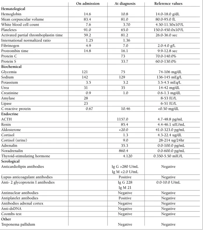
tAble I. lAborAtory results for A pAtIent wIth Aps complIcAted by bIlAterAl AdrenAl hemorrhAgIng
On admission At diagnosis Reference values Hematological
Hemoglobin 14.6 10.8 14.0-18.0 g/dL
Mean corpuscular volume 83.4 81.0 80.0-95.0 fL
White blood cell count 7.6 3.70 4.50-11.50x10⁹/L
Plateletes 91.0 65.0 150.0-450.0x10⁹/L
Activated partial thromboplastin time 59.2 81.2 26.0-36.0 sec
International normalized ratio 1.25 1.36
Fibrinogen 4.9 7.0 2.0-4.0 g/L
Protrombin time 14.8 16.1 9.9-12.8 sec
Protein C 73 70.0-140.0% Protein S 33.7 60.0-130.0% Biochemical Glycemia 121 75 74-106 mg/dL Sodium 142 129 136-145 mEg/L Potassium 3.5 3.2 3.5-4.5 mEq/L Urea 31 35 14-42 mg/dL Creatinine 0.9 1.0 0.6-1.3 mg/dL Amylase 28 8-53 IU/L Lipase 23 6-51 IU/L C-reactive protein 0.67 10.46 <0.50 mg/dL Endocrine ACTH 1157.0 4.7-48.8 pg/mL Renin 85.4 4.4-46.1 uIU/mL Aldosterone <20.0 41.0-323.0 pg/mL Cortisol 1.3 4.3-22.4 ug/dL
Cortisol (urine) 9.0 28-214 ug/24hr
Adrenalin 35.3 0.0-100.0 pg/mL
Noradrenalin 860.4 0.0-600.0 pg/mL
Thyroid-stimulating hormone 4.120 0.350-5.50 mIU/L
Serological
Anticardiolipin antibodies Ig G >280 U/mL Negative
Ig M <2.0 U/mL
Lupus anticoagulant antibodies Positive Negative
Anti- 2 glycoprotein I antibodies Ig G 228 0.0-10.0 U/mL
Ig M 21
Antinuclear antibodies Negative Negative
Antiplatelet antibodies Positive Negative
Antibodies adrenal cortex Negative Negative
Anti-dsDNA Negative Negative
Coombs test Negative Negative
Other
angiogram did not reveal thrombotic or hemorrhagic lesions. The patient was medicated with pethidine and admitted.
Several hours later, vomiting persisted and the pa-tient complained of abdominal pain localized to the left upper quadrant. Seventy-two hours after admis-sion, he began referring acute lumbar pain. He was hy-potensive (BP 96/55 mmHg) and febrile (38ºC) with skin pallor and anemic conjunctivae. Blood work showed he had normocytic, normochromic anemia, thrombocytopenia, prolonged activated partial throm-boplastin time, hyponatremia, and an elevated C-re-active protein (Table I). A renal ultrasound revealed a nodular, heterogeneous, 6-centimeter mass in the up-per extremity of the left kidney. Because of the possi-bility of an acute abdominal complication, blood and urinary cultures were ordered, and empirical antibio -tic treatment with ceftriaxone and gentamicin was ini-tiated.
An abdominal CT scan revealed an extensive left adrenal gland hematoma and a smaller right adrenal gland hematoma (Figure 1).
Primary AI was confirmed by the presence of low serum cortisol levels (1.3 µg/dL after 30 minu tes and 1.4 µg/dL after 60 minutes) after an adrenocorticotro -pic hormone (ACTH) stimulation test with 250 µg of ACTH. Was also observed a low urinary cortisol level, an undetectable aldosterone reading, and high ACTH and renin levels (Table I). Autoimmune serology con-firmed the presence of anticardiolipin and anti- 2 gly-coprotein I antibo dies and negative anti-adrenal cor-tex antibodies.
Treatment began with intravenous hydrocortisone 100 mg, followed up with daily oral hydrocortisone medication 30 mg. Antibiotic treatment was suspen
-ded. Adjustable doses of warfarin were administered to maintain an international normalized ratio between 2.5 and 3.
Since being discharged, the patient has conti nued treatment and maintained a stable condition. Subse-quent abdominal CT scans have revea led a slight duction of the bilateral hematoma. Blood work has vealed that after six months, cortisol insufficiency re-mains, indicating irreversible adrenal damage.
dIscussIon
Still unknown is how APS, a thrombosis-favoring pathology, can lead to adrenal hemorrhaging. Two pos-sible pathogenic mechanisms have been proposed. The first, and most often cited, refers to the unique vascu-larization of the adrenal gland. The adrenal gland has a rich arterial supply, yet limi ted venous drainage. Blood is provided by three main arteries that branch out to form smaller arteries. These smaller arteries pe -ne trate the surface of the gland and run along the zona glomerulosa and zona fasciculata. At the zona reticularis, the innermost region of the adrenal cortex, a ca -pil lary plexus is formed that drains into the medullary sinusoid and eventually into the large central vein6.
The transition from artery to capillary plexus is abrupt, functioning as a “vascular dam” that cau ses the accumulation and stasis of blood1,7- 9. Additionally, the musculature of the central vein consists of eccentric, longitudinal bundles that when contracted form po -ckets of turbulence and local stasis1. These two factors favor a primary venous thrombotic event, leading to increased localized arterial blood pressure, followed by post-infarction hemorrhaging7.
fi gu re 1.Both the right and left adrenal hematomas can be observed in the A) axial, B) coronal, and C) sagittal images. The left adrenal hematoma is clearly the larger of the two.
The second possible pathogenic mechanism refers to the cellular characteristics of the zona fasciculata, the central region of the adrenal cortex. The cells in this region are rich in cholesterol due to a high num-ber of cholesterol trafficking organelles. The mem-branes of these organelles contain lysobiphosphatidic acid, a target of antiphospholipid antibodies. The se -cond proposed pathogenic mechanism suggests that these antibodies, reacting locally, cause the accumula-tion of cholesterol within the cell, leading to cell death and the release of lysosomal proteinases. The proteinases activate endothelial cells, thus favoring coagu -lation and causing microthrombosis10.
A review by Espinosa et al. of 86 cases of AI in pa-tients with APS noted a precipitating factor in 43% of the cases. These factors included surgical procedures, infections, trauma, warfarin withdrawal, and inade-quate anticoagulation5, 7. In the present case, we specu late that the precipitating factor was warfarin withdra -wal. As described earlier, a week prior to this incident, the patient suspended warfarin medication and began enoxaparin. As demonstrated by Palareti and Legnani, warfarin withdrawal results in a “rebound pheno me -non”, creating a hypercoagulant condition11. We belie -ve that an initial -venous thrombotic e-vent occurred, after warfarin withdrawal, followed by hemorrhaging in the adrenal cortex.
The primary symptom manifestations of AI, as lis -ted by Garcia et al., do not exclusively suggest AI. In 66% of cases, abdominal pain localized to the lumbar, epigastric, umbilical, and pericardial region is obser -ved. In 50% of cases, continuing or intermittent fever is observed. In 12% of cases, altered mental status, lethargy, and asthenia is observed, and in 19% of ca -ses hypotension is observed8. Other symptoms include vomiting, weight loss, ileus, and diarrhea12. Because its symptoms are common to other pathologies, AI is difficult to diagnose, a difficulty worsened by the rari -ty of bilateral adrenal hemorrhaging7. Adrenal failure should be suspected in all patients with APS who complain of abdominal symptoms. All patients with bila -teral adrenal hemorrhaging should be screened for an-tiphospholipid antibodies5.
Diagnosis of AI resulting from adrenal hemorrha ging requires suggestive clinical symptoms, radiolo -gical images and hormonal evaluation. Formerly, a ma-jority of bilateral adrenal hemorrhage cases were diagno sed post-mortem8,9. Today, abdo minal CT has become the preferred method to visualize the adrenal gland13. The case illustra ted in this paper demonstrates
how an initially normal abdominal CT scan should not exclude adrenal hemorrhaging. Abdominal CT scans performed in the earlier phases of clinical cases may not reveal abnormalities. CT scans should be repeated to do cument morphological changes in cases where symptoms persist9.
Routine laboratory exams may reveal abnormalities typical of AI due to bilateral hemorrha ging, including hyponatremia, hyperkaliemia, anemia, and prolonged activated partial thromboplastin time13. However, adrenal failure can be confirmed only through hormo nal evaluation. Acute AI is detected by measuring le -vels of the stimulatory hormones, ACTH and renin, and of cortisol and aldosterone, which are produced by the gland in response to stimulatory hormones. Stable or ele vated ACTH and renin levels, accompanied by re-duced cortisol and aldosterone values, as seen in our patient, suggest primary adrenal failure. Since bilate -ral hemorrhaging of the adrenal glands may lead to ir-reversible damage and chronic adrenal failure, it is im-portant to assess long-term adrenal function14. Chro -nic AI is detected by administering ACTH and taking subsequent measurements of plasma cortisol. Cortisol levels below 18 µg/dL suggest chronic failure15. Sixty minutes after adrenal stimulation, we recorded a cor-tisol level of 1.4 µg/dL in our patient.
Based on initial and subsequent blood work and abdo minal CT imaging, we conclude that our patient has chronic, irreversible primary adrenal failu re due to bilateral adrenal hemorrhaging.
Recommended medication includes hormonal re-placement with hydrocortisone and fludrocortisone, as well as anticoagulation therapy to prevent future thrombotic events8,14,16. Six months after the initial event, the patient continued to register undetectable levels of plasma cortisol. One year la ter, the patient re-mains on steroid replacement.
Bilateral adrenal hemorrhaging is a rare complica-tion of APS that frequently leads to AI. Diagnosis of AI is complicated by the overlap of its symptoms with that of other pathologies. Howe ver, prompt diagnosis of AI is needed to avoid serious complications, in-cluding death.
correspondence to Rachel G. Silvério Avenida Rei D.Duarte Serviço de Medicina 2 3504-596 Viseu, Portugal Telefone: 011 351 967 828 387 E-mail: rachel_silverio@hotmail.com
references
1. Presotto F, Fornasini F, Betterle C, Federspil G, Rossato M. Acu-te adrenal failure as the heralding symptom of primary anti-phospholipid syndrome: report of a case and review of the li-terature. European Journal of Endocrinology 2005; 153: 507--514.
2. Andreoli T, Carpenter C, Griggs R, Loscalzo J. Chapter 66: The Adrenal Gland. Friedman T. Cecil Essentials of Medicine, 6th edition. Philadelphia: Saunders, 2004. 603-614.
3. Atsumi T, Furukawa S, Amengual O, Koike T. Antiphospholi-pid antibody associated thrombocytopenia and the paradoxi-cal risk of thrombosis. Lupus 2005; 14: 499-504.
4. Levin J, Branch D, Rauch J. The Antiphospholipid Syndrome. The New England Journal of Medicine 2002; 346: 752-763. 5. Uthman I, Salti I, Khamashta M. Endocrinologic manifestations
of the antiphosholipid syndrome. Lupus 2006; 15: 485-489. 6. Stevens A, Lowe J. Human Histology, 2nd edition. London:
Mosby, 1997. 264-267.
7. Espinosa G, Santos E, Cervera R, et al. Adrenal Involvement in the Antiphospholipid Syndrome: clinical and immunologic characteristics of 86 patients. Medicine (Baltimore) 2003; 82: 106-117.
8. Garcia G, Arberas M, Blanco A, Espiñeira J, Errasti C. Insufi-ciencia suprarrenal aguda por hemorragia suprarrenal bilateral como primera manifestación de um síndrome antifosfolípido. A propósito de um caso y revisión. Anales de Medicina Inter-na 2002; 19: 19-22.
9. Vella A, Nippoldt T, Morris J. Adrenal Hemorrhage: 25-year experience at the Mayo Clinic. Mayo Clinic Proc 2001; 76: 161--168.
10. Berneis K, Buitrago-Téllez C, Muller B, Keller U, Tsakiris D. Case Report. Antiphospholipid syndrome and endocrine da-mage: why bilateral adrenal thrombosis? European Journal of Haematology 2003; 71: 299-302.
11. Palareti G, Legnani C. Warfarin Withdrawal, Pharmacokinetic-Pharmacodynamic considerations. Clinical Pharmacokinetics 1996; 30: 300-313.
12. Espinosa G, Cervera R, Font J, Asherson R. Adrenal involve-ment in the antiphospholipid syndrome. Lupus 2003; 12:569--572.
13. Bermas B, Erkan D, Schur P. Clinical manifestations of the an-tiphospholipid syndrome. St.Vincent s Hospital. http://netac-cess.svhs-ct.org. Updated October 2008.
14. Caron P, Chabannier M, Cambus J, Fortenfant F, Otal P, Suc J. Clinical Case Seminar: Definitive adrenal insufficiency due to bilateral adrenal hemorrhage and primary antiphospholipid syndrome. Journal of Clini cal Endocrinology and Metabolism 1998; 83: 1437-1439.
15. Vaidya B, Chakera AJ, Dick C. Addison s Disease. British Me-dical Journal 2009; 339: b2385.
16. Fujishima N, Komatsuda A, Ohyagi H, et al. Adrenal insuffi-ciency complicated with antiphospholipid syndrome. The Ja-panese Society of Internal Medicine 2006; 963-966.