www.impactjournals.com/oncotarget/ Oncotarget, Vol. 7, No. 19
Exosomes isolation and characterization in serum is feasible in
non-small cell lung cancer patients: critical analysis of evidence
and potential role in clinical practice
Simona Taverna1,2,*, Marco Giallombardo1,3,*, Ignacio Gil-Bazo4, Anna Paola Carreca3, Marta Castiglia3, Jorge Chacártegui3, Antonio Araujo5, Riccardo Alessandro1,2, Patrick Pauwels6, Marc Peeters7 and Christian Rolfo3
1
Department of Biopathology and Medical Biotechnology, Section of Biology and Genetics, University of Palermo, Palermo, Italy
2
Institute of Biomedicine and Molecular Immunology (IBIM), National Research Council, Palermo, Italy
3
Phase I-Early Clinical Trials Unit, Oncology Department, Antwerp University Hospital (UZA) and Center for Oncological Research (CORE) Antwerp University, Wilrijkstraat, Edegem, Antwerp, Belgium
4
Department of Oncology, Clínica Universidad de Navarra, Pamplona, Spain
5
Service of Medical Oncology, Centro Hospitalar do Porto, Instituto de Ciências Biomédicas Abel Salazar, University of Porto, Porto, Portugal
6
Molecular Pathology, Pathology Department, Antwerp University Hospital (UZA) and Center for Oncological Research (CORE) Antwerp University, Wilrijkstraat, Edegem, Antwerp, Belgium
7
Oncology Department, Antwerp University Hospital (UZA) and Center for Oncological Research (CORE) Antwerp University, Wilrijkstraat, Edegem, Antwerp, Belgium
*
These authors have contributed equally to this work
Correspondence to: Christian Rolfo, email: [email protected]
Correspondence to: Simona Taverna, email: [email protected] Keywords: exosomes; NSCLC; liquid biopsies; biomarkers; microRNAs
Received: November 03, 2015 Accepted: January 29, 2016 Published: February 23, 2016
ABSTRACT
Exosomes are nano-sized vesicles of endolysosomal origin, released by several cytotypes in physiological and pathological conditions. Tumor derived exosomes, interacting with other cells of the tumor microenvironment, modulate tumor progression, angiogenic switch, metastasis, and immune escape. Recently, extracellular vesicles were proposed as excellent biomarkers for disease monitoring and prognosis in cancer patients. Non-small cell lung cancer (NSCLC) has a poor 5-year survival rate due to the delay in the detection of the disease. The majority of patients are diagnosed in an advanced disease stage. Exosomes might be promising beneficial tools as biomarker candidates in the scenario of NSCLC, since they contain both, proteins and miRNAs. The clinical case reported in this manuscript is a proof of concept revealing that NSCLC exosomes and sorted miRNAs might constitute, in a near future, novel biomarkers. This review summarizes the role of exosomes in NSCLC, focusing on the importance of exosomal microRNAs in lung cancer diagnosis and prognosis.
INTRODUCTION
Biomarkers constitute one of the most studied tools to understand and diagnose malignancies and to predict the outcome of cancer patients. A valuable biomarker is required to be objectively measured and evaluated as an indicator of physiological or pathological processes
or pharmacological responses to a specified therapeutic
strategy [1].
Extracellular vesicles (EVs) have recently raised a
considerable interest in the field of biomarker discovery. A relevant feature of EV-based biomarker analysis is the
Extracellular vesicles (EVs) are released by several cytotypes, constitutively and/or after cell activation.
Recently, extracellular vesicles have been classified
in different populations, differing in their molecular
composition and subcellular origin. The two populations of
vesicles most studied and characterized are microvesicles and exosomes.
The term exosome was coined by Johnstone et al [3]
in a study to understand the biologic process underlying the differentiation of reticulocytes to mature erythrocytes. The authors observed that maturing reticulocytes
contained large citoplasmatic invaginations filled with
small membrane vesicles of uniform size (40-100 nm).
These vesicles were released in the extracellular space
in order to eliminate transferrin receptor. After ten years, Raposo et al. showed that Epstein-Barr virus
(EBV)-transformed B-lymphocytes are able to secrete exosomes containing molecules that are essential for the adaptive
immune response, specifically, dimers of the Major Histocompatibility Complex class II (MHCII) bound to
antigenic peptides to be presented to T cells [4]. These
vesicles contain a tissue-specific signature composed
of proteins and selectively packaged RNAs. Patients
with cancer show higher concentrations of exosomes
in blood and moreover, these exosomes carry
tumor-specific molecules, such as DNA and their tumor-specific gene
mutations [5]. Therefore, exosomes might become true
predictive and prognostic biomarkers. More specifically in lung cancer, exosomes will be able to identify different
subpopulations of patients according to their molecular
characteristics that may be treated with selective targeted
therapy.
NON-SMALL CELL LUNG CANCER
A poorly early detection, associated with limited efficacy of the treatments for advanced disease, is responsible for the low survival rates in NSCLC patients
[6].
The development of new diagnostic, prognostic and predictive markers could significantly enhance its
early detection outcome. Results from the Collaborative
Advanced Stage Tissue Cancer (CASTLE) network are particularly awaited. In fact, this study aims to collect tumor, plasma and serum samples from stage IV NSCLC
patients before treatment to test a panel of biomarkers in order to predict response (NCT01574300). Early
detection is particularly crucial for tumors without clinical
symptoms during the initial stages of their development,
such as lung cancer. Stage IIIB/IV NSCLC patients have
a poor prognosis and the majority of them, not presenting a drugable molecular alteration, have a median survival
around 12 months using platinum based chemotherapy
doublets [7]. The era of molecular targeted therapy in
lung cancer started in 2004, when activating mutations in the epidermal growth factor receptor (EGFR) and their
correlation with clinical response to EGFR tyrosine kinase inhibitors (TKIs) were discovered [8].
The epidermal growth factor receptor (EGFR), also
named HER1, is the cell-surface receptor for extracellular
protein ligands that are members of the epidermal growth factor family (EGF-family). The EGFR activating mutations are able to confirm sensitivity to EGFR-TKIs. Several randomized clinical trials have shown, both in
Caucasian and Asian population, superior overall response
rates (ORRs) and progression free survival (PFS) for patients receiving EGFR-TKIs (Erlotinib, Gefitinib,
Afatinib), compared to standard chemotherapy in patients
with NSCLC harboring tumors with EGFR activating
mutations [9-11]. EGFR mutations in NSCLC are present in the range of 13% to 18% of Caucasian patients, and in
about 40% of Asian patients [9].
Unfortunately, the majority of EGFR-mutant NSCLC patients that show an initial radiological response to EGFR-TKIs, eventually progress and their tumors develop different mechanisms of resistance [12]. One most frequent known mechanism of resistance to EGFR-TKIs is associated with the appearance of a single recurrent missense mutation, T790M, within the EGFR kinase domain [12]. The presence of this mutation can be
also detected ex novo [13, 14]. The mutation is found in the methionine residue at position 790, causing a steric
hindrance in the interaction with the inhibitor, preventing its binding to the EGFR kinase domain that still preserves catalytic activity [15, 16]. Several strategies are ongoing in
order to overcome these resistance mechanisms, including
third generation EGFR-TKIs, like AZD9291 [17]. A similar scenario occurs for other NSCLC predictive biomarkers. Specifically, the EML4-ALK
fusion gene is a predictive biomarker for response to
first generation ALK-TKIs [18], presenting a significant clinical benefit when used in first-line treatment compared with chemotherapy for EML4-ALK translocated tumors in NSCLC patients [19, 20].
Not with standing, the recent emergent knowledge
on the role of different predictive and prognostic biomarkers makes the molecular analysis of the tissue
mandatory. Due to the small size and the remote localization of many NSCLC tumors, obtaining tissue
for further analysis is a tricky task. Currently, several efforts are ongoing in order to obtain all this biological information through non-invasive means. Circulating
tumor cells (CTCs), circulating tumor DNA (ctDNA) and
exosomes, all different components of the commonly used
concept “liquid biopsies”, are part of this answer [21]. Through new sensitive and faster technologies
for nucleic acids analysis, such as digital droplet PCR
(ddPCR) coupled with Next Generation Sequencing (NGS) technologies, it was possible to detect and sequence ctDNA and CTCs genes in order to exploit them as new
status of the tumor mass [22-24].
The detection of CTCs in the blood of cancer
patients has been usually related to metastatic stages [25, 26]. However, with these new sensitive technologies, it
has been demonstrated that CTCs could be detected also in early stages of several cancer types, such as in lung cancer, thus enhancing their role as predictive and prognostic
biomarkers [27-29]. Recently, CellSearch® system was approved from FDA as first tool for CTCs analysis in order
to have another prognostic biomarker source on metastatic breast cancer [30].
Moreover, it has been demonstrated that is feasible to characterize proteomic and miRNA profiling of
exosomes. Interestingly enough, miRNAs present in the
exosomes, have been correlated with the presence of some specific molecular alterations, such as EGFR mutations,
among others [31].
EXOSOMES IN CANCER PATIENTS:
ORIGIN AND POTENTIAL CLINICAL
INTEREST
Exosomes are spherical nano-sized vesicles that
represent a distinct class of membrane vesicles, with a
density of 1.13-1.19 g/ml and a diameter of 40-100 nm. These vesicles contain a high number of molecular cargo
such as mRNA, miRNAs, proteins and DNA protected by
a lipid bilayer.
Exosomes have an endocytic origin and are released by different cell types in both normal and pathological
conditions [32]. The exosomal composition mirrors the parental cell, but several proteins, such as CD63, CD81, CD9, TSG-101, ALIX, and HSP70, are contained in the
exosomes released by all cytotypes. These proteins are considered exosomal markers and they are commonly used to identify the presence of vesicles as true exosomes [33].
Current models suggest that exosomes are formed
within the endocytic pathway and released from the
plasma membrane via multivesicular bodies (MVBs). MVBs are formed from early endosome maturation, and they can fuse with lysosomes or the plasma membrane. When MVBs fuse with lysosomes, their content will undergo lysosomal degradation. The fusion of MVBs with
the plasma membrane induces the release of intraluminal
vesicles (exosomes) into the extracellular space (Figure 1).
Exosome formation needs the endosomal sorting complex
required for transport (ESCRT) [33].
Figure 1: Schematic model of exosome origin and release. Exosomes derived from the intraluminal vesicles (ILVs) of multivesicular
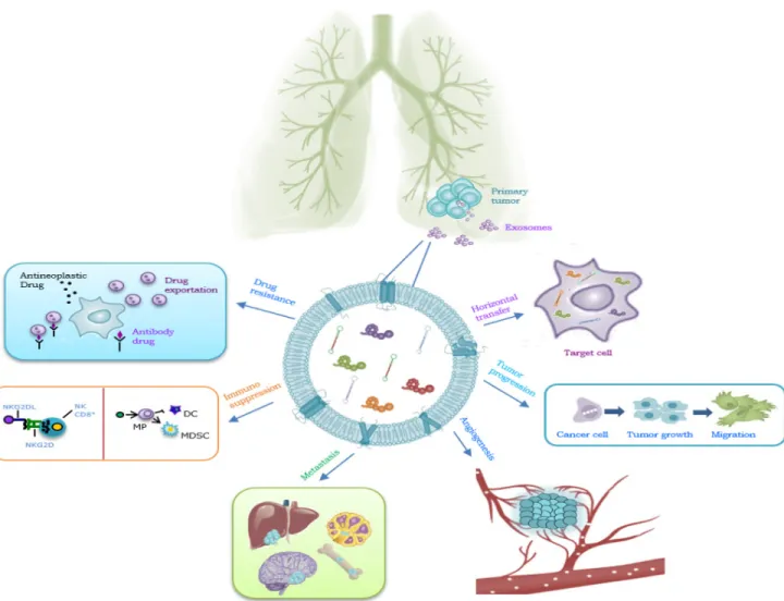
Several studies hypothesize a pleiotropic role for tumor derived exosomes; they may affect the growth and
survival of tumor cells, degradation of the extracellular matrix, stromal remodeling, angiogenesis and drug
resistance as well as modulation of the immune system
[34].
In this field, the exosomes released from macrophage
and NK cells may regulate the tumor invasiveness and its metastatic ability and may have a role in immune cytotoxicity, respectively [35]. It has also been described that mast cell-derived exosomes contain mRNAs and
miRNAs with biological activity, immunological proteins (such as MHC-II, CD86, CD40, CD40L, FcƐ-RI subunits
alpha and beta) and could be involved in B-cell activation,
inflammation process and immunomodulation [36-38].
Extracellular vesicles can also carry drugs and represent a promising strategy for drug delivery. Interestingly, proteins differentially expressed in extracellular vesicles, can be used as biomarkers for early detection, diagnosis
and prognosis of cancer [21].
Specially relevant is the role of extracellular vesicles
in transferring information between cells. That information
may be crucial for tumor progression and angiogenesis
according to a recent published research showing the
involvement of cancer cells-released exosomes in that matter [39]. In fact, several studies support that exosomes may play a relevant role in remodeling the tumor microenvironment and in tumor progression via
an enhanced angiogenesis and the preparation of the
metastatic niche (Figure 2). More specifically, melanoma-derived exosomes have been shown to promote metastasis
through the preparation of the metastatic niche by the
activation of bone marrow-derived progenitor cells [40]. Exosomes have been shown to be involved in several
cellular functions and intrinsic mechanisms of cancer
where they possibly constitute valuable biomarkers [21].
Interestingly enough, although exosomes have been better characterized in peripheral blood samples, they can
be isolated as well from most body fluids, including urine, amniotic fluid, serum, pleural effusion, saliva, breast-milk, cerebrospinal fluid, and nasal secretions [41].
As previously mentioned, the components of
Figure 2: Role of exosomes in NSCLC. Exosomes have a key role in : 1 horizontal transfer of mRNAs and miRNAs from cancer
cells to cells of microenvironment; 2 tumor progression, inducing cells motility; 3 angiogenesis; 4: metastatization; 5: immunosuppression;
exosomes are varied and include proteins, lipids and
different subtypes of RNA, which can be consulted in the ‘Exosome Content Database’ (www.exocarta.org) [42]. Recently, doubled-stranded DNA has been also discovered in exosomes opening a new perspective in this field [43].
Currently, the Exosome Content Database contains
about 4,563 proteins, 1,639 mRNAs, 764 miRNAs
and 194 lipids. Among the most frequently, identified
proteins in exosomes are membrane transport and fusion
proteins, heat-shock proteins, GTPases, MVB biogenesis
proteins, cytoskeletal proteins, metabolic enzymes, signal transduction proteins and carriers. Thanks to proteomic analysis, more than 4,400 different proteins have been detected by the use of mass spectrometry. Certainly, the
specific protein composition depends on the cell type or tissue source [42, 44].
Exosomes are also enriched in lipids that may act as important signaling molecules. They include cholesterol, diglycerides, sphingolipids, phospholipids,
glycerophospholipids and phosphatidylserine, which have
a role in exosomes recognition and internalization [45]. Exosomes are also enriched in bioactive lipids, such as prostaglandins and leukotrienes [46].
In 2007 Valadi et al. discovered exosomal miRNAs
[47], a year after Taylor et al. reported that eight miRNAs,
previously demonstrated as diagnostic markers for ovarian
cancer might be identified in biopsies of ovarian cancer as well as contained in serum exosomes isolated from the same patients. These exosomal miRNAs were not
detectable in normal controls, suggesting that they are easily attainable and may be potentially used as diagnostic
markers, a worthy alternative for biopsy profiling [48].
miRNAs are short single-stranded, endogenous, non-coding RNA molecules that are involved in binding
partial complementary sequences within the 3′-UTR of the target mRNAs. Global gene expression profiles have revealed numerous miRNAs that were deregulated in cancer compared with normal tissues [48]. The miRNAs associated with oncogenesis are also termed “oncomirs”. Depending on the main target, oncomirs are generally
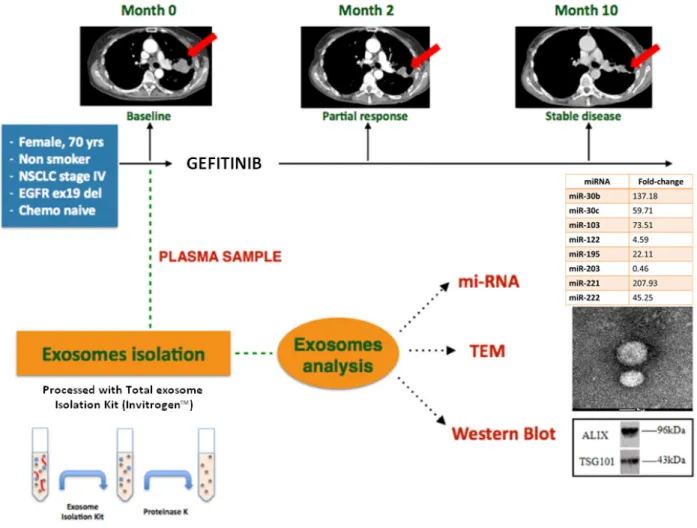
Figure 3: Summary of clinical case report. The upper part of the figure shows the computerized tomography evolution. The patient
achieved a partial response in 2 months, disease has been stable for 8 more months. The lower part shows a short scheme of exosome isolation and the morphological and biochemical analysis of the exosomes collected by the plasma of NSCLC patient. The miRNAs
classified as tumor suppressive and oncogenic miRNAs where, in general, tumor suppressive molecules repress
protein-coding oncogenes and oncogenic miRNAs that
repress protein-coding tumor suppressors. The profile of miRNAs found in exosomes is rather specific, since
particular repertories of miRNAs are selectively sorted
into exosomes, while other miRNAs are usually excluded.
As a good example of this important role, our group recently demonstrated, in vitro and in vivo, a selective sorting of the oncomir miR-21 in exosomes released by
chronic myelogenous leukemia cell lines after curcumin
treatment [49]. Furthermore, unlike other subtypes of RNA [47], exosomal miRNA profile is similar to the miRNA profile of parental cancer cells, increasing the interest of
scientists in the use of exosomal miRNA for diagnosis
of cancer [50, 51]. Moreover, the regulatory properties
attributed to tumor-derived exosomes are essential in shaping the tumor microenvironment and promoting
tumor growth and metastasis [52, 53].
Clear examples of potential clinical applications
can be found in melanoma patients, in which released exosomes were demonstrated to contain high levels of the proteins Caveolin-1 and CD63 [54]. In addition, in
glioblastoma patients, exosomes have been described as
an amenable biomarker for the identification of disease-specific molecules [55]. Que et al. also demonstrated that miR-17-5p and miR-21 were highly expressed in exosomes isolated from patients with pancreatic cancer
(PC). On the one hand, serum exosomal miR-17-5p
was higher in PC patients than in non-PC individuals (with ampullary carcinoma, benign pancreatic tumors or
chronic pancreatitis) and healthy donors. High levels of
miR-17-5p were correlated with the presence of metastasis
and advanced PC stage. On the other hand, the serum
exosomal miR-21 levels in PC were higher than in cohorts of healthy donors and in patients with chronic pancreatitis, but they were not correlated with PC differentiation or
tumor stage [56].
Serum levels of miR-141 and miR-375 are shown to be correlated with tumor progression in prostate cancer [57], showing to be valuable markers in the diagnosis of this tumor type. Moreover, Tanaka et al. showed that exosomal miR-21 expression is upregulated in serum from patients with esophageal squamous cell cancer (ESCC) versus serum from patients with benign diseases, without systemic inflammation. In that case, exosomal miR-21 was positively correlated with tumor progression and
aggressiveness, suggesting that this miRNA might be a
useful target for cancer therapy [58].
Chiba et al. also indicated that exosomes derived
from three colorectal cancer (CRC) cell lines contained
both mRNAs miRNAs, and were delivered into HepG2 and A549 cells. These novel findings indicate that exosomes, containing several RNAs, shuttle between
different cell types, and most probably may regulate gene expression into the recipient cells [59].
MOLECULAR COMPONENTS OF
EXOSOMES IN NSCLC
As previously described, the molecular profile in lung cancer, specifically in NSCLC, plays a crucial
role in prognosis and prediction of the outcome of our
patients. The idea to identify specific molecular patterns in exosomes isolated from NSCLC patients results very attractive. Several exosomal components have been studied. In this section, we will accurately describe the two more relevant ones, proteins and miRNAs.
PROTEINS
NSCLC exosomes contain several tumor-associated proteins, EGFR, KRAS, extracellular matrix metalloproteinase inducer (EMMPRIN), claudins and RAB-family proteins. EGFR is overexpressed in several human cancers and its overexpression correlates with poor
prognosis in a large number of malignancies, including
NSCLC. Exosomal mediated transfer of oncogenic EGFR
from human squamous cell carcinoma to tumor-associated
endothelial cells was shown to activate MAPK and AKT cell signaling pathways and to promote endothelial VEGF expression [60]. In order to study EGFR-containing exosomes in NSCLC, Huang et al [61] compared the exosomes content between tumor biopsies from NSCLC patients with chronic inflammation lung tissues. Eighty percent of the exosomes isolated from those NSCLC samples was positive for surface EGFR by immune staining compared to 2% of the exosomes in chronic inflammatory lung tissue.
The possibility to determine the presence of EGFR
mutations in exosomes could represent a real innovation in the development of exosomes as clinically useful liquid biomarkers, constituting a reliable tool for diagnosis and
treatment selection. EGFR incorporated in exosomes
could generate tollerogenic dendritic cells and Treg cells,
which lead to the suppression of CD8 cells [61]. Exosomes
carry proteins that may not only predict the outcomes of patients, but may actively participate in tumorigenesis via immune system deregulation, as well. Therefore, although
exosomes may also play an important role in immune system regulation, its potential activity against cancer, by
supporting immunotherapy, remains unknown.
Regarding the potential translation of exosomes
into NSCLC diagnostic characterization, Jakobsen et al demonstrated that exosomal proteins are potential diagnostic markers in advanced NSCLC. Plasma isolated from 109 NSCLC patients with advanced stage (IIIa-IV) disease and 110 matched control subjects was analyzed with an Extracellular Vesicle Array (EV Array). This array,
used to phenotype exosomes, contained 37 antibodies
targeting lung cancer-related proteins and was used to
capture exosomes. EV Array constitutes a tool to get
requirement and which can be optimized and adjusted to fit individual sets of samples [62].
Exosomal proteins may well reflect pathological processes associated with the disease. Since some of
those proteins may play a critical role in tumorigenesis they might constitute good diagnostic and prognostic
biomarkers in NSCLC [63].
The idea of obtaining proteomic information
in lung cancer, from other body fluids beyond blood samples, led to Li et al [64] to investigate this extent in
urine samples. In their experiment, urine samples from
eight chemo-naïve NSCLC patients were compared with
samples from ten healthy volunteers. Interestingly enough,
the analysis showed that NSCLC patients were able to release exosomes in urine. Moreover, high expression of leucine-rich α-2-glycoprotein (LRG1) measured by immunohistochemistry in primary tumor, showed a positive correlation rate of 65% with LRG1 presence in urine samples, suggesting that LRG1 in urinary exosomes
might be derived from primary tumor tissue. These
authors concluded that urinary LRG1 may be a candidate biomarker for non-invasive diagnosis of NSCLC in
selected individuals [64].
Other potential clinical use of the study of exosomes in lung cancer patients relays on their suspected role in
the prediction of treatment resistance. For unselected
lung cancer populations, platinum doublet chemotherapy
remains the most beneficial treatment for patients with advanced NSCLC. A recent preclinical study on exosomes
and DNA-damaging Platinum (DDP), an agent that may
cause interstrand and/or intrastrand crosslinks in the
DNA of tumor cells, showed that when A549 cells are exposed to DDP, the expression levels of several miRNAs and mRNAs, usually associated with DDP sensitivity,
drastically change in exosomes, probably mediating the
DDP resistance of A549 cells. Exosomes released by A549 cells during DDP exposure decreased the sensitivity of other A549 cells to DDP, which may be mediated by
miRNAs and mRNAs exchange by exosomes via
cell-to-cell communication [65].
In summary, there are different potential clinical
applications of exosomal proteins in NSCLC as well
as in other malignancies as diagnostic and predictive biomarkers.
MICRORNAS
An important number of clinical trials currently use
miRNAs profiling [66], in fact, miRNA-based therapeutic
approaches are being developed by several pharmaceutical companies.
Recent findings revealed the important regulatory
roles of miRNAs in a complex multistep process of
invasion-metastasis. Overexpression of miR-126 reduced NSCLC cell adhesion, migration, and invasion, which may
be partially due to the regulation of adaptor protein Crk,
also known as p38 or proto-oncogene c-Crk [67].
The expression of miR-34a in the H1299 NSCLC
cell line resulted in massive apoptosis and exogenous delivery of lipid formulated miR-34a reduced tumor size
in a mouse model of NSCLC, suggesting its possible therapeutic potential [68].
An overexpression of 12 specific miRNAs (Table 1) in NSCLC patients tissue compared to normal lung tissue was demonstrated by Yanaihara et al [69], indicating a
potential diagnostic miRNA signature. Among these,
selected miRNAs, miR-146a, targeting EGFR, is
differentially expressed in various cancer histologies. Preclinical experiments performed by this group, in
five different NSCLC cell lines, revealed the miR146a-dependent suppression of cell growth, induction of
apoptosis, inhibition of cell migration, and suppression
of EGFR downstream signaling effectors. Surprisingly, miR-146a was also able to enhance the inhibition of cell proliferation induced by drugs targeting the EGFR. These effects were independent of the EGFR mutation status. miR-146a was also shown to be generally downregulated in lung cancer patients and low levels of this miRNAs correlated with the presence of metastasis [69].
Recently, Rodriguez et al. conducted a prospective analysis of blood and bronchoalveolar lavage (BAL) samples from 30 NSCLC patients and 75 patients without
oncological diseases. They found that exosomes and
miRNAs levels were higher in both plasma and BAL from NSCLC patients. The authors revealed that
miR-Table 1: Selected overexpressed microRNAs discovered in NSCLC
miR-17-3p
miR-21
miR-106a
miR-146
miR-155
miR-191
miR-192
miR-203
miR-205
miR-210
miR-212
126 was only determinated in plasma samples [70]. MiR-126 inhibits the NSCLC cells proliferation via EGF-like domain-containing protein 7 (EGFL7) and targets Sdf-1
cytokine to reduce the recruitment of mesenchymal stem
cells and inflammatory monocytes to primary tumors,
inhibiting lung metastasis appearance [71]. In another
study, five determined miRNAs were selected from a panel of 365 miRNAs in the plasma of 28 NSCLC patients and 20 healthy controls. A validation of these five selected miRNAs (let7f, 20b, 30e-3p, 223and miR-301) was independently performed by real-time PCR in plasma from 78 NSCLC and 48 controls, and correlated with pathologic parameters and survival. Levels of let-7f, miR-20b and miR-30e-3p were decreased in plasma vesicles of NSCLC patients. The levels of let-7f and miR-30e-3p was able to discriminate between two groups of
patients for stage of disease and therefore their surgical
options. More interestingly, plasma levels of miR-30e-3p and let-7f in NSCLC patients has been also associated with poor clinical outcome [72].
The miRNA profile in exosomes is similar to the primary tumor profile and this feature may be a powerful
tool in different aspects such as early diagnosis, prognosis and prediction of response.
After having discussed the role of exosomes in the biology of tumor progression and their potential to translate the preclinical experience into clinical practice, a
clinical case followed by our group will be described and discussed. With this case, we would like to demonstrate
that isolation and characterization of exosomes in patients
with NSCLC is feasible in a daily practice.
CLINICAL CASE
We present a case of a 70 years old woman with no relevant past medical history diagnosed with stage IV NSCLC, (adenocarcinoma histotype) and harboring
an EGFR activating mutation by a deletion in exon 19 (c.2236_2250del15, p.Glu74_Ala750del). The patient was treated with Gefitinib 250 mg/daily. After two months of treatment, a computerized tomography (CT) scan showed
a partial response in the primary tumor. The patient
continued on treatment for a total of 10 months with
stabilization of disease and an improvement in quality of life.
Before the start the treatment with Gefitinib, a blood sample was collected and plasma was separated. The plasma sample was processed with a commercial kit
(Invitrogen™, Carlsbad) to collect exosomes. In order to
characterize the exosomes, we performed a morphological and biochemical analysis. We confirmed exosomes
characterization by performing Transmission Electron
Microscopy (TEM) analysis, in order to show that the purified vesicles of our patient sample had an average size in the range of the exosomal diameter, between 40-100nm, what is in agreement with data from literature [39].
Vesicles were also analysed by Western blotting using specific antibodies for Alix and TSG-101, both well-known exosomal markers. These data confirmed that the vesicles that were purified from plasma of our patient were indeed exosomes (Figure 3).
Based on literature, we selected eight miRNAs, with a documented role in NSCLC [31]. This panel of miRNAs was analyzed by TaqMan® Real-time PCR [58]. Relative changes in miRNA expression between healthy control and patient samples were determined with the ΔΔCt method; miR-1228-3p was selected as stable endogenous
control.
As shown in figure 3, high levels of miR30b, -30c, -103, -195, -221, -222 were encapsulated in exosomes. These results are concordant with the findings of Garofalo et al, indicating that miR-103,-203,-30b-c and -221 -222
have a prognostic value, as a marker, in lung cancer [31].
Yanaihara et al [69] also indicated that the expression of 12 selected miRNAs in exosomes from NSCLC patients could play a role in diagnosis, since they were expressed only in exosomes released by NSCLC cells. We found a downregulation of miR-122 and miR-203, possibly
predictive of more aggressive and metastatic tumors [31].
Circulating miR-122 was expressed at lower levels in patients with EGFR mutated NSCLC compared with wild
type EGFR NSCLC patients [73]. In concordance with these results, our analysis confirmed a downregulation of
this miRNA in exosomes of our EGFR mutant NSCLC
patient.
With this single case, we show that this exosomal miRNAs profile of our patient is in concordance with
the data in the literature. We believe that exosomal miRNAs may have an important role in future diagnostic
and prognostic analysis. Our group is currently working
in this context, to demonstrate the role of exosomes in
monitoring NCSLC patients during the treatment. These observations suggest that NSCLC exosomes and sorted
miRNAs may have an important role in diagnosis and prognosis in a near future. An interesting open question regarding the exosomal application in clinical practice it
could be the standardization of the correlation between
exosomal features and clinical outcomes. In this context,
the idea to exploite the correlation between protein and/or
miRNA levels of exosome (as part of liquid biopsies) and tumor tissue, it could be extremely attractive.
DISCUSSION
Throughout this manuscript, we have described the
role of exosomes in cancer, principally in lung cancer. Exosomes are small vesicles released by cancer cells to the extracellular medium and are able to shuttle proteins and nucleic acids. Exosomes are involved in several processes such as angiogenesis, premetastatic niche preparation and regulation of tumor immune system responses.
non-invasive biomarkers with diagnostic, prognostic and
predictive value. Clinical studies have already described exosomes-associated cancer biomarkers for several cancer
types such as prostate, breast and ovarian cancer as well as
glioblastoma and melanoma.
The isolation of exosomes and its use in the clinic may substitute invasive procedures for diagnosis or for the
follow-up of cancer patients, mainly in NSCLC, where the availability of primary tumor tissue is difficult in most of
the patients. The development of novel biomarkers and
the establishment of a fluid-based early detection system
for lung cancer is crucial to improve the clinical outcome. This approach brings also positive effects like a reduction of complications derived from tumor biopsies, the possibility to anticipate progression and even the
reduction of healthy costs with ambulatory non-invasive techniques. Since the markers must be stable, all these
features make exosomes good candidates for become
biomarkers in NSCLC. In addition, exosomal proteins and
miRNAs are protected from RNase/proteinase-dependent degradation and thus can be stably detected in circulating plasma and serum, making them ideal biomarkers for a number of clinical applications [74]. Another reason to select exosomes as preferable liquid biopsy tool is correlated to their higher concentration in blood of cancer
patients [75] in comparison with CTCs, that could lead
exosomes to be more exploitable for isolation and analysis methods.
For instance, some biomarkers such as PCA3 and TMPRSS2 are mRNAs not easily detected in body fluids, but are found in exosomes in prostate cancer [76]. Compared with traditional biomarkers, exosomes have
other advantages potentially, these vesicles can travel
across the endothelium into the circulation allowing
serum detection. In contrast to invasive biopsy, exosomes
are effective biomarkers in the diversified diagnosis of
personalized medicine [77].
For these reasons, our group is focusing on the
potential predictive and prognostic value of exosomes
collected from NSCLC samples.
The isolation of exosomes from plasma of patients
requires specific expertise, because the amount of samples is often insufficient to perform the purification with
ultracentrifugation. Recently, the increasing number of
commercial kits developed allows researchers to choose
the most suitable one for different analysis. In our clinical
case, the purification of exosomes with a commercial kit in combination with transmission electron microscopy, Western blotting analysis and miRNAs detection allowed
us to demonstrate the presence of exosomes containing
miRNAs in plasma of a NSCLC patient and to confirm the
data of literature regarding the role of exosomal miRNAs. Exosomes are also attractive in cancer therapy, as vehicles for administration of antitumor compounds such as anticancer therapies, siRNAs and antigens to target
recipient cells [78, 79].
Furthermore, in this field, exosomes derived from
immune cells can be used to develop cancer vaccines.
Morse et al reported a phase I study of dendritic-derived exosomes (dexosomes) immunotherapy in patients with advanced NSCLC. Dexosomes were injected in 13 NSCLC patients for 4 weeks but unfortunately inducing only a weak immune response against the tumor [80].
Viaud et al developed a new technology to produce highly
immunogenic dexosomes, currently used in a phase II
clinical trial to test the clinical benefit of dexosomes to maintain immunotherapy in inoperable NSCLC patients that responded to chemotherapy [81].
These perspectives open infinite possibilities for
clinical application. Tremendous multidisciplinary efforts must be done by basic researchers, oncologists and
patients, in order to make all this incredible knowledge
applicable in a real translational oncology scenario.
ACKNOWLEDGMENTS
A particular acknowledgements to Eduardo
Castañón Alvarez from Clínica Universidad de Navarra
and Francesco Liggieri from University Hospital of Antwerp (UZA) for their contributions in this work.
CONFLICTS OF INTERESTS
All authors declare no competing financial interests.
REFERENCES
1. Atkinson AJ J, Colburn WA, DeGruttola VG, DeMets DL, Downing GJ, Hoth DF, Oates J A, Peck CC, Schooley, RT, Spilker BA, Woodcock J, and Zeger SL. Biomarkers and surrogate endpoints: Preferred definitions and conceptual framework. Clin Pharmacol Ther. 2001; 69: 89-95. doi:10.1067/mcp.2001.113989.
2. Boukouris S, and Mathivanan S. Exosomes in bodily fluids are a highly stable resource of disease biomarkers. Proteomics Clin Appl. 2015; 9: 358-67. doi:10.1002/ prca.201400114.
3. Johnstone RM, Adam M, Hammond JR, Orr L, and Turbide
C. Vesicle formation during reticulocyte maturation.
Association of plasma membrane activities with released vesicles (exosomes). J Biol Chem. 1987; 262: 9412-20. 4. Raposo G, Nijman HW, Stoorvogel W, Liejendekker R,
Harding C V, Melief CJ, and Geuze HJ. B lymphocytes secrete antigen-presenting vesicles. J Exp Med. 1996; 183: 1161-72. doi:10.1084/jem.183.3.1161.
6. Jemal A, Siegel R, Ward E, Hao Y, Xu J, and Thun MJ. Cancer Statistics. CA Cancer J Clin. 2009; 59: 225-49. doi:10.3322/caac.20006.
7. Scagliotti G, Hanna N, Fossella F, Sugarman K, Blatter J, Peterson P, Simms, Lorinda S, and Frances A. The differential efficacy of pemetrexed according to NSCLC histology: a review of two Phase III studies. Oncologist. 2009; 14: 253-63. doi:10.1634/theoncologist.2008-0232. 8. Fukuoka M, Wu YL, Thongprasert S, Sunpaweravong P,
Leong SS, Sriuranpong V, Chao TY, Nakagawa K. Chu DT, Saijo N, Duffield EL, Rukazenkov Y, Speake G, et al. Biomarker analyses and final overall survival results from a phase III, randomized, open-label, first-line study of gefitinib versus carboplatin/paclitaxel in clinically selected
patients with advanced non - small-cell lung cancer in Asia (IPASS). J Clin Oncol. 2011; 29: 2866-74. doi:10.1200/ JCO.2010.33.4235.
9. Kosaka T, Yatabe Y, Endoh H, Kuwano H, Takahashi T and Mitsudomi T. Mutations of the epidermal growth
factor receptor gene in lung cancer: Biological and
clinical implications. Cancer Res. 2004; 64: 8919-23. doi:10.1158/0008-5472.CAN-04-2818.
10. Sequist LV, Yang JC-H, Yamamoto N, O’Byrne K, Hirsh V, Mok T, Geater SL, Orlov S, Tsai CM, Boyer M, Su WC, Bennouna J, Kato T, et al. Phase III study of afatinib or cisplatin plus pemetrexed in patients with metastatic lung adenocarcinoma with EGFR mutations. J Clin Oncol. 2013; 31: 3327-34. doi:10.1200/JCO.2012.44.2806.
11. Rosell R, Carcereny E, Gervais R, Vergnenegre A, and Massuti B. Erlotinib versus standard chemotherapy as first-line treatment for European patients with advanced EGFR
mutation-positive non-small-cell lung cancer (EURTAC):
a multicentre, open-label, randomised phase 3 trial. Lancet Oncol 2012;13:239-46.
12. Rolfo C, Giovannetti E, Hong DS, Bivona T, Raez LE, Bronte G, Buffoni L, Reguart NS, Edgardo S, Germonpre P, Taron M, Passiglia F, Van Meerbeeck JP, et al. Novel therapeutic strategies for patients with NSCLC that do not respond to treatment with EGFR inhibitors. Cancer Treat Rev. 2014; 40: 990-1004. doi:10.1016/j.ctrv.2014.05.009. 13. Pao W, Miller VA, Politi KA, Riely GJ, Somwar R,
Zakowski MF, Kris MG, and Varmus H. Acquired resistance of lung adenocarcinomas to gefitinib or erlotinib is associated with a second mutation in the EGFR kinase domain. PLoS Med. 2005; 2: 0225-35. doi:10.1371/journal. pmed.0020073.
14. Shih JY, Gow CH, and Yang PC. EGFR mutation conferring primary resistance to gefitinib in non-small-cell lung cancer. N Engl J Med. 2005; 353: 207-8. doi:10.1056/ NEJM200507143530217.
15. Kobayashi S, Boggon TJ, Dayaram T, Jänne P a, Kocher O, Meyerson M, Johnson BE, Eck MJ, Tenen DG, and Halmos B. EGFR mutation and resistance of non-small-cell lung cancer to gefitinib. N Engl J Med. 2005; 352: 786-92. doi:10.1056/NEJMoa044238.
16. Pao W, and Miller VA. Epidermal growth factor receptor
mutations, small-molecule kinase inhibitors, and
non-small-cell lung cancer: current knowledge and future directions. J Clin Oncol. 2005; 23: 2556-68. doi:10.1200/ JCO.2005.07.799.
17. Hammerman PS, Jänne PA, and Johnson BE. Resistance to Epidermal Growth Factor Receptor Tyrosine Kinase. Clin Cancer Res. 2009; 15: 7502-9. doi:10.1158/1078-0432. CCR-09-0189.
18. Solomon BJ, Mok T, Kim D-W, Wu Y-L, Nakagawa K, Mekhail T, Felip E, Cappuzzo F, Paolini J, Usari T, Iyer S, Reisman A, Wilner KD, et al. First-Line Crizotinib versus Chemotherapy in ALK-Positive Lung Cancer. N Engl J Med. 2014; 371: 2167-77. doi:10.1056/NEJMoa1408440. 19. Friboulet L, Li N, Katayama R, Lee CC, Gainor JF, Crystal
AS, Michellys PY, Awad MM, Yanagitani N, Kim S, Pferdekamper AMC, Li Jie, Kasibhatla S, et al. The ALK
inhibitor ceritinib overcomes crizotinib resistance in
non-small cell lung cancer. Cancer Discov. 2014; 4: 662-73. doi:10.1158/2159-8290.CD-13-0846.
20. Gadgeel L, Riely GJ, Chiappori AA, West HL, Azada MC, Morcos PN, Lee RM, Garcia L, Yu Li, Boisserie F, Laurenzio L, Di Golding S, Sato J, et al. Safety and activity
of alectinib against systemic disease and brain metastases
in patients with crizotinib-resistant ALK-rearranged non-small-cell lung cancer (AF-002JG): results from the dose-finding portion of a phase 1/2 study. Lancet Oncol. 2015;15: 1119-28.
21. Rolfo C, Castiglia M, Hong D, Alessandro R, Mertens I, Baggerman G, Zwaenepoel K, Gil-Bazo I, Passiglia F, Carreca AP, Taverna S, Vento R, Peeters M, et al. Liquid biopsies in lung cancer: The new ambrosia of researchers. Biochim Biophys Acta. 2014; 1846: 539-46. doi:10.1016/j. bbcan.2014.10.001.
22. Toss A, Mu Z, Fernandez S, and Cristofanilli M. CTC enumeration and characterization: moving toward personalized medicine. Ann Transl Med. 2014; 2: 108. doi:10.3978/j.issn.2305-5839.2014.09.06.
23. Diaz LA Jr, and Bardelli A. Liquid biopsies: Genotyping circulating tumor DNA. J Clin Oncol. 2014; 32: 579-86. doi:10.1200/JCO.2012.45.2011.
24. Schwarzenbach H, Hoon DSB, and Pantel K. Cell-free
nucleic acids as biomarkers in cancer patients. Nat Rev
Cancer. 2011; 11: 426-37. doi:10.1038/nrc3066.
25. Alix-Panabier̀es C, and Pantel K. Circulating tumor cells:
Liquid biopsy of cancer. Clin Chem. 2013; 59: 110-8. doi:10.1373/clinchem.2012.194258.
26. Yoneda K, Tanaka F, Kondo N, Hashimoto M, Takuwa T, Matsumoto S, Okumura Y, Tsubota N, Sato A, Tsujimura T, Kuribayashi K, Fukuoka K, Tabata C, et al. Circulating Tumor Cells (CTCs) in Malignant Pleural Mesothelioma (MPM). Ann Surg Oncol. 2013; 4: S472-80. doi:10.1245/ s10434-013-3399-2.
Greystoke A, Ward TH, Ferraldeschi R, Hughes A, Clack G, Ranson M, Dive C, and Blackhall FH. Evaluation and prognostic significance of circulating tumor cells in patients with non-small-cell lung cancer. J Clin Oncol. 2011; 29: 1556-63. doi:10.1200/JCO.2010.28.7045.
28. Cristofanilli M, and Braun S. Circulating tumor cells revisited. JAMA. 2010; 303: 1092-3. doi:10.1146/annurev-med-062310-094219.
29. Tanaka F, Yoneda K, Kondo N, Hashimoto M, Takuwa T, Matsumoto S, Okumura Y, Rahman S, Tsubota N, Tsujimura T, Kuribayashi K, Fukuoka K, Nakano T,
et al. Circulating tumor cell as a diagnostic marker in
primary lung cancer. Clin Cancer Res. 2009; 15: 6980-6. doi:10.1158/1078-0432.CCR-09-1095.
30. Cristofanilli M, Budd GT, Ellis MJ, Stopeck A, Matera J, Miller MC, Reuben JM, Doyle GV, Allard WJ, Terstappen LW, and Hayes DF. Circulating tumor cells, disease
progression, and survival in metastatic breast cancer. N Engl
J Med. 2004; 351: 781-91. doi:10.1056/NEJMoa040766. 31. Garofalo M, Quintavalle C, Di Leva G, Zanca C, Romano
G, Taccioli C, Liu CG, Croce CM and Condorelli G. MicroRNA signatures of TRAIL resistance in human non-small cell lung cancer. Oncogene. 2008; 27: 3845-55. doi:10.1038/onc.2008.6.
32. Théry C, Zitvogel L, and Amigorena S. Exosomes:
composition, biogenesis and function. Nat Rev Immunol.
2002; 2: 569-79.
33. Kowal J, Tkach M, and Théry C. Biogenesis and secretion of exosomes. Curr Opin Cell Biol. 2014; 29: 116-25. doi:10.1016/j.ceb.2014.05.004.
34. Fontana S, Saieva L, Taverna S, and Alessandro R.
Contribution of proteomics to understanding the role of
tumor-derived exosomes in cancer progression: State of the art and new perspectives. Proteomics. 2013; 13: 1581-94. doi:10.1002/pmic.201200398.
35. Benito-Martin A, Di Giannatale A, Ceder S, and Peinado H. The New Deal: A Potential Role for Secreted Vesicles in Innate Immunity and Tumor Progression. Front Immunol. 2015; 6:66. doi:10.3389/fimmu.2015.00066.
36. Carroll-Portillo A, Surviladze Z, Cambi A, Lidke DS, and Wilson BS. Mast cell synapses and exosomes: Membrane contacts for information exchange. Front Immunol. 2012; 3:1-9. doi:10.3389/fimmu.2012.00046.
37. Skokos D, Le Panse S, Villa I, Rousselle J-C, Peronet R, David B, Namane A, and Mécheri S. Mast Cell-Dependent B and T Lymphocyte Activation Is Mediated by the Secretion of Immunologically Active Exosomes. J Immunol. 2001; 166: 868-76. doi:10.4049/ jimmunol.166.2.868.
38. Vincent-Schneider H, Stumptner-Cuvelette P, Lankar D, Pain S, Raposo G, Benaroch P, and Bonnerot C. Exosomes bearing HLA-DR1 molecules need dendritic cells to efficiently stimulate specific T cells. Int Immunol. 2002; 14: 713-22. doi:10.1093/intimm/dxf048.
39. Taverna S, Flugy A, Saieva L, Kohn EC, Santoro A, Meraviglia S, De Leo G, and Alessandro R. Role of
exosomes released by chronic myelogenous leukemia
cells in angiogenesis. Int J Cancer. 2012;130: 2033-43. doi:10.1002/ijc.26217.
40. Peinado H, Alečković M, Lavotshkin S, Matei I, Costa-Silva B, Moreno-Bueno G, Hergueta-Redondo M, Williams C, Santos GG, Nitadori-Hoshino A, Hoffman C, Badal K, Garci BA, et al. Melanoma exosomes educate bone marrow progenitor cells toward a pro-metastatic phenotype through MET. Nat Med. 2013; 18: 883-91. doi:10.1038/nm.2753. 41. Vlassov AV, Magdaleno S, Setterquist R, and Conrad
R. Exosomes: Current knowledge of their composition,
biological functions, and diagnostic and therapeutic
potentials. Biochim Biophys Acta - Gen Subj. 2012; 1820:940-8. doi:10.1016/j.bbagen.2012.03.017.
42. Mathivanan S, Fahner CJ, Reid GE, and Simpson RJ. ExoCarta 2012: database of exosomal proteins, RNA and lipids. Nucleic Acids Res. 2012; 40: D1241-4. doi:10.1093/ nar/gkr828.
43. Thakur BK, Zhang H, Becker A, Matei I, Huang Y, Costa-Silva B, Zheng Y, Hoshino A, Brazier H, Xiang J, Williams C, Rodriguez-Barrueco R, Silva JM, et al. Double-stranded DNA in exosomes: a novel biomarker in cancer detection. Cell Res. 2014; 24: 766-9. doi:10.1038/cr.2014.44. 44. Mathivanan S, Ji H, Tauro BJ, Chen YS, and Simpson RJ.
Identifying mutated proteins secreted by colon cancer cell
lines using mass spectrometry. J Proteomics. 2012; 5; 76 Spec No.:141-9. doi:10.1016/j.jprot.2012.06.031.
45. Zakharova L, Svetlova M, and Fomina AF. T cell exosomes
induce cholesterol accumulation in human monocytes via phosphatidylserine receptor. J Cell Physiol. 2007; 212: 174-81. doi:10.1002/jcp.21013.
46. Subra C, Grand D, Laulagnier K, Stella A, Lambeau G, Paillasse M, De Medina P, Monsarrat B, Perret B, Silvente-Poirot S, Silvente-Poirot M, and Record M. Exosomes account
for vesicle-mediated transcellular transport of activatable
phospholipases and prostaglandins. J Lipid Res. 2010; 51: 2105-20. doi:10.1194/jlr.M003657.
47. Valadi H, Ekström K, Bossios A, Sjöstrand M, Lee JJ, and Lötvall JO. Exosome-mediated transfer of mRNAs and
microRNAs is a novel mechanism of genetic exchange
between cells. Nat Cell Biol. 2007; 9: 654-9. doi:10.1038/
ncb1596.
48. Taylor DD and Gercel-Taylor C. MicroRNA signatures
of tumor-derived exosomes as diagnostic biomarkers
of ovarian cancer. Gynecol Oncol. 2008; 110: 13-21. doi:10.1016/j.ygyno.2008.04.033.
49. Taverna S, Giallombardo M, Pucci M, Flugy A, Manno M, Raccosta S, Rolfo C, De Leo G, and Alessandro
R. Curcumin inhibits in vitro and in vivo chronic
50. Pigati L, Yaddanapudi SCS, Iyengar R, Kim DJ, Hearn SA, Danforth D, Hastings ML, and Duelli DM. Selective release of MicroRNA species from normal and malignant mammary epithelial cells. PLoS One. 2010; 5(10): e13515.
doi:10.1371/journal.pone.0013515.
51. Jaiswal R, Gong J, Sambasivam S, Combes V, Mathys J-M, Davey R, Grau GER, and Bebawy M.
Microparticle-associated nucleic acids mediate trait dominance in cancer.
FASEB J. 2012; 26:420-9. doi:10.1096/fj.11-186817. 52. Wang W, and Lotze MT. Good things come in small
packages: exosomes, immunity and cancer. Cancer Gene. Ther 2014; 21: 139-41. doi:10.1038/cgt.2014.14.
53. Zhao X, Parpart S, Takai A, Roessler S, Budhu A, Yu Z, Blank M, Zhang Ye, Jia HL, Ye QH, Qin LX, Tang ZY, Thorgeirsson SS, et al. Integrative genomics identifies YY1AP1 as an oncogenic driver in EpCAM+ AFP+ hepatocellular carcinoma. Oncogene. 2015; 34(39): 5095-104. doi:10.1038/onc.2014.438.
54. Logozzi M, De Milito A, Lugini L, Borghi M, Calabrò L, Spada M, Perdicchio M, Marino ML, Federici C, Iessi E, Brambilla D, Venturi G, Lozupone F, et al. High levels of exosomes expressing CD63 and caveolin-1 in plasma of melanoma patients. PLoS One. 2009; 4:e5219. doi:10.1371/ journal.pone.0005219.
55. D’Asti E, Garnier D, Lee TH, Montermini L, Meehan B, and Rak J. Oncogenic extracellular vesicles in brain tumor progression. Front Physiol. 2012; 3: 1-15. doi:10.3389/ fphys.2012.00294.
56. Que R, Ding G, Chen J, and Cao L. Analysis of serum
exosomal microRNAs and clinicopathologic features of
patients with pancreatic adenocarcinoma. World J Surg Oncol. 2013; 11:219. doi:10.1186/1477-7819-11-219. 57. Brase JC, Johannes M, Schlomm T, Haese A, Steuber
T, Beissbarth T, Kuner R, and Sültmann H. Circulating miRNAs are correlated with tumor progression in prostate cancer. Int J Cancer. 2011; 128:608-16. doi:10.1002/ ijc.25376.
58. Tanaka Y, Kamohara H, Kinoshita K, Kurashige J, Ishimoto T, Iwatsuki M, Watanabe M, and Baba H. Clinical impact of serum exosomal microRNA-21 as a clinical biomarker in human esophageal squamous cell carcinoma. Cancer. 2013; 119:1159-67. doi:10.1002/cncr.27895.
59. Chiba M, Kimura M, and Asari S. Exosomes secreted
from human colorectal cancer cell lines contain mRNAs, microRNAs and natural antisense RNAs, that can transfer
into the human hepatoma HepG2 and lung cancer A549 cell lines. Oncol Rep. 2012; 28:1551-8. doi:10.3892/ or.2012.1967.
60. Al-Nedawi K, Meehan B, Kerbel RS, Allison AC, and Rak J. Endothelial expression of autocrine VEGF upon
the uptake of tumor-derived microvesicles containing
oncogenic EGFR. Proc Natl Acad Sci U S A. 2009; 106: 3794-9. doi:10.1073/pnas.0804543106.
61. Huang S-H, Li Y, Zhang J, Rong J, and Ye S. Epidermal
growth factor receptor-containing exosomes induce tumor-specific regulatory T cells. Cancer Invest. 2013; 31:330-5. doi:10.3109/07357907.2013.789905.
62. Jakobsen KR, Paulsen BS, Baek R, Varming K, Sorensen BS, and Jørgensen MM. Exosomal proteins as potential
diagnostic markers in advanced non-small cell lung
carcinoma. J Extracell Vesicles. 2015; 4:26659.
63. Park JO, Choi D-Y, Choi D-S, Kim HJ, Kang JW, Jung JH, Lee JH, Kim J, Freeman MR, Lee, KY, Gho YS, and Kim KP. Identification and characterization of proteins isolated
from microvesicles derived from human lung cancer pleural
effusions. Proteomics. 2013; 13: 2125-34. doi:10.1002/ pmic.201200323.
64. Li Y, Zhang Y, Qiu F, and Qiu Z. Proteomic identification of exosomal LRG1: A potential urinary biomarker for detecting NSCLC. Electrophoresis. 2011; 32: 1976-83. doi:10.1002/elps.201000598.
65. Xiao X, Yu S, Li S, Wu J, Ma R, Cao H, Zhu Y, and Feng J.
Exosomes: decreased sensitivity of lung cancer A549 cells
to cisplatin. PLoS One. 2014; 9: 1-6. doi:10.1371/journal. pone.0089534.
66. Nana-Sinkam SP and Croce CM. Clinical applications for microRNAs in cancer. Clin Pharmacol Ther. 2013; 93: 98-104. doi:10.1038/clpt.2012.192.
67. Crawford M, Brawner E, Batte K, Yu L, Hunter MG, Otterson GA, Nuovo G, Marsh CB and Nana-Sinkam SP. MicroRNA-126 inhibits invasion in non-small cell lung
carcinoma cell lines. Biochem Biophys Res Commun.
2008; 373:607-12. doi:10.1016/j.bbrc.2008.06.090. 68. Ma ZL, Hou P, Li Y, Wang D, Yuan T, Wei JL, Zhao
BT, Lou JT, Zhao XT, Jin Y, and Jin YX.
MicroRNA-34a inhibits the proliferation and promotes the apoptosis
of non-small cell lung cancer H1299 cell line by targeting TGFβR2. Tumor Biol. 2015; 36: 2481-90. doi:10.1007/ s13277-014-2861-5.
69. Yanaihara N, Caplen N, Bowman E, Seike M, Kumamoto K, Yi M, Stephens RM, Okamoto A, Yokota J, Tanaka T, Calin GA, Liu CG, Croce CM, et al. Unique microRNA molecular profiles in lung cancer diagnosis and prognosis. Cancer Cell. 2006; 9: 189-98. doi:10.1016/j. ccr.2006.01.025.
70. Rodríguez M, Silva J, López-Alfonso A, López-Muñiz M, Peña C, Domínguez G, García JM, López-Gónzalez A, Méndez M, Provencio M, García V, and Bonilla F. Different exosome cargo from plasma/bronchoalveolar lavage in non-small-cell lung cancer. Genes Chromosom Cancer. 2014; 53: 713-24.
71. Sun Y, Bai Y, Zhang F, Wang Y, Guo Y, and Guo L. miR-126 inhibits non-small cell lung cancer cells proliferation by targeting EGFL7. Biochem Biophys Res Commun. 2010; 391: 1483-9. doi:10.1016/j.bbrc.2009.12.098.
microRNAs in plasma of nonsmall cell lung cancer patients
and correlation with survival. Eur Respir J. 2011; 37: 617-23. doi:10.1183/09031936.00029610.
73. Zhang H, Su Y, Xu F, Kong J, Yu H, and Qian B. Circulating microRNAs in relation to EGFR status
and survival of lung adenocarcinoma in female
non-smokers. PLoS One. 2013; 8: 1-10. doi:10.1371/journal. pone.0081408.
74. Lin J, Li J, Huang B, Liu J, Chen X, Chen XM, Xu YM, Huang LF, and Wang XZ. Exosomes: Novel Biomarkers for Clinical Diagnosis. Sci World J. 2015; 2015:657086. doi:10.1155/2015/657086.
75. Caby MP, Lankar D, Vincendeau-Scherrer C, Raposo G, and Bonnerot C. Exosomal-like vesicles are present in human blood plasma. Int Immunol. 2005; 17: 879-87. doi:10.1093/intimm/dxh267.
76. Nilsson J, Skog J, Nordstrand A, Baranov V, Mincheva-Nilsson L, Breakefield XO, and Widmark A. Prostate
cancer-derived urine exosomes: a novel approach to
biomarkers for prostate cancer. Br J Cancer. 2009; 100: 1603-7. doi:10.1038/sj.bjc.6605058.
77. An T, Qin S, Xu Y, Tang Y, Huang Y, Situ B, Inal JM, and Zheng L. Exosomes serve as tumour markers for personalized diagnostics owing to their important role in cancer metastasis. J Extracell Vesicles. 2015; 4: 27522. doi:10.3402/jev.v4.27522.
78. Alvarez-Erviti L, Seow Y, Yin H, Betts C, Lakhal S, and Wood MJA. Delivery of siRNA to the mouse brain by
systemic injection of targeted exosomes. Nat Biotechnol.
2011; 29: 341-5. doi:10.1038/nbt.1807.
79. Tran T-H, Mattheolabakis G, Aldawsari H, and Amiji M.
Exosomes as nanocarriers for immunotherapy of cancer and
inflammatory diseases. Clin Immunol. 2015; 160: 46-58. doi:10.1016/j.clim.2015.03.021.
80. Morse M a, Garst J, Osada T, Khan S, Hobeika A, Clay TM, Valente N, Shreeniwas R, Sutton MA, Delcayre A, Hsu DH, Le Pecq JB, and Lyerly HK. A phase I study of dexosome immunotherapy in patients with advanced non-small cell lung cancer. J Transl Med. 2005; 3:9. doi:10.1186/1479-5876-3-9.
81. Viaud S, Ploix S, Lapierre V, Théry C, Commere P-H, Tramalloni D, Gorrichon K, Virault-Rocroy P, Tursz T, Lantz O, Zitvogel L, and Chaput N. Updated
technology to produce highly immunogenic dendritic cell-derived exosomes of clinical grade: a critical role of