Comparative effects of the Mandibular Protraction
Appliance in adolescents and adults
Bruno D’Aurea Furquim1, Guilherme Janson2, Laura de Castro Cabrera Cope3, Karina Maria Salvatore Freitas4, José Fernando Castanha Henriques2
Objective: The aim of this study was to compare the skeletal, dental, and soft tissue effects of the Mandibular Protraction Appliance (MPA) application in adolescent and adult Class II malocclusion patients. Methods: The sample comprised the pretreatment and posttreatment lateral cephalograms of 39 subjects presenting Class II malocclusion treated with the MPA and fixed appliances. Sample was divided into two groups: Group 1 comprised 23 subjects (10 male; 13 female), at a mean pretreatment age of 11.75 years, with a mean treatment time of 3.32 years; Group 2 included 16 subjects (7 male; 9 female), at a mean pretreatment age of 22.41 years, with a mean treatment time of 4.24 years. Intergroup comparison of the initial and final stages and treatment changes between the groups was performed with t tests, at p < 0.05. Results: The adults showed less significant amounts of skeletal, dentoalveolar and soft tissue changes than the adolescents. There was significantly greater palatal tipping of the maxillary incisors and retrusion of the upper lip in the adolescents. The adult group showed greater mandibular incisor proclination in the posttreatment stage. Conclusion: Adult patients treated with MPA showed less significant amounts of skeletal, dentoalveolar and soft tissue changes than adolescents.
Keywords: Orthodontics; Functional orthodontic appliances. Corrective orthodontics.
1 Private practice (Maringá/PR, Brazil).
2 Universidade de São Paulo, Faculdade de Odontologia de Bauru, Departamento
de Ortodontia (Bauru/SP, Brazil).
3 Independent researcher currently not affiliated (Dallas/TX, USA).
4 Centro Universitário Ingá Uningá, Departamento de Ortodontia (Maringá/
PR, Brazil).
» Patients displayed in this article previously approved the use of their facial and in-traoral photographs.
Submitted: August 04, 2017 - Revised and accepted: October 30, 2017
DOI: https://doi.org/10.1590/2177-6709.23.3.063-072.oar
How to cite: Furquim BD, Janson G, Cope LCC, Freitas KMS, Henri-ques JFC. Comparative effects of the Mandibular Protraction Appliance in adolescents and adults. Dental Press J Orthod. 2018 May-June;23(3):63-72. DOI: https://doi.org/10.1590/2177-6709.23.3.063-072.oar
» The authors report no commercial, proprietary or financial interest in the products or companies described in this article.
Contact address: Karina Maria Salvatore Freitas
Alameda Octávio Pinheiro Brisolla 9-75, Bauru/SP - CEP: 17.012-901 E-mail: [email protected]
Objetivo: o objetivo do presente estudo foi comparar os efeitos esqueléticos, dentários e em tecidos moles do Aparelho de Protração Mandibular (APM) em pacientes adolescentes e adultos com má oclusão de Classe II. Métodos: a amostra consistiu das telerradiografias pré- e pós-tratamento de 39 indivíduos com má oclusão de Classe II tratados com APM e aparelhos fixos. A amostra foi dividida em dois grupos: o Grupo 1 compreendeu 23 indivíduos (10 homens e 13 mulheres), com idade inicial de 11,75 anos, com um tempo médio de tratamento de 3,32 anos; o Grupo 2 incluiu 16 pacientes (7 do sexo masculino e 9 do sexo feminino), com idade média inicial de 22,41 anos, com um tempo médio de tratamento de 4,24 anos. A comparação intergrupos dos estágios inicial e final e das alterações com o tratamento foi realizada com testes t, considerando-se um nível de significância de p < 0,05. Resultados: os adultos apresentaram quantidades menos significativas de alterações esqueléticas, dentoalveolares e de tecidos moles do que os adolescentes. Houve uma inclinação palatina significativamente maior dos inci-sivos superiores e retrusão do lábio superior nos adolescentes. O grupo de adultos apresentou maior proclinação dos inciinci-sivos inferiores no estágio pós-tratamento. Conclusão: pacientes adultos tratados com APM apresentaram quantidades menos sig-nificativas de alterações esqueléticas, dentoalveolares e de tecidos moles do que os adolescentes.
INTRODUCTION AND STATEMENT OF THE PROBLEM
The number of adults seeking orthodontic treatment
is rising; however,1,2 because there is no growth
poten-tial, certain conditions cannot be resolved with braces alone. Sometimes, surgery and extractions are required
in order to obtain the proper results.3-8
The interest in functional appliances to correct an-teroposterior jaw discrepancies has been emphasized
over the past two decades.7,9-11 Nevertheless, studies
have indicated that a great amount of the corrections seem to occur through dentoalveolar movements rather than through skeletal changes, stimulating some
clini-cians to use them in nongrowing patients.12
The Mandibular Protraction Appliance (MPA),
developed by Coelho Filho13 in 1995, is a handmade
functional appliance to correct Class II malocclusions.
It functions much like the Herbst appliance,14 but has
a smaller design and is attached to the maxillary first molar headgear tube and to the mandibular rectangular archwire. The MPA is an inexpensive, simple and
effec-tive appliance in adolescents.15
Some clinical reports and studies of nongrowing pa-tients treated with the MPA have been described in the
literature,11,16 and one study compared treatment
chang-es of children, adolchang-escents and adult patients, using the
MPA.9 Pontes et al9 found no difference in
anteroposte-rior correction among the three evaluated groups. With the objective of clarifying this matter, the present study aimed to compare the skeletal, dental, and soft tissue ef-fects of the Mandibular Protraction Appliance (MPA) associated to fixed appliances in adolescent and adult Class II malocclusion subjects.
MATERIAL AND METHODS Material
The sample size of each group was calculated based on an alpha significance level of 0.05 and a beta of 0.2 to achieve 80% of power to detect a mean
dif-ference of 0.5o in ANB angle change between the
groups, with 0.5o of estimated standard deviation.17
The sample size calculation showed that 9 patients in each group were needed.
The sample comprising this retrospective study consisted of the pretreatment and posttreatment lateral cephalograms of 39 patients presenting with Class II
malocclusion treated at two different orthodontic pri- Figure 1 - The Mandibular Protraction Appliance.
vate practices. Patients presented mild to moderate Class II malocclusion. All subjects were in the perma-nent dentition up to first molars erupted when MPA was installed. No exclusion criteria related to occlusal result was adopted.
The sample was divided into two groups:
» Group 1 comprised 23 subjects (10 male; 13 female), at a mean pretreatment age of 11.75 ± 1.13 years, treated with the Mandibular Protraction Appliance and fixed ap-pliances (Fig 1), for a mean treatment time of 3.32 ± 1.20 years, at two orthodontic private practices. Class II division 1 was present in 21 subjects and division 2, in 2 patients. Nineteen had bilateral Class II and 4 showed Class II sub-division malocclusion.
» Group 2 comprised 16 subjects (7 male; 9 female), at a mean pretreatment age of 22.41 ± 4.79 years, also treated with the Mandibular Protraction Appliance and fixed appliances (Fig 1), for a mean treatment time of 4.24 ± 2.44 years at two orthodontic private practices. Twelve subjects presented Class II division 1 malocclu-sion and 4 had Class II divimalocclu-sion 2. Class II subdivimalocclu-sion was seen in 5 subjects and bilateral Class II was present in 11 patients.
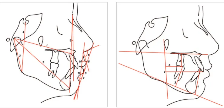
Figure 2 - Skeletal and soft tissue variables: 1) Co-A; 2) Co-Gn; 3) Co-Go; 4) S-Go; 5) NAP; 6) NLA; 7) MLS; 8) UL-E; 9) LL-E; 10) UL-Pog’Sn; 11) LL-Pog’Sn.
Figure 3 - Dentoalveolar variables 1) 1-PM; 2) 1-PTV; 3) 6-PM; 4) 6-PP; 5) 6-PTV.
Statistical analysis
Sex distribution, type of Class II malocclusion (divi-sion 1 or 2; bilateral or unilateral) and number of pa-tients treated by the different clinicians in the groups were compared with Chi-square tests. Application of
t tests requires normal distribution, which was
veri-fied with Kolmogorov-Smirnov tests. Results were not significant for all variables. Therefore, intergroup com-parison of the pretreatment age, treatment time,
pre-treatment (T1) and posttreatment (T2) variables and the
treatment changes (T2-T1) were performed with t tests.
All statistical analyses were performed with Statistica software (Statistica for Windows 6.0; Statsoft, Tulsa,
Okla), and results were considered significant at p < 0.05.
RESULTS
Only the variables 1-PTV and 1-PP showed sys-tematic errors, and the random errors ranged from 0.28 millimeters (MLS) to 2.80 degrees (1.PP).
The groups were comparable regarding sex distribu-tion, types of Class II malocclusion and also regarding the number of patients treated by both clinicians (Ta-ble 1). Only the pre and posttreatment ages were signifi-cantly different between the groups (Table 2).
Methods
Lateral cephalograms were obtained at the pre (T1)
and posttreatment (T2) stages. The anatomic
trac-ing and the location of cephalometric landmarks were manually carried out by a single investigator. These ra-diographs were digitized in grayscale at 300 dpi, in a scanner (Numonics AccuGrid XNT, model A30TL.F (Austin,Texas/USA) and imported into Dentofacial Planner 7.02 software (Toronto, Ontário, Canada); the landmarks were digitized and measurements were per-formed. The magnification factor of the radiographic images, which was between 6% and 9.8%, was cor-rected by the software.
Cephalometric maxillary and mandibular dentoal-veolar variables used are shown in Figures 2 and 3.
Error study
Within a month interval from the first measurement, 15 randomly selected radiographs were re-measured by the same examiner. The random error was calculated
ac-cording to Dahlberg’s formula (Se2= Σd2/2n),18 in which
Se2 is the error variance and d is the difference between
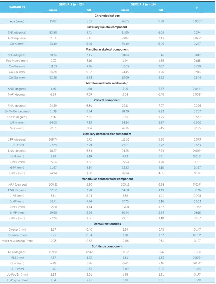
At the pretreatment stage, Group 2 presented significantly greater maxillary retrusion (A-Nperp), mandibular effective and body length (Co-Gn, Go-Gn), and mandibular ramus (Co-Go), when compared to Group 1 (Table 2). Group 1 had a sig-nificantly greater skeletal Class II discrepancy (ANB, and facial convexity, NAP), and a significantly smaller posterior face height (S-Go) than Group 2. Group 2 also presented greater maxillary and mandibular molar extrusion (6-PP, 6-PM) and mesialization (6-PTV, 6-PTV), and showed deeper mentolabial sulcus (MLS) and greater upper (UL-E, UL-Pog’Sn) and lower (LL-E) lip retraction.
At the posttreatment stage, Group 2 showed signifi-cantly greater maxillary retrusion (A-Nperp), smaller apical base discrepancy (ANB) and less convex facial profile (NAP) than Group 1 (Table 3). Group 2 also
presented greater maxillary incisors labial inclination and protrusion (1.NA, 1-NA), greater mandibular inci-sors labial inclination (IMPA), smaller overbite, deeper mentolabial sulcus (MLS) and greater retrusion of the upper lip (UL-E), in relation to Group 1.
The intergroup comparison of treatment changes showed that Group 1 presented significantly greater increase of maxillary (Co-A) and mandibular lengths (Co-Gn, Go-Gn, Co-Go) (Table 4). Group 1 also showed greater increase in lower anterior and poste-rior face heights (LAFH, S-Go), greater palatal tip-ping of the maxillary incisors (1.PP, 1.NA), greater maxillary molar extrusion and mesialization (6-PP, 6-PTV), greater mandibular incisor and molar extru-sion (1-MP, 6-MP), greater mandibular molar me-sialization (6-PTV) and greater upper lip retraction (UL-E), as compared to Group 2.
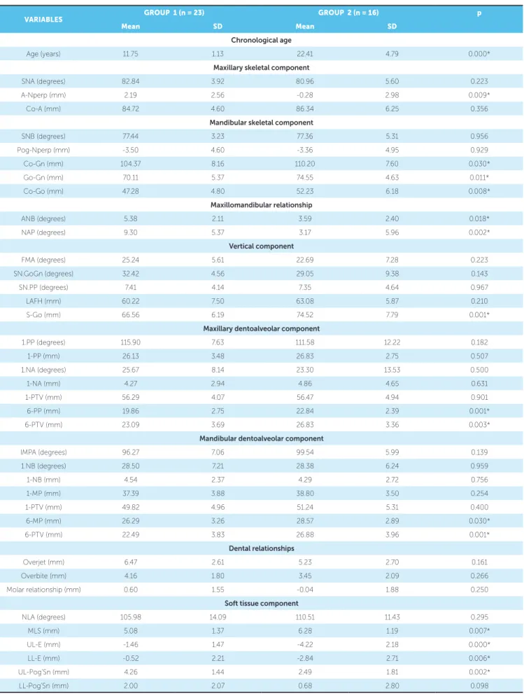
Table 1 - Distribution between the two groups regarding sex, malocclusion type (divisions 1 and 2; bilateral or unilateral) and orthodontist (Chi-square tests).
Group / Sex Female Male Total
1 13 10 23
2 9 7 16
TOTAL 22 17 39
X2 = 0.00
DF= 1 p = 0.986
Group / Division Division 1 Division 2 Total
1 21 2 23
2 12 4 16
TOTAL 33 6 39
X2 = 1.93 DF= 1 p = 0.165
Group Subdivision Bilateral Subdivision Total
1 19 4 23
2 11 5 16
TOTAL 30 9 39
X2 = 1.02 DF= 1 p = 0.312
Group / Orthodontist Orthodontist 1 Orthodontist 2 Total
1 16 7 23
2 11 5 16
TOTAL 27 12 39
Table 2 - Intergroup comparison of pretreatment stage
* Statistically significant for p < 0.05.
VARIABLES GROUP 1 (n = 23) GROUP 2 (n = 16) p
Mean SD Mean SD
Chronological age
Age (years) 11.75 1.13 22.41 4.79 0.000*
Maxillary skeletal component
SNA (degrees) 82.84 3.92 80.96 5.60 0.223
A-Nperp (mm) 2.19 2.56 -0.28 2.98 0.009*
Co-A (mm) 84.72 4.60 86.34 6.25 0.356
Mandibular skeletal component
SNB (degrees) 77.44 3.23 77.36 5.31 0.956
Pog-Nperp (mm) -3.50 4.60 -3.36 4.95 0.929
Co-Gn (mm) 104.37 8.16 110.20 7.60 0.030*
Go-Gn (mm) 70.11 5.37 74.55 4.63 0.011*
Co-Go (mm) 47.28 4.80 52.23 6.18 0.008*
Maxillomandibular relationship
ANB (degrees) 5.38 2.11 3.59 2.40 0.018*
NAP (degrees) 9.30 5.37 3.17 5.96 0.002*
Vertical component
FMA (degrees) 25.24 5.61 22.69 7.28 0.223
SN.GoGn (degrees) 32.42 4.56 29.05 9.38 0.143
SN.PP (degrees) 7.41 4.14 7.35 4.64 0.967
LAFH (mm) 60.22 7.50 63.08 5.87 0.210
S-Go (mm) 66.56 6.19 74.52 7.79 0.001*
Maxillary dentoalveolar component
1.PP (degrees) 115.90 7.63 111.58 12.22 0.182
1-PP (mm) 26.13 3.48 26.83 2.75 0.507
1.NA (degrees) 25.67 8.14 23.30 13.53 0.500
1-NA (mm) 4.27 2.94 4.86 4.65 0.631
1-PTV (mm) 56.29 4.07 56.47 4.94 0.901
6-PP (mm) 19.86 2.75 22.84 2.39 0.001*
6-PTV (mm) 23.09 3.69 26.83 3.36 0.003*
Mandibular dentoalveolar component
IMPA (degrees) 96.27 7.06 99.54 5.99 0.139
1.NB (degrees) 28.50 7.21 28.38 6.24 0.959
1-NB (mm) 4.54 2.37 4.29 2.72 0.756
1-MP (mm) 37.39 3.88 38.80 3.50 0.254
1-PTV (mm) 49.82 4.96 51.24 5.31 0.400
6-MP (mm) 26.29 3.26 28.57 2.89 0.030*
6-PTV (mm) 22.49 3.83 26.88 3.96 0.001*
Dental relationships
Overjet (mm) 6.47 2.61 5.23 2.70 0.161
Overbite (mm) 4.16 1.80 3.45 2.09 0.266
Molar relationship (mm) 0.60 1.55 -0.04 1.88 0.250
Soft tissue component
NLA (degrees) 105.98 14.09 110.51 11.43 0.295
MLS (mm) 5.08 1.37 6.28 1.19 0.007*
UL-E (mm) -1.46 1.47 -4.22 2.18 0.000*
LL-E (mm) -0.52 2.21 -2.84 2.71 0.006*
UL-Pog’Sn (mm) 4.26 1.44 2.49 1.81 0.002*
VARIABLES GROUP 1 (n = 23) GROUP 2 (n = 16) p
Mean SD Mean SD
Chronological age
Age (years) 15.07 1.53 26.65 5.88 0.000*
Maxillary skeletal component
SNA (degrees) 82.80 3.71 81.09 6.05 0.274
A-Nperp (mm) 2.03 2.41 -0.07 3.43 0.029*
Co-A (mm) 88.19 5.26 86.19 6.09 0.277
Mandibular skeletal component
SNB (degrees) 78.34 3.13 78.04 5.14 0.817
Pog-Nperp (mm) -2.32 5.16 -1.94 4.85 0.815
Co-Gn (mm) 111.59 7.76 110.72 7.02 0.720
Go-Gn (mm) 74.28 5.02 74.45 4.76 0.913
Co-Go (mm) 52.28 5.33 53.09 5.52 0.644
Maxillomandibular relationship
ANB (degrees) 4.46 1.68 3.06 2.57 0.044*
NAP (degrees) 6.89 4.59 2.08 6.40 0.009*
Vertical component
FMA (degrees) 24.39 6.39 22.11 7.07 0.296
SN.GoGn (degrees) 31.39 5.84 28.39 8.93 0.207
SN.PP (degrees) 7.66 3.81 6.81 4.75 0.537
LAFH (mm) 64.05 7.83 64.34 5.37 0.900
S-Go (mm) 72.51 7.04 76.16 7.45 0.125
Maxillary dentoalveolar component
1.PP (degrees) 108.74 5.72 112.16 5.80 0.073
1-PP (mm) 27.28 3.79 27.81 2.72 0.633
1.NA (degrees) 18.27 5.54 24.25 7.84 0.007*
1-NA (mm) 2.29 2.19 4.43 3.11 0.015*
1-PTV (mm) 55.54 4.51 55.94 4.70 0.791
6-PP (mm) 21.97 3.14 23.23 2.10 0.167
6-PTV (mm) 24.94 3.80 26.94 4.05 0.119
Mandibular dentoalveolar component
IMPA (degrees) 100.21 5.80 105.19 6.28 0.014*
1.NB (degrees) 32.22 3.70 34.20 4.49 0.136
1-NB (mm) 5.61 2.04 5.55 1.91 0.928
1-MP (mm) 38.41 4.74 37.70 3.16 0.603
1-PTV (mm) 52.88 4.44 53.00 4.57 0.932
6-MP (mm) 29.98 2.96 30.44 2.54 0.618
6-PTV (mm) 27.29 3.86 29.01 4.10 0.187
Dental relationships
Overjet (mm) 2.67 0.40 2.94 0.75 0.147
Overbite (mm) 2.25 0.84 1.38 1.37 0.017*
Molar relationship (mm) -2.35 0.62 -2.06 0.51 0.127
Soft tissue component
NLA (degrees) 109.91 12.42 111.53 11.47 0.680
MLS (mm) 4.57 1.43 5.81 1.33 0.009*
UL-E (mm) -4.02 1.96 -5.46 2.16 0.034*
LL-E (mm) -1.62 2.51 -3.09 2.25 0.065
UL-Pog’Sn (mm) 2.83 2.01 1.98 1.81 0.177
LL-Pog’Sn (mm) 1.64 2.41 0.91 2.05 0.330
Table 3 - Intergroup comparison of posttreatment stage
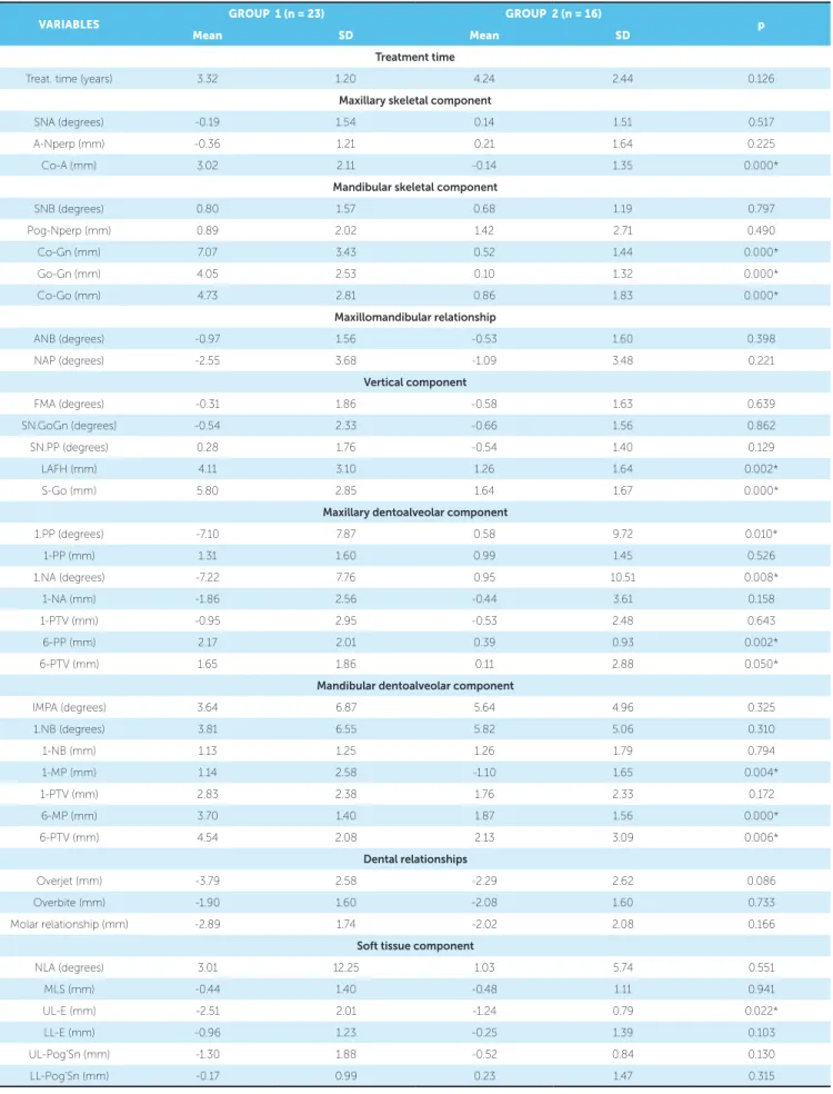
Table 4 - Intergroup comparison of treatment changes.
* Statistically significant for p < 0.05.
VARIABLES GROUP 1 (n = 23) GROUP 2 (n = 16) p
Mean SD Mean SD
Treatment time
Treat. time (years) 3.32 1.20 4.24 2.44 0.126
Maxillary skeletal component
SNA (degrees) -0.19 1.54 0.14 1.51 0.517
A-Nperp (mm) -0.36 1.21 0.21 1.64 0.225
Co-A (mm) 3.02 2.11 -0.14 1.35 0.000*
Mandibular skeletal component
SNB (degrees) 0.80 1.57 0.68 1.19 0.797
Pog-Nperp (mm) 0.89 2.02 1.42 2.71 0.490
Co-Gn (mm) 7.07 3.43 0.52 1.44 0.000*
Go-Gn (mm) 4.05 2.53 0.10 1.32 0.000*
Co-Go (mm) 4.73 2.81 0.86 1.83 0.000*
Maxillomandibular relationship
ANB (degrees) -0.97 1.56 -0.53 1.60 0.398
NAP (degrees) -2.55 3.68 -1.09 3.48 0.221
Vertical component
FMA (degrees) -0.31 1.86 -0.58 1.63 0.639
SN.GoGn (degrees) -0.54 2.33 -0.66 1.56 0.862
SN.PP (degrees) 0.28 1.76 -0.54 1.40 0.129
LAFH (mm) 4.11 3.10 1.26 1.64 0.002*
S-Go (mm) 5.80 2.85 1.64 1.67 0.000*
Maxillary dentoalveolar component
1.PP (degrees) -7.10 7.87 0.58 9.72 0.010*
1-PP (mm) 1.31 1.60 0.99 1.45 0.526
1.NA (degrees) -7.22 7.76 0.95 10.51 0.008*
1-NA (mm) -1.86 2.56 -0.44 3.61 0.158
1-PTV (mm) -0.95 2.95 -0.53 2.48 0.643
6-PP (mm) 2.17 2.01 0.39 0.93 0.002*
6-PTV (mm) 1.65 1.86 0.11 2.88 0.050*
Mandibular dentoalveolar component
IMPA (degrees) 3.64 6.87 5.64 4.96 0.325
1.NB (degrees) 3.81 6.55 5.82 5.06 0.310
1-NB (mm) 1.13 1.25 1.26 1.79 0.794
1-MP (mm) 1.14 2.58 -1.10 1.65 0.004*
1-PTV (mm) 2.83 2.38 1.76 2.33 0.172
6-MP (mm) 3.70 1.40 1.87 1.56 0.000*
6-PTV (mm) 4.54 2.08 2.13 3.09 0.006*
Dental relationships
Overjet (mm) -3.79 2.58 -2.29 2.62 0.086
Overbite (mm) -1.90 1.60 -2.08 1.60 0.733
Molar relationship (mm) -2.89 1.74 -2.02 2.08 0.166
Soft tissue component
NLA (degrees) 3.01 12.25 1.03 5.74 0.551
MLS (mm) -0.44 1.40 -0.48 1.11 0.941
UL-E (mm) -2.51 2.01 -1.24 0.79 0.022*
LL-E (mm) -0.96 1.23 -0.25 1.39 0.103
UL-Pog’Sn (mm) -1.30 1.88 -0.52 0.84 0.130
DISCUSSION
Sample selection and methodology
The lack of a control group that would allow to separate growth changes effects from the MPA ap-pliance effects, especially in Group 1, is a limitation of the present study. However, previous studies also were published with the absence of a control group,
without prejudice of the results.9
The groups were compatible regarding sex distri-bution, malocclusion types (division 1 or 2, and bilat-eral or unilatbilat-eral), and also regarding the orthodontists who conducted the treatments, which can influence the results (Table 1). Only the pre and posttreatment ages were significantly different between the groups, as expected, since the study compares groups with different age ranges (Tables 2 and 4).
Maxillary skeletal component
Adults presented significantly greater maxillary re-trusion (A-Nperp) at the pretreatment stage (Table 2). The effective length of the maxilla significantly in-creased in the adolescent group, which was expected because these patients were still in the growing stage
of craniofacial development20-22 (Co-A; Table 4).
Nev-ertheless, the therapy with the MPA did not seem to have influenced maxillary anterior displacement be-cause the adult group continued to present signifi-cantly greater maxillary retrusion as compared to the adolescent group at the posttreatment stage (Table 3).
Similar results have been previously observed.9,23
Mandibular skeletal component
The adult group presented greater mandibu-lar body (Go-Gn) and effective mandibumandibu-lar length (Co-Gn), as well as mandibular ramus height (Co-Go) at the pretreatment stage, and the adoles-cents had greater increase in these structures during
the treatment period (Tables 2 and 4). The MPA may
have contributed to some of this growth increase.20-22
On the other hand, growth changes in the adult group was negligible so that at the posttreatment stage the sizes of these mandibular structures were similar in the groups (Table 3).
Maxillomandibular relationship
At the pretreatment stage, the adolescent group presented a significantly greater apical base Class II
discrepancy and profile convexity than the adult group (Table 2). The adolescents still presented a significantly greater Class II apical base discrepan-cy and convex facial profile than the adults at the posttreatment stage, although milder than at the pretreatment stage (Tables 3 and 4). Longitudinal comparisons indicate that growth trends are essen-tially similar between Class II division 1 and normal subjects in the various dentofacial parameters com-pared. The differences in mandibular length and po-sition are more evident in the early stages of devel-opment than at the later stages. This may indicate the possibility of a “catch up” period in mandibu-lar growth in Class II division 1 subjects at the later
stages of development.24
Vertical component
Both groups were very similar in the pretreatment stage regarding the growth pattern, with only the adult patients presenting a significantly greater pos-terior face height than the adolescents and therefore a more horizontal growth tendency (Table 2). Howev-er, as the adolescents had significantly greater increase in the lower anterior and posterior face heights, there were no significant differences in any variable of the vertical components at the posttreatment stage (Ta-bles 3 and 4). Most likely these greater increases were consequent to growth in the adolescents because the adult group also experienced slight increases in these variables. These results would be expected, since they
have been shown before.25
Maxillary dentoalveolar component
As it would be expected, in the pretreatment stage the adults had significantly greater maxillary molar dentoalveolar height and mesial positioning than the adolescents, since growth had already been fully
ex-pressed (Table 2).26 During treatment, there was
sig-nificantly greater palatal tipping of the maxillary inci-sors in the adolescents, most likely due to the appli-ance effects, and also greater maxillary molar vertical dentoalveolar development and mesialization, most
probably due to growth.22,26 With these changes, the
Mandibular dentoalveolar component
Similar to the maxillary molars, in the pretreatment stage the adults had significantly greater mandibular molars dentoalveolar height and mesial positioning than the adolescents, because growth had already been fully
expressed (Table 2).22,27 During treatment, the
ado-lescents had greater dentoalveolar development of the mandibular incisors and molars, probably due to the greater vertical development of the alveolar processes on
growing patients, in comparison to adults (Table 4).22
Although the other dentoalveolar changes were not significantly different between the groups, the adult patients had a significantly greater proclination of the mandibular incisors at the posttreatment stage (Table 3). Probably this was the result of a cumulative effect of a greater non-significant mandibular incisor proclination at the pretreatment stage and during treatment that ul-timately produced a significantly greater proclination at the posttreatment stage. Greater dentoalveolar changes may be expected in adult patients under treatment with fixed functional appliances because the skeletal changes
are minimal.7,9 Besides, the adult patients had greater
initial mandibular crowding which could have contrib-uted to some incisor flaring during the initial stages of
leveling and alignment.3,28
Dental relationships
The overjet, overbite and molar relationship were similar in the groups at the pretreatment stage as were the treatment changes (Tables 2 and 4). Only the overbite was significantly smaller in the adult group at the posttreatment stage. Again this result may be the cumulative effect of a non-significantly greater over-bite in the adolescent group in the pretreatment stage, associated to a non-significantly greater reduction in overbite in the adult group.
Soft tissue component
The upper and lower lips were significantly more protruded in the adolescents than in the adults at the pretreatment stage (Table 2), in accordance with
Pecora et al.29 During treatment the upper lip of the
adolescents had significantly greater retraction than in the adult patients (Table 4). However, despite the greater retraction, the upper lip continued to exhibit greater protrusion in the adolescents, in the posttreat-ment period (Table 3).
CLINICAL CONSIDERATIONS
The MPA is indicated in adult patients when they are not willing to cooperate with the use of removable
appliances30 and also when they refuse to go through
extractions or orthognathic surgery. Moreover, the MPA is an affordable option compared to other fixed
devices to correct Class II malocclusions.3,7,10
The amount of skeletal changes was greater in the young group when compared to adult patients, nev-ertheless, it does not mean that the MPA causes more effects on growing subjects when compared to
non-growing patients.9 Probably it was growth, and not
the MPA, the responsible for these differences in the
amount of skeletal changes between the two groups.20
At the posttreatment stage, no differences re-garding mandibular size and position could be seen. This way, MPA has enough potential to warrant its use when indicated even in adult patients, since these dental effects are satisfactory to benefit Class II
mal-occlusion correction.9,31
One has to bear in mind that the results of this study reflect the total treatment period including lev-eling and alignment, active retention and the finish-ing procedures, all performed with fixed appliances. Therefore, it should be emphasized that the chang-es of both groups are the rchang-esults of the joint effects of the MPA and fixed appliances and not the MPA alone. On the other hand, this may be more infor-mative than knowing only the changes produced by the MPA alone, because it provides information of the whole treatment. Patients usually are submitted to complete treatment.
Stability is always a concern with Class II maloc-clusion treatment with fixed functional appliances, especially in adults. Therefore, to increase stability of the anteroposterior correction obtained by the MPA after its removal, Class II elastics were used and their daily use was gradually reduced as stabil-ity was observed in each case. This is a usual active retention alternative employed after using a fixed
functional appliance.10,32 Because use of the MPA
appliance was of 9 months and treatment times were relatively long in both groups, the additional time with Class II elastics use as active retention should, most likely, assure good stability of the
anteropos-terior correction.10,23 Nevertheless, this issue should
CONCLUSIONS
» Adult patients treated with MPA showed significant fewer amounts of skeletal, dentoalveolar and soft tissue changes than adolescents.
» Regarding appliance effects, there was significantly greater palatal tipping of maxillary incisors and retrusion of upper lip in the adolescents. Adults group showed greater mandibular incisor proclination in posttreatment stage.
Authors contributions
Conception or design of the study: BDF. Data acqui-sition, analysis or interpretation: BDF. Writing the arti-cle: BDF, KMSF, LCCC. Critical revision of the artiarti-cle: JFCH, GJ. Final approval of the article: KMSF, JFCH, GJ. Overall responsibility: BDF, KMSF.
1. Harris EF, Vaden JL, Dunn KL, Behrents RG. Effects of patient age on postorthodontic stability in Class II, division 1 malocclusions. Am J Orthod Dentofacial Orthop. 1994 Jan;105(1):25-34.
2. Mathews DP, Kokich VG. Managing treatment for the orthodontic patient with periodontal problems. Semin Orthod. 1997 Mar;3(1):21-38. 3. Gonner U, Ozkan V, Jahn E, Toll DE. Effect of the MARA appliance on
the position of the lower anteriors in children, adolescents and adults with Class II malocclusion. J Orofac Orthop. 2007 Sept;68(5):397-412. 4. Kinzinger G, Diedrich P. Skeletal effects in class II treatment with
the functional mandibular advancer (FMA)? J Orofac Orthop. 2005 Nov;66(6):469-90.
5. Nalbantgil D, Arun T, Sayinsu K, Fulya I. Skeletal, dental and soft-tissue changes induced by the Jasper Jumper appliance in late adolescence. Angle Orthod. 2005 May;75(3):426-36.
6. Paulsen HU, Karle A. Computer tomographic and radiographic changes in the temporomandibular joints of two young adults with occlusal asymmetry, treated with the Herbst appliance. Eur J Orthod. 2000 Dec;22(6):649-56.
7. Ruf S, Pancherz H. Herbst/multibracket appliance treatment of Class II division 1 malocclusions in early and late adulthood. a prospective cephalometric study of consecutively treated subjects. Eur J Orthod. 2006 Aug;28(4):352-60.
8. Ruf S, Pancherz H. Temporomandibular joint remodeling in adolescents and young adults during Herbst treatment: a prospective longitudinal magnetic resonance imaging and cephalometric radiographic investigation. Am J Orthod Dentofacial Orthop. 1999 June;115(6):607-18. 9. Pontes LF, Maia FA, Almeida MR, Flores-Mir C, Normando D. Mandibular
Protraction Appliance Effects in Class II Malocclusion in Children, Adolescents and Young Adults. Braz Dent J. 2017 Mar-Apr;28(2):225-33. 10. Herrera FS, Henriques JF, Janson G, Francisconi MF, de Freitas KM.
Cephalometric evaluation in different phases of Jasper Jumper therapy. Am J Orthod Dentofacial Orthop. 2011 Aug;140(2):e77-84.
11. Furquim BD, Henriques JF, Janson G, Siqueira DF, Furquim LZ. Effects of mandibular protraction appliance associated to fixed appliance in adults. Dental Press J Orthod. 2013 Sept-Oct;18(5):46-52.
12. Johnston LE. If wishes were horses: functional appliances and growth modification. Progress Orthod. 2005;6(1):36-47.
13. Coelho Filho CM. Mandibular protaction appliance for Class II treatment. J Clin Orthod. 1995 May;29(5):319-36.
14. Pancherz H. Treatment of class II malocclusions by jumping the bite with the Herbst appliance. A cephalometric investigation. Am J Orthod. 1979 Oct.;76(4):423-42.
15. Coelho Filho CM. Clinical application of the mandibular protraction appliance in upper lateral agenesy and in asymmetric cases. Tex Dent J. 2002 July;119(7):618-26.
REFERENCES
16. White LW, Coelho Filho CM. Treating Adults with the Mandibular Protraction Appliance. 2003 [Access in: 10 May 2010]. Available from: http://wwwoc-jcom/ jan03/MPA2htm.
17. Stucki N, Ingervall B. The use of the Jasper Jumper for the correction of Class II malocclusion in the young permanent dentition. Eur J Orthod. 1998 June;20(3):271-81.
18. Dahlberg G. Statistical methods for medical and biological students. New York: Interscience; 1940.
19. Houston WJB. The analysis of errors in orthodontic measurements. Am J Orthod. 1983 May;83(5):382-90.
20. Behrents R. Growth in the aging craniofacial skeleton [Monograph No. 17, Craniofacial Growth Series]. Ann Arbor (MI): Center for Human Growth and Development, University of Michigan; 1985.
21. Bishara SE, Jamison JE, Peterson LC, DeKock WH. Longitudinal changes in standing height and mandibular parameters between the ages of 8 and 17 years. Am J Orthod. 1981 Aug;80(2):115-35.
22. Martins DR, Janson G, Henriques JFC, Freitas MR, Almeida RR. Atlas de crescimento craniofacial. São Paulo: Ed. Santos; 1998.
23. Siqueira DF, Almeira RR, Janson G, Brandao AG, Coelho Filho CM. Dentoskeletal and soft-tissue changes with cervical headgear and mandibular protraction appliance therapy in the treatment of Class II malocclusions. Am J Orthod Dentofacial Orthop. 2007 Apr;131(4):447e21-30.
24. Bishara SE, Jakobsen JR, Vorhies B, Bayati P. Changes in dentofacial structures in untreated Class II division 1 and normal subjects: a longitudinal study. Angle Orthod. 1997;67(1):55-66.
25. Stahl F, Baccetti T, Franchi L, McNamara JA, Jr. Longitudinal growth changes in untreated subjects with Class II Division 1 malocclusion. Am J Orthod Dentofacial Orthop. 2008 July;134(1):125-37.
26. Vaden JL, Harris EF, Behrents RG. Adult versus adolescent Class II correction: a comparison. Am J Orthod Dentofacial Orthop. 1995 June;107(6):651-61. 27. Konik M, Pancherz H, Hansen K. The mechanism of Class II correction in late
Herbst treatment. Am J Orthod Dentofacial Orthop. 1997 July;112(1):87-91. 28. Richardson ME. A preliminary report on lower arch crowding in the mature adult.
Eur J Orthod. 1995 June;17(3):251-7.
29. Pecora NG, Baccetti T, McNamara JA Jr. The aging craniofacial complex: a longitudinal cephalometric study from late adolescence to late adulthood. Am J Orthod Dentofacial Orthop. 2008 Oct;134(4):496-505.
30. Faltin KJ, Faltin RM, Baccetti T, Franchi L, Ghiozzi B, McNamara JA Jr. Long-term effectiveness and treatment timing for Bionator therapy. Angle Orthod. 2003 June;73(3):221-30.
31. Coelho Filho CM. Entrevista. Rev Clín Ortod Dental Press. 2003 Out-Nov;2(5):5-11.