Mots-clés: Yarrowia lipolytica, sustratos hidrófobos, gotitas lipídicas, paros celulares, proteínas de la paris celular, DHN-melanina. Investigación de los mecanismos fisiológicos y moleculares que permiten la captación de sustratos hidrofóbicos a través de la pared celular de la levadura Yarrowia lipolytica. En esta etapa, la composición de proteínas, β-glucanos y quitina de la pared celular podría desempeñar un papel importante en la adhesión y absorción de sustratos hidrofóbicos.
También observamos que el perfil espectroscópico de la pared celular del mutante MTYL40-2p se altera según el sustrato hidrofóbico presente en el medio de cultivo. Identificación in silico de los genes putativos de la vía de biosíntesis de DHN-mélanine chez Y.
AVANT PROPOS
INTRODUCTION
Généralités sur la levure Yarrowia lipolytica
- Taxonomie
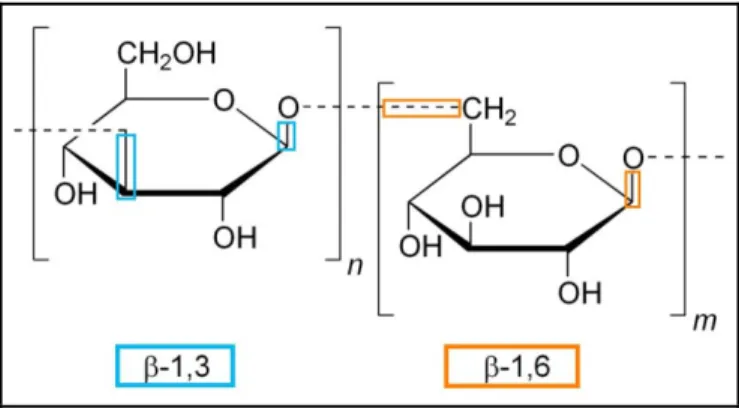
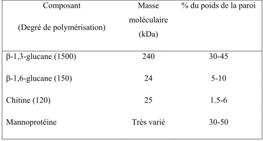
- Composition et organisation de la paroi cellulaire
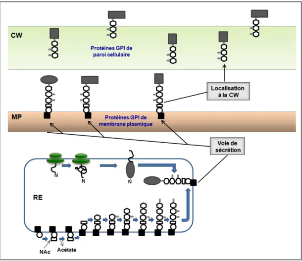
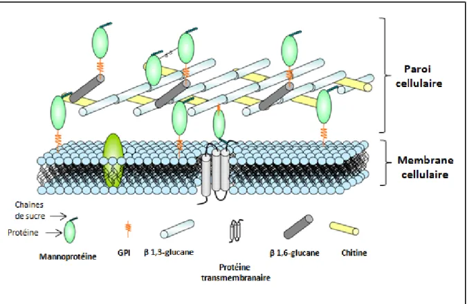
Un classement des principaux composants de la paroi cellulaire (mannoprotéines, β-1,3-glucane, β-1,6-glucane et chitine) et de la membrane est présenté (adapté de Molina et al, 2000). Les mannoprotéines sont des protéines mannosylées liées à la paroi cellulaire de manière non covalente ou covalente. D'autres protéines GPI sont ancrées à la chitine par le β-1,6-glucane, ces protéines peuvent être libérées de la paroi cellulaire par digestion avec des chitinases, des β-1,6-glucanases ou de la (HF)-pyridine (Pitarch et al. al. , 2008).
Ils sont associés à la construction de la paroi cellulaire (Mouyna et al., 2000), au maintien de la stabilité osmotique (Teparić et al., 2004), à la floculation et à la formation de biofilm (Soares 2010), à l'agglutination sexuelle (Lipke et Kurjan, 1992). ) et quelques autres ont des activités enzymatiques, par exemple les phospholipases (Djordjevic, 2010). Les protéines PIR (de l'anglais « Proteins with Internal Repeats ») sont minoritaires dans la paroi cellulaire.
Adaptation de la paroi cellulaire à des conditions externes
Assimilation et bioconversion de substrats hydrophobes par Y. lipolytica
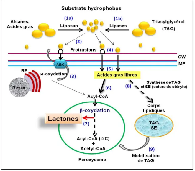
- La prise en charge et le catabolisme des substrats hydrophobes
- Internalisation des substrats hydrophobes
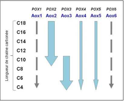
- La β-oxydation chez Yarrowia lipolytica
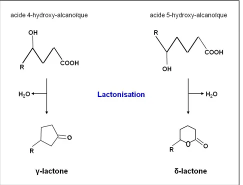
- Les lactones
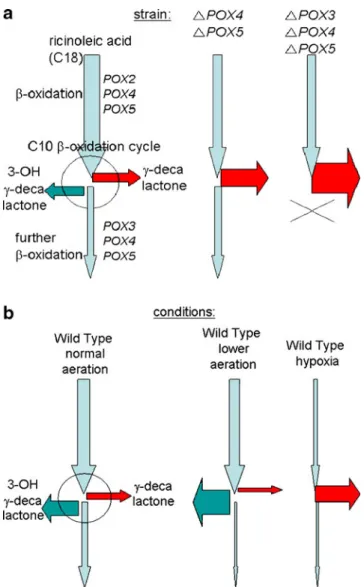
- Biosynthèse et dégradation de lactone par Y. lipolytica : rôle des acyl-CoA
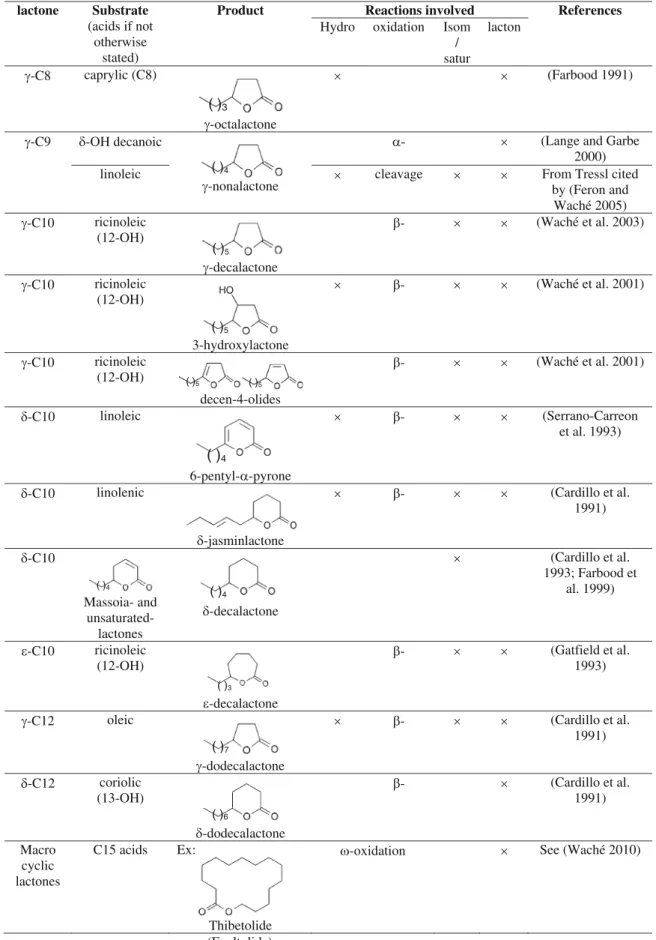
- MINI-REVIEW : Biochemistry of lactone formation in yeast and fungi and
Besides the importance of the different cellular pools, β-oxidation can be channeled, and this is especially the case in the mitochondrial pathway (Eaton et al. 2001). As discussed in Endrizzi et al. 1996), depending on the strain, the lactone or hydroxy acid can be. To this end, Alchihab et al. 2009) have used a psychrophilic strain of the yeast Rhodotorula aurantiaca.
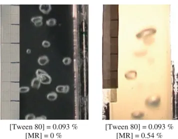
In fact, Gomes et al. 2007) reported that the addition of small amounts of Tween 80 to the medium strongly increases oxygen transfer and thus improves γ-decalactone production by Y. The effect of the hydrophobic phase on kLa can vary according to the range of concentrations (Gomes et al.2007).
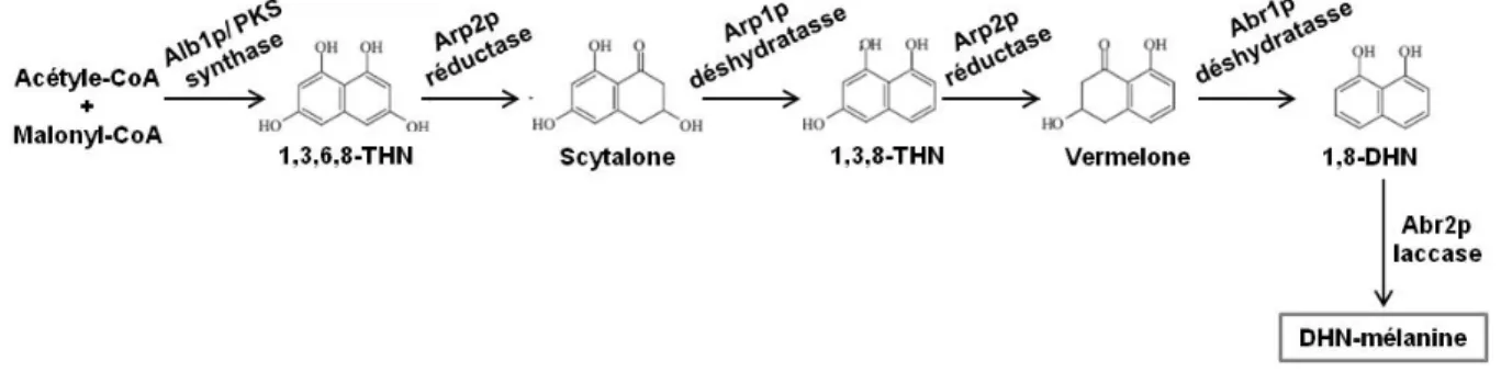
La mélanine chez les champignons
- Voies de synthèse de la mélanine chez les champignons
To go deep into the observation of the cell wall ultrastructure, we performed transmission electron microscopy (TEM) preparations. To get an indication of the cell wall components that can be modified, we performed FTIR spectroscopy. These results indicate a relative decrease in the majority of the cell wall components (proteins and polysaccharides) in cells grown in the presence of methyl oleate.
Although we observed changes in the cell wall by testing the sensitivity of yeast cells to calcofluor white (CFW) and Congo red (CR) dyes. Our results showed that regardless of the composition of the culture medium, cells pre-grown in the presence of methyl oleate were less sensitive to the cell wall weakening effect of CFW. Parts of the investigations were carried out at the Spectral Imaging Platform (PIMS) of the Cell Imaging Facility of Burgundy/Franche Comté (Dimacell).
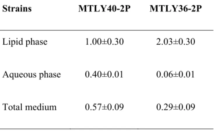
The participation of the proteins of this family is important to maintain the stability of the cell pairs (Sestak et al., 2004). We observed that for MTLY40-2p, 40% of the lactone concentration was present in the aqueous phase compared to the lipid phase. We then monitored the lactone concentration in the lipid and aqueous phases of the medium.
For strain MTLY40-2P, the concentration in the aqueous phase was 40% of the concentration in the lipid phase. Par analyzes in silico nous avons identificado 27 gènes codant des proteines putatives de la paroi cellulaire chez Y. 1997) Physiology and genetics of the dimorphic fungus Yarrowia lipolytica.. 2008) Control of lipid accumulation in the yeast Yarrowia lipolytica. 2009).
OBJECTIF
MATÉRIELS ET MÉTHODES
- Souches de levure
- Milieux de culture et conditions de croissance
- Activation des souches et pré-culture
- Culture des souches
- Microscopie Confocal de Fluorescence et marquage des cellules
- Microscopie Electronique de Transmission (TEM)
- Essais de sensibilité au calcofluor blanc et au rouge Congo
- Test de l’Adhésion Microbienne à l’hexadécane (MATH)
- Cinétique d’adhésion des gouttelettes lipidiques chez les souches W29 et
- Spectroscopie Infrarouge à Transformée de Fourier (FTIR)
- PCR quantitative en Temps Réel
- Extraction et électrophorèse d’ARN
- Traitement à la DNAse
- Contrôle du traitement à la DNase
- Synthèse d’ADNc
- PCR quantitative
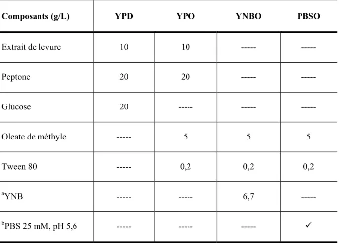
Tandis que pour les analyses de production de mélanine, les souches W29 et MTLY40-2p ont été cultivées dans des milieux de culture contenant différentes huiles comme sources de carbone et de la peptone ou du YNB comme sources d'azote. Les levures issues de la préculture sont récoltées par centrifugation à 6 000 g pendant 5 minutes à 19°C et lavées deux fois à l'eau physiologique. Les cultures y ont été incubées pour la production de lactones et de mélanine.
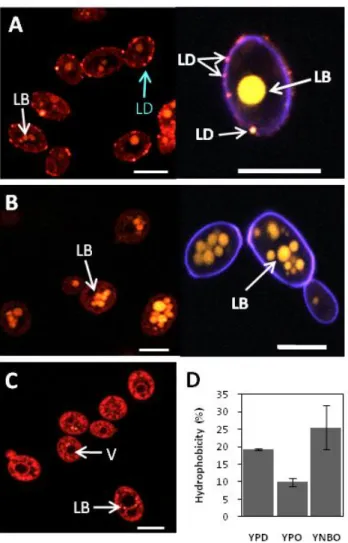
Les sondes utilisées sont : le rouge du Nil, pour le marquage des lipides et le blanc calcofluor, pour le marquage de la paroi cellulaire. Le Calcofluor blanc est un fluorochrome non spécifique qui se lie de manière irréversible à la chitine de la paroi cellulaire fongique. Pour le marquage des lipides, les culots cellulaires ont été remis en suspension dans 998 µl d'eau distillée et 2 µl de rouge de Nil (1 mg/ml) ont été ajoutés à l'abri de la lumière.
Tandis que pour l'observation simultanée des gouttelettes lipidiques et de la paroi cellulaire, nous avons excité le calcolfuor rouge et blanc du Nil à une longueur d'onde de 488 nm. Le test de sensibilité Calcofluor blanc et rouge Congo est un test couramment utilisé pour détecter les changements dans la composition ou la structure de la paroi cellulaire des champignons. Ces colorants anioniques perturbent la construction de la paroi cellulaire en adhérant aux chaînes de chitine naissantes.
Pour analyser la sensibilité de la paroi cellulaire de Yarrowia lipolytica au calcofluor blanc et au rouge Congo, nous avons utilisé des cellules cultivées sur des milieux YPD, YPO et YNBO. Pour déterminer les propriétés d'hydrophobie de la surface cellulaire de la levure cultivée en présence d'oléate de méthyle, nous avons effectué un test d'adhésion microbienne à l'hexadécane (MATH) en utilisant des cellules Y. Les cellules ont été récoltées et remises en suspension au vortex dans 1 ml de TRI®Regeant (SIGMA, Réf : #T9424) et versées dans un microtube contenant environ 800 mg de billes (0,5 mm de diamètre) exemptes de RNase (RF).
RÉSULTATS
Analyse des modifications pariétales et de l’expression des gènes
Analysis of cell wall modifications and CWP genes transcription in
- Résultats complémentaires I
- Cinétiques d’adhésion des gouttelettes lipidiques à la surface cellulaire
- Cinétiques d’expression des gènes de paroi cellulaire spécifiquement induits
- Discussion et conclusion
- Résultats complémentaires II
- Analyses in silico des CWP putatives de Y. lipolytica
Analysis of cell wall modifications and transcription of CWP-encoding genes in Yarrowia lipolytica under hydrophobic growth conditions. Using transmission electron microscopy, no differences were observed at the ultrastructural level of the cell wall between cells grown on glucose and cells grown on methyl oleate. These results suggest that the cell wall is modified in the presence of methyl oleate, but in a different manner depending on the composition of the culture medium.
This result suggests that changes to the cell wall components in response to the presence of methyl oleate make cells more resistant to the cell wall weakening effect of CFW and CR. Magnification of photomicrographs was performed for a better observation of the cell wall (D, E and F). CW) Cell wall; LB) Lipid bodies. This technique has been used to analyze the cell wall in living cells, providing an overview of the main components of the cell wall (Galichet et al., 2001; Adt et al., 2006).
Methyl oleate functional groups (C-H, C=O), amide bands I and II of the protein, and cell wall polysaccharides are shown. A GPI-anchored protein (Gas1/YALI0C06644p) presenting a glycosyl-hydrolase motif is possibly involved in cell wall polymer cross-linking. In contrast, one of the overexpressed gene products in YNBO is the β-1,3-glycosidase YlCrh2p important for cell wall remodeling ( Hwang et al., 2006 ).
Therefore, cell wall biosynthesis, structure, composition and modulation could be interesting tags for biomedical, agricultural and biotechnological improvements. In this area, important advances have been made in the characterization of the cell wall in the conventional yeast Saccharomyces cerevisiae and in the human pathogen Candida albicans. Subsequently, the resistance to CFW and CR of cells precultured in YPO and YNBO results from modifications of the contents of the cell wall components induced by methyl oleate.
A study of yeast cell wall composition and structure in response to growth conditions and mode of cultivation. The cell wall of the human pathogen Candida glabrata: Differential incorporation of novel adhesion-like wall proteins.
Coupled phase distribution of γ-decalactone and melanin, produced
- Résultats complémentaires III
- DISCUSSION GÉNÉRALE
- Modifications de la paroi cellulaire en présence d’oléate de méthyle
- Expression de gènes codant des CWP de Y. lipolytica en présence d’oléate
- Réponse au stress de la paroi cellulaire et biosynthèse de mélanine chez Y
- CONCLUSION
- PERSPECTIVES
- BIBLIOGRAPHIE
Concentration in g/l of γ-decalactone present in different phases of the medium after seven days of culture. The pigment was observed from the third day on the PMO and PMR media on the upper surface of the cultures and in The absorption of the lipid phases produced by the mutant strain was measured after five days of cultivation (Figure II.31).
After four days of culture, the lipid phase of the culture containing Kojic acid was observed to be brown. This result suggests the presence of a compound other than castor oil that could be responsible for the brown pigment in the MTLY40-2p/PCO lipid phase. While in PMR, the cell wall of W29 and MTL40-2p strains showed very similar patterns and concentrations of the surface components.
The O-ester band, the cell wall of the MTLY40-2p strain presented a downshifted band and a new band in the region around 1620 cm-1, very similar to that in PCO medium (Figure II.33D). The results of the kinetics of lactone production can explain both the very high concentration observed in the castor oil phase and the very low concentration in the aqueous phase. So it should be interesting to determine whether the concentration of the oily substrates plays a role in the activation of melanin biosynthesis in Y.
1993) Cloning of the isocitrate lyase (ICL1) gene from Yarrowia lipolytica and characterization of the derived protein.. 1996). Sequential activation of the yeast HOG and SLT2 pathways is required for cell survival to cell wall stress. The two above activation sequences control XPR2 gene expression in the yeast Yarrowia lipolytica. 2003) Molecular evolution of eukaryotic genomes: hemiascomycetous yeast spliceosomal introns. 2004) Characterization of the transcriptional response to cell wall stress in Saccharomyces cerevisiae.. 2006) .
1999 ) Saccharomyces cerevisiae Mid2p is a potential cell wall stress sensor and upstream activator of the PKC1-MPK1 cell integrity pathway. 2003) Modeling mipid accumulation and degradation in Yarrowia lipolytica grown on industrial fats. 2009) Melanin is an essential component for the integrity of the cell wall of Aspergillus fumigatus conidia.