with an Uncoupling of Bone Metabolism in Adult Mice
Yoshiaki Kitaura1,2, Hironori Hojo1,2, Yuske Komiyama1,2, Tsuyoshi Takato1, Ung-il Chung2,3, Shinsuke Ohba2,3*
1Department of Sensory and Motor System Medicine, The University of Tokyo Graduate School of Medicine, Bunkyo-ku, Tokyo, Japan,2Division of Clinical Biotechnology, The University of Tokyo Graduate School of Medicine, Bunkyo-ku, Tokyo, Japan,3Department of Bioengineering, The University of Tokyo Graduate School of Engineering, Bunkyo-ku, Tokyo, Japan
Abstract
Hedgehog (Hh) signaling plays important roles in various development processes. This signaling is necessary for osteoblast formation during endochondral ossification. In contrast to the established roles of Hh signaling in embryonic bone formation, evidence of its roles in adult bone homeostasis is not complete. Here we report the involvement ofGli1, a transcriptional activator induced by Hh signaling activation, in postnatal bone homeostasis under physiological and pathological conditions. Skeletal analyses ofGli1+/2adult mice revealed thatGli1haploinsufficiency caused decreased bone mass with reduced bone formation and accelerated bone resorption, suggesting an uncoupling of bone metabolism. Hh-mediated osteoblast differentiation was largely impaired in cultures ofGli1+/2precursors, and the impairment was rescued by Gli1 expression via adenoviral transduction. In addition, Gli1+/2 precursors showed premature differentiation into osteocytes and increased ability to support osteoclastogenesis. When we compared fracture healing between wild-type and Gli1+/2adult mice, we found that theGli1+/2mice exhibited impaired fracture healing with insufficient soft callus formation. These data suggest thatGli1, acting downstream of Hh signaling, contributes to adult bone metabolism, in which this molecule not only promotes osteoblast differentiation but also represses osteoblast maturation toward osteocytes to maintain normal bone homeostasis.
Citation:Kitaura Y, Hojo H, Komiyama Y, Takato T, Chung U-i, et al. (2014)Gli1Haploinsufficiency Leads to Decreased Bone Mass with an Uncoupling of Bone Metabolism in Adult Mice. PLoS ONE 9(10): e109597. doi:10.1371/journal.pone.0109597
Editor:Pierre J. Marie, Inserm U606 and University Paris Diderot, France
ReceivedJune 2, 2014;AcceptedSeptember 8, 2014;PublishedOctober 14, 2014
Copyright:ß2014 Kitaura et al. This is an open-access article distributed under the terms of the Creative Commons Attribution License, which permits unrestricted use, distribution, and reproduction in any medium, provided the original author and source are credited.
Data Availability:The authors confirm that all data underlying the findings are fully available without restriction. All relevant data are within the paper and its Supporting Information files.
Funding:This work was supported by a Grant-in-Aid for Young Scientists (#23689079), the Center for Medical System Innovation, the Graduate Program for Leaders in Life Innovation, Core-to-Core Program A (Advanced Research Networks), the Funding Program for World-Leading Innovative R&D on Science and Technology, the Center for NanoBio Integration, and the S-innovation Program. The funders had no role in study design, data collection and analysis, decision to publish, or preparation of the manuscript.
Competing Interests:The authors have declared that no competing interests exist. * Email: [email protected]
Introduction
Hedgehog (Hh) signaling is a highly conserved pathway that plays important roles in various development processes. Hh signaling is indispensable for osteoblast formation in endochondral ossification, one of the two ossification processes in mammals, which forms bones in limbs, the trunk, and some head structures [1]. In this context, Indian hedgehog (Ihh) expressed in prehypertrophic chondrocytes is thought to act directly on progenitors in the perichondrium and the bone marrow to induce their differentiation into bone-forming osteoblasts [2–4]. The deletion of Ihh or smoothened (Smo), a transmembrane signaling transducer for Hh, caused no bone collar or primary spongiosa in mice [2,4]. These mutant mice lacked the expression of runt-related transcription factor 2 (Runx2), a key determinant for osteoblasts [5,6] and bone gamma-carboxyglutamate (gra) protein (osteocalcin; official gene symbol,Bglap), a bona fide marker for osteoblasts [1], in the perichondrium.
Gli transcription factors mediate the transcription of target genes downstream of Hh signaling. Gli1, a target gene of Hh signaling, acts as a transcriptional activator, whereas Gli2 and Gli3
can act as both activators and repressors [7]. The activator function of Gli2 and the repressor function of Gli3 were reported to mediate substantial aspects of the action of Ihh on bone development [8,9]. In addition, we found that Gli1 participated in the Hh-mediated osteoblast formation collectively with Gli2 and Gli3 [10,11].
established roles of Hh signaling in embryonic bone formation, the evidence of its roles in adult bone homeostasis is not adequate. In particular, little is known about the roles of Gli1, a potent transcriptional activator induced by Hh signaling, in postnatal bone metabolism.
In the present study, we attempted to examine Hh signaling in the postnatal skeletal system, focusing on the functions of Gli1 in the bone metabolism. We analyzed skeletal phenotypes of Gli1 heterozygous knockout mice postnatallyin vivoand in vitro. We also compared fracture healing between mutant and wild-type (WT) mice. We report thatGli1 haploinsufficiency affects adult bone metabolism, at least partly through the uncoupling of bone formation and bone resorption, leading to a decrease in bone mass and a delay in fracture healing in postnatal skeletons.
Materials and Methods
Animal Experiments
Wild-type C57BL/6J mice were obtained from Charles River Japan, andGli1+/2mice were generated as previously described [15]. All experiments were performed in accord with the protocol approved by the Animal Care and Use Committee of The University of Tokyo (#KA13-5). Mice were kept in individual cages under controlled temperature and humidity with a 12-hr circadian rhythm. They were givenad libitumaccess to food and water. All efforts were made to minimize the suffering of the mice. Euthanasia of mice was performed with an overdose of barbiturates.
Reagents and Vectors
Smoothened agonist (SAG) was purchased from Calbiochem (San Diego, CA; 566660). Receptor activator of NF-kappa-B ligand (RANKL) was purchased from Pepro Tech (Rocky Hill, NJ; 184-01791). Sonic hedgehog (Shh) was purchased from R&D Systems (Minneapolis, MN; 1845-SH). Cyclopamine was pur-chased from Enzo Life Sciences (Farmingdale, NY; BML-GR3334). Plasmids expressing humanGLI1were constructed as previously described [12]. The adenoviral vector expressing humanGLI1-Biotin-3xFLAG-IRES-dsRedwas constructed using the pAd/PL-DEST vector and ViraPower Adnoviral Expression System (Life Technologies, Carlsbad, CA), according to the manufacturer’s instructions. In brief, humanGLI1cDNA carrying Biotin-3xFLAG tag [16] was initially cloned into pCID vectors [17];GLI1-Biotin-3xFLAG-IRES-dsRedwas then transferred into the pENTR1A vector in conjunction with the CAGGS promoter and subjected to adenoviral vector construction using the Gateway system. The pAd/PL-DEST expressing the CAGGS promoter-drivenGLI1-Biotin-3xFLAG-IRES-dsRedwas linearized with Pac I and transfected into 293A cells. After amplification, the virus was stored at280uC. The viral titer was determined by an end-point titer assay using 293A cells.
Cell Culture
C3H10T1/2 cells were obtained from the RIKEN Cell Bank (Ibaraki, Japan). Mouse primary bone marrow stromal cells were isolated from the long bones of 8-week-old male mice. Mouse primary osteoblast precursors were isolated from calvarias of mouse neonates. Cells were cultured in high-glucose Dulbecco’s modified Eagle Medium (DMEM; Sigma-Aldrich, St. Louis, MO) containing 10% fetal bovine serum (FBS) and 1% penicillin/ streptomycin. For osteogenic cultures, the cells were cultured in osteogenic media [18,19] supplemented with Smoothened agonist (SAG). Co-culture experiments using mouse primary bone marrow stromal cells and bone marrow macrophages were performed as
previously described [18]. Thein vitroosteoclast differentiation of RAW264.7 cells was performed as previously described [20]. Adenoviruses expressingGLI1-Biotin-3xFLAG-IRES-dsRed (Ax-Gli1) or GFP (Ax-GFP) were used to infect cells at MOI (multiplicity of infection) 10. Alkaline phosphatase (ALP), von Kossa, and tartrate-resistant acid phosphatase (TRAP) staining were performed as previously described [12,18].
Real-Time RT-PCR
Total RNA extraction and real-time reverse transcription-polymerase chain reaction (RT-PCR) were performed as previ-ously described [21]. All reactions were run in triplicate. The primer sequences are as follows:b-actin, AGATGTGGATCAG-CAAGCAG (forward) and GCGCAAGTTAGGTTTTGTCA (reverse); Alp, GCTGATCATTCCCACGTTTT (forward) and
CTGGGCCTGGTAGTTGTTGT (reverse);Ibsp,
CAGAGGA-GGCAAGCGTCACT (forward) and
CTGTCTGGGTGCCAA-CACTG (reverse); Bglap, AAGCAGGAGGGCAATAAGGT
(forward) and TTTGTAGGCGGTCTTCAAGC (reverse);Gli1, GCACCACATCAACAGTGAGC (forward) and
GCGTCTT-GAGGTTTTCAAGG (reverse); Gli2,
CTGAAGGATTCCTG-CTCGTG (forward) and ACAGTGTAGGCCGAGCTCAT (re-verse);Runx2, CCGCACGACAACCGCACCAT (forward) and
CGCTCCGGCCCACAAATCTC (reverse);Sp7,
ACTCATCC-CTATGGCTCGTG (forward) and
GGTAGGGAGCTGGGT-TAAGG (reverse); Tnfsf11, AGCCATTTGCACACCTCAC
(forward) and CGTGGTACCAAGAGGACAGAGT (reverse); Tbfrsf11b, GTTTCCCGAGGACCACAAT (forward) and
CCA-TTCAATGATGTCCAGGAG (reverse); Dmp1,
CAGTGAG-GATGAGGCAGACA (forward) and
TCGATCGCTCCTGG-TACTCT (reverse); Sost, AAGCCGGTCACCGAGTTGGT
(forward) and GTGAGGCGCTTGCACTTGCA (reverse);
Ptch1, CTGGACTCTGGCTCCTTGTC (forward) and CAA-CAGTCACCGAAGCAGAA (reverse);Ctsk, ACGGAGGCATC-GACTCTGAA (forward) and GATGCCAAGCTTGCGTC-GAT (reverse); and Nfatc1, CCTTCGGAAGGGTGCCTTTT (forward) and AGGCGTGGGGCCTCAGCAGG (reverse).
Luciferase Assay
Cells were plated onto 24-well plates and transfected with 0.4mg of DNA in a mixture containing the reporter plasmids (8639-Gli BS-luc) [22], the control reporter plasmids encoding Renilla luciferase, and effector plasmids (pCMV-myc-GLI1) or adenoviral vectors expressingGLI1. A dual-luciferase assay was performed as previously described [12].
Immunoblot
Whole-cell lysates were isolated using RIPA buffer as previously described [23]. Sodium dodecyl sulfate-polyacrylamide gel elec-trophoresis (SDS-PAGE) and immunoblotting were performed using anti-Gli1 (sc-20687, 1:1000, Santa Cruz Biotechnology, Santa Cruz, CA) and HRP-conjugated anti-FLAG M2 antibodies (A8592-2MG, 1:500, Sigma-Aldrich) as previously described [12].
Radiological analysis
X-ray photographs of the left tibiae of WT andGli1+/2 mice
three-dimensional construction software package TRI/3D-BON (Ratoc System Engineering, Tokyo) was used for quantitative analysis.
Histological analysis
We intraperitoneally injected calcein (0.16 mg per 10 g of body weight; Sigma) into mice 4 days and 1 day before sacrifice. We stained the undecalcified sections of the femur with toluidine blue, von Kossa, and TRAP as previously described [12]. Images were taken using an Axio Imager A1 (Carl Zeiss, Jena, Germany) and processed using AxioVision (Carl Zeiss). Histomorphometric analyses were performed using the HistometryRT CAMERA system. We analyzed five mice for each group.
Fracture model
Ten 8-week-old male mice were used in each group. Under general anesthesia with isofluorane in O2, the left hind limb was
shaved and sterilized for surgery. A 15-mm incision was made longitudinally, and a blunt dissection of the muscle was made to expose the tibia. A transverse osteotomy was performed using disk-shaped dental steel bars at the mid point of the tibia. The fracture was repositioned, and then the full-length of the bone marrow cavity was internally stabilized as previously described [24]. After irrigation with saline, the skin was closed with 4-0 nylon sutures. Fourteen days after surgery, the tibias were harvested from the euthanized mice.
Statistical analysis
The means of groups were compared by an analysis of variance (ANOVA), and the significances of the differences were deter-mined by Student’s t-test. P-values ,0.05 were considered significant.
Results
Gli1 haploinsufficiency causes decreased bone mass in adult mice
As reported, we found that Gli12/2 mice had no gross
abnormalities at birth [11,15]. AlthoughGli12/2pups were born
in Mendelian ratios, the number ofGli12/2adults turned out to
be approximately 10% of the total pups obtained because of their reduced survival rates during the first 10 days after birth (Table 1). In addition, the growth rate of the surviving Gli12/2 mice was
significantly lower than those of both the wild-type (WT) or Gli1+/2 mice, and the body weights of the 8-week-old Gli12/2
mice were approximately 20% lower than those of the WT and Gli1+/2mice (Figure S1).
These findings suggest that the Gli12/2 mice were largely
affected by systemic abnormalities due to complete loss of Gli1 gene. Therefore, to investigate the roles of Gli1 in adult bone
metabolism as directly as possible, we analyzed the skeletal system inGli1+/2 male mice, which had weights and gross appearance
comparable to those of the WT male mice. Micro-computed tomography (m-CT) analyses of distal femurs revealed that the trabecular density of the 8-week-oldGli1+/2 mice was reduced
compared to that of WT mice (Figure 1A). The bone morpho-metric analysis using the micro-CT data supported the finding, as theGli1+/2mice showed less bone mineral density (BMD) along
with decreased parameters for bone formation (Figure 1B). These skeletal phenotypes were also observed in theGli1+/2female mice (Figure S2). In contrast, there was no significant difference in the cortical bone between WT and Gli1+/2 mice (Figure S3), suggesting differences between trabecular and cortical bones with regard to the contribution of Gli1.
The histological analyses of the distal femurs of 8-week-old mice showed that Gli1+/2 mice had reduced trabecular bones compared to the WT mice (Figure 2A, see von Kossa), whereas the growth plate appeared normal (Figure 2A, see toluidine blue). Indeed, the bone volume/tissue volume (BV/TV) and trabecular thickness (Tb.Th) values were significantly decreased inGli1+/2
mice compared to WT mice, as observed in micro-CT-based analyses (Figure 2B). Gli1+/2 mice also had significantly lower
values of osteoid surface/bone surface (OS/BS) and single-labeled surface/bone surface (sLS/BS), which are parameters of the osteogenic capacity (Figure 2B).
Regarding bone resorption, the numbers of TRAP-positive osteoclasts were significantly higher in theGli1+/2mice compared
to the WT mice (Figure 2C and D, see N.Oc/B.Pm). Consistent with this finding, the Gli1+/2 mice showed a trend toward
increased bone resorption capacity parameters, compared to WT mice (Figure 2D, see ES/BS and Oc.S/BS). These data suggest that Gli1 haploinsufficiency causes an uncoupling of bone turnover in adult mice, which leads to decreased bone mass.
Osteoblast differentiation is impaired byGli1 haploinsufficiency
To investigate whether Gli1 haploinsufficiency caused de-creased osteogenic capacity through the impairment of osteoblast differentiation from precursors, we examined osteoblast differen-tiation inex vivocultures of primary bone marrow stromal cells (BMSCs) harvested from 8-week-old WT and Gli1+/2 mice
(Figure 3A and B) and primary osteoblast precursors (OPs) from neonates of each genotype (Figure 3C and D). Given thatGli1 expression is induced upon Hh signaling, we suspected that any difference resulting from the loss of one allele of Gli1would be observed under Hh signaling-activated conditions. We therefore performed the experiments in the presence of Smoothened agonist (SAG), a small molecule that activates Hh signaling. The mRNA expression levels of osteoblast-related genes were lower inGli1+/2
Table 1.Survival rate ofGli1mutant mice during postnatal 10 days.
Genotype Postnatal day
1 10
Gli1+/+ 11 (30.6) 37 (28.5)
Gli1+/2 16 (44.4) 81 (62.3)
Gli12/2 9 (25.0) 12 (9.2)
Total 36 130
BMSCs than in WT BMSCs. In particular, alkaline phosphatase (Alp) and integrin-binding sialoprotein (Ibsp) expression levels were significantly reduced in the Gli1+/2 BMSCs (Figure 3A), which was consistent with our previous finding that Gli1 directly induced the expression of these two genes [11]. ALP activity and matrix calcification, key features of osteoblasts, were also suppressed in theGli1+/2BMSCs as evidenced by ALP and von Kossa staining (Figure 3B). The impaired osteoblast differentiation due to loss of one allele ofGli1was more prominent in the OPs. All of the genes tested here showed significantly lower expression levels in the Gli1+/2OPs compared to WT (Figure. 3C), and ALP activity and calcification were impaired in the Gli1+/2 OPs (Figure 3D). Lastly,Gli1was reduced via the loss of one allele ofGli1in both cell types, although a significant reduction was observed only in the OPs (Figure 3A and C, seeGli1).
The data above suggest that Gli1 haploinsufficiency affects osteoblast differentiation in a cell-autonomous manner. We then attempted to further verify the hypothesis by testing whether the recovery of Gli1expression would rescue the osteoblast pheno-types in Gli1+/2 cells. We prepared an adenoviral vector
expressing Gli1 (Ax-Gli1) (Figure S4A). Gli1 protein expression induced by Ax-Gli1 and its function were confirmed by western blotting using a specific antibody, luciferase reporter assays using the Gli-responsive element, and mRNA expression analyses ofAlp and Ibsp in C3H10T1/2 cells (Figures S4B–D). As shown in Figure 3E, the introduction ofGli1intoGli1+/2OPs induced the expression of Alp, an early osteoblast marker, at a level comparable to that in WT OPs. The expression of Bglap, an osteoblast marker that is expressed at a later stage, was also upregulated but not fully recovered byGli1 introduction. Thus, Gli1 haploinsufficiency in osteoblast precursors is likely to cause
impairment of their differentiation, which underlies reduced bone formation inGli1+/2mice.
The Hh-Gli1 axis is involved in the capacity of osteoblasts/osteocytes to support osteoclastogenesis, but not in osteoclastogenesis itself
Bone homeostasis is achieved by the coupling of bone formation and bone resorption via cross-talk between osteoblasts/osteocytes and osteoclasts. Mature osteoblasts and osteocytes are known to support osteoclastogenesis by expressing the receptor activator of NF-kappa-B ligand (RANKL; official symbol, Tnfsf11 - tumor necrosis factor (ligand) superfamily, member 11) and osteoprote-gerin (OPG; official symbol, Tnfrsf11b - tumor necrosis factor receptor superfamily, member 11b), a stimulator and an inhibitor of osteoclastogenesis, respectively [25–28]. However, Gli1+/2
mice exhibited impaired bone formation and accelerated bone resorption, suggesting an uncoupling state of bone metabolism.
To identify the cellular mechanism underlying the aberrant state inGli1+/2bones, we conducted co-culture experiments using BMSC and bone marrow macrophages (BMMW) derived from either WT or Gli1+/2 mice. The number of TRAP-positive
multinucleated osteoclasts co-cultured withGli1+/2BMMCs was
significantly higher than those with WT BMMCs, regardless of the Gli1 genotypes in the BMMW (Figure 4A and B). Thus, bone-forming cells and their progenitors, not osteoclastic cells, were likely to be responsible for the abnormalities in osteoclastogenesis of theGli1+/2 mice. This led us to analyze BMSCs in terms of
their ability to support the osteoclastogenesis.
In BMSCs harvested from the femurs of 8-week-old Gli1+/2
mice, the mRNA expression of RANKL (Tnfsf11) was signifi-cantly upregulated, whereas that of OPG (Tnfrsf11b) showed a trend toward downregulation compared to WT BMSCs
(Fig-Figure 1. Radiological findings of long bones in wild-type (WT) andGli1+/2mice.(A) Three-dimensional micro-computed tomography (3D-micro-CT) images of the distal femurs of representative 8-week-old WT andGli1+/2 male mice. Sagittal sections, transverse sections, and 3D reconstruction images of the primary spongiosa are shown for each genotype. Bar, 1 mm. (B) Histomorphometric analyses of 3D-micro-CT data. BMD, bone mineral density; BV/TV, bone volume per tissue volume; Tb.Th, trabecular thickness; Tb.N trabecular number parameters. Data are means6SDs of eight male mice per genotype. *p,0.05 vs. WT.
ure 4C). In addition, expression levels of dentin matrix acidic phosphoprotein 1 (Dmp1) and sclerostin (Sost), markers for osteocytes, were higher in theGli1+/2 BMSCs than in the WT
(Figure 4C). This trend was also observed in OPs (Figure S5). Taken together with the downregulation of osteoblast marker genes in Gli1+/2 BMSCs, these findings indicate that Gli1
haploinsufficiency may promote a premature differentiation of osteoblast precursors into osteocytes, which have been reported as a major source of RANKL [25], and these results may indicate that the premature differentiation not only affects bone formation, but also stimulates bone resorption by inducing RANKL expression at a supraphysiological level. Given that the recovery of Gli1expression negated the upregulation of RANKL mRNA expression inGli1+/2 BMSCs (Figure S6), it is also possible that
Gli1not only suppresses the differentiation of osteoblast precursors into osteocytes, but also negatively acts on the transcription of RANKL.
We next investigated the cell-autonomous effects of Hh signaling on osteoclastogenesis using RAW264.7 cells, which have been shown to differentiate into TRAP-positive osteoclasts in the presence of RANKL [20,29]. Treatment with neither Shh nor cyclopamine, an inhibitor of hedgehog signaling, affected the RANKL-induced osteoclast differentiation of RAW264.7 cells (Figures 4D and E) although the cells were responsive to Hh signaling, as indicated by the expression change of Ptch1, a readout of the Hh signaling, upon Shh and cyclopamine (Figure 4F). Finally, we analyzed osteoclastogenesis of primary BMMW harvested from 8-week-old WT or Gli1+/2 mice. Figure 2. Histological findings of adult WT andGli1+/2mice.(A) von Kossa staining and toluidine blue staining of the distal femur sections of representative 8-week-old WT andGli1+/2
male mice. Bar, 100mm. (B) Histomorphometric analyses of bone volume and bone formation parameters
in distal femurs from 8-week-old WT andGli1+/2male mice. OS/BS, osteoid surface per bone surface; MAR, mineral apposition rate; BFR, bone formation rate per bone surface; dLS/BS, double-labeled surface per bone surface; sLS/BS, single-labeled surface per bone surface. Data are means6 SDs of five male mice per genotype. *p,0.05 vs. WT. (C) TRAP staining of the distal femur sections of representative 8-week-old WT andGli1+/2male mice. Bar, 100mm. (D) Histomorphometric analyses of bone resorption parameters in the distal femurs of 8-week-old WT and Gli1+/2male mice. ES/ BS, eroded surface per bone surface; N. Oc/B. Pm, number of osteoclasts per 100 mm of bone perimeter; Oc. S/BS, osteoclast surface per bone surface. In (B) and (D), data are means6SDs of five mice per genotype. *p,0.05 vs. WT.
Expression levels of cathepsin K (Ctsk) and Nfatc1, markers for osteoclasts, were not affected by loss ofGli1in cultures of BMMW with M-CSF and RANKL (Figure 4G).
Thus, the Hh-Gli1 axis is unlikely to mediate osteoclastogenesis in a cell-autonomous manner, which further suggests that the accelerated bone resorption in Gli1+/2 mice is not caused by
defects in osteoclastic cells. Abnormalities in osteoblast precursors due toGli1haploinsufficiency may mediate all the aspects of the
disruption of bone homeostasis inGli1+/2bones, which explains
whyGli1+/2mice have a low bone mass phenotype.
Fracture healing was impaired byGli1haploinsufficiency
We next set out to examine the involvement ofGli1in postnatal bones under a pathological condition, comparing fracture healing between WT and Gli1+/2 mice in a model that was surgically created in the tibias of 8-week-old males. During the fracture
Figure 3. Osteoblast differentiation in cultures of precursor cells from WT andGli1+/2mice.(A) mRNA expression of osteoblast marker genes in 14-day osteogenic cultures of BMSCs in the presence of the Smoothened agonist. mRNA expression was analyzed by real-time RT-PCR. (B) ALP and von Kossa stainings in 14-day osteogenic cultures of BMSCs in the presence of the Smoothened agonist. (C) mRNA expression of osteoblast marker genes in 7-day osteogenic cultures of osteoblast precursors (OPs) isolated from neonatal calvarias, in the presence of the Smoothened agonist. (D) ALP and von Kossa staining in 7-day osteogenic cultures of OPs in the presence of the Smoothened agonist. (E) Rescue of the expression levels of osteoblast marker genes inGli1+/2OPs by the adenoviral overexpression ofGli1in the presence of the Smoothened agonist. WT orGli1+/2 OPs were infected with Ax-GFP (2) or Ax-Gli1 (+) at MOI 10.
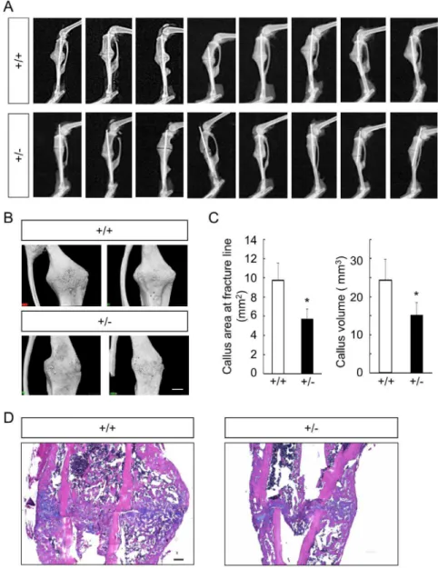
healing process, both intramembranous ossification and endo-chondral ossification are observed, and osteo-chondroprogenitor cells from the periosteum adjacent to fracture sites are major source of cells that contribute to the healing process [30,31]. Fracture healing was evaluated 2 weeks after the surgery, given that bony bridging at the fracture site was typically observed as early as the point [30,31]. In soft X-ray analyses, we observed impairment of callus formation and bone union inGli1+/2mice
compared to WT mice, as well as the variability and reproduc-ibility of the fracture model itself (Figure 5A). Using 3D-micro-CT
analyses, we evaluated the areas of the calluses on horizontal cross-sections at the fracture lines and the volume of the callus. Both the areas and the volumes of the callus in the Gli1+/2 mice were
significantly lower than those in WT mice (Figures 5B and C). We then performed histological analyses on sections stained with the hematoxylin-eosin (H–E) and alcian blue to identify differences in the healing process between WT andGli1+/2mice
(Figure 5D). In WT mice, large volumes of soft callus surrounded by hard calluses were observed around the fracture sites, as previously reported [32] (Figure 5D). However, inGli1+/2mice, Figure 4. Osteoclast differentiation in cultures of bone marrow cells from WT and Gli1+/2 mice. (A) Formation of TRAP-positive multinucleated osteoclasts by the co-culture of BMSCs and BMMWderived from WT orGli1+/2mice. (B) The numbers of osteoclasts expressed as means6SDs of 4 wells per group in (A). *p,0.05 vs. the control group (WT BMSC6WT BMMW). (C) mRNA expression ofRankl,Opg,Dmp1, andSost
in 14-day osteogenic cultures of BMSCs. The mRNA expression was analyzed by real-time RT-PCR.Rankl, receptor activator of nuclear factor-kB ligand;
Opg, osteoprotegerin;Dmp1, dentin matrix acidic phosphoprotein 1;Sost, sclerostin. *p,0.05 vs. WT. (D) Formation of TRAP-positive multinucleated osteoclasts in 5-day cultures of RAW cells in the presence or absence of sonic hedgehog (Shh) and cyclopamine (Cyc). (E) The numbers of osteoclasts in (D) expressed as means6SDs of 4 wells per group. (F) mRNA expression ofPtch1in cultured RAW cells in the presence or absence of Shh and cyclopamine (Cyc). Data are means6SDs of 4 wells per group. (G) mRNA expression of cathepsin K (Ctsk) andNFATc1in osteoclasts derived from WT orGli1+/2BMM
W. Cells were cultured with recombinant M-CSF (10 ng/mL), RANKL (100 ng/mL), and Shh (25 nM). The mRNA expression was analyzed by real-time RT-PCR.
both the soft and hard calluses were reduced compared to those in the WT mice (Figure 5D), suggesting that both endochondral ossification and intramembranous ossification were affected by Gli1haploinsufficiency during the fracture healing process.
Discussion
The present study had six major findings. (1) Adult bone mass was affected by Gli1 haploinsufficiency in mice, although body length and body weight were not. (2) The low bone mass phenotypes were accompanied by impaired bone formation and accelerated bone resorption, that is, an uncoupling of bone metabolism. (3)Gli1haploinsufficiency had a negative impact on Hh-mediated osteoblast differentiation in cultures of precursors. (4) Despite the impairment of osteoblast differentiation, the expression levels ofDmp1andRanklwere upregulated in cultures of Gli1+/2 precursors, suggesting that Gli1 haploinsufficiency
induced the premature differentiation of osteoblasts into osteo-cytes, which have a greater ability to promote osteoclastogenesis than do osteoblasts. (5) Hh-Gli1 was not involved in osteoclasto-genesis in a cell-autonomous manner in vitro. (6) Gli1 haploin-sufficiency affected fracture healing in adult mice. Based on these findings, we propose that Gli1, acting downstream of Hh signaling, not only promotes osteoblast differentiation but also acts as a repressor of osteoblast maturation toward osteocytes to maintain normal bone homeostasis in adult mice.
There are two possible mechanisms underlying the aberrant upregulation of osteocyte marker genes inGli1+/2precursors. The
first possibility is that the overall differentiation of osteoblast precursors is accelerated byGli1haploinsufficiency, although the differentiation program is kept normal. The second possibility is that the program itself is disturbed byGli1haploinsufficiency, and the disturbance may induce the premature differentiation of precursors into osteocytes by skipping proper phases of osteoblast differentiation. The second possibility is more likely, because osteocyte marker genes (Dmp1and Sost) were highly induced in Gli1+/2 cells, despite downregulation of both early and late
osteoblast marker genes (Alp,Ibsp,Runx2,Sp7, andBglap). If the first possibility was the case, Gli1+/2 precursors would show
upregulation of both osteoblast and osteocyte marker genes. It remains to be elucidated howGli1haploinsufficiency disturbs the osteoblast differentiation program, which results in accelerated bone resorption as well as decreased bone formation, i.e., an uncoupling of bone metabolism in development.Gli1, collectively with Gli2 and Gli3, is involved in the specification of osteo-chondroprogenitor cells in the perichondrium into an osteoblast lineage, and it promotes early osteoblast differentiation in a Runx2-independent manner [11]. The removal ofSmoafter the specification ofSp7-positive osteoblast precursors showed normal osteoblast development, suggesting that Hh signaling is not required for the differentiation ofSp7-positive osteoblast precur-sors into osteoblasts [33]. In contrast, the phenotypes ofGli1+/2
mice suggest that Gli1 acts to repress the maturation of osteoblasts into osteocytes in postnatal bones directly or indirectly via a negative feedback loop-like mechanism, in addition to its specifier function. In addition to the repressive effects of Gli1 on the terminal differentiation of osteoblasts, Gli1-mediated negative regulation ofRankltranscription may also explain the enhanced osteoclastogenesis in Gli1+/2 mice. An extensive search for Gli
binding regions in the osteoblast genome will be useful for understanding the precise roles of Gli1 in bone metabolism.
Given that one copy ofGli1allele was removed in all the cells of the mice used in the present study, cells other than skeletal lineages may also be involved in the osteoblast phenotypes seen in the
mutants via systemic factors or direct contact with osteoblastic cells. Indeed, the body weights of the 8-week-oldGli12/2 mice
were about 20% less than those of the WT andGli1+/2 mice,
suggesting systemic abnormalities upon complete loss of Gli1. Stage- and tissue-specific manipulation ofGli1using a floxedGli1 allele would clarify the distinct function ofGli1at different stages of osteoblast development, although such mutant mice are not yet available.
The involvement of Hh signaling in the regulation of bone metabolism has been debated. We previously found thatPtch1+/2
mice and patients with nevoid basal cell carcinoma syndrome, in which Hh signaling was activated by the loss of one copy of the Ptch1 allele, demonstrated high bone mass phenotypes [12], whereas Mak et al. reported that the disruption of the gene in Bglap-positive cells caused low bone mass [13]. At cellular levels, a common phenomenon underlies the contradictory phenotypes. Both osteoblastogenesis and osteoclastogenesis were enhanced in both mutants. Therefore, those different outcomes might be due to an alteration of the balance between enhanced bone formation and bone resorption. These studies and the present investigation have provided consistent evidence of the direct promotion of osteoblastogenesis by Hh signaling in adult mice.
In contrast, there are some arguments with respect to the involvement of Hh signaling in osteoclastogenesis. The studies mentioned above [12,13] and the present one support the concept that Hh signaling stimulates osteoclastogenesis indirectly via the augmentation of osteoblasts or osteocytes. Conversely, Heller et al. described that the inhibition of Smo suppressed osteoclastogenesis in a cell-autonomous manner [34]. The reasons for these conflicting findings should be elucidated with regard to cell type and culture conditions. Joeng et al. recently reported postnatal skeletal phenotypes in mice expressing a constitutively active form ofGli2inSp7-positive cells. The mutants showed osteopenia with decreased bone formation and unaltered bone resorption, although osteoblast differentiation was enhanced in cultures of precursor cells isolated from the mutants [14]. AlthoughGli1is thought to be upregulated in the mutants asPtch1 upregulation was confirmed [14], their skeletal phenotypes are seemingly inconsistent with those in theGli1+/2mice. The discrepancy may
be caused by the difference in populations in which Hh signaling was manipulated or the difference in Gli factors that were manipulated between the studies. Overall, these results suggest that as Joeng et al. mentioned [14], the roles of Hh signaling in bone metabolism depend on its target population, the timing of its activation, and complex regulation by Gli factors downstream of the signaling; the roles are possibly modulated by non-skeletal cells or factors.
We demonstrated roles of Gli1 in the fracture healing process as well as in mouse adult bone homeostasis. A previous report on defects of bone healing in Smo-deleted mice [35] supports the impairment of callus formation in theGli1+/2mice although the
tested models are different between that study and our present investigation. Given that cartilage development was not largely affected by the complete loss ofGli1[11], the reduced size of soft calluses during fracture healing of the Gli1+/2 mice was
matrix deposition [32]. The promotion of proliferation is known to be a direct action of Hh signaling on chondrocytes [36].
When discussing the regulation of the adult skeleton by Hh-Gli signaling under physiological and pathological conditions, we should consider target cell types and sources of Hh ligands. Maeda et al. demonstrated that Ihh secreted from chondrocytes in the growth plate is required for the maintenance of trabecular bones in postnatal mice [37]. Type I collagen-positive cells in bone lining were shown to express Ihh in humans [38]. Both Ihh expressed in soft calluses [39] and sonic hedgehog (Shh) expressed in the periosteum [40] or osteoblasts/osteocytes [41] have been impli-cated in fracture healing. These findings imply that Gli1 is likely to contribute to adult bone metabolism upon the inputs of Ihh and Shh. In addition, given that Hh signaling acts to maintain the skeleton in adults, and some species close the growth plate after
puberty, skeletal components other than growth plate chondro-cytes may produce Hh ligands in this context.
In conclusion, the results of the present study lead us to propose the involvement of Gli1 in postnatal bone homeostasis under physiological and pathological conditions although further studies are necessary to obtain an integrative understanding of the roles of all Gli family members in this context. Collectively with previous studies, the present study also indicates the importance of tissue-and stage-specific manipulation of Hh signaling for the treatment of bone-related disease, as well as the need for a greater understanding of all the actions of Hh signaling on the skeletal tissue throughout life.
Supporting Information
Figure S1 Gross appearance of WT and Gli1 mutant male mice.(A) Gross appearance of WT,Gli1+/2, andGli12/2 Figure 5. Comparison of bone fracture healing between 8-week-old WT andGli1+/2mice.(A) Soft X-ray pictures of the fracture sites of all WT (n = 8) andGli1+/2(n = 8) male mice tested at 2 weeks after the fracture. (B) Representative micro-CT images of the callus in WT andGli1+/2male mice. Bar, 1 mm. (C) The areas of horizontal cross-sections at fracture lines (left) and the volume of the calluses (right) of WT andGli1+/2mice, calculated using 3D-micro-CT data. Data are means6SDs of eight mice per genotype. *p,0.05 vs. WT. (D) H&E and alcian blue double staining of the calluses 2 weeks after the fracture. Bar, 200mm.
male mice at 8 weeks of age. (B) Comparison of postnatal growth between WT,Gli1+/2, andGli12/2male mice. Body weight was
measured on the indicated dates after birth. *p,0.05 vs. WT or Gli1+/2.
(PDF)
Figure S2 Radiological analyses of long bones in female WT and Gli1+/2mice. (A) 3D-micro-CT images of the distal femurs of representative 8-week-old WT andGli1+/2female mice.
Sagittal sections, transverse sections, and 3D reconstruction images of the primary spongiosa are shown for each genotype. Bar, 1 mm. (B) Histomorphometric analyses of the 3D-micro-CT data in (A). BMD, bone mineral density; BV/TV, bone volume per tissue volume; Tb.Th, trabecular thickness; Tb.N, trabecular number parameters. Data are means6SDs of five female mice per genotype. *p,0.05 vs. WT.
(PDF)
Figure S3 Radiological analyses of cortical bones in WT andGli1+/2mice.(A) 3D-micro-CT images of distal femurs of representative 8-week-old WT andGli1+/2male mice. Transverse sections of the primary spongiosa are shown for each genotype. Bar, 500mm. (B) Histomorphometric analyses of 3D-micro-CT data in (A). Cv/Av, cortical bone volume per all bone volume; Cvt, cortical bone thickness; BMD, bone mineral density. Data are means6SDs of ten male mice per genotype.
(PDF)
Figure S4 Construction of adenoviral vector expressing GLI1. (A) Schematic representation of pAd/PL-DEST vectors expressingGLI1. (B) Luciferase reporter assay using the 8639-Gli BS-luc in combination withdsRed,Myc-GLI1, and the construct-ed adenoviral vector (GLI1-IRES-deRconstruct-ed). The luciferase assay was performed 48 hours after transfection in C3H10T1/2 cells. (C) Protein expression of GLI1 in C3H10T1/2 cells transfected with Myc-GLI1 or adenovirally transduced with
GLI1-IRES-dsRed. (D) mRNA expression ofAlpandIbspin C3H10T1/2 cells transfected withMyc-GLI1or adenovirally transduced with GLI1-IRES-dsRed.
(PDF)
Figure S5 mRNA expression ofRankl,Opg,Dmp1, and Sost in 7-day osteogenic cultures of OPs. The mRNA expression was analyzed by real-time RT-PCR. Rankl, receptor activator of nuclear factor-kB ligand;Opg, osteoprotegerin;Dmp1, dentin matrix acidic phosphoprotein 1;Sost, sclerostin. *p,0.05 vs. WT.
(PDF)
Figure S6 Suppression ofRanklexpression in response to the recovery ofGli1expression inGli1+/2BMSCs.(A) Scheme of the experiment. Gli1+/2 BMSCs were cultured in
osteogenic media supplemented with Smoothened agonist (SAG). Cells were infected with either Ax-GFP (control) or Ax-GLI1-IRES-dsRedon Day 4 and cultured for another 7 days. (B) mRNA expression ofRanklon days 0, 4, and 11. The mRNA expression was analyzed by real-time RT-PCR analyses. *p,0.05 vs. Ax-GFP.
(PDF)
Acknowledgments
We thank Drs. Alexandra L. Joyner, Chi-chung Hui, Gen Yamada, Andrew P. McMahon, and Hiroshi Sasaki for providing experimental materials. We are also grateful to Katsue Morii, Harumi Kobayashi, and Nozomi Nagumo for providing technical assistance.
Author Contributions
Conceived and designed the experiments: SO UI. Performed the experiments: Y. Kitaura HH SO. Analyzed the data: Y. Kitaura HH Y. Komiyama TT UI SO. Wrote the paper: Y. Kitaura SO.
References
1. Kronenberg HM (2003) Developmental regulation of the growth plate. Nature 423: 332–336.
2. St-Jacques B, Hammerschmidt M, McMahon AP (1999) Indian hedgehog signaling regulates proliferation and differentiation of chondrocytes and is essential for bone formation. Genes Dev 13: 2072–2086.
3. Chung UI, Schipani E, McMahon AP, Kronenberg HM (2001) Indian hedgehog couples chondrogenesis to osteogenesis in endochondral bone development. J Clin Invest 107: 295–304.
4. Long F, Chung UI, Ohba S, McMahon J, Kronenberg HM, et al. (2004) Ihh signaling is directly required for the osteoblast lineage in the endochondral skeleton. Development 131: 1309–1318.
5. Komori T, Yagi H, Nomura S, Yamaguchi A, Sasaki K, et al. (1997) Targeted disruption of Cbfa1 results in a complete lack of bone formation owing to maturational arrest of osteoblasts. Cell 89: 755–764.
6. Otto F, Thornell AP, Crompton T, Denzel A, Gilmour KC, et al. (1997) Cbfa1, a candidate gene for cleidocranial dysplasia syndrome, is essential for osteoblast differentiation and bone development. Cell 89: 765–771.
7. Ingham PW, McMahon AP (2001) Hedgehog signaling in animal development: paradigms and principles. Genes Dev 15: 3059–3087.
8. Hilton MJ, Tu X, Cook J, Hu H, Long F (2005) Ihh controls cartilage development by antagonizing Gli3, but requires additional effectors to regulate osteoblast and vascular development. Development 132: 4339–4351. 9. Joeng KS, Long FX (2009) The Gli2 transcriptional activator is a crucial effector
for Ihh signaling in osteoblast development and cartilage vascularization. Development 136: 4177–4185.
10. Hojo H, Ohba S, Taniguchi K, Shirai M, Yano F, et al. (2013) Hedgehog-Gli Activators Direct Osteo-chondrogenic Function of Bone Morphogenetic Protein toward Osteogenesis in the Perichondrium. J Biol Chem 288: 9924–9932. 11. Hojo H, Ohba S, Yano F, Saito T, Ikeda T, et al. (2012) Gli1 Protein
Participates in Hedgehog-mediated Specification of Osteoblast Lineage during Endochondral Ossification. Journal of Biological Chemistry 287: 17860–17869. 12. Ohba S, Kawaguchi H, Kugimiya F, Ogasawara T, Kawamura N, et al. (2008) Patched1 haploinsufficiency increases adult bone mass and modulates Gli3 repressor activity. Dev Cell 14: 689–699.
13. Mak KK, Bi Y, Wan C, Chuang PT, Clemens T, et al. (2008) Hedgehog signaling in mature osteoblasts regulates bone formation and resorption by controlling PTHrP and RANKL expression. Dev Cell 14: 674–688. 14. Joeng KS, Long F (2013) Constitutive activation of Gli2 impairs bone formation
in postnatal growing mice. PLoS ONE 8: e55134.
15. Park HL, Bai C, Platt KA, Matise MP, Beeghly A, et al. (2000) Mouse Gli1 mutants are viable but have defects in SHH signaling in combination with a Gli2 mutation. Development 127: 1593–1605.
16. Zhang X, Peterson KA, Liu XS, McMahon AP, Ohba S (2013) Gene regulatory networks mediating canonical Wnt signal-directed control of pluripotency and differentiation in embryo stem cells. Stem Cells 31: 2667–2679.
17. Tenzen T, Allen BL, Cole F, Kang JS, Krauss RS, et al. (2006) The cell surface membrane proteins Cdo and Boc are components and targets of the Hedgehog signaling pathway and feedback network in mice. Dev Cell 10: 647–656. 18. Ogata N, Chikazu D, Kubota N, Terauchi Y, Tobe K, et al. (2000) Insulin
receptor substrate-1 in osteoblast is indispensable for maintaining bone turnover. J Clin Invest 105: 935–943.
19. Akune T, Ogata N, Hoshi K, Kubota N, Terauchi Y, et al. (2002) Insulin receptor substrate-2 maintains predominance of anabolic function over catabolic function of osteoblasts. J Cell Biol 159: 147–156.
20. Ogasawara T, Katagiri M, Yamamoto A, Hoshi K, Takato T, et al. (2004) Osteoclast differentiation by RANKL requires NF-kappaB-mediated downreg-ulation of cyclin-dependent kinase 6 (Cdk6). J Bone Miner Res 19: 1128–1136. 21. Ohba S, Ikeda T, Kugimiya F, Yano F, Lichtler AC, et al. (2007) Identification of a potent combination of osteogenic genes for bone regeneration using embryonic stem (ES) cell-based sensor. Faseb J 21: 1777–1787.
22. Sasaki H, Hui C, Nakafuku M, Kondoh H (1997) A binding site for Gli proteins is essential for HNF-3beta floor plate enhancer activity in transgenics and can respond to Shh in vitro. Development 124: 1313–1322.
23. Ogasawara T, Kawaguchi H, Jinno S, Hoshi K, Itaka K, et al. (2004) Bone morphogenetic protein 2-induced osteoblast differentiation requires Smad-mediated down-regulation of Cdk6. Mol Cell Biol 24: 6560–6568.
25. Nakashima T, Hayashi M, Fukunaga T, Kurata K, Oh-Hora M, et al. (2011) Evidence for osteocyte regulation of bone homeostasis through RANKL expression. Nat Med 17: 1231–1234.
26. Takahashi N, Akatsu T, Udagawa N, Sasaki T, Yamaguchi A, et al. (1988) Osteoblastic cells are involved in osteoclast formation. Endocrinology 123: 2600–2602.
27. Suda T, Takahashi N, Udagawa N, Jimi E, Gillespie MT, et al. (1999) Modulation of osteoclast differentiation and function by the new members of the tumor necrosis factor receptor and ligand families. Endocr Rev 20: 345–357. 28. Udagawa N, Takahashi N, Yasuda H, Mizuno A, Itoh K, et al. (2000)
Osteoprotegerin produced by osteoblasts is an important regulator in osteoclast development and function. Endocrinology 141: 3478–3484.
29. Yamamoto A, Miyazaki T, Kadono Y, Takayanagi H, Miura T, et al. (2002) Possible involvement of IkappaB kinase 2 and MKK7 in osteoclastogenesis induced by receptor activator of nuclear factor kappaB ligand. J Bone Miner Res 17: 612–621.
30. Shimoaka T, Kamekura S, Chikuda H, Hoshi K, Chung UI, et al. (2004) Impairment of bone healing by insulin receptor substrate-1 deficiency. J Biol Chem 279: 15314–15322.
31. Murao H, Yamamoto K, Matsuda S, Akiyama H (2013) Periosteal cells are a major source of soft callus in bone fracture. J Bone Miner Metab 31: 390–398. 32. Gerstenfeld LC, Cullinane DM, Barnes GL, Graves DT, Einhorn TA (2003) Fracture healing as a post-natal developmental process: molecular, spatial, and temporal aspects of its regulation. J Cell Biochem 88: 873–884.
33. Rodda SJ, McMahon AP (2006) Distinct roles for Hedgehog and canonical Wnt signaling in specification, differentiation and maintenance of osteoblast progenitors. Development 133: 3231–3244.
34. Heller E, Hurchla MA, Xiang J, Su X, Chen S, et al. (2012) Hedgehog signaling inhibition blocks growth of resistant tumors through effects on tumor microenvironment. Cancer Res 72: 897–907.
35. Wang Q, Huang C, Zeng F, Xue M, Zhang X (2010) Activation of the Hh pathway in periosteum-derived mesenchymal stem cells induces bone formation in vivo: implication for postnatal bone repair. Am J Pathol 177: 3100–3111. 36. Long F, Zhang XM, Karp S, Yang Y, McMahon AP (2001) Genetic
manipulation of hedgehog signaling in the endochondral skeleton reveals a direct role in the regulation of chondrocyte proliferation. Development 128: 5099–5108.
37. Maeda Y, Nakamura E, Nguyen MT, Suva LJ, Swain FL, et al. (2007) Indian Hedgehog produced by postnatal chondrocytes is essential for maintaining a growth plate and trabecular bone. Proc Natl Acad Sci U S A.
38. Nakase T, Miyaji T, Kuriyama K, Tamai N, Horiki M, et al. (2001) Immunohistochemical detection of parathyroid hormone-related peptide, Indian hedgehog, and patched in the process of endochondral ossification in the human. Histochem Cell Biol 116: 277–284.
39. Vortkamp A, Pathi S, Peretti GM, Caruso EM, Zaleske DJ, et al. (1998) Recapitulation of signals regulating embryonic bone formation during postnatal growth and in fracture repair. Mech Dev 71: 65–76.
40. Miyaji T, Nakase T, Iwasaki M, Kuriyama K, Tamai N, et al. (2003) Expression and distribution of transcripts for sonic hedgehog in the early phase of fracture repair. Histochem Cell Biol 119: 233–237.