Evidence Suggesting That
tularensis
O-Antigen Capsule Contains a Lipid
A-Like Molecule That Is Structurally Distinct
from the More Abundant Free Lipid A
Jason H. Barker1*, Justin W. Kaufman1, Michael A. Apicella2, Jerrold P. Weiss2
1Inflammation Program and Department of Internal Medicine, University of Iowa, Iowa City, IA, United States of America, and Veterans Affairs Medical Center, Iowa City, IA, United States of America,
2Inflammation Program and Department of Microbiology, University of Iowa, Iowa City, IA, United States of America, and Veterans Affairs Medical Center, Iowa City, IA, United States of America
*jason-barker@uiowa.edu
Abstract
Francisella tularensis, the Gram-negative bacterium that causes tularemia, produces a high molecular weight capsule that is immunologically distinct fromFrancisella lipopolysaccha-ride but contains the same O-antigen tetrasacchalipopolysaccha-ride. To pursue the possibility that the cap-sule ofFrancisellalive vaccine strain (LVS) has a structurally unique lipid anchor, we have metabolically labeledFrancisellawith [14C]acetate to facilitate highly sensitive composi-tional analysis of capsule-associated lipids. Capsule was purified by two independent meth-ods and yielded similar results. Autoradiographic and immunologic analysis confirmed that this purified material was largely devoid of low molecular weight LPS and of the copious amounts of free lipid A that the Francisellae accumulate. Chemical hydrolysis yielded [14 C]-labeled free fatty acids characteristic ofFrancisellalipid A but with a different molar ratio of 3-OH C18:0 to 3-OH C16:0 and different composition of non-hydroxylated fatty acids (mainly C14:0 rather than C16:0) than that of freeFrancisellalipid A. Mild acid hydrolysis to induce selective cleavage of KDO-lipid A linkage yielded a [14C]-labeled product that
parti-tioned during Bligh/Dyer extraction and migrated during thin-layer chromatography like lipid A. These findings suggest that the O-antigen capsule ofFrancisellacontains a covalently linked and structurally distinct lipid A species. The presence of a discrete lipid A-like mole-cule associated with capsule raises the possibility thatFrancisellaselectively exploits lipid A structural heterogeneity to regulate synthesis, transport, and stable bacterial surface asso-ciation of the O-antigen capsular layer.
Introduction
Francisella tularensisis a Gram-negative bacterium capable of causing a life-threatening dis-seminated infection after exposure to only a few bacteria [1]. The initial infection induces only limited and delayed host innate immune responses, facilitating bacterial survival and
a11111
OPEN ACCESS
Citation:Barker JH, Kaufman JW, Apicella MA, Weiss JP (2016) Evidence Suggesting That
Francisella tularensisO-Antigen Capsule Contains a Lipid A-Like Molecule That Is Structurally Distinct from the More Abundant Free Lipid A. PLoS ONE 11 (6): e0157842. doi:10.1371/journal.pone.0157842
Editor:Yousef Abu Kwaik, University of Louisville, UNITED STATES
Received:February 25, 2016
Accepted:June 6, 2016
Published:June 21, 2016
Copyright:© 2016 Barker et al. This is an open access article distributed under the terms of the
Creative Commons Attribution License, which permits unrestricted use, distribution, and reproduction in any medium, provided the original author and source are credited.
Data Availability Statement:All relevant data are within the paper.
Funding:This work was supported in whole or part by National Institutes of Health (National Institute of Allergy and Infectious Diseases) Grants 1R01AI104728-01A1 and P01AI044642-12 (www. niaid.nih.gov). The funders had no role in study design, data collection and analysis, decision to publish, or preparation of the manuscript.
multiplication, but the resulting illness, tularemia, can progress to cause life-threatening sepsis [2]. At the cellular level, the bacterium is ingested by phagocytes but rapidly escapes the phago-some and replicates within the cytosol [3]. Among many characteristics ofF.tularensisthat may contribute to its ability to resist or delay triggering of innate host defenses are the synthesis and accumulation of free lipid A species that are very weak agonists of MD-2/TLR4 and expres-sion of an O-antigen capsule [4,5].
Early work on the virulence determinants ofFrancisella tularensisnoted the presence of a thick electron-transparent outer structure that was believed to represent a putative capsule [6]. Subsequent studies noted thatFrancisella tularensisvariants with a rough colony appearance, as opposed to the typical smooth one, possessed impaired virulence or were sensitive to serum [7]. Although these studies suggested the presence of a capsule, none had yet been isolated. More recently, Apicellaet al. generated monoclonal antibodies from crude capsule extracts of F.tularensissubspeciestularensisandholarcticathat recognized a high molecular weight (HMW) polysaccharide. One of these antibodies (clone 11B7) recognized a high molecular weight polysaccharide in immunoblots and localized to the surface of the bacterium as indi-cated by immunoelectron microscopy [4]. Using this antibody to guide further purification and structural characterization, they were able to determine that this HMW polysaccharide contained the identical four-sugar repeat found in the O-antigen of the organism’s lipopolysac-charide (LPS). The O-antigen capsule was not found in significant quantities in culture super-natants, suggesting that it was firmly attached to the bacterium [4]. Although free
polysaccharide does not typically enter the gel during SDS-PAGE [8–10], theFrancisella O-antigen capsule migrates slowly during SDS-PAGE, distinct from faster migrating, lower molecular weight LPS suggesting that the O-antigen capsular polymer contains a covalently attached lipid or protein. To address the possibility that theF.tularensisO-antigen capsule contains a covalently linked lipid moiety, we have in this study utilized metabolic radiolabeling of bacterial fatty acids synthesized during growth in broth culture to permit highly sensitive isolation and characterization of capsule-associated lipids [11].
We now report the presence of lipid covalently-linked to the O-antigen capsular polymers. The fatty acyl constituents recovered have the structural properties characteristic ofF. tularen-sislipid A, but with a composition distinct from the free lipid A that accumulates to high levels in Francisellae. These findings suggest that the O-antigen capsule is a specialized LPS molecule whose high-molecular weight sugar comprises an important surface feature of the bacterium. Further, the observation of distinct lipid A-like structures in the capsule suggests that the Fran-cisellaLPS biosynthetic pathway is somehow selective in determining which lipid A molecules are destined for decoration with capsule.
Materials and Methods
Materials and media
Scientific (Pittsburgh, PA). HPLC-grade H2O was obtained from Sigma-Aldrich. Pyridine was obtained from Fisher Scientific.
Francisella tularensissubsp.holarcticastrain LVS (ATCC strain 29684) was obtained from Dr. Michael Apicella. Stocks were frozen at -80°C and grown overnight (18 h) on cysteine heart agar with sheep blood at 37°C in 5% CO2. We supplemented BHI with [1,2-14C] sodium acetate, to a final concentration of 15μCi/ml. Bacteria were suspended to an OD600 of 0.025–
0.050 and incubated at 37°C with shaking at 200 rpm until the OD600 reached ~0.9 (approxi-mately 8 h).
Isolation of ethanol precipitate, capsule, and lipid A
Because ~60% ofFrancisellaLPS is present as free lipid A, typical hot phenol and water isola-tion of LPS results in considerable losses of lipid A in the phenol phase [11]. Thus, a modified protocol was used to generate an ethanol precipitate (EtOHp) that contains the entirety of FrancisellaLPS and free lipid A (with some phospholipid contamination). Washed bacterial pellets were re-suspended to their original volume with 2% sodium dodecyl sulfate (SDS), 10 mM EDTA, 60 mM Tris base (pH 6.8) and incubated at 95°C for 5 min. The lysate was brought to 37°C and incubated overnight with proteinase K (Sigma-Aldrich), at a final concentration of 50 mg/ml. The treated lysate was diluted with three volumes of 100% EtOH and 1/10 volume of 3 M sodium acetate (pH 5.4) followed by incubation at -20°C overnight. LPS and nucleic acids were pelleted at 12,000gat 4°C and washed twice in -20°C EtOH/sodium acetate as above. Recovered pellets were re-suspended in 50 mM Tris with 5 mM CaCl2(pH 7.9) contain-ing micrococcal nuclease (2000 Kunitz units/ml) and incubated overnight at 37°C.
Isolation of high-molecular weight capsule polysaccharide
Francisellacapsule was isolated by subjecting EtOHp to a modified hot phenol/water extraction followed by water/Triton X-114 partitioning and SDS gel filtration as described previously [4] (S1A Fig). An alternate protocol to isolate capsular polysaccharides from the nuclease-treated EtOHp (S1B Fig) was developed using deoxycholate (DOC)-based gel sieving [12]. EtOHp samples were dissolved in 2% DOC in 0.2 M NaCl, 50 mM Tris, 5 mM EDTA and sonicated for 30 min in a bath sonicator. After centrifugation to remove any insoluble debris, the sample was applied to a 16 mm x 30 cm Sephacryl S-200 column on an Akta Purifier FPLC system (GE Healthcare) at a flow rate of 0.5 ml/min. In order to remove any residual DOC that might interfere with subsequent chromatographic analysis, fractions underwent three rounds of etha-nol precipitation as described above after addition of purified DNA (150μg/ml UltraPure™
Salmon Sperm DNA Solution, Invitrogen) to enhance recovery. To control for any effects of the DOC or ethanol washing on subsequent chromatographic analysis, whole EtOHp was also treated in the same way in parallel with isolated capsule or low molecular weight material. Washed samples were raised in water and stored at 4°C until further analysis.
Weak acid treatment
this CHCl3phase was combined with the first and then washed by adding pre-equilibrated aqueous phase. After vortexing and incubating for 1 h, the organic phase was removed. Organic phase samples were stored at -20°C until analysis.
Sodium dodecyl sulfate-polyacrylamide gel electrophoresis
Samples were dissolved in lithium dodecyl sulfate sample buffer (Invitrogen, Carlsbad, CA, USA) and applied to a 4–12% Bis-Tris gradient gel in 2-(N-morpholino)ethanesulfonic acid buffer (Invitrogen), after which samples were transferred to polyvinylidene difluoride (PVDF) membranes (Millipore Immobilon1FL PVDF Membrane, LiCor Biotechnology, Lincoln, NE). For immunoblotting, the membrane was blocked in 5% dried-milk in Tris-buffered saline with 0.1% Tween-20 (pH 7.5) prior to incubation with mouse anti-F.tularensisLPS mAb (clone FB11) or mouse anti-F.tularensiscapsule (clone 11B7 [4]). Primary Ab was detected using donkey anti-mouse fluorescent antibody (Li-Cor Biotechnology) and imaged using an Odyssey FC Imager (Li-Cor). For imaging by autoradiography, the membrane was dried and subjected to image analysis (PhosphorImager; Molecular Dynamics, Sunnyvale, CA, USA) using a tritium screen (GE Healthcare Life Sciences, Pittsburgh, PA) that permitted quantita-tion of as little as ~100 cpm/band of14C-labeled material after 24 h of exposure.
Thin-layer chromatography (TLC)
Fatty acids of intact bacteria and of various derived fractions were prepared for analysis by chemical hydrolysis using sequential treatment with 4 N HCl and 4 N NaOH at 90°C to release ester- and amide-linked fatty acids from the parent lipids, followed by neutralization and Bligh–Dyer lipid extraction [14]. The released free fatty acids were recovered in the lower (organic) phase. Normal-phase TLC was performed on 0.25-mm HPTLC silica gel 60 (EMD Millipore, Billerica, MA, USA) using hexane/ ethyl acetate/glacial acetic acid (50:50:1 v/v/v) as the solvent system [15]. Reverse-phase TLC used 0.2 mm high-performance-TLC (HPTLC Sil-ica Gel 60, RP-18; EMD Millipore) with acetonitrile/acetic acid (1:1 v/v) as the solvent system [15]. Isolated lipid A species were resolved using normal-phase HPTLC (Silica gel 60, RP-18, Millipore, Billerica, MA) in a solvent system optimized to resolveFrancisellalipid A species: CHCl3:Pyridine:88% Formic Acid:MeOH:H2O (54:46:16:0:5 v/v). Image analysis was per-formed using a tritium screen as described above.
Reverse-phase high-performance liquid chromatography (HPLC)
Hydrolyzed samples containing free fatty acids were dried under nitrogen and dissolved in 2:1 CHCl3:MeOH. Samples were applied to a 5μGrace Prevail organic acid 10 mm x 250 mm col-umn at a flow rate of 1 ml/min on a Waters Alliance e2695 separations module (Waters, Inc. Milford, MA) and collected in 0.25 ml fractions. Samples were eluted in 85% mobile-phase A and 15% mobile phase B for 30 min, where A consisted of MeOH with 0.5% acetic acid and B of water with 0.5% acetic acid. The mobile phase was then changed to 100% A for 20 min. Total cpm in aliquots of various fractions was measured in a Beckman LS 5000TD liquid scin-tillation counter (Beckman Instruments, Inc., Fullerton, CA, USA). Recoveries typically exceeded 75%.
Data presentation and statistics
Results
Identification of lipid A-like lipid associated with HMW O-Ag capsule of
Francisella
LVS
To facilitate detection and characterization of lipid constituents ofFrancisellaO-antigen cap-sule, we metabolically labeled bacteria during growth by supplementing BHI broth with 15μCi/ml [14C]sodium acetate [11]. Initially, capsule was isolated from radiolabeled bacteria (28 million cpm from approximately 7 ml of broth culture) as described previously, by protein-ase K digestion, extraction with hot phenol/water, and further enrichment of the capsule recov-ered in the aqueous phase by water/Triton X-114 partitioning and gel filtration (S1A Fig) [4]. SDS-PAGE and immunoblot revealed the presence of the high molecular-weight O-antigen capsule (Fig 1A, antibody 11B7). For comparison with the isolated capsule, we also analyzed the ethanol precipitate recovered after proteinase K digestion, which contains the entirety of FrancisellaLPS molecules (including free lipid A, which comprises ~60% of the total lipid A
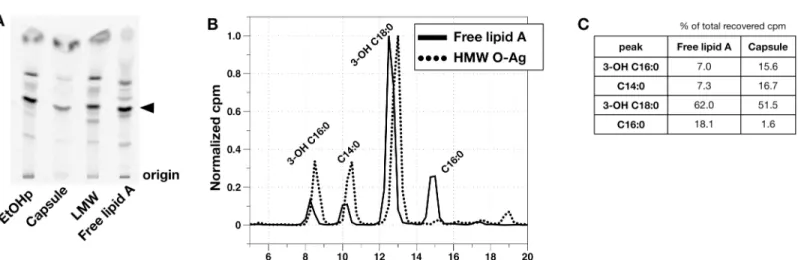
Fig 1. Analysis of O-antigen capsule prepared according to Scheme A, (S1A Fig) SDS-PAGE/immunoblotting for O-Ag capsule (antibody 11B7). The position of a 250 kD molecular weight standard is indicated. B) SDS-PAGE of metabolically labeled EtOHp and capsule, followed by immunoblot for FrancisellaO-antigen (clone FB11, left) or autoradiographic imaging (right). Denoted by asterisks are free lipid A (*), lipid A-core polysaccharide (no O-Ag) (**), and lipid A-core polysaccharide-single O-Ag unit (***) [16]. C) Distribution of cpm after chemical hydrolysis of EtOHp and capsule to release fatty acids. D) HPLC analysis of released [14C]-fatty acids from EtOHp and capsule extracted and recovered in the CHCl3phase after chemical hydrolysis. Recovery of each individual fatty acid species is expressed as % of total recovery of [14C]lipids. Individual fatty acids were identified by comparison to commercial standards (C14:0, C16:0) or LC-MS analysis, as described previously [11].
accumulated by Francisellae) (S1B Fig) [11]. Immunoblot of capsule forFrancisellaLPS O-antigen (O-Ag; FB11 antibody) confirmed the enrichment of higher molecular weight (HMW) polymers containing O-Ag and markedly diminished amounts of lower molecular weight LPS species containing O-Ag vs. EtOHp (Fig 1B, left panel). Examination of these same fractions by autoradiography demonstrated metabolic labeling of the HMW O-Ag capsular polymers and enrichment for higher molecular weight LPS and depletion of free lipid A (Fig 1B, right panel).
Since the O-Ag ofF.tularensiscontains acetylated sugars, further analysis was needed to definitively identify metabolically labeled lipids (e.g., fatty acids) within the HMW O-Ag poly-mers. This was accomplished by sequential 4N HCl/4N NaOH treatments at 90°C to release ester- and amide-linked fatty acids, if present, from the O-Ag polymers followed by Bligh/Dyer extraction to separate radiolabeled sugars from fatty acids in the water/MeOH vs. CHCl3phases, respectively. Comparison of the recovery of radiolabeled material in the water/MeOH vs CHCl3 phases from capsule vs. EtOHp showed a much higher fraction of the recovered radioactive mate-rial from capsule (vs. EtOHp) in the H2O/MeOH phase, consistent with the high degree of enrich-ment of HMW O-Ag polymers in the capsule fractions (Fig 1C). Further characterization of the [14C]-labeled material recovered in the CHCl3phase by HPLC (Fig 1D) revealed [14C] fatty acids that are grossly similar to those present inF.tularensislipid A, which contains three 3-OH fatty acids and one nonhydroxylated fatty acid [5]. Fatty acids recovered from the EtOHp, which con-tains free lipid A in addition to lipid A-core and lipid A-core-O-Ag [11], were comprised primar-ily of the expected 3-OH-fatty acids, 3-OH-18:0 and 3-OH-16:0 (Fig 1D, left). The ratio of cpm in 3-OH C18:0 to that in 3-OH C16:0 in the EtOHp was 5.3, corresponding to a molar ratio of 4.7, consistent with the ratio previously reported forFrancisellalipid A of 5–7:1 [11,17,18]. However, both the ratio of cpm in 3-OH-18:0/3-OH-16:0 (2.2) and the greater prominence of C14:0 and near absence of C16:0 distinguish the fatty acids linked to HMW O-Ag capsule (Fig 1D, right) vs. those present in the EtOHp. Taken together, these findings strongly suggest that the HMW O-Ag polymers that comprise theF.tularensiscapsule contain lipid that closely resembles but is compo-sitionally distinct from the more abundant free lipid A accumulated by these bacteria.
Purification of HMW O-Ag capsule purified from EtOHp by
deoxycholate-based gel sieving
Lipid associated with HMW O-Ag capsule is covalently linked
In order to confirm that isolated HMW O-Ag rich fractions were not contaminated with the free lipid A that is so plentiful in LVS, we subjected HMW capsule to Bligh-Dyer lipid extrac-tion. As expected, a large majority of cpm of the EtOHp partitioned to the organic phase (Fig 2E), which when analyzed by SDS-PAGE and immunoblot for FB11 and autoradiography was greatly enriched for the two fastest-migrating bands corresponding to free lipid A and lipid A + core, with virtually no contaminating O-antigen (Fig 2F). In contrast to the EtOHp, virtu-ally none of the capsule cpm (1.5%) were found in the CHCl3phase (Fig 2E). These results confirm that the capsule fractions resolved by DOC-based gel sieving are free of significant quantities of free lipid, and thus, the lipid A-like material recovered is covalently linked to the HMW O-Ag capsule.
Fatty acid composition of lipid linked to HMW O-Ag capsule differs from
that of the overall EtOHp
DOC-based gel sieving was scaled-up nearly ten-fold to provide sufficient material for further analysis of capsule-associated lipids. EtOHp was isolated from 56 ml of metabolically
Fig 2. Deoxycholate/Sephacryl S-200-based gel sieving of EtOHp to isolate capsule.A) Results of gel sieving of the EtOHp. Recoveries of loaded cpm were typically ~100%. Fractions used for subsequent SDS-PAGE analysis are numbered. B) Capsule (11B7) immunoblot, C) O-antigen (FB11) immunoblot, and (D) autoradiogram, respectively of the indicated fractions. Data shown are representative of at least 4 independent experiments. E) EtOHp and capsule were subjected to Bligh-Dyer extraction and the partitioning of cpm is indicated. Numbers do not add up to 100% due to losses. F) O-antigen immunoblot and autoradiogram of EtOHp and of the extracted lipid fraction following Bligh-Dyer extraction of EtOHp.
radiolabeled late-log LVS cultures and subjected to gel sieving (Fig 3A). Fractions eluting near the void volume were subjected to SDS-PAGE followed by immunoblotting for O-antigen (Fig 3B, top) and autoradiography (Fig 3B, bottom) to confirm enrichment for HMW O-Ag poly-mers. These fractions were combined as were later-eluting fractions, which were enriched in free lipid A and lipid A-core polysaccharide ± short polymers of linked O-Ag (pooled fractions, 46–51, "LMW";Fig 3B). The unfractionated EtOHp was analyzed in parallel after dispersal in DOC sample buffer to control for any effects of residual detergent on the subsequent analysis. The two pooled samples (i.e., HMW capsule and LMW) and the EtOHp were subjected to sequential 4N HCl and 4N NaOH treatments at 90°C followed by Bligh/Dyer extraction to recover released lipids in the CHCl3phase. A significantly smaller fraction of the total cpm of
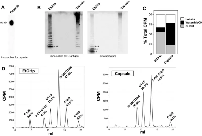
Fig 3. Fatty acid analysis of HMW O-Ag capsule.A) Elution of [14C] labeled species during deoxycholate/Sephacryl S-200 chromatography of radiolabeled EtOHp (larger sample input). B) SDS-PAGE followed by immunoblot (O-Ag; FB11, top pane) and autoradiogram (bottom pane) representing capsule- and LMW-rich species (fractions 25–33 and 46–51, respectively). C) Percent of cpm of the indicated samples recovered in the CHCl3phase after chemical hydrolysis and Bligh-Dyer extraction are indicated.*denotes that recovery from capsule is significantly lower than that from both EtOHp and LMW samples, per Tukey’s test for multiple comparisons (P<0.001). D) HPLC profile of recovered [14C] species in CHCl3phase from EtOHp, nearly O-Ag-free LMW species, and HMW O-Ag capsule. Each curve was normalized so that the peak of the most abundant species (peak 3) was set at 1.0. E) Normal-phase TLC (top panels) and reverse-phase TLC (bottom panels) of the 4 major peaks derived from EtOHp (left) and capsule (right). Francisella-derived and purified commercial FFA were used to identify individual fatty acids, as indicated. NFA, non-hydroxylated fatty acids. 3-OH FA, 3-OH fatty acids. Note that C14:0 and 3-0H-18:0 (peaks 2 and 3) closely migrate on reverse-phase TLC but are readily distinguished by normal phase TLC. F) Relative content of the 4 major FA substituents of the EtOHp, LMW, and capsule samples, expressed as percent of total cpm recovered. These results represent analyses of fractions derived from the same population of metabolically labeled bacteria. G) Similar analyses were performed on three separate EtOHp and capsule preparations from independent batches of labeled bacteria to demonstrate that FA compositional differences between the EtOHp (in which free lipid A is most abundant) and HMW O-Ag capsule are reproducible and, where indicated (*,P<0.05 and**P<0.001 for paired T tests) significant.Leftpanel: ratio of [14C] 3-OH-16:0 to [14C] 3-OH-18:0;centerpanel: ratio of [14C] C14:0 to combined [14C] 3-OH-16:0 + 3-OH-18:0;rightpanel ratio of14[C] C16:0 to combined [14C] 3-OH-16:0 + 3-OH-18:0.
the treated capsule (vs. EtOHp and LMW) was recovered in the CHCl3phase (Fig 3C), consis-tent with the increased abundance of metabolically radiolabeled acetyl sugars of the polar poly-saccharide chains of the HMW O-Ag capsule polymers [4,19].
HPLC of the recovered radiolabeled compounds from the respective CHCl3phases yielded from each sample several peaks (major peaks 1–4,Fig 3D) which were further analyzed by both normal- and reverse-phase TLC to confirm the identity of the fatty acids present in each peak (Fig 3E). These analyses confirmed that the major 3-OH-fatty acid constituents of free and LPS-linked lipid A (3-OH C18:0 and 3-OH C16:0) were also the most prominent compo-nents of capsule-associate lipids. However, as originally observed in HMW O-Ag rich prepara-tions obtained by Triton X-114 partitioning and SDS-based gel sieving (Fig 1), the molar ratio of 3-OH C18:0 to 3-OH C16:0 was much less in capsule than either the starting material (EtOHp) or the pooled LMW fractions (2.7 for capsule vs. 6.4 for EtOHp vs. 7.1 for LMW). In addition, the most abundant non-hydroxylated FA constituent differed significantly between HMW O-Ag capsule (mainly C14:0) and pooled fractions comprising free and LPS-linked lipid A (mainly C16:0,Fig 3D and 3F). When results from three capsule isolations were ana-lyzed and compared to that of the three EtOHp preparations from which the purified HMW O-Ag capsule was derived, the compositional FA differences were again observed, including: increased presence in capsule of 3-OH-16:0 (Fig 3G, left; p<0.05) and C14:0 (Fig 3G, center,
did not reach statistical significance) and decreased presence of C16:0 (Fig 3G, right; p<0.001).
Release of lipid-linked to HMW O-Ag capsule by mild acid treatment
Lipid A is bound to core ± O-Ag polysaccharide via a relatively labile ketoside bond with 3-deoxy-D-manno-oct-2-ulosonic acid (KDO) and can be released after mild acid hydrolysis (in 1% SDS at a pH of 4.5 [13]). To test if the lipid linked to the HMW O-Ag capsule has simi-lar properties, the metabolically radiolabeled HMW O-Ag capsule was treated in the same way. After subjecting capsule to this treatment, approximately 14% of total capsule cpm partitioned to the CHCl3phase, with the majority remaining in the aqueous phase. The radiolabeled lipid liberated by mild acid treatment was then analyzed using an HPTLC system optimized for Francisellalipid A [11]. The major lipid released from the HMW capsule migrated like the major species present in free lipid A and in LMW and EtOHp samples that had been subjected to mild acid treatment (Fig 4A). Fatty acid analysis of the lipid liberated from HMW O-Ag cap-sule by mild acid treatment (Fig 4B and 4C) revealed a composition similar to that of the untreated HMW O-Ag capsule (Fig 3F), indicating that the lipid released by mild acid treat-ment was representative of the major lipid associated with the HMW O-Ag capsule.
Discussion
acid-SDS treatment designed to liberate lipid A that is linked to a (poly)saccharide via a relatively labile ketoside bond with KDO. Finally, the similar migration during TLC of this released lipid in comparison to the major lipid A species of free lipid A and LPS-linked lipid A (Fig 4A) is consistent with its identity as a lipid A-like molecule.
Remarkably, despite the close resemblance of the lipid(s) linked to the HMW O-Ag poly-mers to either free or LPS-linked lipid A, our findings indicate that the HMW O-Ag polymer-linked lipid(s) are structurally distinct. The evidence supporting this conclusion is principally two-fold: i) the difference in the molar ratio of 3-OH-18:0/3-OH-16:0 (ca.>5 in free lipid A/
LPS vs.<3 in the HMW O-Ag polymers (capsule); Figs3Gand4C); and ii) the marked
differ-ence in the most prominent non-hydroxylated fatty acid present (mainly C16:0 in free lipid A/ LPS, C14:0 in HMW O-Ag capsule (Figs3Gand4C). The molar ratio of 3-OH-18:0/3-OH-16:0 and prominence of C3-OH-18:0/3-OH-16:0 we measured in metabolically labeled total free lipid A + LPS correspond closely to published data on the fatty acyl composition ofFrancisellalipid A [5,17, 18,21,22]. Hence, the unusual compositional features of the lipid A-like molecule(s) linked to HMW O-Ag polymers appear to be a unique structural characteristic of these polymers. The near absence of C16:0 in the HMW O-Ag polymers is particularly striking given the promi-nence of this non-hydroxylated fatty acid in all the mass spectroscopic analyses ofFrancisella lipid A reported to date. Taken together, these compositional analyses strongly suggest that the lipid A-like molecule linked to HMW O-Ag polymers is structurally distinct from the bulk lipid A inFrancisellapresent as free lipid A. Other structural differences (e.g., involving charged and/or uncharged polar substituents of lipid A), as well as heterogeneity of capsule-associated lipid, are possible but will require purification of much greater amounts of the HMW O-Ag polymers and liberated lipid for more definitive structural analyses.
The Francisellae produce a remarkable variety of lipid A structures, including multiple iso-baric species with varied fatty acid substitution patterns [21,22]. Analyses of whole bacterial LPS demonstrate the presence of minor populations of lipid A containing both 3-OH and non-hydroxylated fatty acids of varying acyl chain length. Among these are minor species contain-ing the shorter C14:0 non-hydroxylated fatty acid that we find enriched in the HMW O-Ag
Fig 4. Fatty acid analysis of lipid released from HMW O-Ag capsule by mild acid treatment.A) HPTLC of lipids recovered after mild acid hydrolysis of the indicated preparations. The solvent system used was CHCl3:Pyridine:88% Formic Acid:MeOH:H2O (54:46:16:0:5 v/v). The arrowhead indicates the dominant species in free lipid A. Highly enriched free lipid A was derived via Bligh-Dyer extraction of the EtOHp (seeFig 2F). B) HPLC analysis of [14C] fatty acids derived from free lipid A and lipid released by mild acid treatment of HMW O-Ag. C) The four major peaks in B were quantified and expressed as the % of total recovered cpm.
capsule [21,22]. In part, this compositional diversity could reflect promiscuous fatty acid sub-strate properties of the variousFrancisellalipid A acyltransferases. In addition, as exemplified by the differential temperature-dependent activity and expression of twolpxDgenes that pos-sess distinct fatty acid selectivity, lipid A/LPS fatty acid composition can be modulated in response to changing environmental conditions [23].
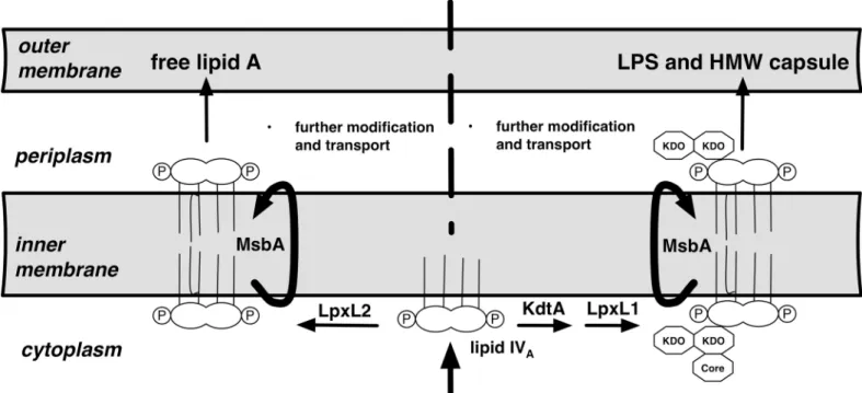
Our finding of distinct lipid A structures in the free lipid A pool ofFrancisellavs. the HMW O-antigen capsule suggests a possible relationship between the fine structure of lipid A and the synthesis and accumulation of HMW O-Ag capsule. In the synthesis of LPS, attachment of the 3-OH-fatty acids to the (di)glucosamine backbone of lipid A precedes attachment of KDO (e.g., lipid IVAinFig 5) whereas subsequent incorporation of non-hydroxylated fatty acid(s) by "late" acyltransferases, trans-bilayer migration across the inner membrane, and transport to the outer membrane typically follow KDO attachment [24]. The remarkable abundance of free lipid A in the Francisellae suggests an unusual capacity of this Gram-negative bacterium to carry out both late acylation and envelope transport in a KDO-independent fashion. Raetz et al. have proposed that one of the two late acyltransferase genes inFrancisellaeis KDO-dependent, which raises the possibility that only a subset of lipid A is subject to modification by this enzyme ([25]; arbitrarily labeled as LpxL1 inFig 5). Fatty acid preference of an individ-ual late acyltransferase inFrancisellahas been suggested by the effects of mutation of one of these late acyltransferases (FTT02323c). Comparison of the lipid A composition of the wild-type and mutant strains revealed a selective loss in the mutant of a minor lipid A subspecies that is strikingly similar in fatty acid composition to the lipid A we have observed linked to HMW O-Ag capsule (1 mol C14:0, 2 mol 3-OH-18:0 and 1 mol 3-OH-16:0) [26]. Apparently in its place, the mutant contained a novel triacyl species, identical to the missing species except for the absence of
Fig 5. Hypothesis of possible mechanistic bases of distinct fatty acid composition of free lipid A vs. lipid A linked to the HMW O-Ag capsule in F.tularensis.Lipid IVAcontains 4 mol of 3-OH fatty acid per mol of lipid A (3-OH-18:0>>3-OH-16:0). LpxL1/2 are homologous "late" acyltransferases that subsequently incorporate the single non-hydroxylated fatty acid ofF.tularensislipid A. Based in part on Raetz et al., we hypothesize that only one (arbitrarily, in this figure, LpxL1) is KDO-dependent and selective for C14:0 whereas LpxL2 is KDO-independent and selective for 16:0 [25]. The different 3-OH-FA composition of free lipid A and lipid linked to HMW O-Ag capsule could reflect the substrate specificity of KdtA that incorporates KDO into lipid IVA.
a myristate. We hypothesize that the selectivity of this late acyltransferase for KDO-containing lipid A could account at least in part for the predominance of C14:0 in the HMW O-Ag capsule (Fig 5). Additionally, the different 3-OH-FA composition of free lipid A vs. lipid A linked to HMW O-Ag capsule could reflect the substrate specificity of KdtA that incorporates KDO into lipid IVA. Whether the lipids associated with lower MW LPS resemble the HMW O-Ag capsule-associated lipid is unknown. Once better separation can be achieved of the abundant free lipid A from short and intermediate-length LPS, it should be possible to determine which structural fea-tures are associated with all KDO-containing LPS vs. only HMW O-Ag capsule.
Ligation of O-Ag to lipid A-core polysaccharide takes place on the outer leaflet of the inner membrane [24], before transport of LPS to the outer membrane. We speculate that synthesis and transport of the HMW O-Ag capsule to the surface ofFrancisellasimilarly requires ligation of the O-Ag to a lipid A-core polysaccharide acceptor on the outer leaflet of the inner mem-brane. In support of this speculation, theFrancisellaO-Ag capsule seems to most closely resemble the group 4 capsules originally characterized inEscherichia coliand classified by Whitfield [27]. Like group 4 capsules, theFrancisellaO-Ag capsule is composed of O-Ag repeats containing acetamido sugars and is dependent upon the Wzy system for polysaccharide polymerization [28,29]. Mutations inFrancisellacore LPS synthetic enzymes and in the O-Ag ligase markedly impair O-Ag capsule production [28,29], further suggesting that the LPS syn-thetic machinery is utilized for HMW O-Ag capsule synthesis. Export of LPS to the outer membrane is carried out by the LPS transport proteins LptABCDEFG [30] that include multi-ple proteins purported to bind specifically to the lipid A portion of LPS [31–33]. If either bind-ing and/or transport efficiency is selective for lipid A of a particular fatty acid composition, the enrichment in HMW O-Ag polymers of lipid A species of shorter chain 3-OH and non-hydroxylated fatty acids could reflect those species that are transported less efficiently, hence allowing more time for synthesis and attachment of the longer O-Ag polymers characteristic of this capsule (Fig 5).
The FB11 mAb used to detect the O-Ag ofFrancisellaLPS and HMW O-Ag capsule recog-nizes the terminal O-Ag unit of each polymer [34]. Thus, each species of differing O-Ag chain length is recognized with similar affinity on immunoblots, providing an estimate of the relative molar abundance of the various O-Ag polymers. Such analyses of the EtOHp fraction that con-tains nearly all the free and LPS-linked lipid A ofFrancisellaas well as the HMW O-Ag poly-mers (Figs1Band2Cand [11]) reveal a bimodal distribution, with the most abundant species being 1) LPS with relatively few O-Ag repeats, and 2) the HMW O-Ag capsular polymers that are recognized by their selective reactivity with the anti-capsular 11B7 mAb (Fig 2B). The selec-tivity of the 11B7 mAb for the HMW O-Ag polymers could reflect the presence of a unique conformational epitope within the HMW O-Ag polymers or the presence of other features independent of the O-Ag repeats. Alternatively, the selectivity could be the result of low-affin-ity interactions with an internal epitope of the O-Ag repeat that requires the abundant presence of this epitope in the longest O-Ag chains to produce a detectable reaction [35]. Whatever the basis of the selective reactivity of the anti-capsular 11B7 mAb, its combined use with the FB11 mAb was key in demonstrating the efficacy of DOC-gel sieving in purification of the HMW O-Ag capsular polymers.
polymers (vs. free and LPS-linked lipid A) and the apparent average chain length of the O-Ag repeats. Purified HMW O-Ag polymers represent<10% of the total radiolabeled material
recovered following DOC-gel sieving,<20% of which is radiolabel derived from the lipid
linked to the capsular polymers. Provided the lipid we have characterized linked to HMW O-Ag capsule is indeed a lipid A, these findings indicate that the lipid A linked to HMW O-Ag capsular polymers represent<~2% of the total lipid A produced and accumulated by growing Francisellaunder the conditions tested, underscoring the sensitivity of the methods used to reveal and partially characterize the lipid linked to the O-Ag capsular polymers. Based on the derived fatty acyl composition of the capsule-linked lipid A-like molecule and the number of acetylated sugars present in the O-Ag repeats, the distribution of radioactivity after sequential 4N HCl/4N NaOH treatments at 90°C and Bligh-Dyer extraction (Fig 3C) suggests an average chain length of>40 O-Ag repeats for the HMW O-Ag capsular polymers. It should be possible
in the future to apply the same combination of methods and reagents under other bacterial growth conditions, including within infected human phagocytic cells [11], to test for regulation ofFrancisellaLPS and O-Ag capsular synthesis and accumulation.
We hypothesize that the association of the HMW O-Ag capsule with a particular subset of lipid A contributes to the means whereby the organism regulates the amount of O-Ag and cap-sule expressed. Studies are underway to isolate short and intermediate-length LPS to determine if the novel lipid is also associated with these species. If shorter LPS species contain distinct lipid A structure from that of the more abundant free lipid A, then it is possible that the bacte-rium uses distinct lipid A structures to regulate the overall balance between free lipid A and core ± O-Ag-containing LPS/ HMW capsule. Given that two late acyltransferases appear to dif-fer in their dependence for KDO in the accepting lipid A (Fig 5), we are generating mutants that ablate the synthesis of these enzymes in strains that we can readily study outside of a BSL3 lab using metabolic labeling (e.g. LVS andF.novicida). If downstream events like flipping lipid A to the periplasm or ligation of O-Ag are selective for lipid A with a particular fatty acid com-position, then these mutants may contain altered proportions of O-Ag capsule or LPS overall. In sum, these strains could provide unique experimental tools in studies of the biogenesis of free lipid A, short chain LPS, and HMW O-Ag capsule and evaluation of their possible roles in the various stages of interactions of Francisellae with the host.
The LVS strain that we have described was derived from aF.tularensissubspeciesholarctica strain in the 1950s [1]. Direct comparisons of the LPS structures from various subspecies ofF. tularensishave shown that the major compositional features of the lipids A are similar [36], but include variations in the degree of substitution of lipid A with phosphate or hexoses [11, 37]. Although the LPS and HMW capsule of LVS is serologically identical to that of the more virulent strains such as Schu S4 (a subspeciestularensisisolate), it remains to be determined if the variations in lipid A structure we have observed are unique to LVS, or to theF.tularensis subspeciesholarctica.
In conclusion, our results identify a lipid that is covalently-associated with HMW Franci-sellaLVS O-Ag capsule. The presence of 3-OH fatty acids, which are unique to lipid A among bacteria, and liberation of this lipid by gentle acid treatment are consistent with it being a lipid A-like molecule. That the fatty acids found in this lipid are distinct from those that are predom-inant in the organism overall suggests thatFrancisellaregulates HMW O-Ag capsule expres-sion in a novel way.
Supporting Information
EtOHp. (TIFF)
Acknowledgments
We would like to acknowledge De-Sheng Zhang for his work developing some of the analytical techniques used herein.
Author Contributions
Conceived and designed the experiments: JHB MAA JPW. Performed the experiments: JHB JWK MAA. Analyzed the data: JHB JPW JWK. Contributed reagents/materials/analysis tools: JHB MAA. Wrote the paper: JHB JPW.
References
1. Sjostedt A. Tularemia: History, epidemiology, pathogen physiology, and clinical manifestations. Ann N Y Acad Sci. 2007; 1105:1–29. PMID:17395726
2. Cowley SC, Elkins KL. Immunity toFrancisella. Front Microbiol. 2011; 2:26. doi:10.3389/fmicb.2011. 00026PMID:21687418
3. Chong A, Celli J. TheFrancisellaintracellular life cycle: toward molecular mechanisms of intracellular survival and proliferation. Front Microbiol. 2010; 1:138. doi:10.3389/fmicb.2010.00138PMID:
21687806
4. Apicella MA, Post DM, Fowler AC, Jones BD, Rasmussen JA, Hunt JR et al. Identification, Characteri-zation and Immunogenicity of an O-Antigen Capsular Polysaccharide ofFrancisella tularensis. PLoS One. 2010; 5:e11060. doi:10.1371/journal.pone.0011060PMID:20625403
5. Gunn JS, Ernst RK. The structure and function ofFrancisellalipopolysaccharide. Ann N Y Acad Sci. 2007; 1105:202–218. PMID:17395723
6. Hood AM. Virulence factors ofFrancisella tularensis. J Hyg (Lond). 1977; 79:47–60.
7. Sandstrom G, Lofgren S, Tarnvik A. A capsule-deficient mutant ofFrancisella tularensisLVS exhibits enhanced sensitivity to killing by serum but diminished sensitivity to killing by polymorphonuclear leuko-cytes. Infection and Immunity. 1988; 56:1194–1202. PMID:3356465
8. Peleg A, Shifrin Y, Ilan O, Nadler-Yona C, Nov S, Koby S et al. Identification of anEscherichia coli operon required for formation of the O-antigen capsule. J Bacteriol. 2005; 187:5259–5266. PMID:
16030220
9. Goldman RC, White D, Orskov F, Orskov I, Rick PD, Lewis MS et al. A surface polysaccharide of Escherichia coliO111 contains O-antigen and inhibits agglutination of cells by O-antiserum. J Bacteriol. 1982; 151:1210–1221. PMID:6179923
10. Peterson AA, McGroarty EJ. High-molecular-weight components in lipopolysaccharides ofSalmonella typhimurium,Salmonella minnesota, andEscherichia coli. J Bacteriol. 1985; 162:738–745. PMID:
3886631
11. Barker JH, Kaufman JW, Zhang DS, Weiss JP. Metabolic labeling to characterize the overall composi-tion ofFrancisellaLipid A and LPS grown in broth and in human phagocytes. Innate Immun. 2014; 20:88–103. doi:10.1177/1753425913485308PMID:23729477
12. Kooistra O, Luneberg E, Knirel YA, Frosch M, Zahringer U. N-Methylation in polylegionaminic acid is associated with the phase-variable epitope ofLegionella pneumophilaserogroup 1 lipopolysaccharide. Identification of 5-(N,N-dimethylacetimidoyl)amino and 5-acetimidoyl(N-methyl)amino-7-acetamido-3,5,7,9-tetradeoxynon-2-ulosonic acid in the O-chain polysaccharide. Eur J Biochem. 2002; 269:560–
572. PMID:11856315
13. Wang X, Ribeiro AA, Guan Z, McGrath SC, Cotter RJ, Raetz CR. Structure and Biosynthesis of Free Lipid A Molecules That Replace Lipopolysaccharide inFrancisella tularensissubsp.novicida. Biochem-istry. 2006; 45:14427–14440. PMID:17128982
14. Katz SS, Weinrauch Y, Munford RS, Elsbach P, Weiss J. Deacylation of lipopolysaccharide in whole Escherichia coliduring destruction by cellular and extracellular components of a rabbit peritoneal inflammatory exudate. Journal of Biological Chemistry. 1999; 274:36579–36584. PMID:10593958
16. Chalabaev S, Kim TH, Ross R, Derian A, Kasper DL. 3-deoxy-D-manno-octulosonic acid (Kdo) hydro-lase identified inFrancisella tularensis,Helicobacter pylori, andLegionella pneumophila. J Biol Chem. 2010; 285:34330–34336. doi:10.1074/jbc.M110.166314PMID:20801884
17. Jantzen E, Berdal BP, Omland T. Cellular fatty acid composition ofFrancisella tularensis. J Clin Micro-biol. 1979; 10:928–930. PMID:521490
18. Whittaker P, Day JB, Curtis SK, Fry FS. Evaluating the use of fatty acid profiles to identifyFrancisella tularensis. J AOAC Int. 2007; 90:465–469. PMID:17474518
19. Vinogradov EV, Shashkov AS, Knirel YA, Kochetkov NK, Tochtamysheva NV, Averin SF et al. Struc-ture of the O-antigen ofFrancisella tularensisstrain 15. Carbohydr Res. 1991; 214:289–297. PMID:
1769021
20. Maitra SK, Schotz MC, Yoshikawa TT, Guze LB. Determination of lipid A and endotoxin in serum by mass spectroscopy. Proc Natl Acad Sci U S A. 1978; 75:3993–3997. PMID:16592555
21. Shaffer SA, Harvey MD, Goodlett DR, Ernst RK. Structural Heterogeneity and Environmentally Regu-lated Remodeling ofFrancisella tularensissubspeciesnovicidaLipid A Characterized by Tandem Mass Spectrometry. J Am Soc Mass Spectrom. 2007; 18:1080–1092. PMID:17446084
22. Schilling B, McLendon MK, Phillips NJ, Apicella MA, Gibson BW. Characterization of Lipid A Acylation Patterns inFrancisella tularensis,Francisella novicida, andFrancisella philomiragiaUsing Multiple-Stage Mass Spectrometry and Matrix-Assisted Laser Desorption/Ionization on an Intermediate Vacuum Source Linear Ion Trap. Anal Chem. 2007; 79:1034–1042. PMID:17263332
23. Li Y, Powell DA, Shaffer SA, Rasko DA, Pelletier MR, Leszyk JD et al. LPS remodeling is an evolved survival strategy for bacteria. Proc Natl Acad Sci U S A. 2012; 109:8716–8721. doi:10.1073/pnas. 1202908109PMID:22586119
24. Raetz CR, Whitfield C. Lipopolysaccharide endotoxins. Annual Review of Biochemistry. 2002; 71:635–
700. PMID:12045108
25. Raetz CR, Guan Z, Ingram BO, Six DA, Song F, Wang X et al. Discovery of new biosynthetic pathways: the lipid A story. J Lipid Res. 2009; 50 Suppl:S103–8. doi:10.1194/jlr.R800060-JLR200PMID:
18974037
26. McLendon MK, Schilling B, Hunt JR, Apicella MA, Gibson BW. Identification of LpxL, a late acyltransfer-ase ofFrancisella tularensis. Infect Immun. 2007; 75:5518–5531. PMID:17724076
27. Whitfield C. Biosynthesis and assembly of capsular polysaccharides inEscherichia coli. Annu Rev Bio-chem. 2006; 75:39–68. PMID:16756484
28. Lindemann SR, Peng K, Long ME, Hunt JR, Apicella MA, Monack DM et al.Francisella tularensisSchu S4 O-antigen and capsule biosynthesis gene mutants induce early cell death in human macrophages. Infect Immun. 2011; 79:581–594. doi:10.1128/IAI.00863-10PMID:21078861
29. Rasmussen JA, Post DM, Gibson BW, Lindemann SR, Apicella MA, Meyerholz DK et al.Francisella tularensisSchu S4 lipopolysaccharide core sugar and O-antigen mutants are attenuated in a mouse model of tularemia. Infect Immun. 2014; 82:1523–1539. doi:10.1128/IAI.01640-13PMID:24452684
30. Dong H, Xiang Q, Gu Y, Wang Z, Paterson NG, Stansfeld PJ et al. Structural basis for outer membrane lipopolysaccharide insertion. Nature. 2014; 511:52–56. doi:10.1038/nature13464PMID:24990744
31. Chng SS, Ruiz N, Chimalakonda G, Silhavy TJ, Kahne D. Characterization of the two-protein complex inEscherichia coliresponsible for lipopolysaccharide assembly at the outer membrane. Proc Natl Acad Sci U S A. 2010; 107:5363–5368. doi:10.1073/pnas.0912872107PMID:20203010
32. Tran AX, Trent MS, Whitfield C. The LptA protein ofEscherichia coliis a periplasmic lipid A-binding pro-tein involved in the lipopolysaccharide export pathway. J Biol Chem. 2008; 283:20342–20349. doi:10. 1074/jbc.M802503200PMID:18480051
33. Sestito SE, Sperandeo P, Santambrogio, Ciaramelli C, Calabrese V, Rovati GE C et al. Functional characterization ofE.coliLptC: interaction with LPS and a synthetic ligand. Chembiochem. 2014; 15:734–742. PMID:24677607
34. Roche MI, Lu Z, Hui JH, Sharon J. Characterization of Monoclonal Antibodies to Terminal and Internal O-Antigen Epitopes ofFrancisella tularensisLipopolysaccharide. Hybridoma (Larchmt). 2011; 30:19–28. 35. Lu Z, Rynkiewicz MJ, Yang CY, Madico G, Perkins HM, Roche MI et al. Functional and structural
char-acterization ofFrancisella tularensisO-antigen antibodies at the low end of antigen reactivity. Monoclon Antib Immunodiagn Immunother. 2014; 33:235–245. doi:10.1089/mab.2014.0022PMID:25171003
36. Hajjar AM, Harvey MD, Shaffer SA, Goodlett DR, Sjostedt A, Edebro H et al. Lack of in vitro and in vivo recognition ofFrancisella tularensissubspecies lipopolysaccharide by Toll-like receptors. Infect Immun. 2006; 74:6730–6738. PMID:16982824

![Fig 3. Fatty acid analysis of HMW O-Ag capsule. A) Elution of [ 14 C] labeled species during deoxycholate/Sephacryl S-200 chromatography of radiolabeled EtOHp (larger sample input)](https://thumb-eu.123doks.com/thumbv2/123dok_br/16361707.190296/8.918.73.862.115.597/analysis-capsule-elution-species-deoxycholate-sephacryl-chromatography-radiolabeled.webp)