Original Article
Development of curcumin liposome formulations using polyol dilution method
Lalana Kongkaneramit
1*, Porntipa Aiemsum-ang
1,2, and Prartana Kewsuwan
31 Department of Pharmaceutical Technology,
2 Research Center for Drug Discovery and Development, Faculty of Pharmacy,
Srinakharinwirot University, Ongkharak, Nakhon Nayok, 26120 Thailand.
3Nuclear Research and Developments, Thailand Institute of Nuclear Technology (Public Organization),
Ongkharak, Nakhon Nayok, 26120 Thailand.
Received: 12 November 2015; Accepted: 16 February 2016
Abstract
This study was aimed to formulate curcumin liposomes (CLs) by using polyol dilution method which is advantageous for no residue of organic solvent. CLs were the mixture of hydrogenated phosphatidylcholine (PC) and cholesterol (CH) at the molar ratio of 9:1. Propylene glycol (PG), glycerin, and polyethylene glycol 400 (PEG-400) were used as polyol solvent. Extrusion was applied after the suspension formed. The amount of polyol and curcumin and preparing temperature were investigated. The obtained suspensions were observed for appearance, size, size distribution, zeta potential, morphology, and percentage of entrapment. The results showed that type and amount of polyol had an impact on both liposomal size and the amount of entrapped curcumin, while preparing temperature was also an important factor. However, the solubility of lipids and drug in a given polyol should be considered because of loading efficiency in the formulation.
Keywords: liposomes, polyol, curcumin, entrapment, TEM
1. Introduction
Liposomes, a drug delivery system, are lipid vesicles in nano- to micro-size range which usually disperse in water. The vesicle membrane is composed of lipid bilayer which has either one or more bilayers (Swarbrick et al., 1994). Liposomal properties, including size, lamellarity, bilayer rigidity, charge, and bilayer surface modifications, are influenced by lipid type, surface charge, and production method. Liposomes are useful in carrying drugs to the target sites, prolonging drug duration of action, protecting drugs against degradation, protecting the patients against side effects or irritation, and solubilizing lipophilic compounds, therefore poorly water-soluble drugs have potential to be used as injection (Crommelin et al., 2003).
In the past, conventional hand shaking or chloroform film method, which is a simple and easy technique, was gener-ally applied. However, its notable disadvantage is the residual organic solvent. Moreover, newer methods, such as the reverse phase evaporation method (REV) and a solvent injec-tion method, also use organic solvents (New, 1990). Not until 1991, Kikuchi et al. (1991, 1994) had firstly introduced the production of liposome by polyol dilution method. By utiliz-ing this method, additional steps and equipment for the removal of solvent were unnecessary. Thus, it has a potential to be transferred to mass production of liposomes (Kikuchi
et al., 1991, 1994).
In 1991, Kikuchi et al. reported that liposomes in isotonic sugar or polyol solution did not get aggregated after moist-heat sterilization. Later in 1994, Kikuchi et al. studied dextran T-40 as a water soluble drug model, for which the percentage of entrapment was 4-18%. Pavelicet al. (1999) reported that liposomes of clotrimazole, metronidazole, and chloramphenicol prepared by such method have the entrap-* Corresponding author.
Email address: [email protected]
ment efficiencies of 93.7, 5.9, and 30.4%, respectively. In 2001, high entrapment efficiency (63%) of calcein, a hydrophilic substance, in liposomal gels was reported. In 2005, acyclovir liposomes were prepared using 5% propylene glycol (PG) and phosphate buffer (pH 7.4), at 60°C. The lipid composition was varied and three cycles of extrusion through 0.4 µm polycarbonate membrane were applied in the study. It was shown that neutral liposomes could entrap only 8% acyclovir while both negatively and positively charged liposomes could entrap up to 25-30% (Pavelicet al., 2001, 2005). As we noticed that the polyol dilution method has many advantages as described above. Entrapment efficiency of both hydro-phobic and hydrophilic substances is proper. Study on opti-mization of compositions and preparation conditions might be useful for liposome formulation of any interested drug.
Curcuma longa Linn., known as turmeric, is a peren-nial herb in Zingiberaceae family. It is a herb and spice that has been used for a long time (Sigh et al., 2010). Turmeric contains many compounds such as curcuminoids (Goel et al., 2008), oils and oleoresin (Sigh et al., 2010). Curcuminoids, the yellow-pigmented fraction with curcumin, demethoxy-curcumin, and bisdemethoxycurcumin as major components, constitutes 3-5% of turmeric. Curcumin (diferuloylmethane), the principal ingredient, has numerous activities such as antioxidant, antimicrobial and anti-inflammatory properties (Jurenka, 2009).
Many reports on delivering efficiency of curcumin liposomes (CLs) have been published. Jung et al. (2006) studied a transfollicular drug delivery system into porcine hair follicles. The results showed that amphoteric and cationic liposomes reached an average relative penetration depth of approximately 70% of the full hair follicle length. Gupta et al.
(2011) indicated that curcumin cream and gel showed less antioxidant and anti-aging activities. However, developed formulation of lipid complex, phyto-vesicle, niosome, and liposome could improve its efficiency. Chen et al. (2012) produced CLs from three lipids, soybean phospholipid, egg yolk phospholipid, or hydrogenated soybean phospholipid. All formulations were found to be similar in particle size and percentage of entrapment while CLs from soybean phospho-lipid showed higher transdermal efficiency (in vitro) and growth-inhibiting activity on B16BL6 melanoma cells.
Although CLs are interesting for their uses, to my best knowledge, CLs produced by polyol dilution method have not been reported. The present study was undertaken to formulate CLs and investigate parameters involved in polyol dilution method. The obtained CLs were observed for size, zeta potential, types of liposome and entrapment efficiency.
2. Methods and Materials
2.1 Reagents and chemical
Curcumin (77%) was purchased from Sigma–Aldrich (St. Louis, MO, USA). Phospholipid (hydrogenated phos-phatidylcholine, Phospholipon®90H) was obtained from
PHOSPHOLIPID GmbH (Köln, Germany). Cholesterol was purchased from Fluka Chemical (Milwaukee, USA). Propylene glycol, glycerin, and polyethylene glycol 400 were purchased from PC Drug Center Ltd. (Bangkok, Thailand). Orthophos-phoric acid (85%), acetonitrile (HPLC grade), and methanol (HPLC grade) were purchased from RCI Labscan Ltd. (Bangkok, Thailand). Triton X-100 was purchased from BDH Chemicals Ltd. (London, England).
2.2 Liposome preparation
A solvent (PG, glycerin, or polyethylene glycol (PEG-400)) was heated up in a beaker to a given temperature (40°C, 60°C, and 80°C) while stirred at 600 rpm. Conditions were controlled by a digital thermal controlled hot plate with magnetic stirrer (RCT basic, IKA®, Malaysia). Subsequently,
cholesterol (CH) and phosphatidylcholine (PC) were dissolved in the solvent, respectively. For CLs, curcumin was added to the solution after the lipids were completely dissolved. After that, purified water was slowly poured into the solution, and the mixture was steadily stirred for 1 hr. Half of the volume of the mixture was extruded for three cycles through an extruder (The LIPEX™ Extruder, Northern Lipid Inc., Canada) with nitrogen gas (150 psi). Polycarbonate membrane with the pore size of 0.2 µm (Whatman® Nuclepore™ Polycarbonate
Track-Etched Membranes, Whatman, UK) was applied. The other half volume of the mixture was kept for characteristic comparison between extrusion and non-extrusion products.
Liposome formulas with or without curcumin were made. The molar ratio of PC and CH and the total lipid con-centration were kept at 9:1 and 5 mg/ml, respectively, for both formulations. The amount of curcumin added to the CLs was varied three levels (25, 250, and 2,500 g/ml).
2.3 Liposome characterization
2.3.1 Appearance observation
Liposome samples were filled in 25-ml glass bottles and kept in dark cabinet at room temperature. Amount of sediment and appearance of supernatant either transparent or translucent were visually observed.
2.3.2 Particle size and zeta potential determination
Particle size and zeta potential of liposomes were determined at 25°C using a particle analyzer (Zetasizer®
NanoSeries, Malvern Instrument Ltd., UK). Samples were diluted with water (1:100) before the measurement. The par-ticle size was measured by dynamic light scattering technique. The intensity of the scattered light was detected at the angle of 173° by a photomultiplier. The polydispersity index (PDI)
2.3.3 Morphological study by transmission electron micro-scopy
Liposome samples diluted with water (1:5) were imaged by transmission electron microscope (TEM) (Jeol JEM-1400 Transmission Electron Microscope; USA). A drop of sample was placed on a formvar-carbon coated copper grid, after that, side of the grid was blotted with filter paper to remove the excess liquid. The grid was air-dried for 3 min. Subse-quently, a drop of phosphotungstic acid solution (2%) was put on the grid for negative staining. The excess liquid was removed and the grid was air-dried again. Samples were investigated at the accelerating voltage range of 80-100 kV.
2.4 Quantitative analysis of curcumin
Amount of curcumin was analyzed using high perfor-mance liquid chromatrography (HPLC; 1260 Infinity Quater-nary LC System, Agilent technologies, USA). Reverse-phase C18 column (150x4.6 mm i.d.; ACE®, Advanced
Chromato-graphy Technologies Ltd., Scotland) was used together with C18 guard column (4.0x3.0 mm i.d.; Security GuardTM,
Phenomenex, USA). The mixture of 0.1% orthophosphoric acid:acetonitrile (45:55, v/v) was used as mobile phase at the flow rate of 1 ml/min. The injection volume was 10 µl and the column temperature was 25°C. The detection wavelength was set at 420 nm. Each sample was filtered through a 0.2-µm membrane filter prior to injection.
Standard curve of curcumin was prepared using stock solution of curcumin (100 µg/ml) in methanol. Aliquots of 0.5, 1.0, 2.0, 3.0, and 4.0 ml of the stock solution were transferred to 10-ml volumetric flasks and adjusted the volume with methanol. The final concentrations of 5, 10, 20, 30, 40 µg/ml of curcumin were obtained.
2.5 Entrapment efficiency
To analyze total curcumin of the liposomal product, 0.5-ml product was added with 1.5 ml of 5% triton-X100 and vortexed for 30 s before analyzing.
To analyze entrapped and unentrapped curcumin of the liposomal product, 12-ml sample was transferred into 15-ml tube which was centrifuged (Sorvall Legend XTR Cen-trifuge, Thermo Scientific, USA) at 4500 rpm and 25°C for 30 min. Ten milliliters of supernatant were transferred to ultra-centrifuge tube for further ultracentrifugation. The obtained pellets (named as pellet 1) were washed with purified water and centrifuged as above conditions for three times. After the last washing, the pellet 1 remained in 1 ml of purified water were taken. Then, 0.2-ml suspension of pellet 1 was added with 1.4 ml of 5% triton-X100 and vortexed for 30s before analyzing.
For the first ultracentrifugation (Sorvall WX Ultra Series Centrifuge, Thermo Scientific, USA), the 10-ml super-natant from above centrifugation was ultra-centrifuged with swinging bucket rotor (TH-641, 6x13.2 ml) at 40,000 rpm
(273,800 g) and 25°C for 30 min. After that, 9.5-ml supernatant was transferred for the second ultracentrifugation. The pellets (named as pellet 2) remained in 0.5 ml of purified water. Then the 0.5-ml suspension of pellet 2 was added with 1.0 ml of 5% triton-X100 and vortexed for 30 s before analyzing.
For the second ultracentrifugation, the 9.5-ml super-natant from the first ultracentrifugation was added with water to 10 ml and centrifuged at 40,000 rpm and 25°C for 60 min. The 9.5-ml supernatant was withdrawn and kept for analysis. The pellets (named as pellet 3) remained in 0.5 ml of purified water. The 0.5-ml suspension of pellet 3 was added with 2.5 ml of 5% triton-X100 and vortexed for 30 s before analyzing.
The amount of total curcumin in the product, entrapped curcumin (total curcumin in pellet 1, 2, 3), and unentrapped curcumin (curcumin in final supernatant) were determined as described in 2.4.
3. Results and Discussion
3.1 Liposome preparation by polyol dilution method
Liposomes without curcumin were developed and investigated for the suitable preparing conditions. The prepa-ration method from Pavelicet al. (2001) was used in the study. Lipids were dissolved in 5% PG and the liposomes were prepared as described. It was found that, among three tem-peratures tested, the sample prepared at 60°C was presented as translucent supernatant with some sediment, while lipids were not dissolved completely in the sample prepared at 40°C. For alternative polyol solvents, glycerin and PEG-400 were used. Lipids were not dissolved in 5% glycerin at all tested temperatures while they were dissolved in 5% 400 only at 80°C. However, the obtained sample from PEG-400 had more sediment than those form PG. Thus, with regard to lipid dissolution, PG was considered suitable for preparing liposomes using the lipid mixture, comprising PC and CH at the molar ratio of 9:1 and 5 mg/ml total lipid, at moderate temperature (60°C).
PG has been used in the formulations of choline or distearoylphosphatidylcholine; egg phosphatidyl-choline and egg phosphatidylglycerol-sodium; egg phos-phatidylcholine and stearylamine (Pavelicet al., 1999, 2001, 2005). For liposomes composed of phospholipids and cholesterol, Kikuchi et al. (1994) had to raise the temperature of PG to 120°C to enhance the dissolution of cholesterol. However, our formulations used very small amount of choles-terol, thus it could be dissolved at 60°C in PG but not dis-solution in glycerin or PEG-400 at such temperature.
The 15% and 20% PG liposome formulations were selected for the production of curcumin liposomes (CLs). To-tal lipid and lipophilic drug in the formulation were used as 5 mg/ml and 0.25 mg/ml, respectively (Manosroi et al., 2004). Moreover, 3 concentrations of curcumin (2,500, 250, and 25 µg/ml) were investigated for CLs preparation. It was found that residual curcumin from 2,500 and 250 µg/ml CLs was observed on the polycarbonate membrane used for extrusion whereas CLs with 25 µg/ml curcumin passed completely through the membrane and no sediment was observed within seven days at RT (Figure 1 (C, D). The solubility of curcumin in PG is 33.39 mg/ml (Setthacheewakul et al., 2010); however CLs with 15-20% PG could not dissolve all curcumin at the concentration of 250 and 2500 µg/ml because PG was also used in lipid solubilization.
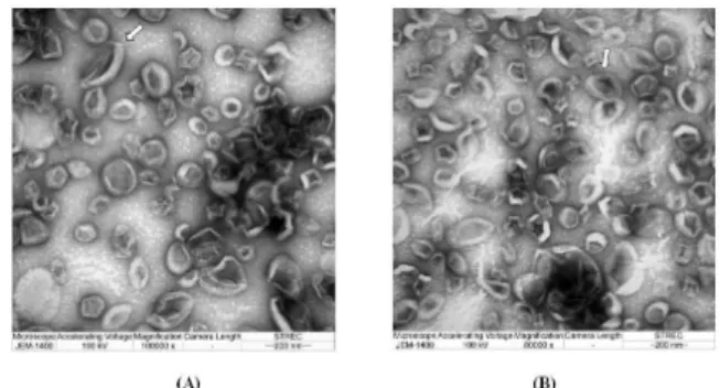
3.2 Size, zeta potential and TEM images
CLs (25 µg/ml curcumin) from 15% and 20% PG, named as CL15 and CL20, respectively had similar size to the pore size of 200 nm of polycarbonate membrane. Zeta potential of the products showed slightly negative values within the range of ±10 mV which were considered nearly neutral (Clogston
et al., 2011). The slightly negative values were attributed to dissociation of curcumin (pKa values of 7.8, 8.5, and 9.0) at
Table 1. Physical appearance of liposomes prepared at 60°C using 5-80% PG.
Non-extrusion Extrusion % PG
pH Supernatant sediment pH Supernatant sediment
5 7.26 + +++ 6.71 ++++ +++
10 7.15 ++ ++ 7.33 ++++ ++
15 6.96 +++ ++ 7.40 ++++
-20 7.17 +++ ++ 7.59 ++++
-30 7.55 ++++ ++++ 7.59 ++++ ++++
40 7.54 ++++ ++++ 7.48 ++++ ++++
60 8.54 Viscous liquid 8.24 Viscous liquid with bubbles 80 Gel
Note : Supernatant: – refers to clear solution; +, ++, +++, ++++, +++++ refer to very little turbidity to very high turbidity. Sediment: – refers to no sediment; +, ++, +++, ++++, +++++ refer to less sediment to a large amount of sediment.
Figure 1. Appearance of liposomes and CLs prepared at 60°C with extrusion. A) liposomes using 15% PG; B) liposomes us-ing 20% PG; C) CLs usus-ing 15% PG; D) CLs usus-ing 20% PG.
Table 2. Size and zeta potential of CLs (25 µg/ml) using 15% and 20% PG.
Percentage of PG Particle size Zeta potential
(%) (nm) (mV)
15 241.7 ± 2.5 -5.485±0.09 20 260.1 ± 0.2 -5.025±0.04
Note: Polydispersity index (PdI) values of the formulations are not more than 0.5.
Figure 2. Images of CL15 and CL20 by TEM. A) CL15 (magnifica-tion 100,000x); B) CL20 (magnifica(magnifica-tion 80,000x); white arrow ( ) indicates liposome with oval shape or with two lipid bilayers.
the neutral pH of the samples (TØnnesen et al., 2002). The data were shown in Table 2.
lipid bilayer) and oligolamellar vesicle (OLV, size > 0.1-1 µm with 2-3 lipid bilayers) (Swarbrick et al., 1994).
3.3 Quantitative analysis of curcumin
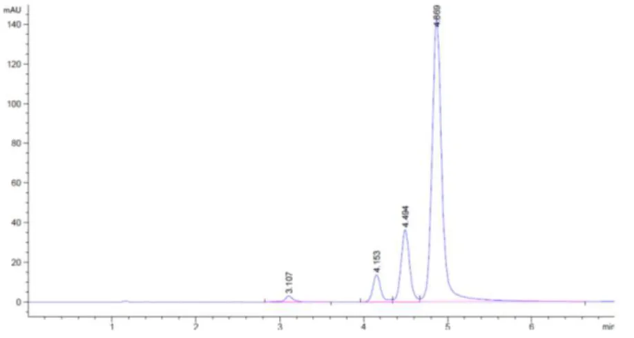
Jangle et al. (2013) reported the HPLC condition for curcumin analysis as followed: C18 column (150x4 mm, 5 m), mobile phase was the mixture of 0.1% orthophosphoric acid: acetonitrile (50:50, v/v), with the flow rate of 1 ml/min. Injec-tion volume, column temperature, and detecInjec-tion wavelength were 5 l, 25°C, and 425 nm, respectively. The retention times of bisdemethoxycurcumin, demethoxycurcumin, and curcumin were shown to be 4.97, 5.62, 6.36 min, respectively.
The modified HPLC conditions, with the mobile phase ratio of 0.1% orthophosphoric acid:acetonitrile (45:55, v/v), injection volume of 10 l, and detection wavelength of 420 nm, were used in this study. A C18 guard column was included for additional column protection. From this condition, the chromatogram showed that curcuminoids peaks were sepa-rated and the peak of curcumin was at 4.869 min (Figure 3). The standard curve was constructed over the concentration range of 5-40 µg/ml. The linear regression equation, with coefficient of determination (r2) of 0.9990, was shown below.
y = 59.74 x - 9.54
y = peak area ; x = curcumin concentration (µg/ml)
3.4 Entrapment efficiency
Curcumin is supposed to be entrapped in the lipid membrane of liposomes due to its lipophilicity. Therefore, to
determine the amount of entrapped curcumin, liposomal pellets were separated from the medium. The regular centri-fuge was used for first separation and the obtained pellets were named as pellet 1. The pellet 1 was washed three times with the same centrifuge. The medium of last washing showed no peak of curcumin. Supernatant obtained from the first separation may have liposomes in very small size thus further centrifugation by ultracentrifuge were done and the pellet 2 and 3 were collected, respectively. Total amount of entrapped curcumin was the sum of curcumin contents found in pellet 1, 2, and 3 while the unentrapped curcumin was that found in the final supernatant. The results were shown in Table 3 and Figure 4.
Percentages of entrapped and unentrapped curcumin of CL15 were 66.5 and 24.5 while those of CL20 were 51.7 and 31.1, respectively. Therefore, using more PG (20%) may effect to increasing of unentrapped curcumin. Since, after dilution with water, PG concentration gradually decreased, hence the liposomes were formed. Most of PG dissolved in the water and some curcumin could soluble in PG. Noticing that CL20 had percentages of unentrapped curcumin higher than that of CL15 because of more PG in the medium. CL15 was the most suitable CLs formulation because of its nano-size range and high percentage of entrapped curcumin.
4. Conclusions
The production of liposomes by polyol dilution method, the solubility of lipid and drug in a given polyol compound is very important. Later, polyol concentration exerts impact on the particle size. Preparing temperature should enhance their solubility while no drug decomposition is resulted. Finally,
Figure 3. HPLC chromatogram of curcuminoids (curcumin peak at 4.869 min).
Table 3. Percentages of curcumin in each sample part.
Curcumin Total curcumin Curcumin in Curcumin in Curcumin in Curcumin in liposomes (%) pellet 1(%) pellet 2(%) pellet 3(%) supernatant(%)
Figure 4. Percentage of curcumin in each sample part.
the curcumin liposomes developed in this study may be useful for many preparations and the hazard from organic solvent contamination is not questionable.
Acknowledgements
We are grateful to Strategic Wisdom and Research Institute, and Faculty of Pharmacy, Srinakharinwirot Univer-sity (SWU) for funding. Our thanks are given to Research Center for Drug Discovery and Development, Faculty of Pharmacy, SWU; Thailand Institute of Nuclear Technology (Public Organization); and Institute of Drug Discovery and Development, Thammasat University for instrumental support.
References
Chen, Y., Wu, Q., Zhang, Z., Yuan, L., Liu, X., and Zhou, L. 2012. Preparation of curcumin-loaded liposomes and evaluation of their skin permeation and pharmaco-dynamics. Molecules. 17(5), 5972-5987.
Cho, N.J., Hwang, L.Y., Solandt, J.J.R., and Frank, C.W. 2013. Comparison of extruded and sonicated vesicles for planar bilayer self-assembly. Materials. 6, 3294-3308. Clogston, J.D. and Patri, A.K. 2011. Zeta Potential
Measure-ment. In Characterization of nanoparticles intended for drug delivery, S.E. McNeil, editor. Humana Press, New York, U.S.A.
Crommelin, D.J.A., Bos, G.W., and Storm, G. 2003. Liposomes: successful carrier systems for targeted delivery of drugs. Business Briefing: Pharmatech. 209-213. Goel, A., Kunnumakkara, A.B., and Aggarwal, B.B. 2008.
Curcumin as “Curecumin”: from kitchen to clinic. Bio-chemical Pharmacology. 75, 787-809.
Gupta, N.K. and Dixit, V.K. 2011. Development and evaluation of vesicular system for curcumin delivery. Archives of Dermatological Research. 303, 89-101.
Jangle, R.D. and Thorat, B.N. 2013. Reversed-phase high-performance liquid chromatography method for analy-sis of curcuminoids and curcuminoid-loaded liposome formulation. Indian Journal of Pharmaceutical Sciences. 75(1), 60-66.
Jung, S., Otberg, N., Thiede, G., Richter, H., Sterry, W., Panzner, S., and Lademann, J. 2006. Innovative liposomes as a transfollicular drug delivery system: penetration into porcine hair follicles. Journal of Investigative Derma-tology. 126, 1728-1732.
Jurenka, J.S. 2009. Anti-inflammatory properties of curcumin, a major constituent of curcuma longa: a review of preclinical and clinical research. Alternative Medicine Review. 14(2), 141-153.
Kikuchi, H., Carlsson, A., Yachi, K., and Hirota, S. 1991. Possi-bility of heat sterilization of liposomes. Chemical and Pharmaceutical Bulletin. 39(4), 1018-1022.
Kikuchi, H., Yamauchi, H., and Hirota, S. 1994. A polyol dilu-tion method for mass producdilu-tion of liposomes. Journal of Liposome Research. 4(1), 71-91.
Manosroi, A., Kongkaneramit, L., and Manosroi, J. 2004. Characterization of amphotericin B liposome formula-tions. Drug Development and Industrial Pharmacy. 30(5), 535-543.
New, R.R.C. 1990. Preparation of liposomes. In Liposomes: a practical approach, R.R.C. New, editors. IRL Press, Oxford, pp 33-104.
Pavelic, Ž., Škalko-Basnet, N., and Jalšenjak, I. 1999. Lipo-somes containing drugs for treatment of vaginal infec-tions. European Journal of Pharmaceutical Sciences. 8, 345-351.
Pavelic, Ž., Škalko-Basnet, N., and Schubert, R. 2001. Lipo-somal gels for vaginal drug delivery. International Journal of Pharmaceutics. 219, 139-149.
Pavelic, Ž., Škalko-Basnet, N., Filipovic´-Grčic, J., Martinac, A., and Jalšenjak I. 2005. Development and in vitro evaluation of a liposomal vaginal delivery system for acyclovir. Journal of Controlled Release. 106, 34-43. Setthacheewakul, S., Mahattanadul, S., Phadoongsombat, N.,
Pichayakorn, W., and Wiwattanapatapee, R. 2010. Development and evaluation of self-microemulsifying liquid and pellet formulations of curcumin, and absorp-tion studies in rats. European Journal of Pharmaceu-tics and BiopharmaceuPharmaceu-tics. 76, 475-485.
Singh, G., Kapoor, I.P.S., Singh, P., de Heluani, C.S., de Lampasona, M.P., and Catalan, C.A.N. 2010. Compara-tive study of chemical composition and antioxidant activity of fresh and dry rhizomes of turmeric (Curcuma longa Linn.). Food and Chemical Toxico-logy. 48, 1026-1031.
Swarbrick, J. and Boylan, J.C. 1994. Liposomes as pharmaceu-tical dosage forms. In Encyclopedia of pharmaceupharmaceu-tical technology, vol. 9, Marcel Dekker, U.S.A., pp 1-9. TØnnesen, H.H., Másson, M., and Loftsson, T. 2002. Studies