Functionalization of zinc ferrite nanoparticles: Influence of
modification procedure on colloidal stability
Marija Milanovi´c
1,∗, Ivan Stijepovi´c
1, Vladimir Pavlovi´c
2, Vladimir V. Srdi´c
11Department of Materials Engineering, Faculty of Technology Novi Sad, University of Novi Sad, Bulevar cara
Lazara 1, 21000 Novi Sad, Serbia
2Faculty of Agriculture, University of Belgrade, Nemanjina 6, 11080 Beograd, Serbia
Received 31 October 2016; Received in revised form 12 December 2016; Accepted 22 December 2016
Abstract
The present work describes the synthesis of zinc ferrite nanoparticles by a chemical coprecipitation method and its surface modification with poly(diallyldimethylammonium chloride) (PDDA), tetramethylammonium hy-droxide (TMAOH) and poly(ethylene glycol) (PEG). The effect of coating on the colloidal stability of obtained systems as well as the effect of modification procedure on the final characteristics of particles were studied. The unmodified and modified nanoparticles were characterized by X-ray diffractometry (XRD), scanning electron microscopy (SEM) and Raman spectroscopy. The particle size distribution and stability of these systems were investigated by dynamic light scattering (DLS) and zeta potential measurements. The results have shown the profound effect of the used modification procedure on colloidal stability of the investigated particles.
Keywords: zinc ferrite, modification, surfactants, PEG, PDDA, TMAOH
I. Introduction
Nanoparticles in solutions present typical colloidal systems, consisting of a continuous phase, which is a dispersed medium (solvent) and a dispersed phase (nanoparticle) [1]. The stability of colloidal solutions is often difficult to achieve. Due to small size of
nanoparti-cles (below 100 nm), these systems are highly reactive, but unstable and they undergo agglomeration. Conse-quently they form large clusters, resulting in increased particle size. This could be very unfavorable, espe-cially in the case of magnetic nanoparticles, where be-sides usual Van der Waals forces, large clusters expe-rience strong magnetic dipole-dipole interactions. One of the ways to prevent agglomeration is modification of the surface of nanoparticles with appropriate surfactant agents [2–4]. A large number of materials can be used for this purpose. They include both inorganic materi-als (silica or gold) and polymers such as chitosan, glu-cose, dextran, poly(ethylene glycol) (PEG), poly(vinyl alcohol) (PVA) etc. Various surfactants, such as sodium oleate, dodecylamine, dopamine, silane agents etc. are also used for modification of surface. By covering the
∗Corresponding author: tel:+381 21 485 3758, fax:+381 21 450 413, e-mail:[email protected]
surface of nanoparticles with appropriate surfactants, particles can be prevented from sticking to each other by two basic modes. The first one is the formation of an electric double layer at the particle surface that cre-ates electrostatic repulsion between the particles, and the second one is coating of particles in order to pre-vent particles touching each other through steric repul-sion [5]. The surface modification of the particles can be physical (by physisorption) or chemical (through cova-lent bonding) and can be done in situ or as a post mod-ification procedure [6,7]. The in situ method involves the addition of surface modifying agents to the reac-tion system already during or even prior to nanoparti-cles formation to avoid aggregation, and to have better control over particle size and shape. Post modification method implies the modification of previously synthe-sized nanoparticles.
For better understanding how modification proce-dure can influence the performance of nanoparticles, we have synthesized zinc ferrite nanoparticles as a model system and functionalized them with different agents.
(TMAOH). Pol(yethylene glycol) is the most used poly-mer for modification of nanoparticles. This polypoly-mer is well soluble in a number of organic polar and apolar solvents, as well as in water. PEG is amphipilic un-charged polymer which prevents aggregation of parti-cles by steric stabilization [8,9]. PDDA is a water sol-uble cationic polyelectrolyte which keeps the nanopar-ticles from aggregating in solutions either by steric or electrosteric interactions [10]. TMAOH is a surfactant whose cationic species are adsorbed at the surface OH groups thus creating an electrostatic repulsion between particles [11].
The present work describes the synthesis of zinc fer-rite nanoparticles by a chemical coprecipitation method and modification with PEG, PDDA and TMAOH in or-der to study the effect of modification procedure on the
final characteristics of particles and their colloidal sta-bility.
II. Experimental
2.1. Synthesis of unmodified zinc ferrite nanoparti-cles
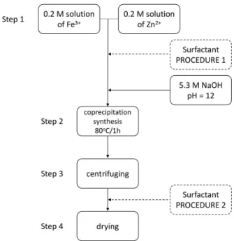
The preparation scheme of zinc ferrite nanoparticles is shown in Fig. 1. Unmodified zinc ferrite nanoparticles were synthesized by coprecipitation method following the four steps:
1. Stoichiometric amounts of Fe(NO3)3·9 H2O (Merck KGaA, Germany) and Zn(NO3)2 · 6 H2O (Merck KGaA, Germany) were dissolved in distilled water, forming 0.2 M solutions and mixed together. 2. Sodium hydroxide was used as a precipitating alkali
until pH value reached 12. The reaction was carried out at 80 °C for 60 min under continuous stirring. 3. The precipitates were separated from the slurry by
centrifuging and washing a number of times with distilled water until the pH reached 7.
4. The obtained nanoparticles were dried at 120 °C/24 h
and finally dry milled in a mortar. The obtained zinc ferrite nanoparticles were denoted as ZF.
2.2. Modification of zinc ferrite nanoparticles
For modification of zinc ferrite nanoparticles, seve-ral surfactants were used such as: PDDA (Sigma Aldrich), TMAOH (Sigma Aldrich) and PEG 600 (Fluka Chemika). In order to investigate the influence of the modification procedure on final powder morphol-ogy, two groups of powders were prepared, using two procedures.
Procedure 1: in the first group, surfactants were
Figure 1. Flowchart representing the preparation of unmodified and modified zinc ferrite nanoparticles by
coprecipitation method
added during the synthesis, after the step 1 in the procedure for preparation of unmodified zinc ferrite nanoparticles. The concentration of surfactant/ferrite
was 5 mmol/g. The following steps 2, 3 and 4 were the
same as described above.
Procedure 2: in the second group, surfactants were
added after the synthesis (after the step 3). Namely, washed zinc ferrite nanoparticles were dispersed in dis-tilled water in the concentration of 6 g/L. The solutions
were treated with surfactants (concentration 5 mmol/g)
and ultrasonically agitated for 60 min, in order to sta-bilize the solution of zinc ferrite nanoparticles. Finally, the powders were obtained following the steps 3 and 4. Sample notations were listed in Table 1.
2.3. Characterization of nanoparticles
The crystal structure of the powders was investigated by X-ray diffraction (XRD) using a Rigaku MiniFlex
600 diffractometer (CuKαradiation,λ=1.5406 Å).
X-ray diffraction patterns were recorded in the range 10–
80° with a scan rate of 0.03°/s. Particle size distribution
was measured by dynamic light scattering (DLS) and the zeta potential of particles was determined by phase analysis light scattering and mixed mode measurement using a Zetasizer Nano ZS with MPT-2 Autotitrator Malvern Instruments, Malvern, United Kingdom. Size and morphology of particles were examined using trans-Table 1. Sample notations of modified zinc ferrite nanoparticles and conditions of surfactant addition
Surfactant During synthesis, procedure 1(T After synthesis, procedure 2 =80 °C, pH=12) (Room temperature, pH=7)
PDDA ZF-PDDA 1 ZF-PDDA 2
PEG ZF-PEG 1 ZF-PEG 2
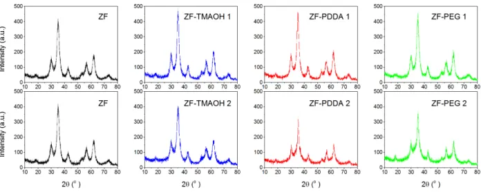
Figure 2. XRD patterns of zinc ferrite nanoparticles unmodified and modified during and after synthesis
mission electron microscopes: JEOL JEM 1400 Plus, operating at 120 kV (for modified zinc ferrite) and a Philips CM20 operating at 200 kV (for unmodified zinc ferrite). Raman spectroscopy of the powders were per-formed at room temperature using a Thermo Fisher Scientific DXR™ Raman Microscope. The device is equipped with DPSS (Diode Pumped Solid State) laser usingλ=780 nm excitation. A CCD camera has been
used as detector. The spectral range 200–1000 cm-1was
examined using a 900 lines/mm grating paired with 10
microscope objective.
III. Results and Discussion
The X-ray patterns of unmodified zinc ferrite nanoparticles, ZF and zinc ferrite nanoparticles modi-fied during and after synthesis are shown in Fig. 2. All samples have the characteristic reflections of the cubic spinel phase with broad peaks indicating the small crys-tallite size. The average cryscrys-tallite size of the samples, calculated by Scherrer’s formula from 311 peak of the spinel phase is around 5 nm. The intensities of the peaks in the samples modified during synthesis (procedure 1) are somewhat higher compared to the samples modified after synthesis (procedure 2) implying the higher crys-tallinity, Fig. 2. When surfactant is added to the sample during synthesis, it promotes a better dispersion of the partially amorphous nanoparticles and enhances the de-gree of spatial arrangement of the matter, thus increas-ing the crystallinity. This effect was proposed by other
authors [11,12]. Moreover, in procedure 1, surfactants together with metal ions undergo coprecipitation reac-tion at 80 °C. In this way, the procedure 1, driven by the temperature, pH, hydrophilicity (PEG) and the charge (cationic groups in TMAOH and PDDA adsorbed onto negatively charged zinc ferrite surfaces), could lead to the steric effect around the particles that promote a
bet-ter arrangement during growing and crystallinity. Further examination of crystallinity and phase
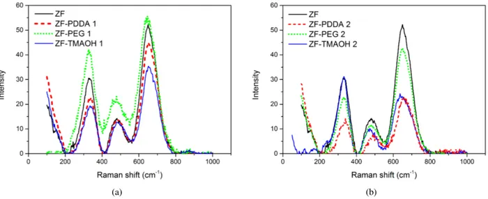
com-position was done by Raman spectroscopy at room tem-perature, Fig. 3. There are five Raman active modes (A1g + Eg + 3T2g) in cubic spinel structure. The
modes above 600 cm-1 mostly correspond to the
mo-tion of oxygen in the tetrahedral MO4 groups and it
can be considered as A1g symmetry. The other low fre-quency modes represent the characteristics of the oc-tahedral sites (FeO6). In the region of 200–1000 cm-1,
all samples presented in Fig. 3, have similar broad Ra-man peaks centered at around 330, 480, and 660 cm-1,
which are associated with Eg, T2g(2) and A1g modes of standard spinel structure ZnFe2O4 [13–15]. The Eg band is due to symmetric bending of Fe–O bonds with peak around 250–360 cm-1, and T2g(2) band is from the
asymmetric stretch of Fe–O bonds with peak around 450–520 cm-1[16]. The broadening of peaks due to the
small particle size was observed. It can be seen from the Raman spectra that used surfactants do not change the structure of the obtained spinel nanoparticles. However, when we compare the used modification procedure, the intensities of the peaks are higher in samples modified during synthesis than in the case of the samples modi-fied after synthesis. It can be concluded that addition of surfactants during the synthesis (procedure 1) results in higher crystallinity of the obtained samples which is in good agreement with the presented XRD results.
In order to examine the effect of modification
deag-(a) (b)
Figure 3. Raman spectra of the zinc ferrite nanoparticles modified during synthesis (a) and after synthesis (b)
glomeration, shifting the particle size distribution to-wards smaller values. Comparing the diagrams shown in Fig. 4, an obvious influence of the type of used sur-factant as well as the type of used procedure on the hy-drodynamic radius of particles could be noted. In both procedures, the smallest average particle size was mea-sured for the samples modified with TMAOH. In addi-tion, the smaller particle size of the samples modified af-ter synthesis, Fig. 4b, compared to the samples modified during synthesis, Fig. 4a, could be a result of more ef-ficient deagglomeration of the particles since ultrasonic agitation is used in procedure 2. The ultrasonic agitation provides larger surface available for surfactant adsorp-tion, resulting in smaller nanoclusters of primary parti-cles [17]. According to DLS measurements, the small-est hydrodynamic radius has a sample modified with TMAOH and ultrasonically treated, whered ∼ 25 nm
(sample ZF-TMAOH 2), Fig. 4b.
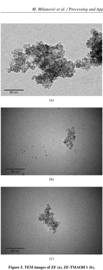
These findings are further verified with the TEM analysis as presented in Fig. 5. The size of unmodified
zinc ferrite primary particles is in accordance with XRD results, but particles are strongly agglomerated, Fig. 5a. However, Figs. 5b and 5c clearly indicate that the degree of agglomeration is reduced when particles are modified with TMAOH which resulted in a lower value of the hy-drodynamic diameter (Figs. 4a,b). Moreover, the pres-ence of the isolated, non-agglomerated particles in ZF-TMAOH 1 sample (Fig. 5b) could be noticed. This can be explained by adsorption of the [(CH3)4N]+ cations
onto the surface of the zinc ferrite nanoparticles that gives rise to repulsive stabilization and prevents the ag-glomeration of particles. A loose connection between particles in ZF-TMAOH 2 sample, Fig. 5c, indicates that ultrasound agitation as well as the type of ions on the nanoparticle surface have profound effect on the
for-mation of soft agglomerates. The particles in such ag-glomerates can be reconnected and regrouped especially when they are dispersed in aqueous media as in DLS measurements, which very well supports the result in Fig. 4b.
(a) (b)
(a)
(b)
(c)
Figure 5. TEM images of ZF (a), ZF-TMAOH 1 (b), ZF-TMAOH 2 (c)
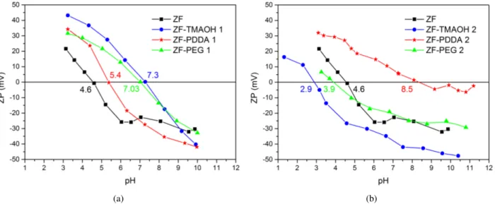
The stability of nanoparticles dispersed in aqueous media was investigated by measuring the zeta poten-tial and results are shown in Fig. 6. Since zeta potenpoten-tial is related to the surface charge on the nanoparticles, its value indicates the degree of electrostatic repulsion be-tween the nanoparticles. If the zeta potential is higher than±30 mV, then it is generally accepted that the parti-cles are electrostatically stable [8]. The isoelectric point (pI) is the point at a particular pH where particles carry no net electric charge on their surfaces.
The isoelectric point of the unmodified zinc ferrite nanoparticles synthesized in this work is around pI ∼
4.6. The observed shifts of pI for modified systems may be associate with successful coating of used surfactant onto the surface of particles. However, it can be noticed that pI of the modified nanoparticles is strongly influ-enced by the modification procedure. This is more ob-vious for TMAOH-modified particles where pI is shift-ing from 7.3 to 2.9 dependshift-ing upon whether the addi-tion step is performed during or after synthesis. The in-crease in pI of ZF-TMAOH 1 sample, with respect to ZF, Fig. 6a, indicates that [(CH3)4N]+cations were
ad-sorbed onto the surface decreasing the negative charge of the zinc ferrite nanoparticles at alkaline pH, which is in accordance with literature data [11]. In comparison to unmodified zinc ferrite nanoparticles with pI ∼4.6,
the pI of ZF-TMAOH 2 sample is decreased to value of 2.9, while the overall zeta potential is more negative in the whole range of pH, Fig. 6b. This result suggests that changing of the ionic strength of the aqueous me-dia (alkaline in procedure 1 to neutral in procedure 2) could change the electrical double layer around the par-ticle which influences the colloidal stability. The small-est value of hydrodynamic diameter of ZF-TMAOH 2 nanoparticles indicates that electrostatic repulsive forces are acting between the particles that opposes the ag-glomeration of these nanoparticles and increases the dis-persion and colloidal stability in water. The zeta poten-tial of solution with ZF-TMAOH 2 nanoparticles at neu-tral pH was found to be−40 mV, suggesting their
stabil-ity in aqueous medium. Moreover, the colloidal stabilstabil-ity of ZF-TMAOH 2 sample is observed for all pH>4.5,
Fig. 6b, while addition of TMAOH during synthesis re-sulted in stable colloids only at high pH, Fig. 6a.
For the PDDA-modified particles the procedure 1 seems to be more effective, Fig. 6a. The large negative
potential value (absolute value above 25 mV) at neutral pH for the solution with ZF-PDDA 1 nanoparticles, im-plies that the particles will have excellent stability in an aqueous medium due to repulsive, interparticle electro-static force. These functionalized particles tend to show a negative potential as the pH value further increases. Thus the surface modification with PDDA was success-ful in this procedure. Solution with ZF-PDDA 2 parti-cles at pH>5 showed extensive aggregation and
conse-quent precipitation, Fig. 6b.
iso-(a) (b)
Figure 6. Zeta potential as a function of pH of zinc ferrite nanoparticles modified during synthesis (a) and after synthesis (b)
electric point is observed at pH∼3.9 while overall zeta
potential is close to the unmodified zinc ferrite particles. Solution with ZF-PEG 2 particles precipitated suggest-ing an unsuccessful coversuggest-ing. Results of DLS measure-ments (Fig. 4b) imply on partially deagglomerated par-ticles (the smaller particle size of ZF-PEG 2 in com-parison to ZF sample), but this is probably governed by ultrasound agitation, as in the case of ZF-PDDA 2 sam-ple.
The profound impact of the pH on the stability of TMAOH and PDDA coatings shows that electrostatic repulsion might be crucial for these types of dispersions [8,18,19].
IV. Conclusion
Single phase spinel zinc ferrite nanoparticles were successfully synthesized by coprecipitation method. These nanoparticles were modified with different
sur-factants in order to investigate the influence of type of used agent as well as the applied modification proce-dure on the final characteristics of nanoparticles. Surfac-tants do not change the structure of the obtained spinel nanoparticles. However, the surface properties of the particles are significantly changed by surfactants. The highest stability of particles in aqueous media is ob-tained with TMAOH. In addition, TMAOH proved to be the most efficient for deagglomeration of particles
(ZF-TMAOH 2 sample). The results have shown that modi-fication protocol (including type of surfactant and type of treatment) influences the final morphology of these particles and especially their size, agglomeration degree and charge of their surface.
Acknowledgment:The authors acknowledge the
finan-cial support from the Serbian Ministry of Education, Science and Technological Development, Project No. III45021, as well as the support from COST Action TD1402, RADIOMAG Project.
References
1. B.I. Kharisov, H. Rasika Dias, O.V. Kharissova, A. Vázquez, Y. Peña, I. Gómez, “Solubilization, disper-sion and stabilization of magnetic nanoparticles in water and non-aqueous solvents: recent trends”,RSC Adv.,4(2014) 45354–45381.
2. B. Kayode Sodipo, A. Abdul Aziz, “Recent advances in synthesis and surface modification of superpara-magnetic iron oxide nanoparticles with silica”, J. Magn. Magn. Mater.,416(2016) 275–291.
3. D. Ling, M.J. Hackett, T. Hyeon, “Surface ligands in synthesis, modification, assembly and biomedical applications of nanoparticles”,Nano Today,9(2014) 457–477.
4. A.K. Gupta, M. Gupta, “Synthesis and surface en-gineering of iron oxide nanoparticles for biomedical applications”,Biomaterials,26(2005) 3995–4021. 5. D.S. Nikam, S.V. Jadhav, V.M. Khot, R.S.
Ningth-oujam, C.K. Hong, S.S. Malic, S.H. Pawar, “Col-loidal stability of polyethylene glycol functionalized Co0.5Zn0.5Fe2O4 nanoparticles: effect of pH, sample
and salt concentration for hyperthermia application”,
RSC Adv.,4(2014) 12662–12671.
6. S. Mallakpour, M. Madani, “A review of current coupling agents for modification of metal oxide nanoparticles”, Prog. Org. Coat., 86 (2015) 194– 207.
7. M. Sapra, A. Ashok Pawar, C. Venkataraman, “A single-step aerosol process for in-situ surface modi-fication of nanoparticles: Preparation of stable aque-ous nanoparticle suspensions”,J. Colloid Interf. Sci.,
464(2016) 167–174.
8. M. Bloemen, W. Brullot, T. Thien Luong, N. Geukens, A. Gils, T. Verbiest, “Improved functional-ization of oleic acid-coated iron oxide nanoparticles for biomedical applications”,J. Nanopart. Res.,14 (2012) 1100.
Schu-bert, “Poly(ethylene glycol) in drug delivery: pros and cons as well as potential alternatives”, Angew. Chem. Int. Ed.,49(2010) 6288–6308.
10. H. Zhang, Y. Lu, H. Liu, J. Fang, “Con-trollable synthesis of three-dimensional branched gold nanocrystals assisted by cationic surfactant poly(diallyldimethylammonium) chloride in acidic aqueous solution”, RSC Adv., 4 (2014) 36757– 36764.
11. Â.L. Andrade, J.D. Fabris, J.D. Ardisson, M.A. Va-lente, J.M.F. Ferreira, “Effect of
tetramethylammo-nium hydroxide on nucleation, surface modification and growth of magnetic nanoparticles”, J. Nano-mater.,2012(2012) 454759.
12. K. Pemartin, C. Solans, J. Alvarez-Quintana, M. Sanchez-Dominguez, “Synthesis of Mn-Zn ferrite nanoparticles by the oil-in-water microemulsion re-action method”, Colloid. Surface. A, 451 (2014) 161–171.
13. J.P. Singh, G. Dixit, R.C. Srivastava, H.M. Agrawal, R. Kumar, “Raman and Fourier-transform infrared spectroscopic study of nanosized zinc ferrite irradi-ated with 200 MeV Ag15+beam”,J. Alloys Compd.,
551(2013) 370–375.
14. Y. Xu, J. Sherwood, Y. Qin, R.A. Holler, Y. Bao, “A general approach to the synthesis and detailed
characterization of magnetic ferrite nanocubes”,
Nanoscale,7(2015) 12641–12649.
15. Z. Wang, D. Schiferl, Y. Zhao, H.St.C. O’Neill, “High pressure Raman spectroscopy of spinel-type ferrite ZnFe2O4”,J. Phys. Chem. Solids,64(2003) 2517–2523.
16. S. Thota, S.C. Kashyap, S.K. Sharma, V.R. Reddy, “Micro Raman, Mossbauer and magnetic studies of manganese substituted zinc ferrite nanoparticles: Role of Mn”,J. Phys. Chem. Solids,91(2016) 136– 144.
17. B. Moji´c, K.P. Giannakopoulos, Ž. Cveji´c, V.V. Srdi´c, “Silica coated ferrite nanoparticles: Influence of citrate functionalization procedure on final par-ticle morphology”, Ceram. Int., 38 (2012) 6635– 6641.
18. R. De Palma, S. Peeters, M.J. Van Bael, H. Van den Rul, K. Bonroy, W. Laureyn, J. Mullens, G. Borghs, G. Maes, “Silane ligand exchange to make hy-drophobic superparamagnetic nanoparticles water-dispersible”, Chem. Mater., 19 [7] (2007) 1821– 1831.