Temporary vascularization on ischemic limbs through
arterial-medullar shunt: an experimental work
Vascularização temporária de membros isquêmicos por meio de shunt arteriomedular:
trabalho experimental
Ronaldo André Poerschke1, Daniela Augustin Silveira2, Peterson Lodi3, Wagner Titton3, Guilherme Marx3, Alexandre Soares Lampert3
Abstract
Background: he authors idealized a temporary shunt between the femoral artery and the medullar canal on long bones to keep the viability of acutely ischemic limbs, while waiting for a deinitive treatment.
Objective: To assess the low on temporary shunts between the femoral artery and the marrow canal of the tibia during two hours in experimental dogs, which had the femoral artery interrupted.
Methods: Two groups with three dogs on the Control Group and six on the Intervention Group were allocated at random. he controls had the right femoral common artery interrupted. he Intervention Group received a shunt between the iliac external artery and the medullar canal of the right tibia in addition. After two hours, the measure of the pH, blood coloration, blood low in sonar Doppler on the ischemic limbs were performed. he lungs were withdrawn in thoracic block for anatomic-pathologic analyses.
Results: he capillary blood pH average of the limb extremities in the Control Group was 6.97 (±0.39) and in the Intervention Group was 7.25 (±0.46), with p<0.001, and the blood coloration in the Intervention Group kept the bright aspect in all animals. he shunts needed in average three irrigations with saline heparin solution to be kept unobstructed. he macroscopic and microscopic evaluation of the pulmonary tissue did not evidence fat emboli.
Conclusion: he artery osseous or artery medullar (marrow) shunt showed to be feasible on the technical point of view in the laboratory.
Keywords: vascular grafting; limb salvage; ischemia.
Resumo
Contexto: Os autores idealizaram um shunt temporário entre a artéria femoral e o canal medular de ossos longos para manter a viabilidade dos membros agudamente isquêmicos, enquanto não é possível estabelecer um tratamento deinitivo.
Objetivo: Avaliar a perviedade de shunts temporários arteriomedulares e a perfusão dos membros, durante duas horas em cães de experimentação, que tiveram a artéria femoral ligada.
Métodos: Alocaram-se aleatoriamente dois grupos, com três cães no Grupo Controle e seis no Grupo Intervenção. Os controles tiveram a artéria femoral comum direita ligada. O Grupo Intervenção, além da ligadura da artéria, recebeu um shunt. Após duas horas, realizou-se a medida de pH dos membros isquêmicos; avaliação do luxo arterial por meio de um sonar Doppler; avaliação da coloração do sangramento na extremidade distal do membro e foram retirados em bloco torácico os pulmões para análise anatomopatológica.
Resultados: A média do pH do sangue capilar das extremidades do membros no Grupo Controle foi de 6,97 (±0,39); no Grupo Intervenção o pH foi de 7,25 (±0,46), com p<0,001; a coloração do sangue no Grupo Intervenção manteve-se com aspecto rutilante em todos os animais. Os shunts
necessitaram, em média, três irrigações com solução salina heparinada para manterem-se pérvios. A avaliação macroscópica e microscópica do tecido pulmonar não evidenciou embolia gordurosa.
Conclusão: O shunt arterio-osteal ou arteriomedular apresentou viabilidade técnica em laboratório. Palavras-chave: enxerto vascular; salvamento de membro; isquemia.
Study carried out at the Laboratory of Experimental Surgery from the Institute of Biological Sciences and Medical School of Universidade de Passo Fundo (UPF) – Passo Fundo (RS), Brazil.
1Vascular and Endovascular Surgeon; MSc in Medical Sciences; Professor of Anatomy at the Medical School of UPF – Passo Fundo (RS), Brazil. 2Professor of Patology at the Medical School of UPF – Passo Fundo (RS), Brazil.
3Medical Students at UPF – Passo Fundo (RS), Brazil.
Financial support: none.
Introduction
There are situations in which early vascular inter-ventions are not possible either due to the geographical distance to transport the victim to a specialized medical center or due to the patient’s critical condition. The au-thors have designed a temporary shunt to maintain per-fusion of ischemic limbs.
Inspired by the techniques of fluid infusion through an intraosseous vascular access1,2 used in Emergency
Rooms3, we formulated the hypothesis that an
iliac-med-ullary shunt could maintain limbs viable in case of acute arterial occlusion until the definitive treatment were in-stituted. To test this hypothesis, we carried out an experi-ment with dogs.
Methods
The experiment was made in compliance with the
Colégio Brasileiro de Experimentação Animal e Sociedade
Brasileira de Ciência de Animais de Laboratório (COBEA/
SBCAL), and approved by the Ethics Committee, linked to the National Ethics Committee in research (proto-col from the Animal Studies Ethics Committee [CEUA] from Universidade de Passo Fundo 00001/2010). All animal experiments were performed under general anesthesia, with additional analgesia. Pre-anesthesia consisted of intramuscular (IM) morphine 1.0 mg/kg 20 minutes before the procedure. The use of Fentanyl was limited to analgesic rescue, if necessary. Anesthesia maintenance was: thiopental 5 mg/kg to keep the ani-mal relaxed, with readministration if necessary; fen-tanyl 1–5 µg/kg every 40 minutes. The airway was secured with an orotracheal tube, and ventilation was maintained with Aire and O2 at 3 L/min. Nine adult
dogs (undefined type) were randomly divided into two groups: the Control Group, with three dogs, and the Intervention Group, with six dogs.
In both groups, basal measurements of arterial blood gasometry (assessed by radiometer ABL 555, USA) and invasive blood pressure measurements were performed, at the beginning and in the end of each experiment.. In the Control Group dogs, the right common femoral artery was ligated at the inguinal crease through a small transversel incision. The dogs were given endovenous heparin in the dose of 100 U/kg. Two hours later, capil-lary blood was collected, and bleeding from a wound at the paw of the ischemic limb was observed. The
animals were then euthanized with sodium thiopental (50 mg/kg) and, five minutes later, intravenous potassi-um chloride (4 mg/kg), and their lungs were removed.
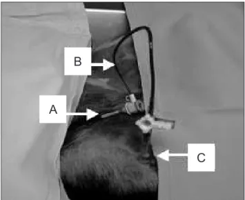
In the Intervention Group, the dogs had their right common femoral arteries ligated at the inguinal region through a transverse incision. They were then given heparin 100 U/kg and a shunt between the right iliac artery and the medullary cavity of the right tibia was performed. In order to perform the temporary shunt, a small pretibial incision was made, followed by exposi-tion of the superior third of the tibia. The bone was then perforated with a 4.5 mm drill through the medul-lary cavity. A plastic cannula was attached to the orifice (Figure 1). Just proximal to the common femoral artery ligature, a 5F catheter was introduced (Figure 1). The system of irrigation was then connected to the cannula, thus establishing the shunt cycle (Figure 1).
Patency of the system was measured every 15 min-utes using Doppler ultrasonography 10 Mhz (Microen, Brazil). This method was also used to evaluate arterial flow in the ischemic limbs. When occlusion of the sys-tem was observed, irrigation with heparin saline solution was performed (1,000 mL/1 mL) until it was solved.
After two hours, blood from the bleeding of the low-er ischemic limb was collected for pH measurement and color assessment. Complete gasometric analysis could not be performed because the volume of blood collected from the animal’s paw was not adequate.
Figure 1. Operation aspect. Catheter (A) inserted in the right external
iliac vein of the dog, irrigation system (B), and cannula (C) placed in the medullary cavity of the right tibia.
B
A
The animals were euthanized using the same proce-dures listed for Control Group, including the removal of the lungs for anatomopathological analysis. The organs were embedded in formaldehyde 10% and sectioned for macroscopic analysis. Ten samples of the pulmonary parenchyma were randomly collected by a blinded pa-thologist. These samples were submitted to the routine histological procedures (Hematoxylin Eosin staining) and to optical microscopy (100 and 400 X) assessment, also performed by a blinded pathologist.
To process these data, the authors used the SPSS 15.0 program and obtained mean values, as well as standard er-rors and deviations.
Results
he characteristics of the individuals of groups did not difer (Table 1). Mean weight was 9.8 kg (±1.8) in the Control Group and 10.5 kg (±3.5) in the Intervention Group. Mean invasive blood pressure was 88.6 mmHg (±12.6) in the Control Group and 91.8 mmHg (±22) in the Intervention Group.
The pH of the sample collected from the femo-ral artery before ligation (pre-intervention) was 7.34 (±0.005) in the Control Group, and 7.33 (±0.005) in the Intervention Group. Initial O2 saturation was 90.6% (±1.4) in the Control Group, and 90.3% (±1.3) in the Intervention Group. The animals remained hemody-namically stable during the procedure.
The shunts presented an average of three occlusions in two hours, but blood flow could be reestablished by
irrigating the system with heparin solution (about 10 to 15 mL).
Doppler ultrasonography showed monophasic low in the distal arteries of the limbs that received shunt.
he blood color from the ischemic limbs ater two hours of the experiment in the Control Group animals was violet and dark, while in the animals from the Intervention Group it was bright red.
Mean capillary blood pH of the ischemic extremity was 6.97 (±0.39) in the Control Group, and 7.25 (±0.46) in the Intervention Group, with p<0.001 (Table 2 e Graph 1).
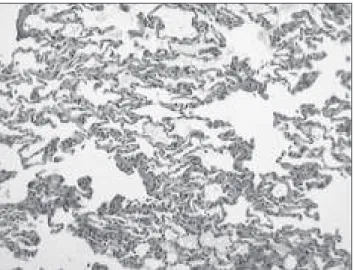
The anatomopathological analysis of the pulmonary parenchyma showed areas of atelectasia and discrete intra-alveolar edema. Neither areas of parenchyma in-farction nor intravascular fat emboli were identified, al-though such emboli could present as cholesterol crystal or intravascular vacuoles (Figures 2 and 3).
Graph 1. pH range in ischemic limbs two hours after arterial occlusion
in both groups.
pH
Group 1: Intervention Group 2: Control
0 1 2
Group 6.9
7.0 7.1 7.2 7.3
Table 2. pH values two hours after the experiment.
Group Mean pH n Standard error p-value
Intervention 7.25 6 0.019 0.001
Control 6.98 3 0.022 0.001
Table 1. Sample characteristics.
Feature Grupo n Mean Standard error p-value
Weight (kg) Intervention 6 10,0 1.46 ns
Control 3 9.9 1.07
SBP (mmHg) Intervention 6 91.8 9.10 ns
Control 3 88.7 6.96
pH Intervention 6 7.33 0.12 ns
Control 3 7.34 0.01
Sat O2 (%)
Intervention 6 90.3 0.55
ns
Control 3 90.6 1.30
Discussion
The temporary shunt was shown to be technically feasible. Shunt occlusions occurred on average three times in two hours of experiment, and could be easily solved by irrigation. This method allowed for partial perfusion of the limb, which was confirmed by tissue pH value, by Doppler flow ultrasonography, and by ob-serving blood color. Lung tissue microscopic analysis showed areas of intra-alveolar edema and atelectasia probably due to respiratory distress, an event observed in both groups, besides the fact that the animals were under anesthesia for four to six hours.
The shunt allows arterial blood flow into the mar-row interstitial cavity, which penetrates the vascular space through the marrow sinusoids4. The
vasculariza-tion of the cortical layer in long bone diaphyses occurs from the marrow to the cortical; the feeding arteries penetrate the marrow, give origin to the longitudinal arteries which, on its turn, give origin to the vessels for the cortical layer of the bone4-7. The adjacent tissues
re-ceive the perfusion through the anastomosis with the periosteum.
Intraosseous access is widely used for emergency treatment in children and adults3, and over 100 drugs
and solutions have been reported to be administered by this means8. Complications are rare. Fat emboli is a
particularly worrisome event because it may cause fat embolism syndrome, that may result in lung and brain damage9,10. Fat embolism was not detected by
macro-scopic and micromacro-scopic evaluations, and is a rare com-plication related to intraosseous infusion of crystalloid solutions3. The fat embolism syndrome is often
associ-ated with long bone fractures and bone tissue manipu-lation, and is caused by the marrow veins’ rupture9-11.
The study initially focused on the technical success of shunting between a great caliber artery and the mar-row cavity of a long bone, and, technically, it was proved to be feasible.
References
1. Drinker CK, Drinker KR, Lund CC. he circulation in the mamma-lian bone marrow. Am J Physiol. 1922;62:1-92.
2. Josefson A. A new method of treatamet intraosseal injections. Acta Med Scand. 1934;81:550-84.
3. Lane JC, Guimarães HP. Acesso venoso intra-óssea em urgências médicas. Rev Bras Terapia Int. 2008;20(1):4-10.
4. Williams W, Dyson B. Gray Anatomia. São Paulo: Guanabara Koogan; 1995.
5. Latarjet M, Riard AR. Anatomia Humana. 3a. ed. Madri: Editora Panamericana; 1999.
6. Pazzaglia UE, Congiu T, Raspanti M, Ranchetti F, Quacci D. Anatomy of the intracortical canal system: scanning electron microscopy study in rabbit femur. Clin Orthop Relat Res. 2009;467:2446-56.
7. Pazzaglia UE, Congiu T, Ranchetti F, Salari M, Dell’Orbo C. Scanning electron microscopy study of bone intracortical vessels using an injection and fractured surfaces technique. Anat Sci Int. 2010;85:31-7.
8. Dubick MA, Holcomb JB. A review of intraosseus vascular access: current status and military application. Mil Med. 2000;165:552-9.
9. De Araújo CAF, Rocha MA, Taia C, Silva LC. Síndrome de embolia gordurosa. Rev Bras Ortop. 1997;32:909-12.
Figure 3. Scarce intra-alveolar protein content (arrows). Optical
micro-scopy at 400x.
Figure 2. Histological slices presenting normal alveoli (HE staining at
10. Filomeno LTB, Carelli CR, Da Silva NC, De Barros Filho TEP, Amatuzzi MM. Embolia gordurosa: Uma revisão para a prática or-topédica atual. Acta Oror-topédica Bras. 2005;13(4):196-208.
11. Corsi PR, Gianini JA, Rasllan S. Embolia gordurosa pós-traumática: revisão da literatura. Acta Cir Bras. 1993;8(3):134-40.
Correspondence Ronaldo André Poerschke Rua Princesa Isabel, 128 CEP 99050-100 – Passo Fundo (RS), Brazil E-mail: [email protected]