Key words:
Gastrointestinal Hemorrhage; Seminoma; Fistula; General Surgery
Int Braz J Urol. 2013; 39: 747-51
__________________
Submitted for publication: June 27, 2012
__________________
Accepted after revision: May 25, 2013
Fistula between arteries and the gastrointestinal tract are a rare cause of gastrointestinal bleeding, but potentially fatal. The recognition and early treatment can modify the patient prognosis. We report a case of a patient with previous surgery for seminoma of cryptor-chidic testicle, with massive lower gastrointestinal bleeding. We performed the diagnosis and surgical treatment of the fistula between left external iliac artery and sigmoid colon. The patient was successfully treated by external iliac artery ligation and left colectomy.
INTRODUCTION
Main causes of massive lower
gastroin-(very rare) and secondary, is complex. Morbidity and mortality are high when associated with blee-ding and sepsis (2-4).
Lower gastrointestinal bleeding as a result of fistula
between the iliac artery and sigmoid colon in patient
with advanced testicular cancer
_______________________________________________
Vanessa Prado dos Santos, Álvaro Razuk Filho, Valter Castelli Júnior, Roberto Augusto Caffaro
Instituto de Humanidades Artes e Ciências Professor Milton Santos, Bahia Federal University (VPS), Bahia; Department of Vascular Surgery, Medical Sciences Faculty of Santa Casa de Sao Paulo (ARfilho, VCJr, RAC), Sao Paulo, Brazil
ABSTRACT
ARTICLE
INFO
the last 7 days. He had been submitted to left or-chiectomy 3 months ago due to neoplasia of criptor-chidic testicle. Pathological exam showed seminoma of left testicle and the patient had also been sub-mitted to three sessions of chemotherapy after or-chiectomy. During physical exam, the patient was pale, tachycardic, eupneic, with a blood pressure of 80x40 mmHg. Hematocrit was 21.3% (normal 36-47%). He was hemodynamically stabilized and submitted to arteriography due to lower intesti-nal bleeding, in order to identify the source and localization of the bleeding. Due to massive blee-ding, scintigraphy was not performed. Initial aor-tography showed extravasation of contrast media in the left external iliac artery, compatible with pseudoaneurism, and probable communication (fistula) with large intestine (Figure-1).
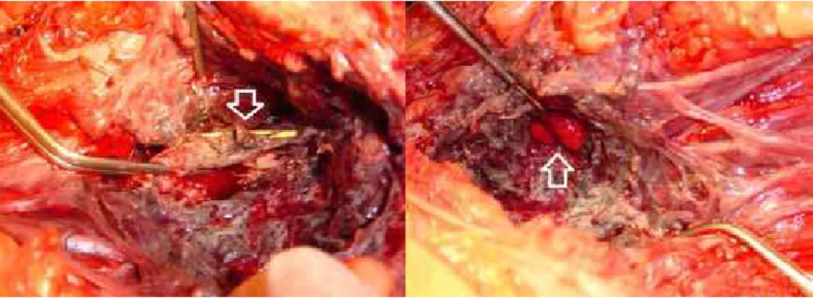
Following arteriography, patient was imme-diately submitted to surgical treatment by vascular and general surgeons. The patient was placed in ri-ght lateral decubitus and a left extraperitoneal access was obtained in order to rapidly control the bleeding from left external iliac artery, the source of the ble-eding. However, an intense periarterial block made the access to left external iliac artery very difficult, in order to control bleeding. So, a right inguinotomy was performed, in order to dissect common femoral artery and to introduce a 7 Fr Fogarty© until aorta, where the balloon was inflated. This maneuver allo-wed the control of the arterial bleeding. In order to identify the left external iliac artery in the middle of the intense local block, a left inguinotomy was also performed and it was also introduced a 7F Fogarty© catheter through left common femoral artery. This catheter allowed the identification of all left external iliac artery that showed several lesion throughout the pelvic block and also allowed total control of bleeding (Figure-2).
In between the inflammatory block of left iliac fossa it was possible to visualize sigmoid co-lon mucosa right to the region of the arterial rup-ture (Figure-2). Due to gross contamination of the abdominal cavity, it was performed ligature of left external iliac artery (proximal and distal stumps) and sigmoidectomy followed by colostomy. Internal left iliac artery was pervious and with good caliber and kept a good perfusion of left leg during arterial clamping, allowing arterial ligature. Patient evolved without complications and was discharged after 10 days. There were no signs of left leg ischemia and
Figure 2 - Intra-operatory image showing extensive destruction or the arterial wall of the left external iliac artery, containing the 7FR Fogarty© catheter (arrow down) and visualization of the sigmoid mucosa (arrow up) (forceps).
the patient was redirected to oncologic treatment. Pathological exam of the surgical specimen showed inflammatory process with fistula to large intestinal and lymphonodal reaction lymphoid hyperplasia.
DISCUSSION
There are very few reports in literature concerning gastrointestinal bleeding related to se-minoma. Leslie et al. related a case of a patient with seminoma with thrombosis of inferior vena cava and melena, due to tumor infiltration of duo-denum (5). Lymphatic dissemination of seminoma was reported by Miller et al. (6), causing retrope-ritoneal mass and duodenal ulcer and gastroin-testinal bleeding. In that case report, the clinical presentation was different, since there was a fistu-la between artery and sigmoid colon, with massi-ve intestinal bleeding and extensimassi-ve arterial wall destruction. The patient was kept under oncolo-gic treatment (that was initiated two months ago) despite absence of tumor in pathological exams. Rupture of external iliac artery was reported in two patients with advanced ovarian tumor, after 11 and 12 days of post-operatory of pelvic lym-phadenectomy (7). Both patients were submitted to ligature of external iliac artery without ische-mic complications of the left leg (7). Ligature of external iliac artery was also reported in a pa-tient with anastomotic pseudoaneurism after renal transplantation (8). This patient was also submit-ted to ligature of external iliac artery without left leg ischemia and complications. Possibly a wide collateral arterial network among the arteries of thorax, pelvis and lower limbs allows refill of fe-moral arteries in case of occlusion of the aorto--iliac sector (9,10). The internal iliac artery of this
Classically, the surgery of fistulae betwe-en arteries and the gastrointestinal tract comprises one arterial ligature and extra-anatomic revascu-larizations due to local contamination (2-4). More recently, endovascular treatment of aorto-enteric fistulae has been proposed in literature, but when the arterial communication is with the colon, the risk of infection and sepsis usually indicates the need of conventional surgery (3). The time betwe-en the first bleeding of an aorto-betwe-enteric fistulae, called sentinel, and the massive bleeding usually takes hours or even months, but the correct diag-nosis during the first bleeding improves progdiag-nosis (4). Although arteriography presents less sensitivi-ty than scintigraphy to localize the source of in-testinal bleeding, a positive exam is able to predict which patients will need surgical treatment (1). During massive intestinal bleeding, the identifica-tion of the localizaidentifica-tion of the bleeding using ar-teriography allows segmental intestinal resection, and better outcome (1). In the present report, the massive intestinal bleeding with hemodynamic alteration lead us to perform an initial and pre-cocious angiography, that correctly identified the source and place of bleeding, allowing an efficient surgical treatment.
CONFLICT OF INTEREST
None declared.
REFERENCES
3. Burks JA Jr, Faries PL, Gravereaux EC, Hollier LH, Marin ML: Endovascular repair of bleeding aortoenteric fistulas: a 5-year experience. J Vasc Surg. 2001; 34: 1055-9.
4. Song Y, Liu Q, Shen H, Jia X, Zhang H, Qiao L: Diagnosis and management of primary aortoenteric fistulas--experience learned from eighteen patients. Surgery. 2008; 143: 43-50. 5. Leslie JA, Stegemann L, Miller AR, Thompson IM: Metastatic
seminoma presenting with pulmonary embolus, inferior vena caval thrombosis, and gastrointestinal bleeding. Urology. 2003; 62: 144.
6. Miller TT, Mendelson DS, Wu LT, Halton KP: Seminoma of the testis presenting as an ulcerating mass of the duodenum. Clin Imaging. 1992; 16: 201-3.
7. Fotopoulou C, Neumann U, Kraetschell R, Lichtenegger W, Se-houli J: External iliac artery ligation due to late postoperative rupture after radical lymphadenectomy for advanced ovarian cancer--two case reports. Eur J Gynaecol Oncol. 2010; 31: 198-200.
8. Silva LF, Marinho ACO, Gonçalves RT, Ferreira FB. Pseudoan-eurisma pós-transplante renal – relato de caso. Rev Col Bras Cir. 2006; 33: 413-15.
9. Vine HS, Sacks BA: ‘Visualization of the distal arterial vessels in complete aortic occlusion. AJR Am J Roentgenol. 1980; 134: 847-8.
10. Krupski WC, Sumchai A, Effeney DJ, Ehrenfeld WK: The im-portance of abdominal wall collateral blood vessels. Planning incisions and obtaining arteriography. Arch Surg. 1984; 119: 854-7.
EDITORIAL COMMENT
This is a rare case of a vascular enteric fistula presenting with massive rectal hemor-rhage. As one may be unfortunate to run into such a challenging case, some points ought to be highlighted:
If diagnosis is suspected, immediate sur-gical aggressive management is required and should not be postponed;
A vascular surgeon presence is greatly appreciated as bleeding control may be very difficult;
Attempt to reconstruct the vessel is not the first option and ligation of the vessel is to be considered even when dealing with a terminal vessel such as the exter-nal iliac artery.
Dr. Ricardo Miyaoka