Inhibitory Activity of (+)-Usnic Acid against
Non-Small Cell Lung Cancer Cell Motility
Yi Yang1, Thanh Thi Nguyen1,2, Min-Hye Jeong1, Florin Crişan3, Young Hyun Yu4, Hyung-Ho Ha4, Kyung Hee Choi4, Hye Gwang Jeong5, Tae Cheon Jeong6, Kwang Youl Lee7, Kyung Keun Kim8, Jae-Seoun Hur1*, Hangun Kim4*
1Korean Lichen Research Institute, Sunchon National University, Sunchon, Republic of Korea,2Faculty of Natural Science and Technology, Tay Nguyen University, Buon Ma Thuot, Vietnam,3Department of Taxonomy and Ecology, Faculty of Biology and Geology, Babeș-Bolyai University, Cluj-Napoca, Romania,
4College of Pharmacy and Research Institute of Life and Pharmaceutical Sciences, Sunchon National University, Sunchon, Republic of Korea,5College of Pharmacy, Chungnam National University, Daejeon, Republic of Korea,6College of Pharmacy, Yeungnam University, Gyeongsan, Korea,7College of Pharmacy, Chonnam National University, Gwangju, Korea,8Medical Research Center for Gene Regulation, Chonnam National University Medical School, Gwangju, Republic of Korea
*hangunkim@sunchon.ac.kr(HK);jshur1@sunchon.ac.kr(JSH)
Abstract
Lichens are symbiotic organisms that produce various unique chemicals that can be used for pharmaceutical purposes. With the aim of screening new anti-cancer agents that inhibit cancer cell motility, we tested the inhibitory activity of seven lichen species collected from the Romanian Carpathian Mountains against migration and invasion of human lung cancer cells and further investigated the molecular mechanisms underlying their anti-metastatic activity. Among them,Alectoria samentosa,Flavocetraria nivalis,Alectoria ochroleuca, and Usnea floridashowed significant inhibitory activity against motility of human lung cancer cells. HPLC results showed that usnic acid is the main compound in these lichens, and (+)-usnic acid showed similar inhibitory activity that crude extract have. Mechanistically,β -catenin-mediated TOPFLASH activity and KITENIN-mediated AP-1 activity were
decreased by (+)-usnic acid treatment in a dose-dependent manner. The quantitative real-time PCR data showed that (+)-usnic acid decreased the mRNA level of CD44, Cyclin D1 and c-myc, which are the downstream target genes of bothβ-catenin/LEF and c-jun/AP-1. Also, Rac1 and RhoA activities were decreased by treatment with (+)-usnic acid. Interest-ingly, higher inhibitory activity for cell invasion was observed when cells were treated with (+)-usnic acid and cetuximab. These results implied that (+)-usnic acid might have potential activity in inhibition of cancer cell metastasis, and (+)-usnic acid could be used for anti-can-cer therapy with a distinct mechanisms of action.
Introduction
Lung cancer is the leading cause of cancer death in developed countries. Due to the lack of effi-cient treatment for advanced disease, the prognosis of lung cancer is still poor, with less than
a11111
OPEN ACCESS
Citation:Yang Y, Nguyen TT, Jeong M-H, Crişan F, Yu YH, Ha H-H, et al. (2016) Inhibitory Activity of (+)-Usnic Acid against Non-Small Cell Lung Cancer Cell Motility. PLoS ONE 11(1): e0146575. doi:10.1371/journal.pone.0146575
Editor:Frédéric André, Aix-Marseille University, FRANCE
Received:September 2, 2015
Accepted:December 18, 2015
Published:January 11, 2016
Copyright:© 2016 Yang et al. This is an open access article distributed under the terms of the Creative Commons Attribution License, which permits unrestricted use, distribution, and reproduction in any medium, provided the original author and source are credited.
Data Availability Statement:All relevant data are within the paper.
Funding:This study was supported by the National Research Foundation of Korea
15% surviving 5 years after diagnosis [1]. Adjacent invasion and distant metastasis are the major causes of cancer-related death [2]. Therefore a search for inhibitors for cancer cell inva-sion and migration ability could reveal a new therapy for cancer treatment. Although herbs have been employed in the treatment of cancers for thousands of years, they remain a very important source of biologically active products. The aim of this study was to identify potential therapeutic agents to improve the survival of patients with lung cancer metastasis.
Lichens are symbiotic organisms that produce a large number of bioactive substances over 800 [3], comprising many classes of compounds: amino acid derivatives, sugar alcohols, ali-phatic acids,γ-,δ- and macrocyclic lactones, monocyclic aromatic compounds, quinones, chromones, xanthones, dibenzofuranes, depsides, depsidones, depsones, terpenoids, steroids, carotenoids [4] and diphenyl ethers [5,6]. Slowly growing organisms in low-resource habitats produce higher levels of defense chemicals [7]. Therefore, lichens are a source of unique chemi-cal agents of which some have already been proved to be effective against various cancerin vitromodels [8]. Here, the current study examined the inhibitory activity of seven lichen spe-cies collected from the Romanian Carpathian Mountains against migration and invasion ability of human lung cancer cells and further investigated the possible molecular mechanisms under-lying their anti-metastatic activity to identify potential compounds for novel anti-metastasis agents.
Material and Methods
Preparation of lichen extracts
Lichen specimens used in this study, collected from Romania in 2011, were identified at the Korean Lichen Research Institute (KoLRI), Sunchon National University, Korea. Briefly, thalli of lichen were collected from Romania in 2011 during the field trip in the National Park Că
li-mani (47°07'28.6"N, 25°13'34.8"E) and the Natural Park Bucegi (45°20'21.7"N, 25°27'41.4"E) organized by Dr. Crişan at Babeş-Bolyai University, Cluj-Napoca, Romania [9]. The permit to
collect lichen specimens from those locations was issued by the Administration of the National Park Călimani and the Administration of the Natural Park Bucegi, with the approval of the
Commission for Protection of Natural Monuments (Romanian Academy). The field studies did not involve any endangered or protected species. The duplicates were deposited into the Korean Lichen and Allied Bioresource Center (KOLABIC) in the Korean Lichen Research Institute (KoLRI), Sunchon National University, Korea. The dried thalli of the lichens were extracted with acetone at room temperature for 48 h. The acetone extracts were then filtered and dried in rotary vacuum evaporator at 45°C. The dry extracts were dissolved in dimethyl-sulfoxide (DMSO) as 5 mg/ml concentration (1000×) for all experiments. Seven Romanian lichen species and their voucher specimen numbers used in this study were listed inTable 1.
High performance liquid chromatography (HPLC) analysis of lichen
material
Acetone extract of lichen thalli at a concentration of 5 mg/ml were subjected to high perfor-mance liquid chromatography (HPLC) analyses (LC-20A; Shimadzu, Kyoto, Japan) on a YMC-Pack ODS-A (150 × 3.9 mm I.D.) reversed-phase column containing fully end-capped C18 material (particle size, 5μm; pore size, 12 nm). Elution was performed at a flow rate of 1
ml/min under the following conditions before subsequent injection: column temperature, 40°C; solvent system, methanol: water: phosphoric acid (80: 20: 1, v/v/v). Analyses were moni-tored by a photodiode array detector (SPD-M20A; Shimadzu) with a range of 190~800 nm throughout the HPLC run. Observed peaks were scanned between 190 and 400 nm. The
Competing Interests:The authors have declared that no competing interests exist.
standard used for salazinic acid (tR= 2.27 ± 0.2 min) was isolated from lichenLobaria
pulmo-naria. Usnic acid used in our study was purchased from Sigma-Aldrich (St. Louis, USA) (329967-5G). Voucher specimens were deposited in the herbarium of the Lichen & Allied Bior-esource Centre at the Korean Lichen Research Institute, Sunchon National University, South Korea.
Liquid chromatography-mass spectrometry (LC-MS) and optical rotation
analaysis of lichen material
LC—MS spectra were recorded on a spectrometer with an electrospray ionization source using Agilent 6460 triple Quadrupole LC/MS. The values of optical rotation were measured at 25°C using Jasco P-1010 polarimeter with a sodium lamp, and described as follows: [α]D, T (c (g/ 100 mL), solvent). Specific rotation of pure (+)-usnic acid (Sigma-Aldrich, St. Louis, USA) is a physical property of at a given wavelength and temperature and can be looked up in literature.
Cell culture
The human lung cancer cells including A549, H460, H1650, and H1975 were cultured in RPMI 1640 culture medium supplemented with 10% fetal bovine serum, 1% Penicillin-Streptomycin solution under a humidified 5% CO2atmosphere at 37°C.
Wound healing assay
A549 cells were plated at a density of 2.5 × 10⁵cells/well on 6-well tissue culture plates (Corn-ing, New York, USA) and grown overnight to confluence. Monolayer cells were scratched with a pipette tip to create a wound. The cells were then washed twice with serum-free RPMI 1640 to remove floating cells and incubated in RPMI1640 culture medium supplemented with 2% FBS with 5μg/ml of the lichen extract or 5μM usnic acid. Photographs of cells were taken at 0,
24, 48, and 72 h after wounding to measure the width of the wound. For each sample, an aver-age of five wound assays was taken to determine the averaver-age rate of migration at a given con-centration of acetone extract or usnic acid. Experiments were repeated at least three times.
Invasion assay
Invasion assays were performed in transwell chambers (Corning, New York, USA) with 8μm
pore size polycarbonate membrane coated with 1% gelatin. Cells were plated at 2 × 10⁵cells/
Table 1. Seven Romanian lichen species used in this study.
Collection No.
Family Lichen species Known lichen substances Reference
RO11025 Parmeliaceae Alectoria samentosa
(-)-Usnic acid, Physodic acid, 8’-O-ethyl-P-alectoronic acid, Alectosarmentin [13]
RO11045 Parmeliaceae Flavocetraria nivalis (±)- Usnic acid, Isousnic acid, Divaricatic acid,p-Hydroxybenzoic acid, Vanillic acid [14–17] RO11084 Parmeliaceae Alectoria
ochroleuca
Diffractaic acid, (-)-Usnic acid, Isousnic acid, Friedelin, Barbatic acid [16,18]
RO11111 Parmeliaceae Bryoria capillaris Alectorialic acid, Barbatolic acid [19]
RO11166 Parmeliaceae Hypogymnia physodes
Atronorin, Physodalic acid, Protocetraric acid, Physodic acid [18,19]
RO11176 Parmeliaceae Usneaflorida (+)-Usnic acid, Barbatic acid, Salazinic acid, Norstic acid,β-Orcinol depsidones, (±)-Thamnolic acid, Stictic acid
[18,20]
RO11209 Parmeliaceae Evernia divaricata Divaricatic acid [19]
well in RPMI1640 containing 0.2% bovine serum albumin in the upper compartment of the chamber with or without 5μg/ml crude lichen extracts. Then RPMI1640 medium with 10μg/
ml fibronectin was added to the lower chamber to serve as a chemotactic agent. After 48-h incubation, the cells in the upper chamber were fixed with Diff Quik kit (Sysmex, Kobe, Japan). Then the cells inside the chamber were mechanically removed from the membrane with a cot-ton swab, and the cells adhering to the under-side of the membrane were stained and counted under light microscope (5 fields per chamber). Each invasion assay was repeated in three inde-pendent experiments. The results are expressed as the mean number of cells migrating per high-power field.
Reporter assay
HEK293T cells were plated into 24-well plates 12 h before transfection. After transfection of the TOPFLASH or AP-1 reporter plasmid with the respective activator,β-catenin or KITENIN, cells were treated with usnic acid for 48 h and then analyzed using a Dual-Luciferase1reporter assay system (Promega, Madison, WI, USA). The Renilla luciferase reporter plasmid (pRL-TK) was used as the internal control for the transfection efficiency. The experiments were per-formed in triplicate, and at least three results from independent experiments were included in the analysis. Fold changes were calculated using values normalized to Renilla luciferase activity.
Quantitative real-time PCR
The quantitative real-time PCR was performed as described previously [9]. Briefly, total RNA was isolated from human lung cancer cells by using RNAiso Plus (TaKaRa, Otsu, Shiga 520–
2193, Japan) according to the manufacturer’s instructions. Total RNA (1μg) from each group
of treated cells was converted to cDNA using a M-MLV reverse Transcriptase kit (Invitrogen, Carlsbad, USA) and SYBR green (Enzynomics, Seoul, Korea). The primers used for real-time PCR were Cyclin D1 (forward)5’-ccgtccatgcggaagatc-3’and (reverse)5’-gaag acctcctcctcgcact-3’; c-myc (forward)5’ -aatgaaaaggcccccaaggtagttatcc-3’and (reverse)5’-gtcgtttccgcaacaagtcctcttc-3’; CD44 (forward)5’-tgcc gctttgcaggtgtat-3’and (reverse)5’-ggcctccgtccgagaga-3’; GAPDH (for-ward)5’-atcaccatcttccaggagcga-3’and (reverse)5’-agttgtcatggatga ccttggc-3’. Real-time PCR reaction and analysis were performed using CFX (Bio-Rad, Hercules, USA).
Affinity Precipitation of Cellular GTPases
The cellular Rac1 and Cdc42 activities were determined using GST-RBD/PBD as previously described [10,11]. Briefly, the cells were lysed in lysis buffer (50 mM Tris, pH 7.4, 1% Triton X-100, 0.5% sodium deoxycholate, 0.1% SDS, 500 mM NaCl, 10 mM MgCl2, and protease
inhibitor mixture). The lysates were incubated with GST- RBD/PBD beads at RT for 1 h. The beads were then washed four times with washing buffer (50 mM Tris, pH 7.4, 1% Triton X-100, 150 mM NaCl, 10 mM MgCl2, and protease inhibitor mixture). The bound Rac1 and
Results
Inhibition of A549 cell motility by lichen extracts
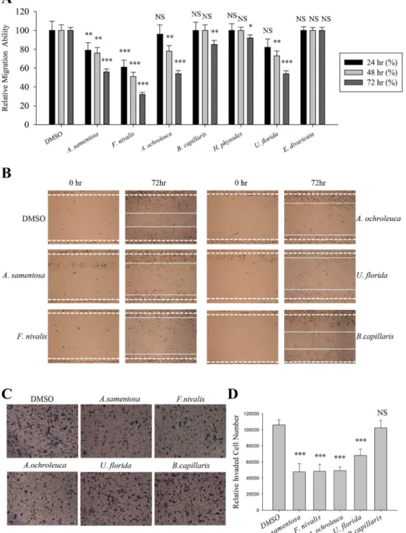
Cell migration plays a crucial role during cancer metastasis. To find the anti-migratory lichen secondary metabolite on human lung cancer cells, wound healing assay was performed among seven acetone extracts of Romanian lichens listed inTable 1. Lichens produce unique polyke-tide secondary metabolites including depsides, depsidones, dibenzofurans, and depsones; and these hydrophobic compounds were normally extracted with acetone [12]. As shown inFig 1A,Alectoria samentosa,Flavocetraria nivalis,Alectoria ochroleuca, andUsnea floridainhibited A549 cell migration at a concentration of 5μg/ml. The length between the edges of the wound
at 72 h with these candidates were significantly wider than that in the DMSO-treated group or the non-active sample (Bryoria capillaris). In particular,F.nivalisshowed more than 60% inhibitory activity compared to control (Fig 1A and 1B).
The effects of acetone extracts ofA.samentosa,F.nivalis,A.ochroleuca, andU.floridaon A549 cell invasion were then determined using transwell chamber invasion assay. As a result, the lichen extracts significantly decreased the invaded cell numbers by as much as 50% com-pared with DMSO orB.capillaries(negative control) (Fig 1C and 1D). These findings demon-strated that acetone extracts ofA.samentosa,F.nivalis,A.ochroleuca, andU.floridainhibited both migration and invasion ability of A549 lung cancer cells.
Usnic acid is the active inhibitor from the lichen species
To identify the components of the acetone extract of lichen,A.samentosa,F.nivalis,A. ochro-leuca, andU.floridaextracts were run on HPLC. As shown inFig 2A, usnic acid was identified as the main compound in all four of these candidates after comparison with the internal stan-dard of purified (+)-usnic acid (Sigma-Aldrich, St. Louis, USA), and these were consistent with previous report (Table 1) [13–20]. Identity of usnic acid and their optical status were analyzed by LC-MS analysis and optical activity analysis, respectively (S1 File). The %intensity of peak for the usnic acid in the candidate lichens at a concentration of 5 mg/ml was obtained by com-paring to that of peak for pure 5 mg/ml usnic acid. It is worth noting that usnic acid content is highest inF.nivalisextract, which may explain its potent inhibitory effect on cell migration (Fig 2A). It was speculated that (-)-usnic acid has similar or more potent inhibitory activity on cell motility as acetone extract ofA.samentosaandA.ochroleucawhich are known to have (-)-usnic acid as their subcomponent [13,16,18] showed similar or more potent inhibitory activity on migration and invasion, respectively, than (+)-usnic acid containingU.florida(Fig 1). In our previous report, we showed that acetone extract of lichenF.cucullataand its compo-nent, usnic acid, inhibited tumorigenicity and motility of cancer cells [9]. In accordance with this, (+)-usnic acid at concentration of 5μM significantly inhibited the migration and invasion
of A549 cells (Fig 2). At this concentration, usnic acid did not show cytotoxicity and/or inhibit cell proliferation (IC50value of usnic acid on A549 cells = 65.3 ± 0.65μM) [9]. As shown inFig
2B–2E, inhibitory activity of (+)-usnic acid at 5μM is as high as 50% at 72 h treatment for
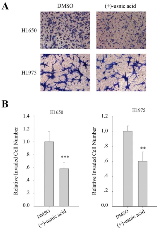
migration and is around 40% at 48 h treatment for invasion. To further examine the inhibitory activity of (+)-usnic acid on the other lung cancer cells, invasion assay was performed using H1650, and H1975 cells. As a result, (+)-usnic acid treatment significantly decreased invaded cell number of H1650 and H1975 cells at a concentration of 5μM (Fig 3A). The quantitative
Fig 1. Inhibition of A549 cell motility by acetone extracts of lichens.(A–B) Quantitative analysis of migration assay of A549 cells treated with 5μg/ml of acetone extracts ofAlectoria samentosa,Flavocetraria nivalis,Alectoria ochroleuca,Bryoria capillaris,Hypogymnia physodes,Usnea floridaandEvernia divaricata(A), and representative images of migration assay of A549 cells treated with the extracts ofA.samentosa,F.nivalis,A.ochroleuca,U.floridaand B.capillaris(B). (C-D) Invasion assay of A549 cells treated with 5μg/ml of acetone extracts ofA.samentosa,F.nivalis,A.ochroleuca,U.floridaandB. capillaris(C), and quantitative analysis of invaded cell numbers in each group (D). Representative images were shown from three independent experiments, n = 3. Data represent mean±S.E.M. (standard error of the mean).***p<0.001; NS, no significant difference compared to 0.01% DMSO-treated A549 cells.
Fig 2. Identification of lichen secondary metabolite from candidate lichens.(A) High performance liquid chromatography (HPLC) analysis of lichen acetone extracts. The %intensity of peak for the usnic acid in the extract at a concentration of 5 mg/ml was obtained by comparing to that of peak for pure 5 mg/ml usnic acid. (B–C) Migration assay of A549 cells treated with 5μM of (+)-usnic acid (B), and quantitative analysis of wound length (C). (D–E) Invasion assay of A549 cells treated with 5μM of (+)-usnic acid (D), and quantitative analysis of invaded cell numbers in each group (E). Representative images are shown from three independent experiments, n = 3. Data represent mean±S.E.M. (standard error of the mean).***p<0.001; NS, no significant difference compared to 0.01% DMSO-treated A549 cells.
Fig 3. (+)-Usnic acid inhibits invasion of H1650 and H1975 human lung cancer cell.(A-B) Invasion assay of H1650, and H1975 cells treated with 5μM of (+)-usnic acid (A), and quantitative analysis of invaded cell numbers in each cell line (B). Representative images are shown from three independent experiments, n = 3. Data represent mean±S.E.M. (standard error of the mean).**p<0.01;***p<0.001; NS, no significant difference compared to 0.01% DMSO-treated A549 cells.
(+)-Usnic acid decreases
β-catenin-mediated TOPFLASH activity and
KITENIN-mediated AP-1 activity
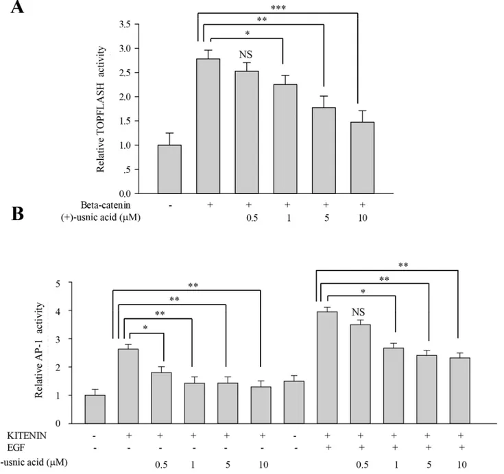
To investigate underlying mechanisms for the inhibitory activity of (+)-usnic acid, we per-formed TOPFLASH and AP-1 reporter assays to assess whether (+)-usnic acid can modulate
β-catenin-mediated and/or KITENIN-mediated signaling activity. As shown inFig 4A, (+)-usnic acid significantly decreased TOPFLASH activity by 18% at 1μM and 37% at 10μM.
As for AP-1 activity, dose-dependent decreases were observed from 0.5μM, and the decrease
was significant, as much as 50% at 10μM. Moreover, (+)-usnic acid also significantly decreased
EGF-activated KITENIN-mediated AP-1 activity by 32% at 1μM and 41% at 10μM (Fig 4B).
To check whether the level of downstream target genes ofβ-catenin/LEF and c-jun/AP-1 were affected by (+)-usnic acid treatment, quantitative real-time PCR analysis was performed. As shown inFig 5, relative expression levels of CD44, cyclin D1, and c-myc were significantly decreased by (+)-usnic acid treatment in lung cancer cells to different extents (Fig 5A–5D). These results suggest that (+)-usnic acid shows inhibitory activity against cell motility through the modulation ofβ-catenin-mediated and KITENIN-mediated signaling activity in lung can-cer cells.
(+)-Usnic acid decreases GTP-Rac1 and -RhoA level
The activities of Rac1 and Cdc42 are involved in mesenchymal mode of migration [21,22]. To determine whether (+)-usnic acid can affect the activities of these proteins in A549 cells, GST pull-down assays were performed using GST-PBD (p21-binding domain). As shown inFig 6, (+)-usnic acid treatment significantly decreased the level of GTP-Rac1 by 22% compared to vehicle-treated cells (Fig 6A). However, no significant change in the level of GTP-Cdc42 was observed by (+)-usnic acid treatment (Fig 6B). RhoA promotes junctional formation, apical constriction, and reduces adhesion and cell spreading [23,24]. To determine whether (+)-usnic acid can affect the activity of RhoA in A549 cells, GST pull-down assays were per-formed using GST-RBD (Rho-binding domain). As shown inFig 6C, (+)-usnic acid treatment significantly decreased the level of GTP-RhoA by 40% compared to vehicle-treated cells. Taken together, these results suggest that (+)-usnic acid inhibits cell motility through the regulation of Rho GTPases.
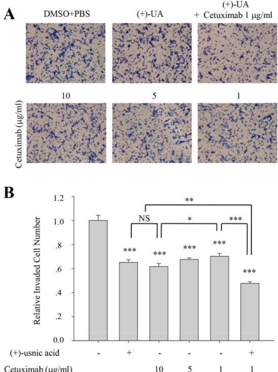
(+)-Usnic acid shows additive inhibitory activity with cetuximab
Cetuximab (Erbitux, C225), monoclonal antibody to epidermal growth factor receptor (EGFR) is used as one of anti-EGFR agents for the treatment of metastatic colon and lung cancer. To examine whether (+)-usnic acid has therapeutic relevancy with cetuximab, invasion assay was performed with various concentration of cetuximab and/or (+)-usnic acid. As shown inFig 7, inhibitory activity of cetuximab was ~30% at 1μg/ml and ~40% at 10μg/ml on A549 cells, and
treatment of 5μM (+)-usnic acid showed similar inhibitory activity with 10μg/ml of cetuximab
(~40% inhibition) on these cells. Interestingly, higher inhibitory activity for cell invasion was observed when the cells were treated with 1μg/ml of cetuximab together with 5μM of
Discussion
Cancer cell acquires biological capabilities including resisting cell death, sustaining prolifer-ative signaling, evading growth suppressors, activating invasion and metastasis, and so forth in developing from early to late stages [26,27], and targeting either of these acquirements can be grouped as‘anticancer’. In this regard, our observations that (+)-usnic acid inhibits migration and invasion ability in lung cancer cells are novel in anticancer activity of (+)-usnic acid. In
Fig 4. (+)-Usnic acid decreasesβ-catenin-mediated TOPFLASH activity and KITENIN-mediated AP-1 activity.(A)β-Catenin-mediated transcriptional activity of TOPFLASH promoter was decreased by (+)-usnic acid treatment. HEK 293T cells were transfected withβ-catenin and TOPFLASH reporter plasmid. After 12 h transfection, cells were treated with (+)-usnic acid for 48h. (B) KITENIN-mediated transcriptional activity of AP1 promoter was decreased by (+)-usnic acid treatment. The HEK 293T cells were transfected with KITENIN and AP-1 reporter plasmid. After 12 h transfection, cells were treated with (+)-usnic acid for 48h with or without EGF. Experiments were performed in at least three independent cultures, n = 3. Data represent mean±S.E.M. (standard error of the mean).*p<0.05;**p<0.01;***p<0.001; NS, no significant difference compared to 0.01% DMSO-treated HEK 293T cells.
addition, our results demonstrated that (+)-usnic acid have specific mechanisms of action for their anticancer activity, and these are quite different from those of previous literature showing cytotoxicity, one of mechanism of actions for the anticancer activity of (+)-usnic acid in various cancer cells.
Hepatotoxicity of usnic acid may restrict their potential medicinal use in cancer therapeutics [28]. However, most of hepatotoxicity in human was observed when high dose of usnic acid was orally administrated as a dietary supplement for the purpose of weight loss [29–31]. In cancer therapeutics, hepatotoxicity can be avoided by adjusting dosage, formulation and/or route of medication. For example, da Silva Santos et al. showed that nano-encapsulation of usnic acid enable to maintain and improve antitumor activity and considerably reduce the hepatotoxicity [32]. Furthermore, it has been shown that supplementation of anti-oxidant, i.e. vitamin E, together with usnic acid could greatly reduce the usnic acid-induced hepatotoxicity in primary cultured mouse hepatocytes [33]. It should also be noted that some of the papers showing hepatotoxicity were carried on HepG2 cells, originated from hepatocellular carcinoma tissue of 15 years male adolescent [34,35]. In our previous paper [9], we demonstrated that usnic acid somehow has selective cytotoxicity in cancer cells when compared to normal cells. In alternative point of views, severe cytotoxicity of usnic acid on HepG2 cells reflects selective and specific anticancer activity of usnic acid on liver cancer in tested concentrations.
Fig 5. (+)-Usnic acid decreases mRNA level of downstream target genes ofβ-catenin/LEF and c-jun/AP-1.(A-D) Quantitative analysis of the mRNA level of CD44, c-myc, and Cyclin D1 in A549 (A), H1650 (B), H1975 (C), and H460 (D) cells treated with 5μM of (+)-usnic acid. Data represent mean±S.E.M. (standard error of the mean), n = 3.*p<0.05;***p<0.001; NS, no significant difference when compared to the 0.01% DMSO-treated group in each cell line.
Fig 6. Regulation of RhoGTPases activity by (+)-usnic acid.(A-C) The levels of GTP-bound Rac1, Cdc42 and RhoA were measured in A549 cells treated with 5μM of (+)-usnic acid. GTP-Rac1 and -Cdc42 were measured using GST-PBD, and GTP-RhoA was measured using GST-RBD. The total amounts of RhoA, Rac1, and Cdc42 were also shown. The relative activities of Rac1 (A), Cdc42 (B), and RhoA (C) were determined as described in Materials and Methods. The data represent the mean±SEM (standard error of the mean), n = 3.**p<0.01;***p<0.001; NS, no significant difference compared to 0.01% DMSO-treated A549 cells.
In our previous report, it was shown that the cytotoxicity of usnic acid is specific to cancer cells such as HT29 (colorectal cancer cells; IC50= 95.2 ± 0.85μM), AGS (gastric cancer cell;
IC50= 15.01 ± 0.52μM), A549 (lung cancer cell; IC50= 65.3 ± 0.65μM), and CWR22Rv-1
(prostate cancer cells; IC50= 24.1 ± 0.63μM), while non-cancer cells such as MDCK (Mardin-Fig 7. (+)-Usnic acid shows additive inhibitory activity with cetuximab.(A-B) Invasion assay of A549 cells treated with 5μM of (+)-usnic acid and/or various concentration of cetuximab (A), and quantitative analysis of invaded cell numbers in each group (B). Representative images are shown from three independent experiments, n = 3. Data represent mean±S.E.M. (standard error of the mean).*p<0.05; **p<0.01;***p<0.001; NS, no significant difference between indicated group.
Darby canine kidney; IC50= 133.04 ± 3.5μM), RIE (rat intestinal epithelial cells; IC50=
126 ± 4.25μM), NIH 3T3 (mouse embryonic fibroblast; IC50= 164.2 ± 3.7μM), and HaCaT
(human keratinocyte; IC50= 185.7 ± 4.8μM) cells, were not severely damaged [9]. Given that
the average circulating blood volume for mice is 72 mL/kg [36] and the molecular weight of usnic acid is 344, LD50value of usnic acid (mouse-oral; 838 mg/kg) in MSDS sheet of usnic
acid can be calculated to 33.8 mM. As inhibitory activity of usnic acid in inhibiting lung cancer cell motility is observed at concentration of 5μM, our results indicated that usnic acid would
be used for anti-metastasis agent with little toxicity at working concentrations.
Usnic acid has a low degree of aqueous solubility, which is an obstacle in the drug develop-ment as it is likely to result in poor bioavailability. To avoid this problem, various approaches can be made for the enhancement of solubility with its own compound by particle size reduc-tion, crystal engineering, salt formareduc-tion, solid dispersion, use of surfactant, complexareduc-tion, and so forth [37]. The selection of the enhancement method should be determined to each drug in required dosage form characteristics. For usnic acid, recent article reported that potassium salt of usnic acid (potassium usnate) has 100% solubility without losing its biological activity at 10μg/ml (approximately 26μM) [38]. Together with the facts that (+) and (-) enantiomeric
forms of usnic acid showed moderate to strong biological activities [39,40], further study is required to evaluate the potential medicinal usage of usnic acid in anticancer therapy in various cancers.
Supporting Information
S1 File. LC-MS Analysis (Figure A in S1 File) and Optical activity Analysis (Figure B in S1 File) of samples used in this study.
(PDF)
Author Contributions
Conceived and designed the experiments: JSH HK. Performed the experiments: YY TTN. Ana-lyzed the data: YY TTN YHY HHH KHC HGJ TCJ KYL KKK JSH HK. Contributed reagents/ materials/analysis tools: MHJ FC. Wrote the paper: YY JSH HK.
References
1. Greenlee RT, Murray T, Bolden S, Wingo PA. Cancer statistics, 2000. CA: a cancer journal for clini-cians. 2000; 50(1):7–33. Epub 2000/03/29. PMID:10735013.
2. Steeg PS. Metastasis suppressors alter the signal transduction of cancer cells. Nature reviews Cancer. 2003; 3(1):55–63. Epub 2003/01/02. doi:10.1038/nrc967PMID:12509767.
3. Huneck S. The significance of lichens and their metabolites. Die Naturwissenschaften. 1999; 86 (12):559–70. Epub 2000/01/22. PMID:10643590.
4. Huneck S, Yoshimura I. Identification of lichen substances: Springer Berlin Heidelberg; 1996. 5. Elix JA, Whitton AA, Sargent MV. Recent progress in the chemistry of lichen substances:
Springer-Vienna; 1984.
6. Fiedler P, Gambaro V, Garbarino JA, Quilhot W. Epiphorellic acids 1 and 2, two diaryl ethers from the lichen cornicularia epiphorella. Phytochemistry. 1986; 25(2):461–5.
7. Coley PD. Effects of plant growth rate and leaf lifetime on the amount and type of anti-herbivore defense: Springer-Verlag; 1988.
8. Kim H, Kim KK, Hur JS. Anticancer activity of lichen metabolites and their mechanisms at the molecular level. In: Upreti DK, Divakar PK, Shukla V, Bajpai R, editors. Modern methods and approaches in lichen systematics and culture techniques. 2: Springer India; 2015. p. 201–8.
suppression of tumorigenic potentials. PloS one. 2014; 9(10):e111575. Epub 2014/11/02. doi:10.1371/ journal.pone.0111575PMID:25360754; PubMed Central PMCID: PMC4216107.
10. Kim H, Han JR, Park J, Oh M, James SE, Chang S, et al. Delta-catenin-induced dendritic morphogene-sis. An essential role of p190RhoGEF interaction through Akt1-mediated phosphorylation. The Journal of biological chemistry. 2008; 283(2):977–87. Epub 2007/11/13. doi:10.1074/jbc.M707158200PMID: 17993462; PubMed Central PMCID: PMC2265781.
11. Kim H, Oh M, Lu Q, Kim K. E-Cadherin negatively modulates delta-catenin-induced morphological changes and RhoA activity reduction by competing with p190RhoGEF for delta-catenin. Biochemical and biophysical research communications. 2008; 377(2):636–41. Epub 2008/10/22. doi:10.1016/j. bbrc.2008.10.030PMID:18930028; PubMed Central PMCID: PMC2614342.
12. Orange A, James PW, White FJ. Microchemical Methods for the Identification of Lichens: British Lichen Society. 2001. p. 101.
13. Gollapudi SR, Telikepalli H, Jampani HB, Mirhom YW, Drake SD, Bhattiprolu KR, et al. Alectosarmen-tin, a new antimicrobial dibenzofuranoid lactol from the lichen, Alectoria sarmentosa. Journal of natural products. 1994; 57(7):934–8. Epub 1994/07/01. PMID:7964789.
14. Bjerke JW, Elvebakk A. Distribution of the lichen genus Flavocetraria (Parmeliaceae, Ascomycota) in the Southern Hemisphere. New Zealand Journal of Botany. 2004; 42(4):647–56.
15. Bjerke JW, Lerfall K, Elvebakk A. Effects of ultraviolet radiation and PAR on the content of usnic and divaricatic acids in two arctic-alpine lichens. Photochemical & Photobiological Sciences. 2002; 1 (9):678–85.
16. Kinoshita Y, Yamamoto Y, Yoshimura I. Distribution of optical isomers of usnic and isousnic acids ana-lyzed by high performance liquid chromatography. Journal of the Hattori Botanical Laboratory. 1997; (83: ):173–8.
17. Zagoskina N, Nikolaeva T, Lapshin P, Zavarzin A, Zavarzina A. Water-soluble phenolic compounds in lichens. Microbiology. 2013; 82(4):445–52.
18. Culberson CF. Chemical and Botanical Guide to Lichen Products: University of North Carolina Press; 1969.
19. Brodo IM, Sharnoff SD, Sharnoff S. Lichens of north America: Yale University Press; 2001. 20. Fiscus SA. A survey of the chemistry of the Usnea florida group in North America. Bryologist.
1972:299–304.
21. Murali A, Rajalingam K. Small Rho GTPases in the control of cell shape and mobility. Cellular and molecular life sciences: CMLS. 2014; 71(9):1703–21. Epub 2013/11/28. doi: 10.1007/s00018-013-1519-6PMID:24276852.
22. Yui Y, Itoh K, Yoshioka K, Naka N, Watanabe M, Hiraumi Y, et al. Mesenchymal mode of migration par-ticipates in pulmonary metastasis of mouse osteosarcoma LM8. Clinical & experimental metastasis. 2010; 27(8):619–30. Epub 2010/09/28. doi:10.1007/s10585-010-9352-xPMID:20872237.
23. Terry S, Nie M, Matter K, Balda MS. Rho signaling and tight junction functions. Physiology (Bethesda). 2010; 25(1):16–26. Epub 2010/02/06. doi:10.1152/physiol.00034.2009PMID:20134025.
24. Etienne-Manneville S, Hall A. Rho GTPases in cell biology. Nature. 2002; 420(6916):629–35. Epub 2002/12/13. doi:10.1038/nature01148PMID:12478284.
25. Bae JA, Yoon S, Park SY, Lee JH, Hwang JE, Kim H, et al. An unconventional KITENIN/ErbB4-medi-ated downstream signal of EGF upregulates c-Jun and the invasiveness of colorectal cancer cells. Clin-ical cancer research: an official journal of the American Association for Cancer Research. 2014; 20 (15):4115–28. Epub 2014/06/05. doi:10.1158/1078-0432.CCR-13-2863PMID:24893630. 26. Hanahan D, Weinberg RA. The hallmarks of cancer. Cell. 2000; 100(1):57–70. Epub 2000/01/27.
PMID:10647931.
27. Hanahan D, Weinberg RA. Hallmarks of cancer: the next generation. Cell. 2011; 144(5):646–74. Epub 2011/03/08. doi:10.1016/j.cell.2011.02.013PMID:21376230.
28. Araujo AA, de Melo MG, Rabelo TK, Nunes PS, Santos SL, Serafini MR, et al. Review of the biological properties and toxicity of usnic acid. Natural product research. 2015:1–14. Epub 2015/02/25. doi:10. 1080/14786419.2015.1007455PMID:25707417.
29. Favreau JT, Ryu ML, Braunstein G, Orshansky G, Park SS, Coody GL, et al. Severe hepatotoxicity associated with the dietary supplement LipoKinetix. Annals of internal medicine. 2002; 136(8):590–5. Epub 2002/04/17. PMID:11955027.
31. Durazo FA, Lassman C, Han SH, Saab S, Lee NP, Kawano M, et al. Fulminant liver failure due to usnic acid for weight loss. The American journal of gastroenterology. 2004; 99(5):950–2. Epub 2004/05/07. doi:10.1111/j.1572-0241.2004.04165.xPMID:15128366.
32. da Silva Santos NP, Nascimento SC, Wanderley MS, Pontes-Filho NT, da Silva JF, de Castro CM, et al. Nanoencapsulation of usnic acid: An attempt to improve antitumour activity and reduce hepatotox-icity. European journal of pharmaceutics and biopharmaceutics: official journal of Arbeitsgemeinschaft fur Pharmazeutische Verfahrenstechnik eV. 2006; 64(2):154–60. Epub 2006/08/11. doi:10.1016/j. ejpb.2006.05.018PMID:16899355.
33. Han D, Matsumaru K, Rettori D, Kaplowitz N. Usnic acid-induced necrosis of cultured mouse hepato-cytes: inhibition of mitochondrial function and oxidative stress. Biochemical pharmacology. 2004; 67 (3):439–51. Epub 2004/03/24. doi:10.1016/j.bcp.2003.09.032PMID:15037196.
34. Aden DP, Fogel A, Plotkin S, Damjanov I, Knowles BB. Controlled synthesis of HBsAg in a differenti-ated human liver carcinoma-derived cell line. Nature. 1979; 282(5739):615–6. Epub 1979/12/06. PMID: 233137.
35. Knowles BB, Howe CC, Aden DP. Human hepatocellular carcinoma cell lines secrete the major plasma proteins and hepatitis B surface antigen. Science. 1980; 209(4455):497–9. Epub 1980/07/25. PMID: 6248960.
36. Diehl KH, Hull R, Morton D, Pfister R, Rabemampianina Y, Smith D, et al. A good practice guide to the administration of substances and removal of blood, including routes and volumes. Journal of applied toxicology: JAT. 2001; 21(1):15–23. Epub 2001/02/17. PMID:11180276.
37. Savjani KT, Gajjar AK, Savjani JK. Drug solubility: importance and enhancement techniques. ISRN pharmaceutics. 2012; 2012:195727. Epub 2012/07/26. doi:10.5402/2012/195727PMID:22830056; PubMed Central PMCID: PMC3399483.
38. Martins MC, Silva MC, Silva LR, Lima VL, Pereira EC, Falcao EP, et al. Usnic acid potassium salt: an alternative for the control of Biomphalaria glabrata (Say, 1818). PloS one. 2014; 9(11):e111102. Epub 2014/11/07. doi:10.1371/journal.pone.0111102PMID:25375098; PubMed Central PMCID: PMC4222767.
39. Ingolfsdottir K. Usnic acid. Phytochemistry. 2002; 61(7):729–36. Epub 2002/11/28. PMID:12453567. 40. Cocchietto M, Skert N, Nimis PL, Sava G. A review on usnic acid, an interesting natural compound. Die