Cancer Cells
Varisa Pongrakhananon, Preedakorn Chunhacha, Pithi Chanvorachote*
Department of Pharmacology and Physiology, Faculty of Pharmaceutical Sciences and Cell-based Drug and Health Product Development Research Unit, Chulalongkorn University, Bangkok, Thailand
Abstract
The migratory capability of cancer cells is one of the most important hallmarks reflecting metastatic potential. Ouabain, an endogenous cardiac glycoside produced by the adrenal gland, has been previously reported to have anti-tumor activities; however, its role in the regulation of cancer cell migration remains unknown. The present study has revealed that treatment with ouabain at physiological concentrations is able to inhibit the migratory activities of human lung cancer H292 cells. The negative effects of ouabain were found to be mediated through the suppression of migration regulatory proteins, such as focal adhesion kinase (FAK), ATP-dependent tyrosine kinase (Akt), and cell division cycle 42 (Cdc42). We found that the observed actions of ouabain were mediated via a reactive oxygen species (ROS)-dependent mechanism because the addition of ROS scavengers (N-acetylcysteine and glutathione) could reverse the effect of ouabain on cell migration. Furthermore, ouabain was shown to inhibit the spheroidal tumor growth and decrease the cancer cell adhesion to endothelial cells. However, the compound had no significant effect on anoikis of the cells. Together, these findings shed light on the understanding of cancer cell biology by exploring the novel function of this endogenous human substance.
Citation:Pongrakhananon V, Chunhacha P, Chanvorachote P (2013) Ouabain Suppresses the Migratory Behavior of Lung Cancer Cells. PLoS ONE 8(7): e68623. doi:10.1371/journal.pone.0068623
Editor:Vladimir V. Kalinichenko, Cincinnati Children’s Hospital Medical Center, United States of America
ReceivedFebruary 4, 2013;AcceptedMay 30, 2013;PublishedJuly 10, 2013
Copyright:ß2013 Pongrakhananon et al. This is an open-access article distributed under the terms of the Creative Commons Attribution License, which permits unrestricted use, distribution, and reproduction in any medium, provided the original author and source are credited.
Funding:This research was supported by the Thailand Research Fund and Ratchadaphiseksomphot Endowment Fund. The funders had no role in study design, data collection and analysis, decision to publish, or preparation of the manuscript.
Competing Interests:The authors have declared that no competing interests exist. * E-mail: [email protected]
Introduction
Understanding the molecular basis of cancer cells in response to biologically derived substances is considered an essential aid to discovering novel molecular targets for drug therapies. Substantial evidence has indicated that ouabain, a sodium/potassium pump inhibitor, is present in human plasma and tissues in the range of 0.002-0.77 nM [1–3]. In addition, ouabain was found to be up-regulated in several pathologies including cardiac failure [1,4], renal failure [5] and hypertension [3,6]. Recently, we have provided evidence indicating that ouabain sensitizes tumor necrosis factor-related apoptosis-inducing ligand (TRAIL)-medi-ated lung cancer cell apoptosis, and this finding suggests that ouabain may affect cancer cell biology [7].
In lung cancer, metastasis has become the most interesting area of research because the high death rate of this disease is associated with cancer metastasis [8]. During cell spreading, the cancer cells must have the ability to migrate from their original sites into the nearby circulatory system. Increased kinase activity of focal adhesion kinase (FAK), a key signaling pathway controlling cell motility, potentiates tumorigenesis and metastasis [9]. Such alterations were also found during the acquisition process of metastatic cancer cells [10,11]. FAK controls signal transduction from integrin-enriched focal adhesion sites of the cell-extracellular matrix interaction [12]. Regarding the regulation of cancer cell migration, FAK phosphorylation at Tyr-397 and the recruitment of Src family kinases are important processes that initiate migration [12–13]. Additionally, the activated status of several migratory regulators such as ATP-dependent tyrosine kinase (Akt)
and cell division cycle 42 (Cdc42) [14,15] are critical for the process of cell movement. Several studies have indicated that activation of Akt enhances the capability of cancer cells to migrate and invade [15,16]. Akt that is localized at the edge of moving cells interacts with actin-binding proteins and induces actin remodeling and the formation of membrane protrusions, facilitating cell motility [15]. This concept was confirmed by a study showing that the down-regulation of Akt using an antisense technique causes a dramatic inhibition of cancer cell invasionin vitro[17] andin vivo
[18]. Additionally, the interaction of FAK and ERK1/2 has been shown to control cell motility [19–20]. Recently, the Rho family of small guanosine triphosphatases (GTPases), especially Cdc42, has been shown to play an essential role in actin reorganization and filopodia formation. The expression level of Cdc42 was found to be up-regulated in many cancers including lung cancer [21–22], and Cdc42 induction was shown to enhance cancer migration [23].
Figure 1. Characterization of human lung cancer H292 cell migration.A: Confluent monolayers of H292 cells were wounded using a 1-mm-wide tip and allowed to migrate for 0–24 h. The wound space was visualized under a microscope, and the relative migration levels compared with the 0 h time point were calculated. Data represent the means6SD (n = 4). *P,0.05 vs. value at the 0 h time point. B: H292 cell migration was examined by a transwell assay for 0–24 h. Migratory cells at the basolateral side of the membrane were stained with Hoechst 33342 for 30 min and visualized under a fluorescence microscope. Data were plotted as an average number of cells in each field and represent the means6SD (n = 4). *P,0.05 vs. value at 0 h time point. C: H292 cells were treated with various concentrations of ouabain (0–5,000 pM) for 24 h. Cell survival was determined by a 3-(4,5-dimethly-thiazol-2-yl)-2,5-diphenyl tetrazolium bromide (MTT) assay. The viability of untreated cells was represented as 100%. Data represent the means6SD (n = 4). *P,0.05 vs. untreated control cells. D: Apoptotic cell death was evaluated using Hoechst 33342 staining. Data represent the means6SD (n = 4). *P,0.05 vs. untreated control cells. E: Cellular apoptosis was determined by DNA content analysis using flow cytometry.
doi:10.1371/journal.pone.0068623.g001
Materials and Methods
Cells and Reagents
The non-small cell lung cancer (NSCLC) cell lines H292 and H460 were obtained from the American Type Culture Collection (Manassas, VA). The cells were cultured in RPMI 1640 supplemented with 5% fetal bovine serum (FBS), 2 mM L-glutamine and 100 units/mL penicillin/streptomycin. The cells were incubated in a 5% CO2 environment at 37uC. Ouabain, dichlorofluorescein diacetate (DCFH2-DA), poly 2-hydroxyethyl-methacrylate (poly-HEMA), 3-(4,5-dimethyl-thiazol-2-yl)-2,5-di-phenyl tetrazolium bromide (MTT) and phalloidin tetramethylr-hodamine B isothiocyanate were obtained from Sigma Chemical, Inc. (St. Louis, MO). Propidium iodide (PI) and Hoechst 33342 were obtained from Molecular Probes, Inc. (Eugene, OR). Antibodies for pan-Akt, p473-Akt, FAK, p397-FAK, Cdc42, caveolin-1 andb-actin, as well as peroxidase-conjugated secondary antibodies were obtained from Cell Signaling Technology, Inc. (Danvers, MA).
Cell Viability and Apoptosis Assays
Cell viability was evaluated using the MTT assay. After the indicated treatments, the cells were incubated with 500mg/ml of MTT at 37uC for 4 h. An intensity reading of the MTT product was measured at 550 nm using a microplate reader, and the percentage of viable cells was calculated in relation to control cells. Apoptosis was determined by Hoechst 33342/PI staining and DNA content analysis. The cells were washed and incubated with 10mg/ml Hoechst 33342 and 5mg/ml PI for 30 min. Nuclei condensation and DNA fragmentation of apoptotic cells and PI-positive necrotic cells were visualized and scored by fluorescence microscopy (Olympus IX51 with DP70). For sub G0 DNA content analysis, after the specified treatment, the cells were trypsinized, washed with phosphate-buffered saline (PBS) and fixed in 70% ethanol at 37uC for 3 h. After washing with phosphate-buffered saline, the cells were incubated in a PI solution containing 0.1% Triton-X, 1mg/ml RNase and 1 mg/ml propidium iodide at room temperature for 30 min. The DNA content was analyzed using flow cytometry (FACSort, Becton Dickinson, Rutherford, NJ).
ROS Detection
Intracellular ROS levels were determined by flow cytometry using DCFH-DA as a fluorescent probe. Briefly, the cells were incubated with 10mM DCFH2-DA for 30 min at 37uC, after which they were washed, trypsinized, resuspended in phosphate-buffered saline, and immediately analyzed for fluorescence intensity by a microplate reader.
Migration Assay
Migration was determined by wound healing and transwell assays. For the wound healing assay, a monolayer of cells was cultured in a 24-well plate, and a wound space was made with a 1-mm-wide tip. After rinsing with PBS, the cell monolayers were incubated with the indicated treatments and allowed to migrate for 24 h. Micrographs were taken under a phase contrast microscope (Olympus DP70, Melville, NY), and the wound spaces were measured from 10 random fields of view using Olympus DP controller software. Quantitative analysis of cell migration was performed using an average wound space from those random fields of view, and the percentage of change in the wound space was calculated using the following formula: % change = (average space at time 0 h) - (average space at time 24 h)/(average space at time 0 h)6100. Relative cell migration was calculated by dividing
the percentage change in the wound space of treated cells by that of the control cells in each experiment. For the transwell assay, the cells were seeded at a density of 56104cells/well onto the upper chamber of a transwell (8mm pore size) in a 24-well plate in serum-free medium and incubated with various concentrations of ouabain. RPMI medium containing 10% FBS was added to the lower chamber. Following the incubation, the non-migrated cells in the upper chamber were removed by cotton-swab wiping, and the cells that migrated to the underside of the membrane were stained with 10mg/ml Hoechst 33342 for 10 min and visualized and scored under a fluorescence microscope (Olympus IX51 with DP70).
Invasion Assay
An invasion assay was performed using a 24-well transwell unit with polycarbonate (PVDF) filters (8mm pore size). The mem-brane was coated with 0.5% matrigel on the upper surface of the chamber overnight at 37uC in a humidified incubator. The cells were plated at a density of 26104cells per well into the upper chamber of the transwell unit in serum-free medium. Medium containing 10% FBS was added to the lower chamber of the unit. After incubation with specific test agents for 24 h at 37uC, the medium in the upper chamber was aspirated, and the cells on the upper side of the membrane were removed with a cotton swab. The cells that invaded to the underside of the membrane were stained with 10mg/ml Hoechst 33342 for 10 min, visualized and scored under a fluorescence microscope.
Cell Morphology and Filopodia Characterization
Cell morphology was investigated by a phalloidin-rhodamine and sulforhodamine B staining assay. The cells were seeded at a density of 104cells/well onto a 96-well plate overnight. The cells were treated with various concentrations of ouabain for 24 h. The cells were then washed with PBS, fixed with 4% paraformaldehyde in PBS for 10 min at 37uC, permeabilized with 0.1% Triton-X100 in PBS for 4 min, and blocked with 0.2% BSA for 30 min. Then, the cells were incubated with either 1:100 phalloidin-rhodamine in PBS or 0.4% sulforhodamine B in 1% acetic acid for 15 min, rinsed 3 times with PBS, and mounted with 50% glycerol. Cell morphology was then assessed by fluorescent imaging (Olympus IX51 with DP70). Filopodia protrusion was represented as the average number of filopodia/cell relative to untreated cells in each field.
In vitro 3D Tumorigenesis Assay
In vitro 3D tumorigenesis was performed in a matrigel-coated 96-well plate. A plate was coated with 0.5% matrigel and left for solidification overnight at 37uC. The cells were suspended in culture medium containing 4% matrigel and ouabain (0–30 pM), and plated at a density of 103 cells/well onto a matrigel-coated plate. Medium containing ouabain (0–30 pM) was replaced every 3 days. After 10 days, the cells were visualized and scored by image analyzer under microscope.
Anoikis Assay
anoikis cells were visualized and scored by fluorescence micros-copy (Olympus IX51 with DP70).
Anchorage-independent Growth Assay
Anchorage-independent growth was determined by colony formation assay in soft agar. Briefly, cells from 6-well plate
Figure 2. Ouabain inhibits lung cancer cell migration and invasion.A: Confluent monolayers of H292 cells were wounded using a 1-mm-wide tip and incubated with ouabain (0–30 pM) for 24 h. The wound space was analyzed and represented as the migration level relative to the change of the untreated cells. B: Migratory cells were stained with Hoechst 33342 for 30 min, visualized under a fluorescence microscope and represented as an average number of cells in each field. C: Cell invasion was evaluated using a transwell coated with matrigel as described under Materials and Methods. After 24 h, the cells that invaded across the membrane were visualized under a fluorescence microscope and represented as an average number of cells in each field. Data represent the means6SD (n = 4). *P,0.05 vs. untreated control cells.
doi:10.1371/journal.pone.0068623.g002
monolayer cultures were prepared into a single-cell suspension. Cells were suspended in RPMI containing 10% FBS and 0.33% low melting temperature agarose, and 26104cells were plated in a 35 mm dish over a layer of solidified RPMI/10% FBS/0.6%
agarose. The cells were fed every three days by adding 200ml of RPMI/10% FBS. Colonies were stained using 10mg/ml Hoechst 33342 for 30 min, visualized and scored by image analyzer under fluorescent microscope (Olympus IX51 with DP70).
Figure 3. Ouabain attenuates filopodia formation.A: H292 cells were treated with 30 pM ouabain (OB) or left untreated as control cells for 24 h. The cells were stained with either sulforhodamine B or phalloidin and visualized under a fluorescence microscope at 406. Filopodia protrusions are indicated by arrows and are represented as an average number of filopodia per cell in each field relative to untreated cells. Data represent the means6SD (n = 4). *P,0.05 vs. untreated control cells. B: After the indicated treatment, the cells were collected and analyzed for cell division cycle 42 (Cdc42) expression by Western blotting. The blots were reprobed withb-actin to confirm equal loading. The immunoblot signals were quantified by densitometry, and mean data from independent experiments were normalized to the results. The bars are the means6SD (n = 4). *P,0.05 vs. untreated control cells.
Monolayer Cell Adhesion Assay
HUV-EC-C were stimulated with 10 ng/ml IL-1b for 0–4 h. After indicated treatment, H292 cells (2.56104 cells/ml) were added onto a semi-confluent monolayer culture of HUV-EC-C, incubated for 20 min at 37uC with rotation at 120 rpm, and washed extensively to exclude nonspecific cell attachment. The number of attached cells was counted directly under a fluorescence microscope.
Western Blotting
After specific treatments, the cells were incubated in lysis buffer containing 20 mM Tris-HCl (pH 7.5), 1% Triton X-100, 150 mM sodium chloride, 10% glycerol, 1 mM sodium orthovanadate, 50 mM sodium fluoride, 100 mM phenylmethylsulfonyl fluoride
and a commercial protease inhibitor cocktail (Roche Molecular Biochemicals) for 30 min on ice. The cell lysates were collected, and the protein content was determined using the Bradford method (Bio-Rad Laboratories, Hercules, CA). Equal amounts of protein from each sample (60mg) were denatured by heating at 95uC for 5 min with Laemmli loading buffer and subsequently loaded onto a 10% SDS-polyacrylamide gel. After separation, the proteins were transferred onto 0.45-mm nitrocellulose membranes (Bio-Rad). The transferred membranes were blocked for 1 h in 5% nonfat dry milk in TBST (25 mM Tris-HCl (pH 7.5), 125 mM NaCl, 0.05% tween 20) and incubated with the appropriate primary antibodies at 4uC overnight. The membranes were washed twice with TBST for 10 min and incubated with horseradish peroxidase-coupled isotype-specific secondary anti-bodies for 1 h at room temperature. The immune complexes were
Figure 4. Effect of ouabain on migratory-related proteins.A: H292 cells were treated with ouabain (0–30 pM) for 24 h. The cells were collected and analyzed for focal adhesion kinase (FAK), phospho-FAK (p-FAK, Tyr397), ATP-dependent tyrosine kinase (Akt), phospho-Akt (p-AKT, Ser473), and caveolin-1 (Cav-1) expression by Western blotting. The blots were reprobed withb-actin to confirm equal loading. B: The immunoblot signals were quantified by densitometry, and mean data from independent experiments were normalized to the results. The bars are the means6SD (n = 4). *P,0.05 vs. untreated control cells.
doi:10.1371/journal.pone.0068623.g004
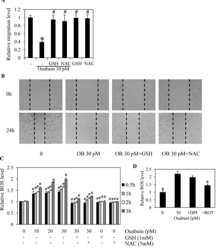
Figure 5. Ouabain inhibits cancer cell migration through reactive oxygen species (ROS) generation.A: Confluent monolayers of H292 cells were wounded by using a 1-mm-wide tip and incubated with antioxidants, 1 mM cell-permeable glutathione (GSH) or 5 mM N-acetyl cysteine (NAC) in the presence or absence of ouabain (0–30 pM) for 24 h. The wound space was analyzed and represented as the migration level relative to the change of the untreated cells. B: Cell migration was captured using a microscope. C: The cells were pretreated with either 1 mM GSH or 5 mM NAC for 30 min in the presence or absence of 30 pM ouabain or ouabain (0–30 pM) alone for 0–3 h. The cells were then incubated with dichlorofluorescein diacetate (DCFH2-DA) for 30 min, and the fluorescence intensity was analyzed by a microplate reader. Mean intensity was
normalized to untreated control cells and represented as relative ROS levels. Data points are means6SD (n = 4). *P,0.05 vs. untreated control cells.
#P
detected by enhancing with a chemiluminescence substrate (Supersignal West Pico; Pierce) and quantified using analyst/PC densitometry software (Bio-Rad).
Statistical Analysis
Mean data from independent experiments were normalized to the results from cells in the control group. All of the experiments were repeated at least four times. A statistical analysis between two groups was verified by Student’s t test, and to compare to multiple groups, an analysis of variance (ANOVA) with a post-hoc test was conducted. A P-value of less than 0.05 was considered statistically significant.
Results
Migratory Characteristic of Non-small Cell Lung Cancer H292 Cells
To test the effect of ouabain on lung cancer cell migration, we first characterized the migration profile of H292 cells using wound migration and transwell assays. Figs. 1A and B show that H292 cells migrated across the wound space in a time-dependent manner, and the results were consistent with those obtained from the transwell migration assay.
Next, a cytotoxicity experiment was performed to test whether biological concentrations of ouabain affect H292 cell viability. The cells were cultured in the absence or presence of ouabain (10–5000 pM), and the viability of the cells was determined using an MTT
Figure 6. Effect of ROS generated by ouabain on migration-related proteins.A: H292 cells were pretreated with either 1 mM GSH or 5 mM NAC for 30 min in the presence or absence of ouabain (30 pM) for 24 h. The cells were collected and analyzed for focal adhesion kinase (FAK), phospho-FAK (p-FAK, Tyr-397), ATP-dependent tyrosine kinase (Akt) and phospho-Akt (p-AKT, Ser-473) expression by Western blotting. The blots were reprobed withb-actin to confirm equal loading. B: The immunoblot signals were quantified by densitometry, and mean data from independent experiments were normalized to the results. The bars are the means6SD (n = 4). *P,0.05 vs. untreated control cells.#P
,0.05 vs. 30 pM ouabain-treated cells.
doi:10.1371/journal.pone.0068623.g006
assay at 24 h. The results indicate that concentrations of ouabain at endogenous levels (2–770 pM) caused neither toxic nor proliferative effects on these lung cancer cells (Fig. 1C). While the concentrations of this substance up to 1000 nM induced no apoptosis, 5000 nM ouabain significantly reduced cell viability (Fig. 1 C) and mediated cell death detected by a Hoechst 33342 nuclear staining assay (Fig. 1D). In addition, the cells were treated with 0–500 pM ouabain for 24 h, and the cells were subjected to flow cytometry for sub G0fraction determination. Fig. 1E shows that treatment of the cells with 10–500 pM ouabain caused no significant alteration of the sub G0fraction in comparison to that of the untreated control cells. These results suggest that concentrations of ouabain found in human plasma and tissues do not affect the viability of H292 cells.
Ouabain Inhibits Lung Cancer Cell Migration and Invasion
Endogenous concentrations of ouabain were further tested for their possible effect on cancer cell migration. The cells were
treated with 0–30 pM ouabain and used in migration assays for 24 h. A wound migration assay showed that ouabain suppressed H292 cell migration in a dose-dependent manner compared with the untreated control cells (Fig. 2A). At the concentration of 20 pM, ouabain caused an approximate 50% reduction in the migratory activity of the cells. Additionally, the transwell migration assay provided supporting data indicating that ouabain signifi-cantly inhibited cell migration (Fig. 2B). Although the defined mechanisms and regulation of cancer cell migration and invasion are not completely known, studies have suggested that there are associated signaling pathways [24]. The effect of ouabain on the invasive potential of cancer cells was further tested using an extracellular matrix-coated transwell assay. The cells were added to the transwell and treated with the indicated concentrations of ouabain for 24 h. Fig. 2C shows that ouabain treatment substantially reduced the number of invaded H292 cells compared with untreated control cells with an approximate 50% reduction found in response to 20 pM ouabain.
Figure 7. Ouabain inhibition of non-small cell lung cancer (NSCLC) H460 cell migration.A: Confluent monolayers of H460 cells were wounded by using a 1-mm-wide tip and incubated with ouabain (0–30 pM) for 24 h. The wound space was analyzed and represented as the migration level relative to the change of the untreated cells. Data are the means6SD (n = 4). *P,0.05 vs. untreated control cells. B. Under the same treatment conditions, the cells were collected and analyzed for focal adhesion kinase (FAK), phospho-FAK (p-FAK, Tyr-397), ATP-dependent tyrosine kinase (Akt), phospho-Akt (p-AKT, Ser-473) and caveolin-1 (Cav-1) expression by Western blotting. The blots were reprobed withb-actin to confirm equal loading. The immunoblot signals were quantified by densitometry, and mean data from independent experiments were normalized to the results. The bars are the means6SD (n = 4). *P,0.05 vs. untreated control cells.
Figure 8. Ouabain suppresses spheroidal tumor growth and cancer cell adhesion to endothelial cells.A: H292 cells were suspended in culture medium containing 4% matrigel and ouabain (0–30 pM) and plated onto matrigel-coated plate. After 10 days, colonies were visualized under microscope with 406(upper row) and 46magnifications (lower row), respectively. Value was represented as average diameter and number of colonies in each field relatively to control cells using image analyzer. Data are the means6SD (n = 4). *P,0.05 vs. untreated control cells. B: H292 were pretreated with various concentration of ouabain (0–30 pM) for 24 h, then, detached H292 cells were subjected to a monolayer culture of HUV-EC-C endothelial cells. The interaction of H292 and HUV-HUV-EC-C cells was examined in the presence of IL-1b(10 ng/ml). Phase-contrast microscopic pictures of representative experiments are shown. Data are means6SD (n = 4), *P,0.05 vs. nontreated control cells. C: H292 cells were detached and treated with ouabain (0–30 pM). Cells were stained with Hoechst for 30 min and visualized under fluorescent microscope. Data are the means6SD (n = 4). *P,0.05 vs. untreated control cells at each time point. D: H292 cells were mixed with medium containing ouabain (0–30 pM) and subjected to soft agar colony formation assay for 2 weeks as describe under ‘‘Material and Methods’’. Colonies were observed under fluorescent microscope. Data are the means6SD (n = 4). *P,0.05 vs. untreated control cells.
doi:10.1371/journal.pone.0068623.g008
Ouabain Reduces Filopodia Formation in H292 Cells Plasma membrane protrusions called filopodia increase during cell movement, and the formation of filopodia is involved in cancer cell migration and metastasis [25]. Having shown that ouabain inhibited the migration and invasion of the cells, we further investigated the effect of this substance on the presence of filopodia in H292 cells. The cells were cultured in the presence or absence of ouabain (30 pM) for 24 h, and filopodia protrusion was captured under fluorescence and phase-contrast modes of a microscope using phalloidin and sulforhodamine B assays, respectively. Fig. 3A shows that H292 cells in the moving state exhibited a substantial number of filopodia protrusions that was dramatically reduced in response to ouabain treatments. These results suggest that the reduction of filopodia in the cells may, at least in part, play a role in the negative regulatory role of ouabain. Sufficient evidence has indicated that filopodia formation in cancer cells involves the Cdc42 protein [26–28]. Because the expression level of Cdc42 was shown to be tightly associated with the formation of filopodia, we further investigated the mechanism of ouabain in the regulation of filopodia in these cells by determining the cellular expression of Cdc42. The results indicate that the expression level of Cdc42 dramatically decreased in response to ouabain treatments (Fig. 3B). While an adequate level of this protein was detected in untreated cells, 30 pM ouabain nearly abolished the Cdc42 expression. This information suggests that ouabain may reduce the migratory activity of these cells via Cdc42 attenuation.
Ouabain Reduces Activation of FAK, Akt, and ERK via a ROS-dependent Mechanism
To elucidate the mechanisms of cancer cell motility inhibition by ouabain, the present study determined the expression and activated levels of proteins involved in cell migration. The cells were treated with non-toxic concentrations of ouabain or left untreated, and Akt, activated Akt (phosphorylated Akt at Ser473) [29], FAK, activated FAK (phosphorylated FAK at Tyr-397) [24] and Cav-1 levels were determined by western blot analysis. Fig. 4 shows that treatment of the cells with ouabain (0–30 pM) substantially down-regulated the expression of activated Akt and activated FAK while preserving their non-activated counterparts. These results suggest that ouabain interfered with the activated level of the proteins and possibly regulated cell migration and invasion by inhibiting Akt and FAK downstream pathways. Recently, we and others have indicated the considerable role of Cav-1 on lung cancer invasive and migratory activities. We further tested whether ouabain attenuates cell motility through decreased Cav-1 levels. Unexpectedly, the level of Cav-1 in response to ouabain treatment was not altered (Fig. 4).
We have shown that the migration of cancer cells is tightly associated with the oxidative status of cells. Previous data indicated that the migratory activity of cancer cells was significantly attenuated in response to antioxidant treatments [30]. To clarify the possible regulatory effect of ouabain in this context, the cells were treated with antioxidants in the presence or absence of ouabain and their migratory behavior and cellular ROS levels were assessed. Figs. 5A and 5B show that treatment with the known antioxidants cell-permeable glutathione (GSH) and N-acetylcysteine (NAC) significantly increased migration of the ouabain-treated cells, confirming the role of ROS in the migratory activity of cells that is suppressed by ouabain. In addition, intracellular ROS levels were determined in the cells incubated with ouabain, GSH, and NAC. ROS detected by a specific oxidative probe showed that cellular ROS increased in response to ouabain treatments (10–30 pM), compared with untreated control,
whereas neither NAC nor GSH alone had an effect on either cell motility or endogenous ROS levels. Importantly, addition of GSH and NAC in the ouabain-treated cells significantly suppressed the ROS production triggered by ouabain (Fig. 1C). Furthermore, we provided a link between cellular ROS and the activation status of Akt and FAK. The cells were pre-treated with either GSH or NAC in the presence or absence of ouabain and the levels of Akt, activated Akt, FAK and activated FAK were determined as described above. The results indicate that administration of the antioxidants reversed the down-regulating effect of ouabain on activated Akt and activated FAK, whereas the non-phosphorylat-ed forms of both proteins were unaffectnon-phosphorylat-ed (Fig. 6). These findings not only provide evidence of a pro-oxidant effect of ouabain, but they also indicate the possible mechanism of ouabain in inhibiting cell migration via a ROS-dependent mechanism.
Ouabain Reduces Migration and Invasion of NSCLC H460 Cells
To test whether ouabain was able to inhibit migration in other lung cancer cells, we treated the human non-small cell lung cancer cell line H460 with ouabain (0–30 pM) and subjected the cells to a migration assay. Notably, the MTT results using the H460 cells indicated that treatment with ouabain at 0–30 pM caused no significant effect on the viability of the cells (data not shown). Consistent with the results obtained from the H292 cells, treatment of the H460 cells with ouabain at the indicated concentrations significantly attenuated cell migration in a dose-dependent manner (Fig. 7A). Additionally, the expression of proteins and their activated counterparts were determined. Western blotting results indicated that treatment with ouabain similarly altered activated Akt, activated FAK, and activated ERK, while the non-activated proteins and Cav-1 levels were not affected by the treatment (Fig. 7B).
Ouabain Suppresses Tumor Growth in 3D Tumor Spheroid Condition and Inhibits Cell detachment
Having shown the role of ouabain in cancer cell migration, we next investigated the potential regulation of cancer metastasis by ouabain. The effect of ouabain on key metastasis steps which are anoikis, cell detachment, and growth in 3D tumor spheroid condition were evaluated. Because previous studies have shown that in vitro 3D tumorigenesis assay provides proximate in vivo
cancer condition and the cells grown as a spheroid activate signaling pathways associated with cancer progression and metastasis in vivo (16, 30–31), we therefore investigated whether
condition compared with nontreated control (Fig. 8D). These results indicated the potential effect of ouabain as a promising anti-metastasis agent.
Discussion
While the normal migration of cells is required for several physiological processes, including tissue repair and regeneration and embryonic development, this cell behavior also drives the pathological progression of cancer metastasis [31]. In lung cancer, a high degree of metastasis has been shown to cause high mortality. Therefore, therapeutic means that suppress cancer cell metastasis are considerably important in the lung cancer-related field. A previous study demonstrated the role of ouabain, a human hormone, on the sensitization of lung cancer cells to TRAIL-induced apoptosis [7]. Ouabain levels in human plasma and tissue distribution from different studies seem to be varied in the range of 0.002–0.77 nM [1–3]. Herein, we demonstrate that endogenous concentrations of ouabain play a role in inhibiting lung cancer cell migration and invasion.
The migration of cancer cells involves multiple cellular signals, and most of them involve the invasive function of the cells. While the exact mechanisms are still being investigated, several proteins including FAK, Akt and Cav-1 have been shown to control motility in cancer cells. Among several regulatory proteins, tyrosine kinases, such as focal adhesion kinase (FAK), have been shown to play central roles in cell motility by transmitting signals to the downstream cellular machinery via their kinase activity [24]. Phosphorylation of FAK at Tyr-397 is a prerequisite for its activated state, and phosphorylated FAK was shown to be elevated in highly motile and invasive cancer cells [32]. In the present study, we demonstrated that phosphorylated FAK was reduced in ouabain-treated cells. Although FAK has been indicated as a redox-sensitive protein and increased ROS levels from exogenous H2O2 enhance FAK activation [33], it was also reported that oxidative stress caused inactivation of several protein tyrosine phosphatases including FAK [34]. Treatment with oxidized low density lipoprotein (oxLDL) shows an increase in endogenous ROS levels, while pretreatment with the antioxidant NAC was able to abrogate this effect in the oxLDL-treated group, consequently triggering FAK activation and promoting cell motility [34]. Our observation supports the later concept that, in certain conditions, ROS play a negative role in FAK activation [34].
Even though many pieces of evidence support the function of the mentioned proteins separately and the pathways regulating cancer cell motility are still largely unknown, a substantial number of studies have determined their regulatory impact on each other. Activated FAK was shown to activate Akt and ERK, and these activations play a key role in cell migration [35]. Based on these data, it is possible that ouabain may suppress FAK activation and then cause a reduction in Akt function through its downstream effectors [24,35].
Recently, Cav-1, a major protein of the plasma membrane domain named caveolae, has been reported to potentiate cancer cells’ ability to resist anoikis [36–37] and to migrate and invade [30]. However, results from the present study indicated that ouabain attenuated the migratory activity of H292 and H460 cells via a Cav-1-independent mechanism (Figs. 4 and 7B).
Furthermore, we provided information regarding effects of ouabain on key steps of cancer metastasis including anoikis, detachment, growth in 3D condition, and cancer cell adhesion to endothelial cells. Tumor Growth in 3D was proposed to mimic the pathological condition of the cancer in vivo and several studies
indicated that the cancer cells grown in a spheroid exhibits signaling pathways and cell behaviors observed in the process of cancer metastasisin vivo [38–40]. Ouabain was shown to inhibit
several key steps of cancer metastasis, namely cell growth in 3D tumorigenesis assay and adhesion to endothelial cells (Fig. 8). However, we found that ouabain has insignificant effect on cancer cell anoikis. These findings have supported the anti-metastasis potentials of ouabain in the physiological concentrations.
Because information regarding endogenous substances that play a significant role in cancer cell behaviors could contribute to the knowledge of the disease, the present study provides important information that may support a better understanding of lung cancer and lead to the development of novel therapies.
Acknowledgments
The authors wish to thank Dr. Yon Rojanasakul and Mr. Krich Rajprasit, a proofreader.
Author Contributions
Conceived and designed the experiments: VP P. Chanvorachote. Performed the experiments: VP P. Chanvorachote. Analyzed the data: VP P. Chanvorachote. Contributed reagents/materials/analysis tools: VP P. Chunhacha P. Chanvorachote. Wrote the paper: VP P. Chunhacha P. Chanvorachote.
References
1. Gottlieb SS, Rogowski AC, Weinberg M, Krichten CM, Hamilton BP, et al. (1992) Elevated concentrations of endogenous ouabain in patients with congestive heart failure. Circulation 86: 420–425.
2. Hamlyn JM, Blaustein MP, Bova S, DuCharme DW, Harris DW, et al. (1991) Identification and characterization of a ouabain-like compound from human plasma. Proc Natl Acad Sci U S A 88: 6259–6263.
3. Manunta P, Messaggio E, Ballabeni C, Sciarrone MT, Lanzani C, et al. (2001) Salt sensitivity study group of the Italian society of hypertension plasma ouabain-like factor during acute and chronic changes in sodium balance in essential hypertension. Hypertension 38: 198–203.
4. Nicholls MG, Lewis LK, Yandle TG, Lord G, McKinnon W, et al. (2009) Ouabain, a circulating hormone secreted by the adrenals, is pivotal in cardiovascular disease. fact or fantasy? J Hypertens 27: 3–8.
5. Kariya K, Sano H, Yamanishi J, Saito K, Furuta Y, et al. (1986) A circulating Na+-K+ATPase inhibitor, erythrocyte sodium transport and hypertension in patients with chronic renal failure. Clin Exp Hypertens A 8: 167–183. 6. Manunta P, Stella P, Rivera R, Ciurlino D, Cusi D, et al. (1999) Left ventricular
mass, stroke volume, and ouabain-like factor in essential hypertension. Hypertension 34: 450–456.
7. Chanvorachote P, Pongrakhananon V (2012) Ouabain down-regulates Mcl-1 and sensitizes lung cancer cells to TRAIL-induced apoptosis. Am J Physiol Cell Physiol (November 21, 2012) doi:10.1152/ajpcell.00225.2012.
8. Mehlen P, Puisieux A (2006) Metastasis: a question of life or death. Nat Rev Cancer 6: 449–458.
9. Itoh K, Yoshioka K, Akedo H, Uehata M, Ishizaki T, et al. (1999) An essential part for Rho-associated kinase in the transcellular invasion of tumor cells. Nat Med 5: 221–225.
10. Chen JS, Huang XH, Wang Q, Chen XL, Fu XH, et al. (2010) FAK is involved in invasion and metastasis of hepatocellular carcinoma. Clin Exp Metastasis 27: 71–82.
11. Hiratsuka S, Goel S, Kamoun WS, Maru Y, Fukumura D, et al. (2011) Endothelial focal adhesion kinase mediates cancer cell homing to discrete regions of the lungs via E-selectin up-regulation. Proc Natl Acad Sci U S A 108: 3725–3730.
12. Mitra SK, Hanson DA, Schlaepfer DD (2005) Focal adhesion kinase: in command and control of cell motility. Nat Rev Mol Cell Biol 6: 56–68. 13. Lietha D, Cai X, Ceccarelli DF, Li Y, Schaller MD, et al. (2007) Structural basis
for the autoinhibition of focal adhesion kinase. Cell 129: 1177–1187. 14. Huang Q, Shen HM, Ong CN (2005) Emodin inhibits tumor cell migration
through suppression of the phosphatidylinositol 3-kinase-Cdc42/Rac1 pathway. Cell Mol Life Sci 62: 1167–1175.
15. Kim D, Kim S, Koh H, Yoon SO, Chung AS, et al. (2001) Akt/PKB promotes cancer cell invasion via increased motility and metalloproteinase production. FASEB J 15: 1953–1962.
16. Scaltriti M, Baselga J (2006) The epidermal growth factor receptor pathway: a model for targeted therapy. Clin Cancer Res 12: 5268–5272.
17. Pu P, Kang C, Li J, Jiang H (2004) Antisense and dominant-negative AKT2 cDNA inhibits glioma cell invasion. Tumour Biol 25: 172–178.
18. Pu P, Kang C, Li J, Jiang H, Cheng J (2006) The effects of antisense AKT2 RNA on the inhibition of malignant glioma cell growth in vitro and in vivo. J Neurooncol 76: 1–11.
19. Huang C, Jacobson K, Schaller MD (2004) MAP kinases and cell migration. J Cell Sci 117: 4619–4628.
20. Webb DJ, Donais K, Whitmore LA, Thomas SM, Turner CE, et al. (2004) FAK-Src signalling through paxillin, ERK and MLCK regulates adhesion disassembly. Nat Cell Biol 6: 154–161.
21. Chen QY, Jiao DM, Yao QH, Yan J, Song J, et al. (2012) Expression analysis of Cdc42 in lung cancer and modulation of its expression by curcumin in lung cancer cell lines. Int J Oncol 40: 1561–1568.
22. Liu M, Lang N, Qiu M, Xu F, Li Q, et al. (2011) miR-137 targets Cdc42 expression, induces cell cycle G1 arrest and inhibits invasion in colorectal cancer cells. Int J Cancer 128: 1269–1279.
23. Johnson E, Seachrist DD, DeLeon-Rodriguez CM, Lozada KL, Miedler J, et al. (2010) HER2/ErbB2-induced breast cancer cell migration and invasion require p120 catenin activation of Rac1 and Cdc42. J Biol Chem 285: 29491–29501. 24. Hsia DA, Mitra SK, Hauck CR, Streblow DN, Nelson JA, et al. (2003)
Differential regulation of cell motility and invasion by FAK. J Cell Biol 160: 753–767.
25. Machesky LM (2008) Lamellipodia and filopodia in metastasis and invasion. FEBS Lett 582: 2102–2111.
26. Mattila PK, Lappalainen P (2008) Filopodia: molecular architecture and cellular functions. Nat Rev Mol Cell Biol 9: 446–454.
27. Nobes CD, Hall A (1995) Rho, rac, and cdc42 GTPases regulate the assembly of multimolecular focal complexes associated with actin stress fibers, lamellipodia, and filopodia. Cell 81: 53–62.
28. Shen Q, Rahn JJ, Zhang J, Gunasekera N, Sun X, et al. (2008) MUC1 initiates Src-CrkL-Rac1/Cdc42-mediated actin cytoskeletal protrusive motility after ligating intercellular adhesion molecule-1. Mol Cancer Res 6: 555–567.
29. Yuan TL, Cantley LC (2008) PI3K pathway alterations in cancer: variations on a theme. Oncogene 27: 5497–5510.
30. Luanpitpong S, Talbott SJ, Rojanasakul Y, Nimmannit U, Pongrakhananon V, et al. (2010) Regulation of lung cancer cell migration and invasion by reactive oxygen species and caveolin-1. J Biol Chem 285: 38832–40, 2010.
31. Thiery JP, Acloque H, Huang RY, Nieto MA (2009) Epithelial-mesenchymal transitions in development and disease. Cell 139: 871–890.
32. Miyazaki T, Kato H, Nakajima M, Sohda M, Fukai Y, et al. (2003) FAK overexpression is correlated with tumor invasiveness and lymph node metastasis in oesophageal squamous cell carcinoma. Br J Cancer 89: 140–145. 33. Usatyuk PV, Natarajan V (2005) Regulation of reactive oxygen species-induced
endothelial cell-cell and cell-matrix contacts by focal adhesion kinase and adherens junction proteins. Am J Physiol Lung Cell Mol Physiol 289: L999– 1010.
34. Park YM, Febbraio M, Silverstein RL (2009) CD36 modulates migration of mouse and human macrophages in response to oxidized LDL and may contribute to macrophage trapping in the arterial intima. J Clin Invest 119: 136– 145.
35. Meng XN, Jin Y, Yu Y, Bai J, Liu GY, et al. (2009) Characterization of fibronectin-mediated FAK signaling pathways in lung cancer cell migration and invasion. Br J Cancer 101: 327–334.
36. Chanvorachote P, Nimmannit U, Lu Y, Talbott S, Jiang BH, et al. (2009) Nitric oxide regulates lung carcinoma cell anoikis through inhibition of ubiquitin-proteasomal degradation of caveolin-1. J Biol Chem 284: 28476–28484. 37. Chunhacha P, Chanvorachote P (2012) Roles of caveolin-1 on anoikis resistance
in non-small cell lung cancer. Int J Physiol Pathophysiol Pharmacol 4: 149–155. 38. Kim JB (2005) Three-dimensional tissue culture models in cancer biology. Semin
Cancer Biol 15(5): 365–77.
39. Nyga A, Cheema U, Loizidou M (2011) 3D tumour models: novel in vitro approaches to cancer studies. J Cell Commun Signal 5(3): 239–48.