The DAF-16/FOXO Transcription Factor Functions as a
Regulator of Epidermal Innate Immunity
Cheng-Gang Zou.*, Qiu Tu., Jie Niu., Xing-Lai Ji, Ke-Qin Zhang*
Laboratory for Conservation and Utilization of Bio-Resources, Yunnan University, Kunming, Yunnan, China
Abstract
TheCaenorhabditis elegansDAF-16 transcription factor is critical for diverse biological processes, particularly longevity and stress resistance. Disruption of the DAF-2 signaling cascade promotes DAF-16 activation, and confers resistance to killing by pathogenic bacteria, such asPseudomonas aeruginosa,Staphylococcus aureus, andEnterococcus faecalis. However,daf-16
mutants exhibit similar sensitivity to these bacteria as wild-type animals, suggesting that DAF-16 is not normally activated by these bacterial pathogens. In this report, we demonstrate that DAF-16 can be directly activated by fungal infection and wounding in wild-type animals, which is independent of the DAF-2 pathway. Fungal infection and wounding initiate the Gaq signaling cascade, leading to Ca2+ release. Ca2+ mediates the activation of BLI-3, a dual-oxidase, resulting in the production of reactive oxygen species (ROS). ROS then activate DAF-16 through a Ste20-like kinase-1/CST-1. Our results indicate that DAF-16 in the epidermis is required for survival after fungal infection and wounding. Thus, the EGL-30-Ca2+ -BLI-3-CST-1-DAF-16 signaling represents a previously unknown pathway to regulate epidermal damage response.
Citation:Zou C-G, Tu Q, Niu J, Ji X-L, Zhang K-Q (2013) The DAF-16/FOXO Transcription Factor Functions as a Regulator of Epidermal Innate Immunity. PLoS Pathog 9(10): e1003660. doi:10.1371/journal.ppat.1003660
Editor:Frederick M. Ausubel, Massachusetts General Hospital, Harvard Medical School, United States of America
ReceivedApril 4, 2013;AcceptedAugust 10, 2013;PublishedOctober 17, 2013
Copyright:ß2013 Zou et al. This is an open-access article distributed under the terms of the Creative Commons Attribution License, which permits unrestricted use, distribution, and reproduction in any medium, provided the original author and source are credited.
Funding:This work was supported in part by a grant (2013CB127500 and 2012CB722208) from National Basic Research Program of China, a grant from the National Natural Science Foundation of China (311171365) and a grant from Yunnan Department of Science and Technology (2009CI045).The funders had no role in study design, data collection and analysis, decision to publish, or preparation of the manuscript.
Competing Interests:The authors have declared that no competing interests exist.
* E-mail: [email protected] (CGZ); [email protected] (KQZ)
.These authors contributed equally to this work.
Introduction
All organisms are in constant contacts with a variety of microorganisms. The innate immune system in hosts provides the first line of defense against these microorganisms. During the last decade, studies usingCaenorhabditis elegansas a model host have revealed the involvement of evolutionarily conserved signaling pathways in the innate immune response to microbial infection and injury, including the DAF-2/DAF-16 insulin-like signaling pathway [1,2].C. elegansDAF-2 is orthologous to the mammalian insulin/insulin-like growth factor-1 receptor [3] anddaf-2mutants exhibit increased resistance to pathogenic bacteria, such as
Pseudomonas aeruginosaandStaphylococcus aureus[4]. Under standard growth conditions, DAF-2 initiates a kinase cascade that leads to the phosphorylation and cytoplasmic retention of its downstream effector DAF-16, the ortholog of mammalian Forkhead box O (FOXO) transcription factors [5,6,7]. A reduction in DAF-2 signaling leads to the dephosphorylation of DAF-16, allowing its nuclear translocation and transcriptional activation [5,6]. The pathogen-resistant phenotype of daf-2 mutants is suppressed by mutations indaf-16, suggesting a crucial role for DAF-16 in innate immunity against bacteria [4]. As a transcriptional factor, activated DAF-16 mediates a variety of genes that are positive regulators of innate immunity against pathogenic bacteria [8,9]. In
Drosophilaand human tissues, FOXOs also induce the expression of a variety of antimicrobial peptides, such as drosomycin and defensins [10], suggesting that the role for FOXOs as innate immunity regulators is highly conserved across species.
Although DAF-16 is involved in immune responses to pathogenic bacteria including P. aeruginosa, S. aureus, Enterococcus faecalisandSalmonella enterica,daf-16 mutants are not significantly more susceptible than wild-type worms to the killing mediated by these bacteria [4,11]. Interestingly, a previous study shows that although the knock-down ofdaf-16by RNAi in wild-type worms does not affect susceptibility to P. aeruginosa PA14, intestinal-specific knock-down ofdaf-16leads to enhanced susceptibility to PA14 [7]. These results suggest that DAF-16 in the intestine, but not in the whole worms, is required for resistance to PA14 infection. One reasonable explanation is that loss of DAF-16 in the intestine, in combination with loss of DAF-16 in other tissues, has an overall neutral effect on resistance to PA14 infection. Meanwhile, two recent studies demonstrate that two bacterial pathogens enteropathogenic Escherichia coli (EPEC) and Bacillus thuringiensis induce DAF-16 nuclear translocation, respectively [12],[13]. These results contradict the previous notion that DAF-16 is activated by something other than pathogens [11,14]. More importantly,daf-16 mutants are more sensitive to the two bacterial pathogens [12,15,16]. However, the mechanism under-lying DAF-16 activation by these bacterial pathogens remains unclear.
Pathogenic bacteria, includingP. aeruginosa,S. aureus,E. faecalis,
elegans infected with D. coniospora [23] and predicted DAF-16 transcriptional target genes [8], we found that there was a significant overlap between D. coniospora-upregulated genes and DAF-16 target genes. These findings prompted us to examine the role of DAF-16 in the innate immune response to fungal infection. After exposure ofC. eleganstoD. coniosporaandC. rosea, we found thatdaf-16 mutants were more susceptible than wild-type worms to killing by fungi. Further studies indicated that fungal infections resulted in the activation of DAF-16 as a consequence of the production of reactive oxygen species (ROS). Similar results were obtained with nematodes subjected to physical injury. Our data demonstrate that DAF-16 can act in a tissue-specific way in the epidermis as an active regulator of immune responses to fungal infection and physical injury.
Results
Fungal infection and physical wounding activate DAF-16 independently of the insulin/IGF-1 pathway
Under standard growth conditions, DAF-16 is distributed predominately throughout the cytoplasm of all tissues [5,6,7]. We compared previously identified DAF-16 target genes [8] to published microarray analysis of gene expression in response toD. coniospora infection [23]. 48 of the genes up-regulated by D. coniospora are also targets of DAF-16 (Figure 1A, Table S1), significantly more than expected by chance (Fisher’s exact test,
P,0.0001). To further confirm these results, we randomly selected eight of these genes and determined their expression by qPCR (Figure 1B). The expression of these eight genes was significantly elevated after D. coniospora infection. However, daf-16 mutation suppressed the up-regulation of these eight genes induced byD. coniospora. These results suggest that nematophagous fungi could activate the transcription activity of DAF-16 in wild-type worms under standard growth conditions.
To test this hypothesis, we monitored the cellular translocation of 16 using transgenic worms that express a functional DAF-16::GFP fusion protein. The status of DAF-16 localization was categorized as cytosolic, intermediate, or nuclear (Figure 1C). We observed that exposure toD. coniosporaorC. roseainduced DAF-16 nuclear localization (Figure 1C). In contrast, infection with P. aeruginosaPA14 orS. aureusATCC 25923 failed to cause increased
DAF-16 nuclear accumulation (Figure 1C), consistent with previous studies [7]. Recent studies have demonstrated that fungal infection and epidermal injury activate similar signaling pathways in C. elegans [19,24]. Infection by nematophagous fungi causes nematode cuticle damage [18,19,20,21,22]. We have previously reported a unique fungal structure, called the spiny ball, on the vegetative hyphae of the fungusCoprinus comatusthat damages the nematode cuticle [25]. To investigate the response to physical wounding of the cuticle, we exposed worms to purifiedC. comatus
spiny balls. After nematodes were added to NGM plates containing purified spiny balls (approximately 10,000/plate), DAF-16 nuclear localization was observed (Figure 1C). Mean-while, the expression of the eight genes was significantly up-regulated in wild-type worms, but not indaf-16(mu86) mutants, after treatment with spiny balls (Figure 1B). We also tested one of classic targets of DAF-16, sod-3, using transgenic worms that express Psod-3::GFP. We found that infection of D. coniospora or treatment with spiny balls up-regulated the expression of Psod-3::GFP(Figure 1D). Knock-down ofdaf-16by RNAi inhibited the expression ofPsod-3::GFPinduced byD. coniosporaor spiny balls. It should be noted that, similar to fungal infection (Figure S1A and S1B in Text S1), mutation indaf-2(e1370)also induced DAF-16 nuclear translocation predominately both in the hypodermis and the intestine without fungal infection (Figure S1C in Text S1). Meanwhile, we found that either epidermal- or intestinal-specific knock-down ofdaf-16 by RNAi suppressed the expression of the eight DAF-16 target genes (Figure S2A and S2B in Text S1). Taken together, these results demonstrate that infection by nematophagous fungi and physical wounding activate DAF-16 inC. elegans.
Reduced signaling in the DAF-2 pathway results in the nuclear accumulation of DAF-16 [5,6]. P. aeruginosa PA14 infection up-regulates the expression of the insulin-like agonist ins-7, thus activating the DAF-2 insulin-like signaling [7,26]. This is one of the mechanisms by which PA14 suppresses nuclear accumulation of DAF-16. It is tempting to speculate that in contrast to bacterial infection, fungal infection reduces expression of insulin-like agonists, thereby leading to the activation of DAF-16. Unexpect-edly, like P. aeruginosa PA14, D. coniospora also up-regulated the expression ofins-7(Figure 1E). We thus examined the effect of ins-7 on DAF-16 translocation and the immune phenotypes, and found that mutations inins-7did not alter DAF-16 translocation and the survival of worms after D. coniospora infection and treatment with spiny balls (Figure S3A–C in Text S1). In addition, the expression ofins-1, an antagonist of DAF-2 signaling [27,28], was down-regulated afterD. coniosporainfection (Figure 1E). These results suggest that similarly to bacterial infection, fungal infection also activates DAF-2 insulin-like signaling, probably by altering the expression of insulin-like peptides. Thus, the activation of DAF-16 does not result from reduced signaling in the DAF-2 pathway, suggesting that other mechanisms exist for the activation of DAF-16 following fungal infection.
DAF-16 in the epidermis is required for the immune response to fungal infection and physical injury
Since fungal infection activated DAF-16, we determined the survival rates of daf-16(mu86) mutants after infection by D. coniospora and C. rosea. We found that daf-16(mu86) mutants exhibited enhanced susceptibility to killing by D. coniospora
(Figure 2A) andC. rosea(Figure S4A in Text S1). Similar results were obtained from worms bydaf-16RNAi (Figure S4B and S4C in Text S1). These results indicate that DAF-16 is directly involved in controlling fungal resistance in wild-type animals. Meanwhile, we also examined the survival of worms in the presence of spiny
Author Summary
balls. daf-16(mu86) animals were more sensitive than wild-type animals to physical injury (Figure 2B).
Unlike pathogenic bacteria that mainly infect the intestine, nematophagous fungi infect the epidermis [23]. To determine tissue-specific activities of DAF-16 in the regulation of immune responses to fungal infection and physical injury, we knocked downdaf-16by RNAi in the intestine, the epidermis, and muscle, respectively. We found that epidermal-specific knock-down of daf-16 resulted in enhanced sensitivity to D. coniospora infection (Figure 2C) and physical injury by spiny balls (Figure 2D). The epidermal-specific knock-down of daf-16 did not alter DAF-16 nuclear translocation in the intestine after D. coniospora infection (Figure S5A and S5B in Text S1). In contrast, intestinal- or muscular-specificdaf-16 RNAi had no effect on sensitivity toD. coniosporainfection (Figure 2E, Figure S6A in Text S1) and spiny
balls (Figure 2F, Figure S6B in Text S1). In addition, expression of
daf-16 under control of an epidermal (dpy-7) promoter [29] enhanced the resistance to D. coniospora infection and physical injury in wild-type animals (Figure S7A and S7B in Text S1). We conclude that DAF-16 functions within the epidermis of nematode to promote immune responses to fungal infection and physical wounding.
ROS production is induced through BLI-3 during fungal infection and physical wounding
Accumulating evidence suggests that the levels of ROS in tissues are induced in response to physical wounding in human epithelial keratinocytes [30,31,32], the tail fin of zebrafish larvae [30,31,32], and the Drosophila embryo epidermis [30,31,32]. Since oxidative stress induces the activation of DAF-16 in C. elegans [33], we
Figure 1. DAF-16 is activated by fungal infection and physical injury.(A) Venn diagrams comparing the overlaps in genes activated byD. coniosporaand the target genes of DAF-16. (B) qPCR analysis of expression of DAF-16 target genes in wild-type (N2), anddaf-16(mu861)mutants 24 h afterD. coniospora(DC) infection or treatment with spiny balls. *P,0.05, N2+DC or N2+SB relative to N2. (C) DAF-16 translocation assay. Transgenic worms expressing DAF-16::GFP were treated withP. aeruginosaPA14 (PA),S. aureus(SA),D. coniospora(DC) andC. rosea(CR), and spiny balls (SB). After 12 h of treatment, the DAF-16::GFP expression pattern was observed. DAF-16 is present in cytosolic (Cyt), intermediate (Int), or nuclear (Nuc) fractions. Quantification of DAF-16 distribution. These results are means6SD of four experiments. *P,0.05 versus control (N2). n = 100–110 nematodes per condition. (D) Expression ofPsod::GFPwas up-regulated in WT animals exposed toD. coniosporaor spiny balls for 12 h.daf-16RNAi inhibited the expression ofPsod-3::GFPinduced byD. coniosporaor spiny balls. (E) The mRNA levels of DAF-2 insulin-like signaling ligands,ins-7and ins-1, in wild-type animals exposed toP. aeruginosaPA14 andD. coniosporafor 12 h, respectively. These results are means6SD of four experiments. *P,0.05versuscontrol (N2).
doi:10.1371/journal.ppat.1003660.g001
hypothesized that the production of ROS is one of the mechanisms underlying DAF-16 activation by fungal infection and physical injury. To test this idea, we first determined the levels of ROS using 29,79-dichlorodihydrofluorescein diacetate (H2DCFDA), a fluorescent dye that has been used to detect the ROS levels inC. elegans[34,35,36]. We found that the levels of ROS were dramatically elevated during fungal infection and treatment with spiny balls (Figure 3A).
Recent studies demonstrate that dual oxidases (DUOXs) mediate ROS production during wound responses in zebra fish larvae and Drosophila embryos [31,32]. InC. elegans, there are two DUOX homologs. BLI-3/Ce-DUOX-1 is the major enzyme responsible for the production of ROS [37]. Since mutations in
bli-3and the standard feeding RNAi with construct based onbli-3
result in a severe blistered phenotype [38,39], we tested the worms subjected to RNAi in a 1/10 dilution as described by Chavez et al. [38,39]. qPCR analysis demonstrated that that knock-down of bli-3in a 1/10 dilution reduced more than 50%bli-3mRNA levels (Figure S8 in Text S1). We found that the knock-down ofbli-3by RNAi significantly reduced the ROS levels induced byD. coniospora
and spiny balls (Figure 3A), indicating that BLI-3 is involved in the increase in ROS levels in these processes. Furthermore, the nuclear accumulation of DAF-16 was markedly reduced bybli-3
RNAi after infection ofD. coniosporaand treatment with spiny balls, respectively (Figure 3B). Similarly, knock-down ofbli-3by RNAi markedly inhibited the expression ofPsod-3::GFPinduced by D.
Figure 2. DAF-16 in the epidermis is required for resistance to fungal infection and physical injury.(A–B)daf-16mutants were sensitive to fungal infection and spiny balls. Fraction ofdaf-16(mu86)and wild-type animals are plotted as a function of time exposure toD. coniospora(A), and spiny balls (B).P,0.001 relative to wild-type animals. (C–D) Contribution of epidermal DAF-16 to sensitivity by fungi and injury. Epidermal-specific RNAi ofdaf-16significantly reduces survival rate of worms exposed toD. coniospora(C), and spiny balls (D).P,0.01 relative to control with empty vector (NR222). (E–F) Intestinal-specificdaf-16RNAi had no effect on sensitivity toD. coniosporainfection (E), and physical injury (F).
coniosporaand spiny balls (Figure 3C). Taken together, these results suggest that ROS production is essential for the activation of DAF-16 after fungal infection and physical injury.
Meanwhile,bli-3RNAi enhanced susceptibility to killing byD. coniospora and spiny balls (Figure 3D and 3E). However, in daf-16(mu86)background, knock-down ofbli-3by RNAi did not cause an increase in susceptibility to D. coniospora and spiny balls
compared todaf-16(mu86)mutants alone.bli-3is mainly expressed in the epidermis of nematodes [38]. We thus used tissue-specific RNAi to reduce bli-3 function only in the adult epidermis. As expected, after D. coniospora infection and treatment with spiny balls, epidermal-specific RNAi ofbli-3significantly suppressed the production of ROS (Figure S9A in Text S1), inhibited nuclear accumulation of DAF-16 (Figure S9B in Text S1), and reduced
Figure 3. BLI-3-mediated ROS formation plays a crucial role in DAF-16 nuclear accumulation.(A) The levels of ROS were elevated in wild-type worms after fungal infection and treatment with spiny balls. Worms exposed toD. coniospora(DC), and spiny balls (SB) for 8 h. The levels of ROS were detected by DCF-DA. The induction of ROS byD. coniosporaand spiny balls were abolished by knock-down ofbli-3RNAi. The data are expressed as percent of control (N2). These results are means6SD of four experiments. *P,0.05versuscontrol (N2). (B) DAF-16 nuclear translocation was diminished bybli-3RNAi. Transgenic worms expressing DAF-16::GFP subjected tobli-3RNAi were treated withD. coniospora(DC) and spiny balls (SB). After 12 h of treatment, the DAF-16::GFP expression pattern was observed. The lower part shows quantification of DAF-16 distribution. n = 100–110 nematodes per condition. These results are means6SD of four experiments. *P,0.05versuscontrol (DC and SB). (C) Expression ofPsod::GFPwas up-regulated in WT animals exposed toD. coniosporaor spiny balls for 12 h.bli-3RNAi inhibited the expression ofPsod-3::GFPinduced byD. coniospora. *P,0.05versuscontrol (N2). (D and E) Contribution of BLI-3 to fungal infection and physical injury sensitivity.bli-3RNAi significantly reduced the survival rate of wild-type worms exposed toD. coniospora(D) and spiny balls (E).P,0.001 relative to control with empty vector. However,bli-3RNAi did not enhanced susceptibility ofdaf-16(mu86)mutants to killing byD. coniospora(D) and spiny balls (E).
doi:10.1371/journal.ppat.1003660.g003
survival rate of worms (Figure S9C and S9D in Text S1). In contrast, intestinal-specific knock-down ofbli-3had no such effects (Figure S9E and S9F in Text S1). These results suggest that BLI-3 functions within the epidermis to promote ROS formation in response to fungal infection and physical injury.
BLI-3 is a dual oxidase, which has a NADH oxidase activity and a peroxidase activity [38]. The mutant bli-3(e767) encodes a protein that lacks the peroxidase domain, but retains its ability to produce ROS.bli-3(e767)mutants exhibited similar sensitivity to killing by D. coniospora and spiny balls as did wild-type animals (Figure S10A and S10B in Text S1), indicating that the peroxidase activity of BLI-3 is not crucial for resistance to fungal infection and physical injury.
The IP3-ITR-1/Ca2+signaling functions upstream of BLI-3 to regulate DAF-16 nuclear accumulation
How does fungal infection activate DUOX1? BLI-3 contains a Ca2+
-responsive EF hand domain [38], implicating that Ca2+ probably plays a role in regulating the activity of BLI-3 for ROS production in response to fungal infection. We thus determined Ca2+
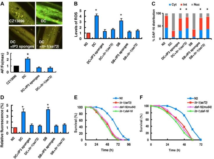
release using the nematode strain carrying Ca2+ sensor GCaMP3 under the control of epidermal-specific promoters. As shown in Figure 4A,D. coniosporainfection induced an increase in GCaMP fluorescence. These results indicate that fungal infection induces Ca2+
release in the epidermis. Increases in intracellular Ca2+ are initiated by the phospholipase C (PLC) family of enzymes, which hydrolyze phosphatidylinositol 4,5-diphosphate (PIP2) to produce inositol 1,4,5-trisphosphate (IP3) and diacylgly-cerol [40]. Since IP3 and its receptor IP3R/ITR-1 contribute to the epidermal Ca2+release after needle wounding [29], we tested the role of the IP3/ITR-1 signaling in Ca2+
release after D. coniosporainfection. We used worms overexpressing N-terminal IP3 binding domains (‘‘IP3 sponges’’ (cz12690)) in the epidermis [29]. IP3 sponges function as a dominant negative regulator to disturb IP3 signaling. We observed that IP3 sponges led to a decrease in GCaMP fluorescence. GCaMP fluorescence was reduced in itr-1(sa73)mutants (Figure 4A). Thus, abolishment of the IP3/ITR-1 signaling cascade inhibited Ca2+
release after fungal infection. Next, we tested whether Ca2+
release is required for the formation of ROS and DAF-16 nuclear accumulation after fungal infection and physical injury. An increase in the production of ROS and DAF-16 nuclear accumulation was essentially abolished in worms expressing IP3 sponges in the epidermis after fungal infection and physical injury (Figure 4B and 4C). Meanwhile, blockage of Ca2+
release by mutations initr-1also suppressed the production of ROS and DAF-16 nuclear accumulation afterD. coniosporainfection and treatment with spiny balls (Figure 4B and 4C). Finally, we found that overexpression of IP3 sponges or knock-down ofitr-1by RNAi markedly inhibited the expression of
Psod-3::GFP induced by Drechmeria coniospora and spiny balls (Figure 4D). These results demonstrate that the IP3/ITR-1/ Ca2+
signaling cascade is genetically upstream of BLI-3 for DAF-16 activation.
We asked whether blockage of Ca2+
signaling could influence the survival rate after fungal infection and physical injury. Indeed, worms expressing IP3 sponges in the epidermis were more susceptible than wild-type worms to killing mediated by D. coniosporaand spiny balls, respectively (Figure S11A and S11B in Text S1). Furthermore, mutations initr-1(sa73)shifted the survival curve to mimic the daf-16(mu86) phenotype after D. coniospora
infection (Figure 4E) and treatment with spiny balls (Figure 4F). However, the survival curve for daf-16(mu86); itr-1(sa73) double mutants was similar to that ofdaf-16(mu86)mutants. InC. elegans,
itr-1is expressed in many tissues, including the epidermis [41]. We
found that epidermal-specific RNAi ofitr-1significantly reduced the survival of worms after infection ofD. coniosporaand treatment with spiny ball (Figure S12A and S12B in Text S1). In contrast, intestinal-specific knock-down ofitr-1had no such effects (Figure S12C and S12D in Text S1). These results indicate that the IP3/ ITR-1 pathway functions within the epidermis to promote innate immunity against fungal challenge and physical injury.
EGL-30 and EGL-8 regulate DAF-16 nuclear accumulation Since needle wounding inC. eleganstriggers an EGL-30-EGL-8 signaling cascade, leading to the release of Ca2+[29], we tested whether the Gaq protein EGL-30 and the phospholipase C (PLCb) EGL-8 were also required for the production of ROS and DAF-16 nuclear accumulation upon fungal infection and physical injury. We found that the formation of ROS was reduced in egl-30(n686)oregl-8(n488)mutants after infection ofD. coniosporaand treatment with spiny balls (Figure 5A). To confirm the role of egl-30andegl-8in the activation of DAF-16, we crossed theegl-30or
egl-8 mutations into the transgenic worms that express DAF-16::GFP fusion protein. As shown in Figure 5B, the nuclear accumulation of DAF-16::GFP was reduced inegl-30(n686)or egl-8(n488)mutants compared to control worms after infection ofD. coniosporaand treatment with spiny balls. Similarly, mutations in
egl-30oregl-8significantly suppressed the expression ofPsod-3::GFP
induced byDrechmeria coniosporaand spiny balls (Figure 5C). Bothegl-30(n686)and egl-8(n488)mutants were more sensitive than wild-type worms to killing by D. coniospora (Figure 5D) or spiny balls (Figure 5E), respectively. However, mutations inegl-30
andegl-8did not alter thedaf-16(mu86)phenotype.daf-16(mu86); egl-30(n686) or daf-16(mu86); egl-8(n488) double mutants were indistinguishable fromdaf-16(mu86) for sensitivity to D. coniospora
(Figure 5D) or spiny balls (Figure 5E), suggesting that these genes function in a common pathway. InC. elegans,egl-30is expressed in many tissues, including the epidermis [42]. In contrast, egl-8, which is predominantly expressed in neurons, has been shown to act genetically downstream of egl-30 [43]. We found that epidermal-specific, rather than intestinal-specific, knock-down of
egl-30oregl-8significantly reduced the survival rate of nematodes afterD. coniosporainfection and treatment with spiny balls (Figure S13A–D in Text S1). In addition, epidermal-specific expression of
egl-30 oregl-8 was sufficient to rescue immune-deficient pheno-types in egl-30(n686) and egl-8(n488) mutants to D. coniospora
infection and physical injury, respectively (Figure S14A and S14B in Text S1). These results suggest that the EGL-30-EGL-8 pathway functions within the epidermis to promote innate immunity against fungal challenge and wound response.
CST acts downstream of BLI-3 to regulate the nuclear accumulation of DAF-16
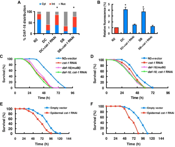
It has been reported that the mammalian Ste20-like kinase-1 (MST1) mediates oxidative stress-induced activation of FOXO transcription factors [44]. In C. elegans, CST-1, the ortholog of mammalian MST1, promotes life-span extension in a DAF-16-dependent manner [44]. Thus, we hypothesized that CST-1 might function analogously to MST1 as an activator of DAF-16. To test this idea, we assayed the effect ofcst-1 knock-down on DAF-16 activation by induced byD. coniosporaand spiny balls.cst-1 knock-down by RNAi led to a significant reduction incst-1expression (Figure S15 in Text S1).cst-1 RNAi significantly suppressed the nuclear accumulation of DAF-16 (Figure 6A), but did not influence the production of ROS induced by D. coniospora and spiny balls (Figure S16 in Text S1). Similarly, knock-down ofcst-1
by RNAi significantly inhibited the expression of Psod-3::GFP
suggest that CST-1 acts upstream of DAF-16, but downstream of BLI-3 in response to fungal infection and wounding.
Knock-down of cst-1 by RNAi reduced the survival of nematodes afterD. coniosporainfection (Figure 6C) and treatment with spiny balls (Figure 6D). However, the survival of daf-16(mu86);cst-1 RNAi was comparable to that of daf-16(mu86)
mutants (Figure 6C and 6D). These data suggest that daf-16 is epistatic to cst-1.cst-1 is mainly expressed in the epidermis, tail, vulva, and sensory neurons in the head [44]. We found that epidermal-specificcst-1RNAi resulted in enhanced sensitivity after
D. coniosporainfection (Figure 6E) and treatment with spiny balls (Figure 6F), whereas intestinal-specific cst-1 RNAi did not affect the survival of worms (Figure S17A and S17B in Text S1). These results indicate that cst-1is required for innate immunity in the epidermis.
A previous study demonstrated that BAR-1, the ortholog to mammalian b-catenin, is required for oxidative stress-induced DAF-16 activity inC. elegans[33]. BAR-1 also plays a positive role
inC. elegansintestinal immunity toS. aureus[45]. Thus,bar-1might be expected to act genetically downstream of bli-3 to activate DAF-16. However, the nuclear accumulation of DAF-16 was not altered in thebar-1(ga80)mutants afterD. coniosporainfection and treatment with spiny balls (Figure S18 in Text S1). These results suggest that BAR-1 is not involved in the activation of DAF-16 upon fungal infection and wound response.
Discussion
In a variety of animals, the epidermis may represent a first line of defense against pathogenic infection and physical injury. The key finding in this study is that the DAF-16/FOXO transcription factor is a direct regulator of immune responses associated with epidermal damage. Our data also provides a novel molecular mechanism by which DAF-16 is activated by pathogenic fungi and wounding, and that the pathway is independent of the DAF-2 insulin-like signaling pathway.
Figure 4. The IP3-ITR-1-Ca2+pathway regulates DAF-16 nuclear accumulation.(A) Epidermal GCaMP was induced after fungal infection. IP3 sponges overexpression and mutations initr-1inhibited GCaMP fluorescence afterD. coniospora infection (DC), respectively. These results are means6SD of four experiments. The right part shows quantification of GCaMP fluorescence levels. The data are expressed as percent of the strain CZ13896. *P,0.05versuscontrol (CZ13896). (B) IP3 sponges overexpression and mutations initr-1suppressed the levels of ROS afterD. coniospora infection. *P,0.01versuscontrol (N2). (C) Quantification of DAF-16 distribution. n = 100–110 nematodes per condition. These results are means6SD of four experiments. *P,0.05versuscontrol (DC and SB). (D) IP3 sponges overexpression and mutations initr-1inhibited the expression ofPsod-3::GFP induced byD. coniosporainfection and spiny balls for 12 h. *P,0.05versuscontrol (N2). (E–F)itr-1(sa73)mutants exhibited increased susceptibility after infection ofD. coniospora(E) and treatment with spiny balls (F).P,0.001 relative to control with empty vector. However, mutations initr-1did not enhanced susceptibility ofdaf-16(mu86)mutants to killing byD. coniospora(E) and spiny balls (F).
doi:10.1371/journal.ppat.1003660.g004
A sustained production of ROS following injury has been observed in human cells, the zebrafish and Drosophila tissues [30,31,32]. Recent studies demonstrate that these processes are mediated by DUXOs [31,32]. In the current study, we observe that ROS production is mediated by BLI-3 in the epidermis after fungal infection and physical injury. Our study indicates that ROS production is crucial for resistance to fungal infection and physical injury inC. elegans, supporting the idea that injury-induced ROS production is an important regulator of tissue regeneration [46]. Furthermore, our results demonstrate that ROS production is required for activation of DAF-16, which, in turn is essential for resistance to fungal infection and physical injury inC. elegans. Two recent studies indicate that knock-down ofbli-3by RNAi leads to enhanced susceptibility toE. faecalis[39,47]. However, thedaf-16
mutants exhibit a comparable degree of susceptibility toE. faecalis -mediated killing as wild-type worms [4,48]. These results indicate that the protective effect of BLI-3 on E. faecalis infection is not mediated through DAF-16.
Impairment in release of Ca2+
abolished ROS production upon fungal challenge and wounding, suggesting that BLI-3 enzymatic activity is dependent on Ca2+
. The EF-hand calcium-binding motif inC. elegansBLI-3 has a relatively low amino acid identity (41%) and similarity (61%) to the human DUOX1, casting doubt
as to whether calcium binding is required for C. elegans BLI-3 function [38]. However, the EF-hand calcium-binding motif in
DrosophilaDUOX1 also shares a relatively low identity (42%) and similarity (66%) to the human DUOX1. Because ROS-producing
Drosophila DUOX1 enzymatic activity depends on intracellular Ca2+
through binding to the EF-hand domains [49,50], it is plausible that Ca2+
modulates the enzymatic activity ofC. elegans
BLI-3.
The PI3K-Akt-FOXO signaling pathway is evolutionarily conserved from nematodes to mammals [10,51]. In mammalian cells, protein kinase Akt, a downstream effector of the insulin-signaling pathway, phosphorylates two sites (Thr32 and Ser252) on the FOXO3 protein leading to its nuclear exclusion and inactivation [52]. Likewise,P. aeruginosasuppresses the activity of DAF-16 by activating DAF-2 insulin-like signaling [7,26]. However, our data demonstrate that causal involvement of diminished DAF-2 insulin-like signaling in the activation of DAF-16 by fungal infection is unlikely, suggesting that alternative mechanisms are involved. A previous study has demonstrated that oxidative stress activates FOXO3 through an MST1-mediated mechanism [44]. Under oxidative stress, MST1 phosphorylates FOXO3 at Ser207 and the phosphorylation of FOXO3 in turn induces its dissociation from 14-3-3 proteins and translocation to
Figure 5. The Gaq-PLCbsignaling is required for DAF-16 nuclear accumulation.(A)egl-30(n686)oregl-8(n488)mutants displayed reduced the production of ROS induced byD. coniosporainfection (DC) and spiny balls (SB). These results are means6SD of four experiments. *P,0.05versus control (N2). (B) Quantification of DAF-16 distribution. n = 100–110 nematodes per condition. These results are means6SD of four experiments. *P,0.05versuscontrol (DC and SB). (C) Mutations inegl-30 and egl-8inhibited the expression ofPsod-3::GFPinduced byD. coniosporainfection and treatment with spiny balls or 12 h. *P,0.05versuscontrol (N2). (D–E) Mutations inelg-30andegl-8reduced the survival rate of worms exposed toD. coniospora(D) and spiny balls (E).P,0.001 relative to control with wild-type worms. However, mutations inegl-30oregl-8did not enhanced susceptibility ofdaf-16(mu86)mutants to killing byD. coniospora(D) and spiny balls (E).
the nucleus [44]. Although knock-down of daf-16 by RNAi completely inhibits the ability of CST-1 to extend life span inC. elegans, whether CST-1 activates DAF-16 under oxidative stress remains unclear. Our data indicate that ROS mediates activation of DAF-16 in response to epidermal damage in a CST-dependent manner. These results support a model in which the evolutionarily conserved MST/CST pathway functions in parallel with the insulin signaling pathway to regulate FOXO/DAF-16 by oxida-tive stress [44].
The epidermis forms a protective barrier against physical damage and pathogen entry [53,54]–[55]. An intimate relationship between wound repair and innate immunity is widely accepted [56]. Previous studies have shown that epidermal immune responses to fungal infection and physical wounding share some of the same signals and mediators in C. elegans [19,24]. For instance, Ga12/
GPA-12 acts, together with the two phospholipases EGL-8 and PLC-3, upstream of the PKC-TIR-1-p38 MAPK pathway, to induce a set of thenlpgenes encoding antimicrobial peptides (AMPs) in response to fungal challenge and needle wounding [19,24]. A recent study has shown that the EGL-30-EGL-8 signaling pathway triggers epidermal Ca2+
release through IP3 and its receptor ITR-1 after wounding [29]. In this study, our results indicate that DAF-16 is activated by EGL-30-Ca2+
upon fungal infection and physical injury. Since epidermal DAF-16 is required for innate immune response to fungal infection and physical injury, it is an important immune effector of EGL-30-Ca2+
in the epidermis. However, mutations indaf-16do not alter the expression of AMPs induced by fungal infection, which is consistent with the observation that the EGL-30-Ca2+
pathway appears not to be involved in the up-regulation of AMPs after wounding [29].
Figure 6. CST regulates the nuclear accumulation of DAF-16.(A)cst-1RNAi diminished DAF-16 nuclear translocation byD. coniospora infection (DC) and treatment with spiny balls (SB). n = 100–110 nematodes per condition. These results are means6SD of four experiments. *P,0.05 versuscontrol (DC and SB). (B) Knock-down ofitr-1inhibited the expression ofPsod-3::GFPinduced byD. coniosporainfection and spiny balls for 12 h. *P,0.05versuscontrol (N2). (C and D)cst-1RNAi reduced the survival rate of nematodes exposed toD. coniospora(C) and spiny balls (D).P,0.001 relative to control with empty vector. However,cst-1RNAi did not enhanced susceptibility ofdaf-16(mu86)mutants to killing byD. coniospora(C) and spiny balls (D). (E and F) Epidermal-specific RNAi ofcst-1significantly reduced the survival rate of worms exposed toD. coniospora(E) and spiny balls (F).P,0.001 relative to control with empty vector.
doi:10.1371/journal.ppat.1003660.g006
Because FOXOs are conserved from worms to humans, it is of great interest to investigate whether FOXOs are involved in epidermal innate immunity in other species (e.g., humans). FOXOs have been shown to mediate the induction of antimicro-bial peptides, such as defensins, both in Drosophila and human tissues [10]. Accumulating evidence indicates that defensins, the major skin-derived antimicrobial peptides, not only act as endogenous antibiotics, but also participate in additional roles such as promoting wound repair [57,58]. Meanwhile, inhibition of PI3K, a component of insulin/insulin-like growth factor signaling, by LY294002 (a specific inhibitor of PI3K), strongly accelerates scratch closure in human keratinocytes [59]. Because reduced signaling of the insulin/insulin-like growth factor pathway leads to the activation of FOXO transcription factors, these results imply that FOXOs are probably involved in keratinocyte wound healing. A recent study has investigated epidermal gene expression in wounded skin from three donors and examined transcription factor binding sites (TFBS) in the promoters of the 100 most differentially expressed genes [60]. Highly significant overrepre-sentations of TFBS for FOXO transcription factors are identified. These data suggest that FOXOs are possibly involved in controlling the epidermal gene expression during the proliferative phase of wound healing. Thus, the DAF-16/FOXO transcription factor that functions as an effector of innate immunity in epidermal tissues seems to be evolutionarily conserved in various animal species including worms, insects and mammals.
In summary, our findings suggest that DAF-16 is directly involved in innate immunity in the epidermis. EGL-30/Ca2+
/BLI-3/ROS/CST-1 signaling represents a novel pathway to regulate DAF-16 activity (see model in Figure 7), which is functionally independent of the DAF-2 insulin-like signaling pathway. Based on these findings, we propose that FOXO/DAF-16 could be a novel target for the treatment of epidermal damage.
Materials and Methods
Nematode strains
The followingC. elegansstrains were used in this study: N2 (wild-type), daf-16(mu86), bli-3(e767), itr-1(sa73), bar-1(ga80), egl-30(n686), egl-8(n488), ins-7(ok1573), TJ356-daf-16::gfp(zIs356 (pDAF-16::DAF-16-GFP;rol-6)), muIs84 (Psod-3::gfp), NR222 (rde-1(ne219); kzIs9[pKK1260(plin-12::nls::gfp), pKK1253(plin-26::rde-1),
rol-6]); and NR350(rde-1(ne219); kzIs20[pDM#715(phlh-1::rde-1), pTG95(psur-5::nls::GFP), rol-6]) were kindly provided by the Caenorhabditis Genetics Center (CGC). The CZ13896 ( Pcol-19-GCaMP (juIs319)), CZ12690 (Pcol-19-Superspronge (juEx3052)), and CZ15386 (egl-8(sa47V;egl-8(juEX4257)) strains were kindly provid-ed by Dr. Andrew D. Chisholm (University of California San Diego). The strain GR1353 (daf-2(e1370) III; mgIs41[daf-16::gfp]) and the strain for intestinal-specific RNAi(sid-1(qt9); Is[vha-6pr::sid-1]; Is[sur-5pr::GFPNLS])were kindly provided by Dr. Gary Ruvkun (Massachusetts General Hospital, Harvard Medical School).
Mutants and transgenic strains were backcrossed three times into the N2 strain used in the laboratory. All strains were maintained on nematode growth media (NGM) and fed with E. coli strain OP50.
Infection with fungi and bacteria
Standard conditions were used for C. elegans growth at 20uC [61]. Synchronized populations of worms were cultivated at 20uC until the mid-L4 stage. For all pathogen assays, 75mg/ml of fivefluoro-29-deoxyuridine (FUdR) was added to the assay plates to abolish the growth of progeny.
Killing assays with D. coniospora: 50–60 L4 nematodes were transferred to fresh plates seeded with heat-killed E. coli OP50, with,1.06108D. coniosporaspores at 25uC. Killing assays withC. rosea: ,1.06108 spores of C. rosea were inoculated onto plates containing heat-killedE. coliOP50 for 1–2 days at 28uC, and the infection experiments were started by adding 50–60 nematodes to each plate at 25uC. The number of living worms was counted by using a light microscope at time intervals. Immobile nematodes unresponsive to touch were scored as dead. One-sided rank log tests were used to the statistical significance of the differences between treatments.
Killing assays withP. aeruginosa:P. aeruginosa PA14 (a gift from Dr. Kun Zhu, Institute of Microbiology, CAS) was cultured in Luria broth (LB), then seeded on slow-killing plates, which contain modified NGM (0.35% instead of 0.25% peptone). PA14 was incubated first for 24 h at 37uC and then for 24 h at 25uC. The infection experiments were started by adding 50–60 nematodes to each plate at 25uC. Killing assays withS. aureus:S. aureusATCC 25923 (a gift from Dr. Wen-Hui Lee, Kunming Institute of Zoology, CAS) was cultured in tryptic soy broth (TSB, BD, Sparks, MD), then seeded on plates containing modified NGM (0.35% instead of 0.25% peptone). The infection experiments were started by adding 50–60 nematodes to each plate at 25uC.
Physical injury by spiny balls
Spiny balls were purified by a previously described method [25]. The spiny ball suspension was adjusted to ,1.06105 per ml. 100ml of the spiny ball suspension was thoroughly added to on plates containing modified NGM with heat-killed E. coli OP50. The infection experiments were started by adding 50–60 nematodes to each plate at 25uC. Mobile and immobile nematodes were counted every 12 h after their addition.
RNA interference
RNAi bacterial strains containing targeting genes were obtained from the Ahringer RNAi library [62]. RNAi feeding experiments were performed on synchronized L1 larvae at 20uC for 40 h. L4 larvae or young adult worms were used in immunity assays. The strain NR222 was used in epidermis-specific RNAi, the strain (sid-1(qt9); Is[vha-6pr::sid-1]; Is[sur-5pr::GFPNLS])was used in intestine-specific RNAi , and the strain NR350 was used in muscular-specific RNAi.
DAF-16 nuclear localization assay
After 12 h of fungal infection or treatment with spiny balls, the worms were immediately mounted in M9 onto microscope slides. The slides were viewed using a Zeiss Axioskop 2 plus fluorescence microscope (Carl Zeiss, Jena, Germany) with a digit camera. The status of DAF-16 localization was categorized as cytosolic localization, nuclear localization when localization is observed throughout the entire body from head to tail, or intermediate localization when there is a visible nuclear localization but one not as complete as nuclear [63]. The number of worms with each level of nuclear translocation was counted.
Quantitative real-time RT-PCR analysis
Construction of transgenes
Thedpy-7andclo-19genes have shown to be expressed in the epidermis [29]. The Pdpy-7:daf-16 fusion gene was chemically synthesized, and obtained from Generay Biotech Co. (Shanghai, China). The DNA fragment contains a 436 bp ofdpy-7promoter fragment (corresponding to nucleotide2436 to21 relative to the translational start site), a 1530 bp of thedaf-16cDNA, a 729 bp of the GFP cDNA and a 234 bp of the 39-UTR ofunc-54. The Pcol-19:egl-30fusion gene was constructed as follows. A 2838 bp of col-19promoter fragment was obtained by PCR onC. elegansgenomic DNA using primers 59-GCT CTA GAG CAT CGT CAC ATT CTG TCT-39and 59-TCC CCC GGG GGC TTT CCA TCG TCT CC-39followed by XbaI and SmaI digestion. The fragment was inserted into XbaI and SmaI digested pPD95.79, resulting in plasmid pPDegl. A 1116 bp fragment of egl-30 cDNA was amplified by PCR onC. elegansgenomic DNA using primers 59 -TCC CCC GGG TTG TTC TAT TCG CTG GCT T-39and 59 -GGG GTA CCC CAA GTT GTA CTC CTT CAG ATT AT-39
followed by SmaI and KpnI digestion. The fragment was inserted into SmaI and KpnI digested pPDegl vector.
ThePdpy-7:daf-16fusion gene fragment or the vector containing
Pcol-19::egl-30 fusion gene was co-injected with rol-6 plasmid (pRF4) into gonads of wild-type and egl-30(n686) animals by standard techniques [64]. The transgenic worms carrying Pdyp-7::daf-16 or Pcol-19::egl-30 were confirmed in prior to each pathogenesis assay.
Measurement of ROS
The ROS levels were detected by 29,79 -dichlorodihydrofluor-escein diacetate (DCF-DA) as a probe as described previously with modifications [34,35,36]. Briefly, after infected with pathogens or treated with spiny balls for 8 h, about 1000 worms from each group were collected in M9 buffer and washed three times to eliminate conidia. Then, the worms were transferred to a 1.5-mL tube containing 150ml PBS with 1% Tween 20, and immediately frozen in liquid nitrogen. After thawing at room temperature, the worms were subjected to sonication (Branson Sonifier 250; VWR Scientific, Suwanee, GA). Samples were vortexed, and superna-tants were collected after centrifugation. The supernatant containing 10mg protein was transferred into 96-well plates, and incubated with 15mL of 100mM DCF-DA in PBS at 37uC in a Spectra Max M5 fluorescent microplate reader (Molecular Devices, Sunnyvale, CA) for quantification of fluorescence at excitation 485 nm and emission 530 nm. Samples were read kinetically every 20 min for 2.5 h.
Analysis of Ca2+in the epidermis using GCaMP fluorescence
To analyze Ca2+
in the epidermis, GCaMP fluorescence imaged was obtained using confocal microscopy (Zeiss LSM-510) with a 406objective, as described previously [29]. Briefly, average
fluorescence was determined in ten equivalent regions of interest (ROI), five centered on the epidermal cell and five in the
Figure 7. A proposed mechanism ofC. elegansDAF-16 activation by fungal infection and physical injury.Fungal infection or physical injury in the epidermis activates an unknown G protein-coupled receptor (GPCR), which in turn activates EGL-30 (Gaq). EGL-30 may positively modulate EGL-8 (PLCb) activity, resulting in the production of inositol 1,4,5-trisphosphate (IP3). IP3 then binds to the IP3 receptor ITR-1 located in the endoplasmic reticulum membrane to initiate the release of Ca2+
stored in this organelle. The released Ca2+
may activate BLI-3 activity through its Ca2+
-sensitive EF hand domain to produce ROS. Subsequently, ROS induces the CST-mediated accumulation of DAF-16 in the nucleus to promote innate immunity against fungal infection and physical injury.
doi:10.1371/journal.ppat.1003660.g007
background. Baseline fluorescence (F0) and induction fluorescence (Ft) were obtained by averaging fluorescence in five ROIs in the epidermis then subtracting the average of five ROIs in the background before and after fungal infection or injury. GCaMP fluorescence was normalized to an internal control, Pcol-19 -tdTomato. The change in fluorescenceDF was expressed as the ratio of change with respect to the baseline [(Ft–F0)/F0]. Raw data from fluorescent microscopy were then analyzed using ImageJ.
Statistical analysis
Differences in survival rates were analyzed using the log-rank test. Differences in gene expression, distribution of DAF-16, and fluorescence intensity were assessed by performing a one-way ANOVA followed by a Student-Newman-Keuls test. Data were analyzed using SPSS11.0 software (SPSS Inc.). To test for significant overlap between different gene lists, a Fisher’s exact test was used.
Supporting Information
Table S1 The target genes of DAF-16 up-regulated byD. coniosporainfection.When compared DAF-16 target genes to published microarray analysis of gene expression induced byD. coniospora infection, 48 of the genes up-regulated by D. coniospora
were found to be targets of DAF-16. (DOC)
Table S2 The primers are used for real-time PCR.This table lists all primers for real-time PCR analysis.
(DOC)
Text S1 Supporting Figures. Figure S1. Fungal infection and mutation in daf-12 induce DAF-16 nuclear translo-cation.(A) Wild-type worms in the absence of D. coniospora. (B) Wild-type worms were exposed toD. coniosporafor 12 h. (C)daf-2
mutants were growth under normal conditions without D. coniospora. Figure S2. Both epidermal- and intestinal-specific knock-down of daf-16 by RNAi suppress the expression of DAF-16 target genes. (A) qPCR analysis of expression of DAF-16 target genes in NR222 strains (CTR), CTR 24 h afterD. coniosporainfection (CTR+DC), and CTR subjected todaf-16RNAi afterD. coniosporainfection (CTR+DC+epidermal
daf-16 RNAi). (B) qPCR analysis of expression of the intestinal-specific RNAi strain sid-1(qt9);Is[sur-5::GFP]; alxIs7[VHA-6p::SID-1::SL2::GFP] (CTR), CTR 24 h after D. coniospora infection (CTR+DC), and CTR subjected todaf-16RNAi afterD. coniospora
infection (CTR+DC+epidermal daf-16 RNAi). *P,0.05, CTR+DC relative to CTR+DC+ daf-16 RNAi. Figure S3. Genetic loss ofins-7has no effect on DAF-16 transloca-tion and the survival of worms after D. coniospora
infection and physical injury.(A) Mutation inins-7(ok1573)
did not influence the nuclear accumulation of DAF-16 afterD. coniosporainfection (DC) and treatment with spiny balls (SB). (B and C) Mutation in ins-7(ok1573) did not affect the survival of nematodes after D. coniospora infection (B) and treatment with spiny balls (C).Figure S4. DAF-16 is required for resistance to fungal infection.(A)daf-16(mu86)mutants were sensitive to
C. roseainfection. (B–C)daf-16RNAi reduced the survival rate of nematodes exposed toD. coniospora(B) andC. rosea(C).P,0.001 relative to wild-type animals.Figure S5. Epidermal-specific knock-down ofdaf-16reduces the expression ofdaf-16in the hypodermis. (A) NR222 strains were exposed to D. coniospora for 12 h. (B) NR222 strains subjected to daf-16 RNAi were exposed toD. coniospora for 12 h. Figure S6. Muscular-specific daf-16 RNAi has no effect on sensitivity to D.
coniospora infection and spiny balls. (A) NR350 strains were exposed toD. coniospora. (B) NR350 strains were exposed to spiny balls. After 12 h of treatment, the DAF-16::GFP expression pattern was observed.Figure S7. Elevateddaf-16expression in the epidermis confers resistance to fungal infection and physical injury.Overexpression ofdaf-16under epidermal (dpy-7) promoter increased survival of nematodes afterD. coniospora
infection (A) and treatment of spiny balls (B).Figure S8. Knock-down ofbli-3in a 1/10 dilution results in a decrease in the expression ofbli-3. qPCR analysis ofbli-3 expression in WT worms subjected tobli-3 RNAi in a 1/10 dilution.Figure S9. BLI-3 in the epidermis is required for ROS production, DAF-16 nuclear accumulation, and resis-tance to fungal infection and physical injury. (A) Epidermal-specific RNAi ofbli-3in a 1/10 dilution significantly reduced the production of ROS induced byD. coniospora(DC) or spiny balls (SB). (B) Epidermal-specific RNAi ofbli-3 in a 1/10 dilution significantly reduced DAF-16 nuclear accumulation induced byD. coniospora or spiny balls. (C–D) Epidermal-specific RNAi ofbli-3in a 1/10 dilution significantly reduced the survival rate of nematodes exposed toD. coniospora(C) or spiny balls (D). (E–F) Intestinal-specific RNAi ofbli-3in a 1/10 dilution did not influence the survival of nematodes afterD. coniosporainfection (E) and treatment with spiny balls (F).Figure S10. The peroxidase activity of BLI-3 is not crucial for resistance to fungal infection and physical injury. (A–B) The mutantbli-3(e767)
encoding a protein that lacks the peroxidase domain exhibited similar sensitivity to killing byD. coniosporainfection (A) and spiny balls (B) as wild-type animals. Figure S11. A disturbance of IP3 signaling reduces the survival of worms after fungal infection and physical injury. (A–B) Worms overexpressing IP3 sponges (cz12690) in the epidermis exhibited enhanced susceptibility to killing by D. coniospora (A) and spiny balls (B). Figure S12. ITR-1 in the epidermis is required for resistance to fungal infection and physical injury.(A–B) Epidermal-specific RNAi ofitr-1significantly reduced the survival rate of worms afterD. coniospora infection (A) or treatment with spiny balls (B). (C–D) Intestinal-specific knock-down ofitr-1did not influence the survival of nematodes afterD. coniosporainfection (C) and treatment with spiny balls (D). Figure S13. Epidermal-specific, rather than intestinal-Epidermal-specific, knockdown of
egl-30 or egl-8 suppresses innate immunity. (A–B) Epidermal-specific knock-down ofegl-30oregl-8by RNAi reduced the survival of nematodes exposure toD. coniospora infection (A) and spiny balls (B). (C–D) Intestinal-specific knock-down ofegl-30
or egl-8 did not influence the survival of nematodes after D. coniosporainfection (C) and treatment with spiny balls (D).Figure S14. EGL-30 and EGL-8 in the epidermis are required for resistance to fungal infection and physical injury.(A– B) Epidermal-specific expression of egl-30 rescued immune-deficient phenotypes in egl-30(n686) mutants to D. coniospora
infection (A) and treatment with spiny balls (B). (C–D) Epider-mal-specific expression of egl-8 (cz15896) rescued immune-deficient phenotypes in egl-8(n488) mutants to D. coniospora
infection (C) and treatment with spiny balls (D). Figure S15.
cst-1RNAi significantly reduces the expression ofcst-1. After worms were subjected tocst-1RNAi,the expression ofcst-1
rate of nematodes afterD. coniospora infection (A) and treatment with spiny balls (B). Figure S18. BAR-1 is not involved in DAF-16 activation upon fungal infection and physical injury. Mutations in bar-1(ga80) did not influence the nuclear accumulation of DAF-16 after D. coniospora infection (DC) and treatment with spiny balls (SB).
(PDF)
Acknowledgments
We are grateful to Drs. Jianping Xu (McMaster University, Canada), and FM Ausubel for their critical reading of this manuscript. We thank Drs. AD Chisholm, G Ruvkun, and the Caenorhabditis Genetics Center for nematode strains; Drs. WH Lee and K Zhu for bacteria strains.
Author Contributions
Conceived and designed the experiments: CGZ KQZ. Performed the experiments: QT JN. Analyzed the data: CGZ QT. Contributed reagents/ materials/analysis tools: XLJ. Wrote the paper: CGZ KQZ.
References
1. Aballay A, Ausubel FM (2002) Caenorhabditis elegans as a host for the study of host-pathogen interactions. Curr Opin Microbiol 5: 97–101.
2. Irazoqui JE, Urbach JM, Ausubel FM (2010) Evolution of host innate defence: insights from Caenorhabditis elegans and primitive invertebrates. Nat Rev Immunol 10: 47–58.
3. Kimura KD, Tissenbaum HA, Liu Y, Ruvkun G (1997) daf-2, an insulin receptor-like gene that regulates longevity and diapause in Caenorhabditis elegans. Science 277: 942–946.
4. Garsin DA, Villanueva JM, Begun J, Kim DH, Sifri CD, et al. (2003) Long-lived C. elegans daf-2 mutants are resistant to bacterial pathogens. Science 300: 1921. 5. Lin K, Hsin H, Libina N, Kenyon C (2001) Regulation of the Caenorhabditis elegans longevity protein DAF-16 by insulin/IGF-1 and germline signaling. Nat Genet 28: 139–145.
6. Henderson ST, Johnson TE (2001) daf-16 integrates developmental and environmental inputs to mediate aging in the nematode Caenorhabditis elegans. Curr Biol 11: 1975–1980.
7. Evans EA, Kawli T, Tan MW (2008) Pseudomonas aeruginosa suppresses host immunity by activating the DAF-2 insulin-like signaling pathway in Caenor-habditis elegans. PLoS Pathog 4: e1000175.
8. Murphy CT, McCarroll SA, Bargmann CI, Fraser A, Kamath RS, et al. (2003) Genes that act downstream of DAF-16 to influence the lifespan of Caenorhabditis elegans. Nature 424: 277–283.
9. Singh V, Aballay A (2006) Heat-shock transcription factor (HSF)-1 pathway required for Caenorhabditis elegans immunity. Proc Natl Acad Sci U S A 103: 13092–13097.
10. Becker T, Loch G, Beyer M, Zinke I, Aschenbrenner AC, et al. (2010) FOXO-dependent regulation of innate immune homeostasis. Nature 463: 369–373. 11. Troemel ER, Chu SW, Reinke V, Lee SS, Ausubel FM, et al. (2006) p38 MAPK
regulates expression of immune response genes and contributes to longevity in C. elegans. PLoS Genet 2: e183.
12. Anyanful A, Easley KA, Benian GM, Kalman D (2009) Conditioning protects C. elegans from lethal effects of enteropathogenic E. coli by activating genes that regulate lifespan and innate immunity. Cell Host Microbe 5: 450–462. 13. Wang J, Nakad R, Schulenburg H (2012) Activation of the Caenorhabditis
elegans FOXO family transcription factor DAF-16 by pathogenic Bacillus thuringiensis. Dev Comp Immunol 37: 193–201.
14. Shapira M, Hamlin BJ, Rong J, Chen K, Ronen M, et al. (2006) A conserved role for a GATA transcription factor in regulating epithelial innate immune responses. Proc Natl Acad Sci U S A 103: 14086–14091.
15. Anyanful A, Dolan-Livengood JM, Lewis T, Sheth S, Dezalia MN, et al. (2005) Paralysis and killing of Caenorhabditis elegans by enteropathogenic Escherichia coli requires the bacterial tryptophanase gene. Mol Microbiol 57: 988–1007. 16. Hasshoff M, Bohnisch C, Tonn D, Hasert B, Schulenburg H (2007) The role of
Caenorhabditis elegans insulin-like signaling in the behavioral avoidance of pathogenic Bacillus thuringiensis. FASEB J 21: 1801–1812.
17. Pukkila-Worley R, Ausubel FM, Mylonakis E (2011) Candida albicans infection of Caenorhabditis elegans induces antifungal immune defenses. PLoS Pathog 7: e1002074.
18. Couillault C, Pujol N, Reboul J, Sabatier L, Guichou JF, et al. (2004) TLR-independent control of innate immunity in Caenorhabditis elegans by the TIR domain adaptor protein TIR-1, an ortholog of human SARM. Nat Immunol 5: 488–494.
19. Pujol N, Cypowyj S, Ziegler K, Millet A, Astrain A, et al. (2008) Distinct innate immune responses to infection and wounding in the C. elegans epidermis. Curr Biol 18: 481–489.
20. Tong A, Lynn G, Ngo V, Wong D, Moseley SL, et al. (2009) Negative regulation of Caenorhabditis elegans epidermal damage responses by death-associated protein kinase. Proc Natl Acad Sci U S A 106: 1457–1461.
21. Yang J, Wang L, Ji X, Feng Y, Li X, et al. (2011) Genomic and proteomic analyses of the fungus Arthrobotrys oligospora provide insights into nematode-trap formation. PLoS Pathog 7: e1002179.
22. Zou CG, Tao N, Liu WJ, Yang JK, Huang XW, et al. (2010) Regulation of subtilisin-like protease prC expression by nematode cuticle in the nematopha-gous fungus Clonostachys rosea. Environ Microbiol 12: 3243–3252. 23. Engelmann I, Griffon A, Tichit L, Montanana-Sanchis F, Wang G, et al. (2011)
A comprehensive analysis of gene expression changes provoked by bacterial and fungal infection in C. elegans. PLoS One 6: e19055.
24. Ziegler K, Kurz CL, Cypowyj S, Couillault C, Pophillat M, et al. (2009) Antifungal innate immunity in C. elegans: PKCdelta links G protein signaling and a conserved p38 MAPK cascade. Cell Host Microbe 5: 341–352. 25. Luo H, Liu Y, Fang L, Li X, Tang N, et al. (2007) Coprinus comatus damages
nematode cuticles mechanically with spiny balls and produces potent toxins to immobilize nematodes. Appl Environ Microbiol 73: 3916–3923.
26. Kawli T, Wu C, Tan MW (2010) Systemic and cell intrinsic roles of Gqalpha signaling in the regulation of innate immunity, oxidative stress, and longevity in Caenorhabditis elegans. Proc Natl Acad Sci U S A 107: 13788–13793. 27. Kodama E, Kuhara A, Mohri-Shiomi A, Kimura KD, Okumura M, et al. (2006)
Insulin-like signaling and the neural circuit for integrative behavior in C. elegans. Genes Dev 20: 2955–2960.
28. Pierce SB, Costa M, Wisotzkey R, Devadhar S, Homburger SA, et al. (2001) Regulation of DAF-2 receptor signaling by human insulin and ins-1, a member of the unusually large and diverse C. elegans insulin gene family. Genes Dev 15: 672–686.
29. Xu S, Chisholm AD (2011) A Galphaq-Ca(2) signaling pathway promotes actin-mediated epidermal wound closure in C. elegans. Curr Biol 21: 1960–1967. 30. Firth JD, Uitto VJ, Putnins EE (2008) Mechanical induction of an epithelial cell
chymase associated with wound edge migration. J Biol Chem 283: 34983– 34993.
31. Niethammer P, Grabher C, Look AT, Mitchison TJ (2009) A tissue-scale gradient of hydrogen peroxide mediates rapid wound detection in zebrafish. Nature 459: 996–999.
32. Razzell W, Evans IR, Martin P, Wood W (2013) Calcium flashes orchestrate the wound inflammatory response through DUOX activation and hydrogen peroxide release. Curr Biol 23: 424–429.
33. Essers MA, de Vries-Smits LM, Barker N, Polderman PE, Burgering BM, et al. (2005) Functional interaction between beta-catenin and FOXO in oxidative stress signaling. Science 308: 1181–1184.
34. Strayer A, Wu Z, Christen Y, Link CD, Luo Y (2003) Expression of the small heat-shock protein Hsp16-2 in Caenorhabditis elegans is suppressed by Ginkgo biloba extract EGb 761. FASEB J 17: 2305–2307.
35. Wu Y, Wu Z, Butko P, Christen Y, Lambert MP, et al. (2006) Amyloid-beta-induced pathological behaviors are suppressed by Ginkgo biloba extract EGb 761 and ginkgolides in transgenic Caenorhabditis elegans. J Neurosci 26: 13102– 13113.
36. Zhou B, Yang L, Li S, Huang J, Chen H, et al. Midlife gene expressions identify modulators of aging through dietary interventions. Proc Natl Acad Sci U S A 109: E1201–1209.
37. Benedetto A, Au C, Avila DS, Milatovic D, Aschner M (2010) Extracellular dopamine potentiates mn-induced oxidative stress, lifespan reduction, and dopaminergic neurodegeneration in a BLI-3-dependent manner in Caenorhab-ditis elegans. PLoS Genet 6:e1001084.
38. Edens WA, Sharling L, Cheng G, Shapira R, Kinkade JM, et al. (2001) Tyrosine cross-linking of extracellular matrix is catalyzed by Duox, a multidomain oxidase/peroxidase with homology to the phagocyte oxidase subunit gp91phox. J Cell Biol 154: 879–891.
39. Chavez V, Mohri-Shiomi A, Garsin DA (2009) Ce-Duox1/BLI-3 generates reactive oxygen species as a protective innate immune mechanism in Caenorhabditis elegans. Infect Immun 77: 4983–4989.
40. Rhee SG (2001) Regulation of phosphoinositide-specific phospholipase C. Annu Rev Biochem 70: 281–312.
41. Walker DS, Ly S, Gower NJ, Baylis HA (2004) IRI-1, a LIN-15B homologue, interacts with inositol-1,4,5-triphosphate receptors and regulates gonadogenesis, defecation, and pharyngeal pumping in Caenorhabditis elegans. Mol Biol Cell 15: 3073–3082.
42. Bastiani CA, Gharib S, Simon MI, Sternberg PW (2003) Caenorhabditis elegans Galphaq regulates egg-laying behavior via a PLCbeta-independent and serotonin-dependent signaling pathway and likely functions both in the nervous system and in muscle. Genetics 165: 1805–1822.
43. Suo S, Kimura Y, Van Tol HH (2006) Starvation induces cAMP response element-binding protein-dependent gene expression through octopamine-Gq signaling in Caenorhabditis elegans. J Neurosci 26: 10082–10090.
44. Lehtinen MK, Yuan Z, Boag PR, Yang Y, Villen J, et al. (2006) A conserved MST-FOXO signaling pathway mediates oxidative-stress responses and extends life span. Cell 125: 987–1001.
45. Irazoqui JE, Ng A, Xavier RJ, Ausubel FM (2008) Role for beta-catenin and HOX transcription factors in Caenorhabditis elegans and mammalian host epithelial-pathogen interactions. Proc Natl Acad Sci U S A 105: 17469–17474. 46. Love NR, Chen Y, Ishibashi S, Kritsiligkou P, Lea R, et al. (2013) Amputation-induced reactive oxygen species are required for successful Xenopus tadpole tail regeneration. Nat Cell Biol 15: 222–228.
47. Hoeven R, McCallum KC, Cruz MR, Garsin DA (2011) Ce-Duox1/BLI-3 generated reactive oxygen species trigger protective SKN-1 activity via p38 MAPK signaling during infection in C. elegans. PLoS Pathog 7: e1002453. 48. Chavez V, Mohri-Shiomi A, Maadani A, Vega LA, Garsin DA (2007) Oxidative
stress enzymes are required for DAF-16-mediated immunity due to generation of reactive oxygen species by Caenorhabditis elegans. Genetics 176: 1567–1577. 49. Ha EM, Oh CT, Bae YS, Lee WJ (2005) A direct role for dual oxidase in
Drosophila gut immunity. Science 310: 847–850.
50. Ha EM, Lee KA, Park SH, Kim SH, Nam HJ, et al. (2009) Regulation of DUOX by the Galphaq-phospholipase Cbeta-Ca2+pathway in Drosophila gut immunity. Dev Cell 16: 386–397.
51. Kenyon C (2005) The plasticity of aging: insights from long-lived mutants. Cell 120: 449–460.
52. Van Der Heide LP, Hoekman MF, Smidt MP (2004) The ins and outs of FoxO shuttling: mechanisms of FoxO translocation and transcriptional regulation. Biochem J 380: 297–309.
53. Martin P (1997) Wound healing–aiming for perfect skin regeneration. Science 276: 75–81.
54. Galko MJ, Krasnow MA (2004) Cellular and genetic analysis of wound healing in Drosophila larvae. PLoS Biol 2: E239.
55. Mace KA, Pearson JC, McGinnis W (2005) An epidermal barrier wound repair pathway in Drosophila is mediated by grainy head. Science 308: 381–385. 56. Jackson JA, Friberg IM, Little S, Bradley JE (2009) Review series on helminths,
immune modulation and the hygiene hypothesis: immunity against helminths and immunological phenomena in modern human populations: coevolutionary legacies? Immunology 126: 18–27.
57. Braff MH, Gallo RL (2006) Antimicrobial peptides: an essential component of the skin defensive barrier. Curr Top Microbiol Immunol 306: 91–110. 58. Auvynet C, Rosenstein Y (2009) Multifunctional host defense peptides:
antimicrobial peptides, the small yet big players in innate and adaptive immunity. FEBS J 276: 6497–6508.
59. Fitsialos G, Chassot AA, Turchi L, Dayem MA, LeBrigand K, et al. (2007) Transcriptional signature of epidermal keratinocytes subjected to in vitro scratch wounding reveals selective roles for ERK1/2, p38, and phosphatidylinositol 3-kinase signaling pathways. J Biol Chem 282: 15090–15102.
60. Roupe KM, Alberius P, Schmidtchen A, Sorensen OE (2010) Gene expression demonstrates increased resilience toward harmful inflammatory stimuli in the proliferating epidermis of human skin wounds. Exp Dermatol 19: e329–332. 61. Brenner S (1974) The genetics of Caenorhabditis elegans. Genetics 77: 71–94. 62. Kamath RS, Ahringer J (2003) Genome-wide RNAi screening in Caenorhabditis
elegans. Methods 30: 313–321.
63. Oh SW, Mukhopadhyay A, Svrzikapa N, Jiang F, Davis RJ, et al. (2005) JNK regulates lifespan in Caenorhabditis elegans by modulating nuclear translocation of forkhead transcription factor/DAF-16. Proc Natl Acad Sci U S A 102: 4494– 4499.