Avaliação da densidade microvascular em astrocitomas em
adultos correlacionada com SPECT-MIBI
Avaliação da densidade microvascular em astrocitomas em
adultos correlacionada com SPECT-MIBI
Dissertação apresentada à Universidade
Estadual Paulista - Faculdade de Medicina
de Botucatu para obtenção do Título de
Mestre Profissional pelo programa de
mestrado profissionalizante em Pesquisa e
Desenvolvimento (Biotecnologia Médica).
Orientador: Prof. Dr. Euclides Timóteo da
Rocha
Avaliação da densidade microvascular em astrocitomas em adultos correlacionada com SPECT-MIBI / Sandro Pantoja Cavalcante. – Botucatu : [s.n.], 2009.
Dissertação (mestrado) – Faculdade de Medicina de Botucatu, Universidade Estadual Paulista, 2009.
Orientador: Prof. Dr. Euclides Timóteo da Rocha Assunto CAPES: 21400000
1. Distúrbios da microcirculação. 2. Cérebro – Tumores.
CDD 616.81
Ao Dr. Euclides Timóteo da Rocha, orientador e amigo, pela confiança depositada durante esta trajetória, e pelo exemplo de professor atencioso.
AoDr. Paulo Prata e à Drª Scylla Duarte Prata que iniciaram o projeto do Hospital São Judas Tadeu de Barretos-SP.
Ao Dr. Edmundo Carvalho Mauad, incentivador e introdutor da pós-graduação na Fundação Pio XII.
Ao Sr. Henrique Duarte Prata pelo apoio ao desenvolvimento dos programas de pós-graduação da Fundação Pio XII.
Aos Drs. José Reynaldo Walther de Almeida, Carlos Afonso Clara e Carlos Roberto de Almeida Júnior, pela colaboração diária, compreensão, amizade e por acreditarem no meu trabalho e contribuírem diariamente para minha formação pessoal e profissional.
Ao Dr. Cristovam Scapulatempo Neto, cuja contribuição foi fundamental para o desenvolvimento desta dissertação.
Aos profs. e amigos formados durante minha residência em neurocirurgia no Conjunto Hospitalar do Mandaqui, em especial ao Dr. Paulo Augusto da Silva Dumont (in memoriam).
Objetivos: Avaliar a densidade microvascular (DMV) em astrocitomas de baixo grau (ABG), astrocitomas anaplásicos (AA) e glioblastoma multiforme (GBM) por imuno-histoquímica, correlacionando com índices de captação pela SPECT com SESTAMIBI (MIBI). Métodos: Estudo transversal com coleta retrospectiva que avaliou 48 pacientes, com faixa etária de 20 a 73 anos, com o diagnóstico de tumores cerebrais ditos ABG (somente os difusos), AA e GBM admitidos no Hospital de Câncer da Fundação Pio XII de Barretos. As SPECT-MIBI foram classificadas como alteradas ou normais inicialmente pela análise visual. Também foram analisadas de forma semiquantitativa através do desenho de regiões de interesse (RI) com a obtenção de um índice para correlacionar com os parâmetros da DMV. Esta foi determinada com o emprego de anticorpo anti-CD34. Resultados: Os GBM, AA e ABG representaram 50%, 16,7% e 33,3% da amostra, respectivamente. Treze exames foram visualmente normais, e 35 considerados alterados. A DMV média teve diferença significativa entre os AA e ABG (p=0,040), mas não entre as SPECT-MIBI normais e alteradas. Os índices de contagem média obtidos através da análise semiquantitativa das SPECT-MIBI não apresentaram correlação com a DMV. Entre os GBM não foi encontrada nenhuma significância, exceto pela maior probabilidade de encontrar-se exames alterados neste tipo histológico. Conclusão: A DMV demonstrou relação com o grau histológico entre os AA e ABG, mas os índices de captação das SPECT-MIBI não apresentaram correlação com a DMV.
AIM: To evaluate the microvascular density (MVD) in low-grade astrocytomas (LGA), anaplastic astrocytomas (AA) and glioblastoma multiforme (GBM) by immunohistochemistry technique using anti-CD34, correlated with SPECT-MIBI uptake parameters. Methods: This is a cross-sectional study with retrospective assessment data which evaluated 48 subjects, ages ranging from 20 to 73 years, all with diagnosis of brain tumors known as LGA (only diffuse type), AA and GBM who were admitted to the Hospital de Cancer da Fundação Pio XII de Barretos. The SPECT-MIBI images were initially classified as normal or altered by visual analysis. Then they were also considered for semiquantitative analysis through drawing of anatomical regions of interest (ROI) resulting in an index to correlate with the MVD parameters. DMV was determined with the use of monoclonal antibody anti-CD34. Results: GBM, AA and LGA represented 50%, 16.7% and 33.3% of the sample, respectively. Thirteen images were visually normal, while 35 were considered abnormal. There were significant differences in MVD between AA and LGA (p = 0.04), but not between the normal and abnormal SPECT-MIBI. The mean counts obtained by semiquantitative analysis from SPECT-MIBI showed no correlation with MVD. Among GBM subjects it was not seen any significance, except for being most likely to find this histological test as abnormal. Conclusions: MVD had relationship with the histological grade between AA and LGA, but there was no correlation with SPECT-MIBI.
Figura 1. Gama-câmera da Fundação Pio XII, onde foram realizadas as SPECT- MIBI...16
Figura 2. SPECT-MIBI com captação moderada………21
Table 1. Description of the clinical, surgical and anatomopathological data and visual analysis of the SPECT-MIBI results……….64
Table 2. Description of mean count parameters and MVD………...65
Table 3. Difference in mean MVD between the categories………..66
Table 4. Difference in mean MVD between anaplastic astrocytomas and low-grade astrocytomas, and between examinations with normal and abnormal uptake………..67
Table 5. Correlations of quantitative variables with MVD……….68
Table 6. Likelihood of abnormal examination result in relation to histological
AA Astrocitoma anaplásico ABG Astrocitoma de baixo grau BHE Barreira hematoencefálica CD Cluster of differenciation
CM Contagem média
DMV Densidade microvascular FSCr Fluxo sanguíneo cerebral regional GBM Glioblastoma multiforme
HCB Hospital de Câncer de Barretos – Fundação Pio XII
IM Índice mibi
IR Índice de retenção
KPS Karnofsky performance scale MBq Mega Becquerel
MDR Multi-drug resistance MIB-1 Mindbomb homolog
MO Microscopio óptico
OMS Organização Mundial de Saúde PET Positron Emission Tomography Pgp Glicoproteína P
RI Região de interesse RM Ressonância magnética SESTAMIBI 2-metoxi isobutil isonitrila SNC Sistema Nervoso Central
SPECT Single Photon Emission Computed Tomography TC Tomografia Computadorizada
99mTC Tecnécio-99-metaestável T/L Tumor / Lado contra lateral VEGF Vascular endotelial growth factor
Dedicatória Agradecimentos Lista de figuras Lista de tabelas Lista de quadros Lista de abreviaturas Resumo
1. INTRODUÇÃO... 1.1 Astrocitomas... 1.2 Diagnóstico por imagem... 1.2.1 Descrição geral dos métodos de imagem... 1.2.2 Descrição metodológica da SPECT... 1.2.3 Descrição metodológica da PET... 1.3 Barreira hematoencefálica... 1.4 Aplicações do SESTAMIBI em oncologia... 1.4.1 Farmacocinética do SESTAMIBI... 1.4.2 Mecanismo de captação e usos em oncologia... 1.4.3 Relação com a GLICOPROTEÍNA-P... 1.4.4 Forma de análise das imagens... 1.4.5 Emprego em tumores cerebrais... 1.5 Fatores determinantes de prognóstico... 1.5.1 Densidade microvascular... 1.5.2 Métodos de avaliação da densidade microvascular... 1.6 Imuno-histoquímica... 2. OBJETIVOS DA DISSERTAÇÃO... 3. MÉTODOS... 3.1 Delineamento do estudo... 3.1.1 Critérios de inclusão... 3.1.2 Critérios de exclusão... 3.2 Aquisição e análise das imagens cerebrais...
3.3.2 Reações imuno-histoquímicas... 3.3.3 Análise microscópica das reações imuno-histoquímicas... 3.4 Análise estatística... 4. RESULTADOS, DISCUSSÃO E CONCLUSÃO... 5. REFERÊNCIAS... 6. ARTIGO CIENTÍFICO... 7. ANEXOS...
1. INTRODUÇÃO
Desde os trabalhos, e descobertas, pioneiras de Harvey e Cushing em tumores cerebrais tem havido uma verdadeira revolução tanto na manipulação cirúrgica quanto diagnóstica. Tal melhora na capacidade de imageamento tem permitido localizações anatômicas mais precisas, assim como melhor definição funcional de lesões tumorais residuais, com claro impacto na conduta médica. Além disto, diversos estudos têm trazido à luz um melhor entendimento da oncogênese molecular de muitos tipos de tumores cerebrais resultando em um leque maior de possíveis alvos para terapia (Brem, Sawaya, 2004).
1.1 ASTROCITOMAS
gigantes. Estes tumores, histologicamente caracterizam-se por atipia nuclear, atividade mitótica, proliferação microvascular e necrose. Neste sistema de graduação a proliferação microvascular é considerada uma proliferação de células endoteliais multilaminar, ou seja, uma determinação qualitativa e não quantitativa do número de vasos (Lois et al., 2007).
1.2 DIAGNÓSTICO POR IMAGEM
1.2.1 DESCRIÇÃO GERAL DOS MÉTODOS DE IMAGEM
Existem métodos de imageamento analíticos e funcionais. Entre os métodos analíticos os mais empregados são a tomografia computadorizada (TC) e a ressonância magnética (RM). Os métodos analíticos ou morfológicos fornecem informações anatômicas de melhor resolução que os métodos funcionais, sendo amplamente empregados para diagnósticos e orientação cirúrgica. Mas são insuficientes para o seguimento de pacientes que sofreram múltiplas modalidades de tratamento ou várias formas de irradiação pelo fato de apresentarem limitações em distinguir efeitos benignos do tratamento da recorrência tumoral (Rachinger et al., 2005)
1.2.2 DESCRIÇÃO METODOLÓGICA DA SPECT
As técnicas para obtenção de imagens funcionais como a PET e a SPECT já estão disponíveis há algum tempo, e têm sido empregadas na neurologia em estudos de pesquisa, mas também na prática clínica para fins de diagnóstico e de seguimento clínico pós-terapia. Os equipamentos gama-câmeras convencionais (Figura 1) são dotados de um ou vários detetores que absorvem contribuições, contagens radioativas, em múltiplas projeções gerando posteriormente imagens, mapas bi ou tridimensionais dos níveis de radiação detectados no cérebro que representam a medida da função metabólica. As gama-câmeras estão posicionadas em pedestal (gantry) que realiza a função rotacional, orbitando em volta de um objeto estacionário com a face da câmera voltada para o objeto. Após a reconstrução dos dados brutos, a apresentação das imagens pode ser feita nos cortes transaxial, sagital e coronal (English, Brown, 1996).
1.2.3 DESCRIÇÃO METODOLÓGICA DA PET
Os equipamentos PET além de mais sofisticados são mais caros. São dotados de vários pequenos detetores, habitualmente dispostos em anel, o que facilita o estudo de múltiplos cortes. Dentre os isótopos emissores de pósitron mais comumente empregados estão o flúor-18, o carbono-11, o oxigênio-15 e o nitrogênio-13. Geralmente, estes isótopos são usados para marcar moléculas de glicose ou água, e um número cada vez maior de outras moléculas disponíveis. Este artifício possibilita a mensuração dos parâmetros fisiológicos como metabolismo e densidades de neuro-receptores (Bundinger, 1998). Como trata-se de um procedimento de custos elevados há uma dificuldade para o seu emprego pleno na rotina clínica. A PET apresenta sensibilidade e especificidade superiores à RM para diagnosticar recorrência tumoral (Rachinger et al., 2005), e novas moléculas têm sido desenvolvidas com o intuito de incrementar o repertório de opções tanto diagnósticas, quanto para seguimento ou monitoramento clínico, aplicados à oncologia (Chen et al., 2006). Entretanto, há uma técnica alternativa que é uma adaptação para possibilitar a realização de exames tanto com os emissores de pósitron quanto com os emissores gama de baixa energia, geralmente adotados em estudos convencionais com a técnica SPECT. Esta metodologia híbrida foi amplamente suplantada pela PET, e atualmente é pouco utilizada devido à baixa relação custo benefício (Jarrit, Acton, 1996).
1.3 BARREIRA HEMATOENCEFÁLICA
indiscriminada de substâncias do sangue para o SNC. No entanto, algumas substâncias têm passagem livre como água e gases. Outras como glicose e aminoácidos sofrem transporte ativo. Há uma relação estreita entre perfusão e metabolismo em condições fisiológicas, e mesmo patológicas. Algumas exceções são observadas em alguns tumores e infarto subagudo. Logo, na maioria das situações o suprimento de oxigênio e glicose adequado é feito para diferentes regiões do cérebro, levando em conta a atividade metabólica o que torna o fluxo sanguíneo cerebral regional (FSCr) diretamente relacionado à atividade neuronal (Lang et al., 1988).
1.4 APLICAÇÕES DO SESTAMIBI EM ONCOLOGIA
1.4.1 FARMACOCINÉTICA DO SESTAMIBI
O 2-metoxi isobutil isonitrila ou sestamibi (MIBI) é um radiofármaco lipofílico e catiônico que entra na célula por difusão passiva dirigido pelo potencial transmenbrana negativo, e é retido preferencialmente nas mitocôndrias (Carvalho et al., 1992; Piwinica-Worms et al., 1992). A atividade mitocondrial nos tumores é habitualmente alta, gerando um aumento no gradiente transmembrana, o que propicia o acúmulo do MIBI no tecido tumoral de forma significativa. O tecnécio, que é usado para marcar o MIBI, é um isótopo emissor gama, habitualmente fornecido por geradores, que proporciona a liberação de fótons após decaimento radioativo (English, Bromw, 1996).
1.4.2 MECANISMO DE CAPTAÇÃO E USOS EM ONCOLOGIA
com a percentagem de frações de células durante a fase S da mitose e o nível de aneuploidia, tendo um papel na determinação do potencial proliferativo e prognóstico destes tumores (Nagamachi et al., 2001; Baldari et al., 2002; Ak et al., 2003). Além disso, a intensidade da captação do MIBI também correlaciona-se com a taxa de sobrevida e com a expressão do MIB-1, que é um índice proliferativo de tumores cerebrais (Baldari et al., 2002). Ou seja, a captação pode apresentar relação com o suprimento vascular, grau de malignidade e viabilidade tumoral (Bagni et al., 1995).
Fatores como suprimento sanguíneo e o tempo de permanência intralesional são importantes e influenciam a captação do traçador. Vale reforçar que a quebra da BHE é crucial para os tumores cerebrais (Staudenherz et al., 2002; Staudenherz et al., 2004). Todavia a carga catiônica, propriedades lipofílicas e o conteúdo mitocondrial podem ter um importante papel na captação do MIBI sugerindo que o radiofármaco pode ser distribuído passivamente e retido por células tumorais malignas (Baldari et al., 2002). O papel de outros fatores teciduais como o pH, o potencial de membrana e os sítios de ligação não estão completamente definidos. Entre estes fatores, a alta vascularização tumoral é um forte indicador prognóstico em tumores sólidos.
Os exames de imagem que analisam a fisiologia do encéfalo são procedimentos diagnósticos decisivos na avaliação de pacientes com tumores cerebrais primários e metastáticos. Estes exames têm como finalidade auxiliar na localização mais adequada de sítios para biópsias, na monitorização do tratamento e também para diferenciar a recorrência tumoral da radionecrose (Soler et al., 1998; Baldari et al., 2002). Além disso, a cintilografia com MIBI é uma ferramenta não invasiva que auxilia na diferenciação das lesões malignas das não malignas (Minutoli et al., 2003; Minutoli et al., 2005), na avaliação do grau do tumor (Baillet et al., 1994; Baldari et al., 2002), e da resposta terapêutica (Sasajima et al., 2007).
1.4.3 RELAÇÃO COM A GLICOPROTEÍNA-P
qual promove a retirada do MIBI da célula. Funciona como uma bomba transmembrana presente em células capilares endoteliais de cérebros normais, em muitos tumores, e age expelindo drogas das células, sendo responsável pela resistência a múltiplos agentes quimioterápicos. A Pgp é expressa por uma variedade de gliomas de alto e baixo grau (Andrews et al., 1997; Baldari et al., 2002; Goethals et al., 2003).
Shibata et al. (2002) encontraram uma densidade maior de Pgp em tumores recorrentes. Apesar dos tumores de mais alta malignidade apresentarem maiores índices de MIBI do que o grupo de baixa malignidade, nenhuma clara correlação negativa entre os índices MIBI e a Pgp foi reconhecida no seu estudo.
1.4.4 FORMA DE ANÁLISE DAS IMAGENS
Figura 2- SPECT-MIBI com captação moderada
1.4.5 EMPREGO EM TUMORES CEREBRAIS
O MIBI tem sido estudado extensivamente pelo seu potencial de visualizar tumores primários, metastáticos e recorrentes. Bagni et al. (1995) relataram que em astrocitomas a intensidade de captação do MIBI, comparando baixo e alto grau, tem relação com o grau histológico do tumor. Por outro lado, Andrews et al. (1997) avaliaram com SPECT-MIBI a presença da expressão do gene MDR-1 em gliomas. Foram comparados seis casos de gliomas com o emprego de MIBI e Tálio, e foi estabelecida uma relação entre captação e o gene MDR-1. Entretanto, Bénard et al. (2003) relataram que tal expressão gênica apresenta relação inversa com o grau de malignidade dos gliomas, indicando que não pode ser relacionada à quimioresistência nestes tumores.
al., 2006). Essa quantificação, hoje somente é possível objetivamente por análises de lâminas através de métodos diferentes (Weidner et al., 1991; Offersen et al., 2002) os quais apresentam limitações determinadas pela heterogeneidade destes tumores, tanto no mesmo paciente como em pacientes diferentes, assim como dentro do mesmo grau histológico (Birlik et al., 2006). Apesar de haver um número crescente de publicações acerca do valor prognóstico da vascularização tumoral, com dados de imuno-histoquimica, ainda não há densidade de informação relacionada a métodos de imagem, em especial a SPECT-MIBI. Dessa forma, abre-se a perspectiva de estudos que procurem caracterizar o papel desta metodologia e o seu impacto na avaliação de prognóstico.
1.5 FATORES DETERMINANTES DE PROGNÓSTICO
O grau histológico (I / II versus III / IV), idade ( 40 versus > 40), Karnofsky performance scale (KPS) antes da radioterapia ( 80 versus < 80) e ressecção total são fatores independentes de prognóstico para os tumores cerebrais (Wu et al., 2004). Observamos na prática clínica que existem pacientes com características semelhantes, mas que apresentam evoluções diferentes, e pacientes com bom prognóstico mas com evoluções catastróficas.
Por esta ótica, os recentes avanços obtidos pela biologia molecular têm aumentado o conhecimento científico quanto aos mecanismos de progressão tumoral dos astrocitomas e leva a suplementar o grau histológico com novos potenciais marcadores de prognóstico, sejam estes genéticos (Ohgaki, Kleihues, 2005; Ohgaki, Kleihues, 2007) ou histológicos. Alguns estudos têm procurado avaliar o possível papel da quantificação dos vasos intratumorais adicionando informação prognóstica à rotina histológica dos astrocitomas (Leon et al., 1996; Yao et al., 2005; Birlik et al., 2006).
1.5.1 DENSIDADE MICROVASCULAR
relacionado ao crescimento tumoral, à invasão e à metástase (Birlik et al., 2006). Há consideráveis evidências experimentais que indicam que o crescimento tumoral é dependente da angiogênese. Deste modo, após um novo tumor ter obtido um pequeno tamanho de poucos milímetros em diâmetro, seu novo crescimento requer a indução de novos capilares sanguíneos (Folkman, 1990). Além disso, a angiogênese tem sido relacionada com a capacidade de metastatização e o prognóstico em neoplasias malignas em diversos sítios anatômicos (Weidner et al., 1991; Tjalma et al., 1998). Embora a metastatização seja a característica mais marcante das neoplasias malignas, os resultados em estudos realizados em carcinomas da boca têm se mostrado conflitantes quanto à importância da densidade microvascular (DMV) e sugerem que o método, desenvolvido em outras neoplasias, necessita de adaptação (Amar et al., 2002). Alguns dos microvasos contêm eritrócitos o que indica perfusão, contudo, a histologia não permitiu a distinção entre vasos perfundidos e não perfundidos (Staudenherz et al., 2004).
Weidner et al. (1991) foram os primeiros autores a relatar o valor prognóstico da estimativa da angiogênese em secções histológicas de 49 cânceres primários da mama, in situ ou invasivos. Neste estudo, propuseram um método quantitativo simples e reprodutível para quantificar a DMV através do uso da imuno-histoquímica em que demonstraram a relação entre a DMV e o risco de desenvolver metástases. A maioria dos investigadores apóia uma correlação inversa entre a DMV e o prognóstico (Weidner et al., 1991; Mineo et al., 2004), mas alguns autores não dão suporte a esta afirmação(Tanaka et al., 2001; Offersen et al., 2001). Amar et al. (2002) não encontraram diferença significativa na DMV entre carcinomas de língua metastáticos e não metastáticos, enquanto que Shivakumar et al. (2008) mostraram haver correlação significativa somente nos estádios precoces de carcinomas de mama. Yao et al. (2005) mostraram haver correlação entre a DMV e a sobrevida pós-operatória em astrocitomas.
adição, a DMV mostrou ser um indicador independente de prognóstico, somente em adultos.
Por fim, a DMV tem sido considerada como um indicador prognóstico em uma variedade de neoplasias humanas e o seu aumento apresenta correlação com menor sobrevida global e tempo livre de doença. A DMV determinada por CD105 e CD34 correlaciona-se inversamente com a sobrevida global em pacientes com carcinoma gástrico, mas tem expressões distintas em lesões benignas (Ding et al., 2006). Mineo et al. (2004) demonstraram que a expressão elevada de VEGF, DMV elevada quantificada através dos marcadores CD105 e CD34 e a invasão tumoral vascular são bons marcadores de angiogênese e estão significativamente relacionados a uma taxa de sobrevida menor.
1.5.2 MÉTODOS DE AVALIÇÃO DA DENSIDADE MICROVASCULAR
A literatura cita diferentes métodos de avaliação da DMV, entre eles o método descrito por Weidner et al. (1991) no qual a contagem dos microvasos é feita visualmente somente com o auxílio de microscópio óptico (MO). A contagem de Chalkley utilizando MO adiciona ao método anterior a grade de ChalKley que facilitaria a contagem (Offersen et al., 2002). Existem os analisadores de imagens que são programas utilizados em métodos ditos semi-automáticos, e que ainda não excluem a participação humana na escolha dos campos analisados (Tjalma et al., 1998). A escolha destes campos pode ser feita de modo automático sem a participação do homem (Kim et al., 2003). Um resumo destes métodos encontra-se no quadro 1.
Quadro 1- Métodos de avaliação da DMV
MÉTODO REFERÊNCIA UTILIZA
Análise direta de lâminas Weidner et al., 1991 Microscópio óptico somente
Contagem de Chalkley Offersen et al., 2002 Adiciona recurso a lente no microscópio
Semi-automáticos Tjalma et al., 1998 Programas analisadores de imagens
Automáticos Kim et al., 2003 Imagens scaneadas de secções totais do tumor DMV – densidade microvascular
1.6 IMUNO-HISTOQUÍMICA
A imuno-histoquímica dentro do estudo da vascularização tumoral, é utilizada para analisar marcadores vasculares (determinantes antigênicos ou epítopos), entre os quais, o fator de crescimento do endotélio vascular (VEGF) e a DMV determinada pela expressão de CD31, CD34 e CD105 em material tumoral arquivado (Mineo et al., 2004).
neoplásico (Traweek et al., 1991; Heimburg et al., 1997; Ding et al.,2006). Estudos da função do CD34 indicam que sua expressão nas células endoteliais pode participar da adesão leucocitária durante o processo inflamatório e a localização de células progenitoras na medula óssea (Krause et al., 1996).
O anticorpo primário anti-CD34 reage positivamente com o endotélio das artérias, veias e capilares notando-se uma expressão mais intensa em pequenos vasos (Heimburg et al., 1997). Existem diferentes anticorpos monoclonais no mercado, entre eles o CD105, chamado de anticorpo anti-célula endotelial tumoral específico que teria maior afinidade com células endoteliais ativadas. Por outro lado, os anticorpos anti CD34 e CD31, são designados anticorpos pan-células endoteliais os que teriam capacidade de reagir com ambos os endotélios, tanto o normal quanto o ativado (Tanaka et al., 2001; Mineo et al., 2004; Ding et al.,2006). Apesar destas características, sugerindo uma superioridade do anti-CD105 para estudar a microvasculatura tumoral, os resultados de trabalhos têm demonstrado que quando associados ao prognóstico os dados ainda são divergentes (Tanaka et al., 2001; Mineo et al., 2004). Além disso, os astrocitomas de baixo grau incorporam vasos pré-existentes enquanto o GBM desenvolve novos vasos (Folkerth, 2004). A quantificação dessa vascularização é limitada pela heterogeneidade celular presente nos tumores cerebrais (Folkerth, 2004; Birlik et al.,2006), pela existência de necrose fazendo com que alguns autores contra-indiquem o emprego do método imuno-histoquímico em tumores com necrose (Shivakumar et al., 2008). Alguns autores relatam que em decorrência das situações acima descritas as biópsias não forneceriam material adequado para este tipo de estudo (Leon et al., 1996).
2. OBJETIVOS
3. MÉTODOS
3.1. DELINEAMENTO DO ESTUDO
É um estudo do tipo transversal, baseado em dados de prontuário com coleta de forma retrospectiva, desenvolvido nos departamentos de Cirurgia Oncológica, Medicina Nuclear e Anatomia Patológica do Hospital de Câncer da Fundação Pio XII de Barretos (HCB). Foi aprovado sem restrição pelo Comitê de Ética em Pesquisa do HCB sob o n° 080/2007.
Foram coletadas também informações quanto ao sintoma de apresentação e KPS na admissão.
3.1.1CRITÉRIOS DE INCLUSÃO
Foram incluídos os doentes maiores de 18 anos admitidos no HCB com diagnóstico de ABG (somente astrocitomas difusos), AA e GBM com localização supratentorial. Para inclusão, fez-se necessária a presença de dados clínico-morfológicos nos prontuários obtidos no Serviço de Arquivo Médico e Estatística (SAME) da instituição, bem como, cada paciente ter realizado uma SPECT-MIBI antes de qualquer intervenção clínica ou cirúrgica no HCB. Além disso, também foram analisados os blocos de parafina e incluídos apenas os espécimes que estivessem em condições adequadas para o estudo imuno-histoquímico.
3.1.2 CRITÉRIOS DE EXCLUSÃO
3.2 AQUISIÇÃO E ANÁLISE DAS IMAGENS CEREBRAIS
Os exames foram adquiridos 15 minutos após a administração intravenosa de 720 MBq de MIBI-Tc99m, em equipamento gama câmara equipado com dois detetores de alta resolução espacial, MILLENIUM VG, empregando-se matriz de 128x128 com 360º de rotação, 3º cada frame, o que possibilitou a aquisição de 120 frames bi-dimensionais do cérebro com 20 segundos por frame.
As imagens resgatadas foram reconstruídas em uma matrix de 128x128 tendo sido empregado o Butterworth filtered back-projection com cutoff=0.25 e Nyquist frequency order=5, de acordo com Soler et al (1998), através de um protocolo semi-automatizado fornecido pelo fabricante. A interpretação dos exames foi realizada por um médico nuclear experiente do Departamento de Medicina Nuclear do HCB, sem conhecimento do diagnóstico do paciente, utilizando imagens transaxiais reconstruídas na escala de cinza. Para comparação com a captação em relação às glândulas salivares foram utilizadas, em alguns casos, imagens coronais.
A interpretação destes exames foi realizada tanto de forma visual quanto semiquantitativa. A análise visual procurou classificar os exames inicialmente em normais e alterados. A partir daí, os estudos alterados foram sub-classificados como alterado discreto (quando houvesse captação definida, mas menor que a do couro cabeludo), alterado moderado (quando houvesse captação similar a do couro cabeludo) e alterado acentuado (quando houvesse captação maior que a do couro cabeludo e/ou igual à glândula salivar).
3.3 AVALIAÇÃO DOS DADOS PELA HISTOPATOLOGIA
3.3.1 CLASSIFICAÇÃO HISTOPATOLÓGICA
Após identificação dos blocos de parafina adequados nos arquivos da patologia do HCB, foram confeccionadas lâminas que foram coradas em hematoxilina-eosina para revisão dos diagnósticos e graduação histopatológica por dois observadores, de acordo com os critérios da OMS (Lois et al., 2007).
3.3.2 REAÇÕES IMUNO-HISTOQUÍMICAS
Foram realizados cortes de 3 micra de espessura a partir dos blocos de cada sujeito para realização da reação imuno-histoquímica. Os cortes foram aderidos em lâminas previamente silanizadas (3-aminopropyl-triethoxilane, SIGMA A-3648, USA), e a desparafinização foi realizada em estufa a 60º C por 12 horas.
Foi empregada a técnica da estreptavidina-biotina-peroxidase e utilizado o AcM anti-CD34 class II, clone QBEnd-10, código m7165, titulação 1;800, DAKO CYTOMATION, Glostrup – Denmark. Sequencialmente, a recuperação antigênica foi feita em panela PASCAL com citrato em pH 6.0. A amplificação foi realizada com o system ADVANCETM HRP, DAKO CYTOMATION.
3.3.3 ANÁLISE MICROSCÓPICA DAS REAÇÕES IMUNO-HISTOQUÍMICAS
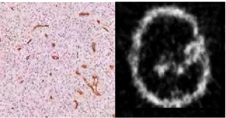
Para a análise da DMV foi adotada a técnica descrita por Weidner at al. (1991), com algumas modificações. Sem conhecimento prévio dos diagnósticos, dois observadores simultaneamente, e sob MO utilizaram um microscópio, modelo OLYMPUS BX 41, com dupla observação. Em cada lâmina, inicialmente foi empregado um aumento de 40X para identificar áreas de maior concentração de vasos imunomarcados (hot spot). Três hot spots em cada lâmina
200X para fotografar o campo óptico, ou seja, um total de 3 campos foram fotografados em cada tumor. Foi utilizado o software Adobe® Photoshop
7.0 para abrir e ampliar as imagens, e a contagem dos vasos foi realizada na tela do computador. Os microvasos individuais (arteríolas e vênulas) foram contados em cada uma destas áreas, sendo a densidade microvascular determinada pela relação do total de microvasos contados dividido pelos 3 campos estudados (Staudenherz et al., 2004)
Qualquer célula endotelial ou grupamentos de células endoteliais (clusters) CD34 positivos, que estavam claramente separados das células tumorais e outros elementos gliais foram considerados como único microvaso contável. Aqueles que pareciam ser derivados do mesmo vaso, se separados, também foram contados. Cada lúmen fixado foi contado como um único microvaso contável, se não havia lúmen, mas se somente uma única célula positiva CD34 fosse visível, esta célula também era interpretada como um simples microvaso (Weidner et al., 1991; Staudenherz et al., 2004; Birlik et al., 2006). Os dois observadores concordaram quanto à escolha e à contagem dos hot spots.
3.4 ANÁLISE ESTATÍSTICA
Foi realizada a análise descritiva dos dados por meio de freqüências e porcentagens, medidas de tendência central, média e mediana, e dispersão, desvio padrão mínimo e máximo. O teste de Komolgorov-Smirnov foi aplicado para verificar a aderência das variáveis quantitativas à curva normal. Para a variável dependente DMV foi aplicado este teste de acordo com as subdivisões de cada categoria demográfica, clínica e de intervenção (Field, 2005). Para a associação entre as variáveis qualitativas, anátomo-patológica e análise visual, foi aplicado o teste exato de Fisher.
4. RESULTADOS, DISCUSSÃO E CONCLUSÃO
5. REFERÊNCIAS
Ak I, Gulbas Z, Altinel F, Vardareli E. TC-99 MIBI uptake and its relation to the proliferative potential of brain tumors. Clin Nucl Med. 2003;28(1):29-33.
Andrews DW, Das R, Kim S, Zhang J, Curtis M. Technetium-MIBI as a glioma imaging agent for the assessment of multidrug resistance. Neurosurgery 1997;40(6):1323-32.
Amar A, Giovanini AF, Rosa MP, Yamassaki HO, Carvalho MB, Rapoport A. Densidade Microvascular no carcinoma de língua. Rev Assoc Med Bras. 2002;48(3):204-8.
Bagni B, Pinna L, Tamarozzi R, Cattaruzzi E, Marzola MC, Bagni I et al. SPET imaging of intracranial tumours with 99Tcm-sestamibi. Nucl Med Commun. 1995;16(4):258-64.
Baillet G, Albuquerque L, Chen Q, Poisson M, Delattre JY. Evaluation of single-photon emission tography imaging of supratentorial brain gliomas with technetium-99m sestamibi. 1994;21(10):1061-6.
Baldari S, Pecorella R, Cosentino S, Minutoli F. Investigation of brain tumours with 99mTc-MIBI SPET. Q J Nucl Med. 2002;46(4):336-45.
Bénard F, Romsa J, Hustinix R. Imaging gliomas with positron emission tomography and single-photon emission computed tomography. Semin Nucl Med. 2003;33(2):148-62.
Birlik B, Canda S, Ozer E. Tumour vascularity is of prognostic significance in adult, but not paediatric astrocytomas. Neuropathol Appl Neurobiol. 2006;32(5):532-8.
Brem H, Sawaya R. Brain Tumors: General Considerations. In: Winn HR. Youmans neurological surgery. 5th ed. Philadelphia: Saunders; 2004. Chapter 29. p.659-60.
Carvalho PA, Chiu ML, Kronauge JF, Kawamura M, Jones AG, Holman BL, et al. Subcellular distribution and analysis of technetium-99-MIBI in isolated perfused rat Hearts. J Nucl Med 1992;33(8):1516-22.
Chen W, Silverman DH, Delaloye S, Czernin J, Kamdar, Pope W et al. 18-FDOPA PET imaging of brain tumors: comparison syudy with 18F-FDG PET and evalçuation of diagnostic accuracy. J Nucl Med. 2006;47(6):904-11.
Collier BD, Greenberg M, Tikofsky RS, Hellman RS. Use of SPECT to distinguish brain tumor from persistent activity at a craniotomy site. Clin Nucl Med. 1987;12(3):226-8.
Ding S, Li C, Lin S, Yang Y, Liu D, Han Y et al. Comparative evaluation of microvessel density determined by CD34 or CD105 in benign and malignant gastric lesions. Humpath. 2006;37(7 ):861-6.
English RJ, Brown S.E. SPECT: Single Photon Emission Computed Tomography a primer. 3 ed. New York, Society of Nuclear Medicine, 1996.
Field A. Discovering statistic using SPSS, Second Edition. California: SAGE, 2005.
Folkman J. What is the evidence that tumors are angiogenesis dependente? J Natl Cancer Inst. 1990;82(1):4-6.
Folkerth RD. Histologic measures of angiogenesis in human primary brain tumors. Cancer Treat Res. 2004;117:79-95.
Goethals I, De Winter O, Dierckk R, Annovazzi A, Signore A, Van de Wiele C. False-negative Tc-99 Scintigraphy in histopathologically proved recurrent high-grade oligodendroglioma. Clin Nucl Med. 2003;28(4):299-301.
Guenova M, Balatzenko G. CD133-2 (AC141) expression analysis in acute leukemia immunophenotyping in correlation to CD34 and P-glycoprotein. Hematology. 2008;13(3):137-41.
Jarrit PH, Acton PD. PET imaging using gamma camera systems: a review. Nucl Med Commun. 1996;17(9):758-66.
Kim NT, Elie N, Plancoulaine B, Herlin P, Coster M. An original approach for quantification of blood vessels on the whole tumor section. Anal Cell Pathol. 2003;25(2):63-75.
Kleihues P, Louis DN, Scheithauer BW, Rorke LB, Reifenbe5rg G, Burger PC, et al. The WHO classification of tumors of the nervous system. J Neuropathol Exp Neurol 2002;61(3):215-25.
Krause DS, Fackler MJ, Civin CI, May WS. CD34: Structure, biology, and clinical utility. Blood. 1996;87(1):1-13.
Lang W, Podreka I, Suess E, Müller C, Zeitlhofer J, Deecke L. Single photon emission computerized tomography during and between seizures. J Neurol. 1988; 235(5):277-84.
Leon SP, Folkerth RD, Black PM. Microvessel density is a prognostic indicator for patients with astroglial brain tumors. Cancer 1996; 77(22):362-72.
Lois D.N, Ohgaki H, Wiestler O.D, Cavenee W.K (Eds): WHO classification of tumors of the central nervous system. IARC: Lyon 2007.
Mineo TC, Ambrogi V, Baldi A, Rabitti C, Bollero P, Vincenzi B et al. Pronostic impact of VEGF, CD31, CD34, and CD105 expression and tumor vessel invasion after radical surgery for IB-IIA non-small cell lung cancer. J Clin Pathol. 2004; 57(6):591-7.
Minutoli F, Angileri FF, Consentino S, Pecorella GR, Cardali S, De Divittis O et al. 99mTc-MIBI SPECT in distinguishing neoplastic from nonneoplastic intracerebral hematoma. J Nucl Med. 2003;44(10):1566-73.
Minutoli F, Angileri FF, Conti A, Herberg A, Aricó D, Baldari S, et al. Timing of examination affects reliability of 99mTc-methoxyisobutylisinitrile SPECT in distinguishing neoplastic from nonneoplastic brain hematomas. J Nucl Med 2005; 46(4):574-9.
marker in glioma – a comparative study with 201TL. Neuroradiology 2001; 43(12):1023-30.
Offersen B, Pfeiffer P, Hamilton-Dutoit S, Overgaard J. Patterns of angiogenesis in nonsmall-cell lung carcinoma. Cancer. 2001; 91(8):1500-9.
Offersen B., Sorensen F., Yilmaz M., Knoop A., Overgaard J. Chalkley estimates of angiogenesis in early breast cancer. Acta Oncol. 2002; 41(7):695-703.
Ohgaki H, Kleihues P. Population-based studies on incidence, survival rates, and genetic alterations in astrocytic and oligodendroglial gliomas. J Neuropathol Exp Neurol 2005; 64(6):479-89.
Ohgaki H, Kleihues P. Genetic pathways to primary and secondary glioblastoma. Am J Pathol 2007; 170(5):1445-53.
Piwinca-Worms D, Kronauge JF, Chiu ML. Enhancement by tetraphenylborate of technetium-99m-MIBI uptake kinetics and accumulation in cultured chick myocardial cells. J Nucl Med 1991; 32(10)1992-9.
Pardridge WM. CNS drug design based on principles of blood-brain barrier transport. J Neurochem.1998; 70(5):1781-92.
Rachinger W, Goetz C, Pöpperl G, Gildehaus FJ, Kreth FW, Holtmannspötter M et al. Positron emission tomography with O-(2-[18F]Fluoroethyl)-I-tyrosine versus magnetic resonance imaging in the diagnosis of recurrent gliomas. Neurosurgery. 2005; 57(3):505-11.
Sasajima T, Shimada N, Naitoh Y, Takahashi M, Hu Y, Satoh T, et al. 99mTc-MIBI imaging for prediction of therapeutic effects of second-generation MDR1 inhibitors in malignant brain tumors. Int J Cancer. 2007; 121(12):2637-45.
Shibata Y, Matsumura A, Nose T. Effect of expression of P-glycoprotein on Technetium-99m methoxyisobutylisonitrile single photon emission computed tomography of brain tumors. Neurol Med Chir. 2002; 42(8):325-30.
Soler C, Beauchesne P, Maatougui K, Schimitt T, Barrai F, Michel D et al. Technetium-99m sestamibi brain single-photon emission tomography for detection of recurrent Gliomas after radiation therapy. Eur J Nucl Med. 1998; 25(12):1649-57.
Staudenherz A, Fazeny B, Marosi C, Nasel C, Hoffmann M, Puig S et al. Does 99mTc-Sestamibi in high-grade malignant brain tumors reflect blood-brain barrier damage only? Neuroimage. 2000; 12(1):109-11.
Staudenherz A, Wolfsberg S, Killer M, Nasel C, Puig S, Marosi C et al. Microvessel density is not crucial for scintigraphic visulization of brain tumors using 99mTc-MIBI. Microvasc Res. 2004; 67(3):218-22.
Tanaka F, Otake Y, Yanagihara K, Kawano Y, Miyahara R, Li M et al. Evaluation of angiogenesis in non-small cell lung cancer: Comparison between anti-CD34 antibody and anti-CD105 antibody. Clin Cancer Res. 2001; 7(11):3410-5.
Tjalma W, Van Marck E, Weyler J, Dirix L, Van Daele A, Goovaerts G et al. Quantification and prognostic relevance of angiogenic parameters in invasive cervical cancer. Br J Cancer. 1998; 78(2):170-4.
Traweek ST, Kandalaft PL, Metha P, Battifora H. The human hematopoietic progenitor cell antigen (CD34) in vascular neoplasia. Am J Clin Pathol 1991; 96(1):25-31.
Weidner N, Semple JP, Welch WR, Folkman J. Tumor angiogenesis and metastasis –correlation in invasive breast carcinoma. N Eng J Med. 1991; 324(1):1-8.
Wu SX, Deng ML, Li QQ, Zhao C, Lu TX, Li FY et al. Prognostic analysis of patients with cerebral glioma treated with radiotherapy. Ai Zheng. 2004; 23(11 Suppl):1561-6.
6. ARTIGO CIENTÍFICO SUBMETIDO AO ANNALS OF SURGICAL
ONCOLOGY
EVALUATION OF THE MICROVASCULAR DENSITY IN ASTROCYTOMAS IN ADULTS, CORRELATED USING SPECT-MIBI
Cavalcante, Sandro. Almeida, José. Clara, Carlos. Peres, Stela. Moriguchi, Sonia Rocha, Euclides.
EVALUATION OF THE MICROVASCULAR DENSITY IN
ASTROCYTOMAS IN ADULTS, CORRELATED USING SPECT-MIBI.
S P Cavalcante, MD. Neuro-oncology division, Department of Surgery, Hospital de Câncer - Fundação Pio XII, Barretos, São Paulo, Brazil; and Blood Transfusion Center, Medical School, São Paulo State University (UNESP), 18618-000 Botucatu, São Paulo, Brazil
J R W de Almeida, MD, MSc. Neuro-oncology division, Department of Surgery, Hospital de Câncer - Fundação Pio XII, Barretos, São Paulo, Brazil;
C A Clara, MD, Neuro-oncology division, Department of Surgery, Hospital de Câncer - Fundação Pio XII, Barretos, São Paulo, Brazil;
C S Neto, MD. Department of Pathology, Hospital de Câncer - Fundação Pio XII, Barretos, São Paulo, Brazil;
S V Peres, MSc. Epidemiologist in the Research Support Group, Hospital de Câncer - Fundação Pio XII, Barretos, São Paulo, Brazil;
S M Moriguchi, MD, PhD, Department of Nuclear Medicine, Hospital de Câncer - Fundação Pio XII, Barretos, São Paulo, Brazil;
E T da Rocha, MD, PhD. Department of Nuclear Medicine, Hospital de Câncer - Fundação Pio XII, Barretos, São Paulo, Brazil; and Blood Transfusion Center, Medical School, São Paulo State University (UNESP), 18618-000 Botucatu, SP, Brazil.
ABSTRACT
Introduction: Microvascular density (MVD) may be an additional prognostic marker for astrocytomas, but the heterogeneity of these tumors limits its use. Thus, imaging examinations such as SPECT-MIBI may take on an indirect role in these evaluations.
Objectives: The aim of this study was to evaluate MVD in astrocytomas, using the immunohistochemical technique with anti-CD34 monoclonal antibodies to obtain a relationship between these data and the parameters obtained from SPECT-MIBI.
Methods:This cross-sectional study evaluated 48 patients with brain tumors such as low-grade astrocitoma (LGA), anaplastic astrocytoma (AA) and glioblastoma multiforme (GBM) who were admitted to Hospital de Câncer, Fundação Pio XII, Barretos. All of them underwent brain SPECT-MIBI before any treatment. MVD was determined under an optical microscope, by counting microvessels on slides from each case. SPECT-MIBI images were analyzed visually and semiquantitatively.
Results: GBM, AA and LGA represented 50%, 16.7% and 33.3%, respectively. There were 13 normal and 35 abnormal SPECT-MIBI images. There were significant differences in MVD between AA and LGA (p = 0.040), but not between normal and abnormal SPECT-MIBI. The mean counts from SPECT-MIBI did not correlate with MVD. Among GBM cases, there were no significant findings, except for greater likelihood of abnormal histological tests. Conclusion: MVD had a relationship with histological grade, i.e. between AA and LGA, but it did not have any correlation with SPECT-MIBI.
INTRODUCTION
Among the tumors that affect the brain, gliomas of astrocytic, oligodendroglial and ependymal origin are responsible for more than 70% of the occurrences.1 Of these, astrocytomas are the most frequent histological type: they are classified according to their malignant potential and graded on the basis of the histological criteria recommended by the World Health Organization (WHO).2 Over the last few years, knowledge of the tumor progression mechanisms for astrocytomas has increased, and this had led to supplementation of the histological grade with new potential prognostic markers. Among these, there has been increasing interest in the role of quantification of the vessels in astrocytomas, with ongoing investigations.3 In 1991, Weidner4 was the first to report on the prognostic value of measurements of angiogenesis, using the model of primary breast tumors. Many investigators have supported the notion of an inverse correlation between microvascular density (MVD) and prognosis,5-7 while others have refuted this.8 MVD has also been shown to be a prognostic indicator for postoperative survival among patients with astrocytomas.3,9,10 However, there have been limitations on quantifying the vascularization of brain tumors because of the cell heterogeneity found in these tumors.9 Other authors have demonstrated significant differences in MVD between tumors in children and adults.3
neoplastic and non-neoplastic lesions,11,13 allows the tumor grade to be evaluated11 and assists in predicting the therapeutic response.11,14,15 The intensity of uptake is related not only to breaking the blood-brain barrier but also to tumor metabolic activity.11,16,17 Thus, MIBI may be used to evaluate the biological characteristics of brain tumors, and it has a role in determining the proliferative potential and prognosis for these tumors.11,18,19 Because of its molecular characteristics, MIBI is preferentially removed in mitochondria.20,21 Since the mitochondrial activity in tumors is usually high, this generates an increase in the transmembrane gradient, which favors its accumulation in tumor tissue significantly.
Vascularization is important as a prognostic factor in many types of neoplasia,3-5,22,23 but such evaluations are only possible today through direct analysis of slides using different methods4,22 that present limitations determined by the heterogeneity of these tumors.3 Imaging methods have been discussed as interesting tools for evaluating the vascularization in brain tumors, and SPECT-MIBI is among these.16,17 This method presents a significant correlation between MIBI uptake rates and length of survival, according to the aggressiveness of the recurrent malignant gliomas.24 However, the amount of robust information on its relationship with MVD is still insufficient, thus opening up perspectives regarding this approach.
METHODS
STUDY POPULATION AND PROTOCOL
A study of cross-sectional type was conducted in which demographic and KPS (Karnofsky Performance Scale) data were gathered retrospectively from 48 patients (29 men and 19 women; mean age 48.8 years, SD = 15.9, range from 20 to 73 years) who had been admitted to Hospital de Câncer, Fundação Pio XII, Barretos. None of these patients had undergone any previous surgical or therapeutic procedure. Only cases of supratentorial gliomas that had been diagnosed as astrocytomas of LGA (Only difuse astrocytomas were selected), AA or GBM type, in accordance with criteria established by WHO,2 were included. All of these patients underwent brain SPECT with MIBI before any procedure. Tumor tissue specimens of adequate quantity were available in paraffin blocks, for all of these patients. This study received the approval of our hospital’s ethics committee.
ACQUISITION OF BRAIN SPECT IMAGES WITH MIBI
The examinations were initially performed by visual analysis and classified as normal or abnormal. The latter were then subdivided into abnormal images of mild intensity (when uptake could be discerned, but less than in the scalp), moderate intensity (when the uptake was as intense as the scalp) and marked intensity (when the uptake was equivalent to the salivary glands). Semiquantitative analysis was performed to investigate the mean count in the tumor area and in a mirror area in the contralateral hemisphere.14,16,24,26 This made it possible to obtain an index (tumor/contralateral side index, T/CL), as the ratio between these measurements. HISTOPATHOLOGICAL CLASSIFICATION
Slides stained with hematoxylin-eosin were produced from the paraffin blocks, in order to review the diagnoses and the histopathological grade. These reviews were performed by two experienced pathologists, following the WHO criteria.2
IMMUNOHISTOCHEMICAL REACTIONS
Sections of three microns in thickness were cut from the paraffin blocks from each case, to perform immunohistochemical reactions. These were mounted on slides and were silanized (3-aminopropyltriethoxilane, SIGMA A-3648, USA) and deparaffinized in a heated chamber at 60º C for 12 hours.
The streptavidin-biotin-peroxidase technique was used, with anti-CD34 class II monoclonal antibodies (clone QBEnd-10, code m7165, titration 1:800; Dako Cytomation, Glostrup, Denmark). The antigen recovery was done on a Pascal panel, using citrate at pH 6.0. The amplification was performed using the AdvanceTM HRP system (Dako Cytomation).
MICROSCOPIC ANALYSIS OF THE IMMUNOHISTOCHEMICAL REACTIONS
the areas of greatest concentration of immunolabeled vessels (hot spots). At each hot spot, 200X magnification was used to photograph the optical field. A total of three fields were photographed, one in each hot spot of each tumor. The Adobe® Photoshop
7.0 software was used to open and amplify the images. The vessels were counted on the computer screen. The individual microvessels (arterioles and venules) were counted in each of these areas, and the MVD was determined as the ratio of the total number of microvessels counted divided by the three fields examined.17
Any CD34-positive endothelial cell or groupings of endothelial cells (clusters) that were clearly separated from the tumor cells and other glial elements were considered to be a single countable microvessel. Those that appeared to be derived from the same vessel, if separate, were also counted. Each fixed lumen was counted as a single countable microvessel. If there was no lumen, but only a single CD34-positive cell visible, this cell was also interpreted as a single microvessel.3,4,17
STATISTICAL ANALYSIS
A descriptive analysis was performed on the data, in terms of frequencies and percentages, central trend and dispersion measurements, means, medians, standard deviations, minimums and maximums. The Kolmogorov-Smirnov test was applied to investigate the adherence of the quantitative variables to a normal distribution curve. For the dependent variable of MVD, this test was applied in accordance with the subdivisions of each demographic, clinical and interventional category.27 For associations between qualitative variables relating to anatomopathology and visual analysis, Fisher’s exact test was applied.
MVD, the Mann-Whitney test was applied when the independent variable had two categories and the Kruskal-Wallis test, when the independent variable had more than two categories. To investigate correlations between the dependent variable (MVD) and the tumor/contralateral side (T/CL) index, Spearman’s correlation was used. In this study, statistical significance level was set as p < 0.05. Data input, consistency and analysis were performed using the SPSS software for Windows, version 16.0.
RESULTS
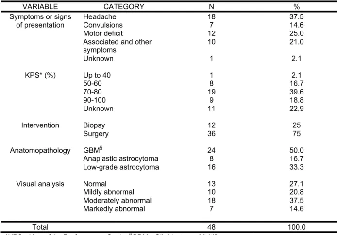
As shown in Table 1, the single forms of clinical presentation most commonly found were headache (37.5%), motor deficit (25%) and convulsions (14.6%). Most of these patients (81.3%) presented KPS of 70% or more. Among the paraffin-block specimens, 75% were obtained from surgical procedures with full or partial macroscopic resection of the tumor, and the remainder came from biopsies. GBM accounted for 50% (24 cases) of the sample, while the LGA and AA groups represented 33.3% (16) and 16.7% (8), respectively. Visual analysis of the SPECT-MIBI examinations did not find any abnormality in 27.1% (13 examinations), while the remaining 72.9% (35 examinations) presented mild, moderate or marked abnormalities.
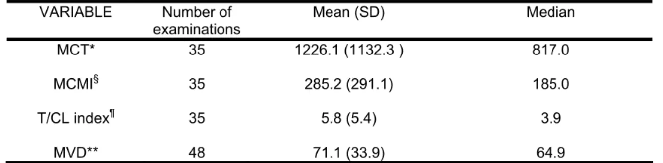
Among these 35 abnormal examinations, the mean count in the tumor was 1226.1 and in the mirror image, it was 285.2. The mean T/CL index was 5.8. The mean number of vessels found in the 48 slides was 71 vessels per field (Table 2).
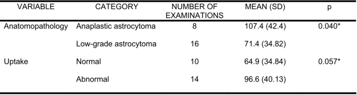
The difference in mean MVD between the AA and LGA was significant (p = 0.040). However, there was no significant difference in mean MVD between normal and abnormal examinations (p = 0.057) (Table 4). A similar analysis for the GBM group did not reach significance (p = 0.295), (result not shown in the tables). Correlation between the T/CL index and MVD was also nonsignificant (Table 5).
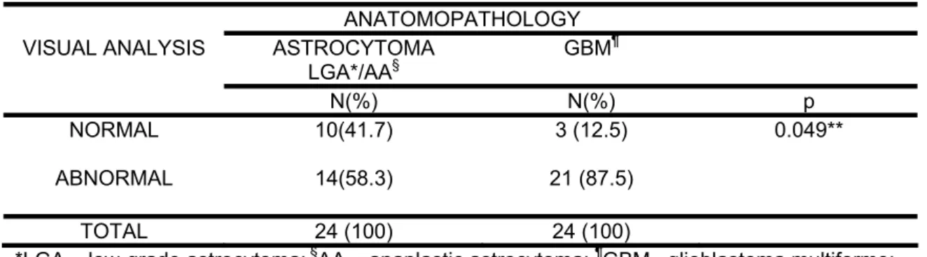
On the other hand, Table 6 shows that there was a greater likelihood that the GBM group would present abnormal examinations in relation to AA and LGA (87.5% versus 58.3%; p = 0.049 ).
DISCUSSION
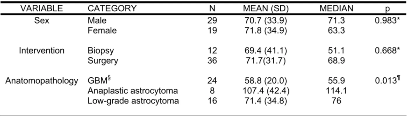
In the present study, data on 48 adult patients with supratentorial astrocytomas were evaluated in an attempt to establish a relationship between MVD in these tumors and analysis using SPECT-MIBI. Sex did not influence MVD, and this had not previously been discussed in the literature. GBM accounted for 50% of the sample evaluated (Table 1), although there was no randomization. Despite the lower diagnostic accuracy provided by biopsies,28 which suggests that studies with such samples should be regarded separately,9 in our study there was no significant difference in mean MVD between the samples obtained through biopsy and those obtained through surgery (Table 3).
3). In 2005, Yao10 found a significant difference only between GBM and LGA, while Leon,9 in 1996, showed that there was a significant association between microvascular grade and histology, considering only the AA and GBM. Furthermore, the latter author also showed that the microvascular grade and MVD were significant predictors of postoperative survival and that the vascular grade was more intense, which was explained by the existence of glomeruloid vascular structures. In fact, these structures are clusters of vessels that influence the choice of hot spots, given that the microvascular grade is higher in these areas. Nonetheless, they are counted as single vessels at higher magnification, thereby probably underestimating the count of individual microvessels.9 Extensive areas of necrosis also bring additional explanations, although in the present study, there was continual concern about avoiding areas that were close to necrosis. The existence of necrosis makes immunohistochemical analyses difficult,29 while MVD has failed to show any correlation with histological grade in tumors presenting associations with necrosis.30 Tumor necrosis, and in particular large-scale ischemic necrosis, is significantly more frequent in primary GBM.31,32 Therefore, the presence of glomeruloid formations and necrosis, which were elements observed in the present study, may furnish explanations regarding why there is underestimation of microvessel counts in GBM. It could be seen that the identification of hot spots, from the manner in which this was done, might not faithfully represent the appropriate area for counting the vessels inside the tumor. This would particularly be so in GBM, in which the heterogeneity characteristic of brain tumors seemed to be more evident.
which implies a lack of standardization for this purpose and leads to additional difficulties in establishing the real role of vessel quantification as prognostic information.
It is known that 95% of GBM start out as GBM, and only 5% of them originate from LGA or AA. Thus, GBM may be primary or secondary, and these constitute distinct entities that affect patients at different ages and develop through different genetic routes. However, LGA and AA represent a continuum within the disease, with similar genetic characteristics, thus differing from GBM, which has separate genetic characteristics.35,36 In the present study, out of the 48 specimens evaluated, there were 24 GBM, which were studied separately. It is important to note that because of the presence of extensive areas of necrosis, it was not possible to establish a correlation between MVD and GBM.
The mechanisms for MIBI uptake are still not well defined, although some authors have reported that MIBI uptake did not depend on MVD and only reflected the rupturing of the blood-brain barrier.16,17 Nonetheless, the existence of methodological inconsistencies in quantifying MVD means that its true value in relation to MIBI uptake cannot be stated with certainty. MIBI has been widely used to differentiate radionecrosis from tumor recurrence,11 but there were three cases of GBM in the present study in which no abnormal areas of uptake were viewed (Table 6). A similar finding was described previously,12,17 which indicates the need for caution in interpreting these examinations.
There was a significant difference in mean MVD between LGA and AA, but significant difference was not seen between uptake and MVD (=0.057) (Table 4). However, we believe that the sample size may have influenced this result and that evaluation of a larger sample would make it possible to find significance in this tumor group. Staudenhers,17 in 2004, stated that the MVD is not crucial for scintigraphic viewing of brain tumors using 99mTc-MIBI. In their sample, there were four cases, of which three were astrocytomas and one was an oligodendroglioma. There were no associations between histological grade and MVD, and only a descriptive analysis was made.
We observed that there was a greater likelihood that GBM cases would present abnormal SPECT-MIBI results, in relation to tumors of lower grade (Table 6). Histologically, GBM are characterized by intense vascular proliferation,2 but no association with such data could be confirmed by current immunohistochemical methods because of the limitations cited earlier.
ACKNOWLEDGEMENTS
REFERENCES
1. Kleihues P, Louis DN, Scheithauer BW, Rorke LB, Reifenberg G, Burger PC, et al. The WHO classification of tumors of the nervous system. J Neuropathol Exp Neurol 2002;61(3):215-25.
2. Lois DN, Ohgaki H, Wiestler OD, Cavenee WK (Eds): WHO classification of tumors of the central nervous system. IARC: Lyon 2007.
3. Birlik B, Canda S, Ozer E. Tumour vascularity is of prognostic significance in adult, but not paediatric astrocytomas. Neuropathol Appl Neurobiol 2006;32(5):532-8.
4. Weidner N, Semple JP, Welch WR, Folkman J. Tumor angiogenesis and metastasis – correlation in invasive breast carcinoma. N Eng J Med 1991;324(1):1-8.
5. Tanaka F, Otake Y, Yanagihara K, Kawano Y, Miyahara R, Li M et al. Evaluation of angiogenesis in non-small cell lung cancer: Comparison between anti-CD34 antibody and anti-CD105 antibody. Clin Cancer Res 2001;7(11):3410-5.
6. Offersen B, Pfeiffer P, Hamilton-Dutoit S, Overgaard J. Patterns of angiogenesis in nonsmall-cell lung carcinoma. Cancer 2001;91(8):1500-9.
7. Ding S, Li C, Lin S, Yang Y, Liu D, Han Y, et al. Comparative evaluation of microvessel density determined by CD34 or CD105 in benign and malignant gastric lesions. Humpath 2006;37(7 ):861-6.
9. Leon SP, Folkerth RD, Black PM. Microvessel density is a prognostic indicator for patients with astroglial brain tumors. Cancer 1996;77(22):362-72.
10. Yao Y, Kubota T, Takeuchi H, Sato K. Prognostic significance of microvessel density determined by an anti-CD105/endoglin monoclonal antibody in astrocytic tumors: Comparison with an ant-CD31 monoclonal antibody. Neuropathology 2005;25(3):201-6.
11. Baldari S, Pecorella R, Cosentino S, Minutoli F. Investigation of brain tumours with 99mTc-MIBI SPECT. Q J Nucl Med 2002;46(4):336-45.
12. Goethals I, De Winter O, Dierckk R, Annovazzi A, Signore A, Van de Wiele C. False-negative Tc-99m scintigraphy in histopathologically proved recurrent high-grade oligodendroglioma. Clin Nucl Med 2003;28(4):299-301.
13. Minutoli F, Angileri FF, Conti A, Herberg A, Aricò D, Baldari S, et al. Timing of examination affects reliability of 99mTc-methoxyisobutylisonitrile SPECT in distinguishing neoplastic from nonneoplastic brain hematomas. J Nucl Med 2005;46(4):574-9.
14. Andrews DW, Das R, Kim S, Zhang J, Curtis M. Technetium-MIBI as a glioma imaging agent for the assessment of multidrug resistance. Neurosurgery 1997;40(6):1323-32.
15. Sasajima T, Shimada N, Naitoh Y, Takahashi M, Hu Y, Satoh T, et al. 99m Tc-MIBI imaging for prediction of therapeutic effects of second-generation MDR1 inhibitors in malignant brain tumors. Int J Cancer 2007;121(12):2637-45.
17. Staudenherz A, Wolfsberg S, Killer M, Nasel C, Puig S, Marosi C et al. Microvessel density is not crucial for scintigraphic visualization of brain tumors using 99mTc-MIBI. Microvasc Res 2004;67(3):218-22.
18. Nagamachi S, Jinnouchi S, Nabeshima K, Nishii R, Flores II L, Kodama T, et al. The correlation between 99mTc-MIBI uptake and MIB-1 as a nuclear proliferation marker in glioma – a comparative study with 201TL. Neuroradiology 2001;43(12):1023-30.
19. Ak I, Gulbas Z, Altinel F, Vardareli E. TC-99m MIBI uptake and its relation to the proliferative potential of brain tumors. Clin Nucl Med 2003;28(1):29-33. 20. Carvalho PA, Chiu ML, Kronauge JF, Kawamura M, Jones AG, Holman BL, et
al. Subcellular distribution and analysis of technetium-99-MIBI in isolated perfused rat Hearts. J Nucl Med 1992;33(8):1516-22.
21. Piwinca-Worms D, Kronauge JF, Chiu ML. Enhancement by tetraphenylborate of technetium-99m-MIBI uptake kinetics and accumulation in cultured chick myocardial cells. J Nucl Med 1991;32(10)1992-9.
22. Offersen B, Sorensen F, Yilmaz M, Knoop A, Overgaard J. Chalkley estimates of angiogenesis in early breast cancer. Acta Oncol 2002;41(7):695-703.
23. Mineo TC, Ambrogi V, Baldi A, Rabitti C, Bollero P, Vincenzi B et al. Prognostic impact of VEGF, CD31, CD34, and CD105 expression and tumor vessel invasion after radical surgery for IB-IIA non-small cell lung cancer. J Clin Pathol 2004;57(6):591-7.
25. Soler C, Beauchesne P, Maatougui K, Schimitt T, Barrai F, Michel D et al. Technetium-99m-sestamibi brain single-photon emission tomography for detection of recurrent Gliomas after radiation therapy. Eur J Nucl Med 1998;25(12):1649-57.
26. San Pedro EC, Yilmaz M, Liu HG, Rosenfeld SS, Mountz JM. A new semiquantitative method for comparing brain tumor uptake of Tc-99m sestamibi and TL-201. Clin Nucl Med 1999;24(11):868-73.
27. Field A. Discovering statistic using SPSS, Second Edition. California: SAGE, 2005.
28. Glantz MJ, Burger PC, Herdon JE 2nd, Friedman AH, Cairncross JG, Vick NA, et al. Influence of the type of surgery on the histologic diagnosis in patients with anaplastic gliomas. Neurology 1991;41(11):1741-4.
29. Aronsson DE, Muhr C. Quantification of sensitivity of endothelial cell markers for the astrocytoma and oligodendroglioma tumors. Anticancer Res 2002;22(1A):343-6
30. Shivakumar S, Prabhakar BT, Jayashree K, Rajan MG, Salimath BP. Evaluation of serum vascular endothelial growth factor (VEGF) and microvessel density (MVD) as prognostic indicators in carcinoma breast. J Cancer Res Clin Oncol 2008 (in press).
31. Tohama Y, Gratas C, Van Meir EG, Desbaillets I, Tenan M, Tachibana O, et al. Necrogenesis and Fas/APO-1 (CD95) expression in primary (de novo) and secondary glioblastomas. J Neuropathol Exp Neurol 1998;57(3):239-45.
33. Netto GC, Bleil CB, Hilbig A, Coutinho LM. Immunohistochemical evaluation of the microvascular density through the expression of TGF- (CD 105/endoglin) and CD 34 receptors and expression of the vascular endothelial growth factor (VEGF) in oligodendrogliomas. Neuropathology 2008;28(1):17-23.
34. Vaquero J, Zurita M, Coca S, Oya S, Morales C. Prognostic significance of clinical and angiogegenesis-related factors in low-grade oligodendrogliomas. Surg Neurol 2000;54(3):229-34.
35. Ohgaki H, Kleihues P. Population-based studies on incidence, survival rates, and genetic alterations in astrocytic and oligodendroglial gliomas. J Neuropathol Exp Neurol 2005;64(6):479-89.
36. Ohgaki H, Kleihues P. Genetic pathways to primary and secondary glioblastoma. Am J Pathol 2007;170(5):1445-53.
37. Folkerth RD. Descriptive analysis and quantification of angiogenesis in human brain tumors. J Neurooncol 2000;50(1-2):165-72.
Table 1. Description of the clinical, surgical and anatomopathological data and visual analysis
of the SPECT-MIBI results.
VARIABLE CATEGORY N %
Symptoms or signs Headache 18 37.5
of presentation Convulsions 7 14.6
Motor deficit 12 25.0
Associated and other
symptoms 10 21.0
Unknown 1 2.1
KPS* (%) Up to 40 1 2.1
50-60 8 16.7
70-80 19 39.6
90-100 9 18.8
Unknown 11 22.9
Intervention Biopsy 12 25
Surgery 36 75
Anatomopathology GBM§ 24 50.0
Anaplastic astrocytoma 8 16.7
Low-grade astrocytoma 16 33.3
Visual analysis Normal 13 27.1
Mildly abnormal 10 20.8
Moderately abnormal 18 37.5
Markedly abnormal 7 14.6
Total 48 100.0
Table 2. Description of mean count parameters and MVD
VARIABLE Number of
examinations Mean (SD) Median
MCT* 35 1226.1 (1132.3 ) 817.0
MCMI§ 35 285.2 (291.1) 185.0
T/CL index¶ 35 5.8 (5.4) 3.9
MVD** 48 71.1 (33.9) 64.9
Table 3. Difference in mean MVD between the categories
VARIABLE CATEGORY N MEAN (SD) MEDIAN p
Sex Male 29 70.7 (33.9) 71.3 0.983*
Female 19 71.8 (34.9) 63.3
Intervention Biopsy 12 69.4 (41.1) 51.1 0.668*
Surgery 36 71.7(31.7) 68.9
Anatomopathology GBM§ 24 58.8 (20.0) 55.9 0.013¶
Anaplastic astrocytoma 8 107.4 (42.4) 114.1 Low-grade astrocytoma 16 71.4 (34.8) 76
Table 4. Difference in mean MVD between anaplastic astrocytomas and low-grade
astrocytomas, and between examinations with normal and abnormal uptake.
VARIABLE CATEGORY NUMBER OF
EXAMINATIONS
MEAN (SD) p
Anatomopathology Anaplastic astrocytoma 8 107.4 (42.4) 0.040*
Low-grade astrocytoma 16 71.4 (34.82)
Uptake Normal 10 64.9 (34.84) 0.057*
Abnormal 14 96.6 (40.13)
Table 5. Correlations of quantitative variables with MVD
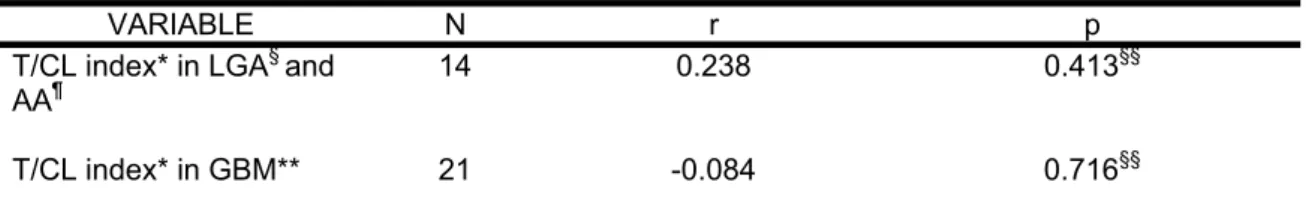
VARIABLE N r p
T/CL index* in LGA§ and AA¶
14 0.238 0.413§§
T/CL index* in GBM** 21 -0.084 0.716§§
Table 6. Likelihood of abnormal examination result in relation to histological type.
ANATOMOPATHOLOGY VISUAL ANALYSIS ASTROCYTOMA
LGA*/AA§
GBM¶
N(%) N(%) p
NORMAL 10(41.7) 3 (12.5) 0.049**
ABNORMAL 14(58.3) 21 (87.5)
TOTAL 24 (100) 24 (100)