A murine model of
xenotransplantation of human
glioblastoma with imunosupression
by orogastric cyclosporin
Alexandre M. Cunha1,2, Fernanda S. Nascimento1, Jane C.O.F. Amaral3, Sandra Konig3, Crhistina M. Takiya4, Vivaldo M. Neto3,5,
Eduardo Rocha6, Jorge P.B.M. Souza1
ABSTRACT
Several animal experimental models have been used in the study of malignant gliomas. The objective of the study was to test the efficacy of a simple, reproducible and low cost animal model, using human cells of glioblastoma multiforme (GBM) xenotransplantated in subcutaneous tissue of Wistar rats, immunosuppressed with cyclosporin given by orogastric administration, controlled by nonimunosuppressed rats. The animals were sacrificed at weekly intervals and we have observed gradual growth of tumor in the immunosuppressed group. The average tumor volume throughout the experiment was 4.38 cm3 in the immunosuppressed group, and 0.27 cm3 in the control one (p<0.001). Tumors showed histopathological hallmarks of GBM and retained its glial identity verified by GFAP and vimentin immunoreaction. Immunosuppression of rats with cyclosporin was efficient in allowing the development of human glioblastoma cells in subcutaneous tissues. The model has demonstrated the maintenance of most of the histopathological characteristics of human glioblastoma in an heterotopic site and might by considered in research of molecular and proliferative pathways of malignant gliomas.
Key words: animal model, Wistar rats, cyclosporin, glioblastoma, xenotransplant.
Modelo murino de xenotransplante de glioblastoma humano com imunossupressão utilizando ciclosporina orogástrica
RESUMO
Vários modelos animais têm sido avaliados no estudo dos gliomas e até o momento nenhum pôde ser considerado ideal. O objetivo deste trabalho é verificar a eficácia de um modelo animal simples, reprodutível e de baixo custo. Utilizamos células humanas de glioblastoma multiforme (GBM) xenotransplantadas em ratos Wistar, submetidos a imunossupressão com ciclosporina administrada por via orogástrica. Células tumorais foram implantadas no tecido subcutâneo dos ratos imunossuprimidos com ciclosporina, sendo o controle feito em ratos não imunossuprimidos. Os animais foram sacrificados em intervalos semanais e foi observado crescimento progressivo do tumor no grupo imunossuprimido. A média do volume tumoral em todo o experimento foi de 4,38 cm3 no grupo imunossuprimido e 0,27 cm3 no grupo controle (p<0,001). Os tumores apresentavam características histopatológica do GBM e mantinham sua identidade glial, verificadas por imunoreação para GFAP e vimentina. A imunossupressão dos ratos com ciclosporina
Correspondence Alexandre Martins Cunha Rua Prof. Rodolpho Rocco 225 21941-913 Rio de Janeiro RJ - Brasil E-mail: [email protected] [email protected]
Received 7 February 2010 Received in final form 23 July 2010 Accepted 30 July 2010
1Division of Neurosurgery, Department of Surgery, Clementino Fraga Filho University Hospital, Federal University of Rio de
Janeiro, Rio de Janeiro RJ, Brazil; 2Division of Neurosurgery, Department of Surgical Specialties, Pedro Ernesto University
Hospital, State University of Rio de Janeiro, Rio de Janeiro RJ, Brazil; 3Department of Anatomy, Institute of Biomedical
Sciences, Federal University of Rio de Janeiro, Rio de Janeiro RJ, Brazil; 4Department of Histology and Embriology, Institute
of Biomedical Sciences, Federal University of Rio de Janeiro, Rio de Janeiro RJ, Brazil; 5National Institute for Translational
Neuroscience, Ministry of Science and Technology, Brazil; 6Division of Nephrology, Department of Internal Medicine,
foi eficiente em permitir o desenvolvimento do glioblastoma no tecido subcutâneo. Uma vez que o presente modelo mantém a maioria das características histopatológicas do glioblastoma humano, ele pode ser considerado em estudos que avaliem as vias moleculares e proliferativas dos gliomas malignos.
Palavras-chave: modelo animal, ratos Wistar, ciclosporina, glioblastoma, xenotransplante.
Glioblastoma multiforme (GBM) is the most frequent primary brain tumor and is classiied by the World Health Organization (WHO) in the group of difusely iniltrative astrocytomas, representing the most malignant subtype of them1-9. Understanding the mechanisms of angiogen-esis, cellular migration and proliferative pathways might improve therapeutic development for the treatment of GBM10-12. Scientiic knowledge of the biology of the gli-omas has been evolving with the use of animal models13. An ideal animal model for the study of gliomas must have some deined characteristics: growth rate and ma-lignancy characteristics of the tumor should be repro-ducible; the time of the tumor induction should be rela-tively short and the survival time should be standardized; the tumor should present intraparenchymal growth that simulates glioma, showing invasion, neovascularity and no encapsulation; the tumor should grow well in culture and must be safe for the laboratorial handling; and cheap and small species must be preferable14,15. Although many animal models are available, none of them ills all above the described characteristics14,15.
he model of orthotopic xenotransplantation with hu-man tumor cells in anergic or imunosuppressed animals is the best way to simulate the growth of human glio-mas16. he models of heterotopic transplant outside the central nervous system are frequently used, and the sub-cutaneous xenografts are useful and reproducible mod-els that allow the study of molecular biology and genetic alterations in GBM17.
hese models of subcutaneous implantation allow a fast tumor growth and an easy evaluation of the size and volume of the tumor, without need of the animal’s sac-riice. Moreover, no major histopathological diference has been demonstrated between the tumors implanted in subcutaneous and in cerebral tissue18. Because of dif-ference in the pattern and vascular architecture between the models of cerebral and subcutaneous implantation, and also of the absence of blood-brain barrier in the sub-cutaneous models, care has to be taken in extrapolating the results of studies with subcutaneous models to hu-man brain gliomas19,20.
he main objective of the present work was to verify the utility of an experimental model of human glioblas-toma cells xenotransplantation in subcutaneous tissue of Wistar rats, immunosuppressed with cyclosporine given by orogastric administration.
METHOD Tumor cells
Tumour sample (GBM-95) was obtained from a pa-tient with GBM treated at the Neurosurgery Division from University Hospital Clementino Fraga Filho and ex-perimental use was approved by Ethical Committee from Brazilian Ministry of Health. he protocol of this study was also approved by the Committee of Ethics in Animal Researches of Department of Surgery of Federal Universi-ty of Rio de Janeiro. Microscopically dissected tumor was transferred to tissue culture lasks containing culture
dium DMEM (Dubelcco’s modiied eagle medium) with 10% of fetal bovine serum (FBS) and maintained in a stan-dard tissue culture incubator at 37ºC and 5% CO2. Brief-ly, when cellular growth achieved near 90% conluence, the cells were submitted to successive passages with tryp-sin treatment and then centrifuged and the pellet was re-suspended with dimetilsulfoxide (DMSO) and FBS. hey were maintained at a temperature of –20ºC for one hour and then stored in liquid nitrogen. We have previously described this detailed protocol in other study21.
Cells were defrosted in DMEN with 10% FBS and cul-tured until use to xenotransplantation (Fig 1A). To do this, 106 cells were used and we have used cells cultured in 14th passage.
Immunocytochemistry
Immunocytochemistry was performed as described previously21. Briely, cultured cells plated on glass cov-erslips were ixed with 4% paraformaldehyde for 30 min and permeabilized with 0.1% Triton X-100 in phosphate-bufered saline (PBS) for 5 min at room temperature. Af-ter blocking, cells were incubated with primary antibod-ies overnight at 4ºC temperature, followed by PBS washes and incubation with speciic secondary antibody conju-gated with rhodamine: alexa luor 488 goat anti-mouse, invitrogen, molecular probes; and luorescein: alexa lu-or 546 goat anti-rabbit, invitrogen, molecular probes. Pri-mary antibodies used and dilutions were as follows: rab-bit anti-cow, glial ibrillary acidic protein (GFAP), 1:500, Dako; monoclonal mouse, anti-vimentin, 1:200, Dako. In all immunostaining-negative controls, reactions were per-formed by omitting the primary antibody. No reactivi-ty was observed when the primary antibody was absent.
Experimental design
Adult male Wistar rats weighting from 229 to 337 grammas were selected for the experiment. he animals were maintained in cages with free access to food and wa-ter. he rats were divided in two groups. he immuno-suppressed group with twenty animals and the control group, not immunosuppressed, with ifteen rats. he im-munossuppression was made by daily administration of cyclosporine through orogastric tube. he cyclosporine was diluted in olive oil and given at a dose of 5 mg/kg. Immunosuppression was initiated 48 h prior the implan-tation of tumor cells and maintained until sacriice of the animals. he rats in the control group received only olive oil for the same period.
he rats were anesthetized by intraperitoneal injec-tions of ketamine (10 mg/kg) and xylazine (2 mg/kg), and 106 GBM cells from 14th passage, were injected in a 1 ml solution in subcutaneous tissue of the right lanks - in-guinal region - of the animals.
he rats were sacriiced at weekly intervals, using the same anesthetic protocol, from the end of the irst to the fourth week of experiment. Five rats were sacriiced each week in the immunosuppressed group and in the control group three rats were sacriiced at the end of irst week, and four at the end of the next three weeks.
Tumor mass was identified, dissected, removed in bloc, measured and placed in 10% neutral bufered
for-malin. When no lesion was identiied, subcutaneous tis-sue was isolated and kept in the same formalin solution. Tumor volume was estimated by multiplying anterior-posterior, lateral-lateral and superior-inferior measures of the lesion, and the result divided by two.
After resection of the lank tumor, abdomen and tho-racic cavities were inspected and kidneys, liver, spleen and lungs were removed for macroscopic metastatic veriication.
Histopathologic analysis
Tissues removed from the animals were main-tained in the 10% neutral bufered formalin for 96 hours. Afterwards, specimens were processed to hematoxy-lin and eosin (HE) staining and inal neuropathological analysis was done in order to verify the presence of the glial component of the tumor and histopathological characteristics.
Immunohistochemistry
Biotin-streptavidin-peroxidase immunohistochem-istry was performed in 3-4 mm thickness parain tumor sections. Sections were immunostained with anti-GFAP and anti-vimentin monoclonal primary antibodies (Dako, CA). he universal immunostaining system streptavidin-peroxidase kit (Coulter, Fullerton, CA) was used to de-velop the reaction. Hematoxylin was used to stain and counterstain parain tumor sections and mounted with Permount. he primary antibody was omitted to provide negative controls.
Statistical analysis
Statistical analyses of the tumor volume differenc-es between the two groups were made with t-tdifferenc-est and Mann-Whitney rank sum test by Sigmastat program, with a statistical signiicance of 5% (p<0.05).
RESULTS
Immunocytochemistry
Macroscopy
In the non-immunosuppressed samples there was no measurable tumor growth in the irst week. By the end of the second week there was tumor in all rats. At the third week, two animals had a small tumor and two others did
not have any lesion. At the fourth week none of the rats had macroscopic tumor.
In the immunosuppressed group we have observed tumor in four of the ive rats in the irst week. We have veriied presence of tumor in all ive animals in the fol-lowing weeks with progressive increase of mass volume toward the fourth week. At the fourth week all the rats had large lesions with evident central necrosis. he le-sions were well circumscribed without iniltration of ad-jacent tissues in both groups - immunosuppressed (Fig 2A and 2B), and control one.
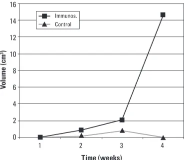
he tumor volume measures have showed signiicant diference between the two groups in all experiment. he rate of the tumor growth was larger in the immunosup-pressed group throughout all the four weeks. Tumor vol-ume average in the immunosuppressed group was 4.38 cm3 and in the control group was 0.27 cm3. Comparing these data of the two groups throughout all the experi-ment, regardless the timing, there was a signiicant sta-tistical diference between them (p<0.001).
A progressive increase of the tumor volume was ver-iied in both groups until the third week. At the fourth week there was regression of tumor in the control group and marked growth of tumor volume in the
immunosup-Fig 2. Tumor from rat after fourth week of immunosupresion. [A] Big and well cir-cumscribed lesion, without infiltration of adjacent tissues. [B] The same lesion show-ing central necrosis. [C] Histopathologic staining showing mitosis and nuclear pleo-morphism (HE 40×). [D] Pseudopalisading necrosis (HE 20×). [E] Immunohistochem-istry showing GFAP expression (40×). [F] Immunohistochemistry showing vimentin expression (40×).
Fig 3. Average tumor volume in the both groups.
16
14
12
10
8
6
4
2
0
V
o
lu
me (c
m
3)
Time (weeks)
Immunos.
pressed group, with signiicant statistical diference be-tween de two groups (p=0.0059) (Fig 3).
We have not detected any metastasis to lungs, spleen, liver or kidneys in none of the animals.
Histopathology
Histopathological analysis revealed presence of glial neoplasia in all the removed lesions. he tumors were characterized by elongated cells on a ibrillar background, with mitosis and nuclear pleomorphism (Fig 2C). hey were well vascularized and inlammatory cells were seen at the periphery of the lesions. We observed the pres-ence of central necrosis, including pseudopalisading ne-crosis, more evident in the largest lesions at the fourth week (Fig 2D).
here was no evidence of neoplasic tissue in the sub-cutaneous implantation area from animals that did not have macroscopic tumor.
Immunohistochemistry
he immunohistochemistry are strongly positive for GFAP and vimentin in lesions of both groups, demon-strating the glial nature of the developed tumors in rats (Fig 2E and 2F).
DISCUSSION
Animal models are a fundamental step in the study of neoplasias and xenotransplantation have been used with several conigurations for human GBM15,22-26. Our design had the objective of testing a simple and practical experi-mental model, using Wistar rats immunosuppressed with cyclosporin for GBM human cells implantation in the in-guinal subcutaneous tissue.
Cyclosporin promotes immunosuppression through its highly selective ability to inhibit the activation of T cells, by means of the inhibition of the calcineurin, and is largely studied in organs transplantation in order to de-crease the index of rejection27,28. It has been demonstrat-ed that cyclosporin inhibits the growth of murine glioma cells in vitro in dose dependent mechanism29. Apoptosis of tumoral cells, when exposed to the cyclosporin, seems to be mediated by the gene p5330.
Inhibition of the tumoral cell proliferation was not ob-served in our model as well as induction of apoptosis. Our results clearly demonstrated that cyclosporin immuno-suppression was related to tumor growth. A possible ex-planation for these diferences is the fact that we have used the drug in doses next to the same of ones for clinical uti-lization (5 mg/kg/day) through orogastric administration in opposite of direct cell exposition as in vitro studies29,30.
Immunosupression of the animals receiving cyclo-sporin was clearly evident after observing GBM lesions in all the animals by the end of the fourth week,
where-as in the control group there wwhere-as complete involution of the tumors. he diference of tumor volume between both immunosuppressed and control groups, by the end of the fourth week, was statistically signiicant (p=0.0059). Moreover, the rate of the tumor growth was larger in the immunosuppressed group throughout all the four weeks. Comparing the tumor volume of the two groups, there was a significant difference between them (p<0.001), showing that the cyclosporin administration was deter-minant for the tumor development.
Our results have been similar to a previous work, where human glioblastoma cells were transplanted into brain and ocular region of cyclosporin immunosup-pressed rats, by daily intraperitoneal injections at a dose of 5 mg/kg26. Xenotransplantation models of human glio-blastoma multiforme in rats immunosuppressed with cy-closporin have also been used in the study of tumor inva-sion and in the development of new drugs, but we have found no articles in English literature where cyclosporin was given by orogastric route to the rats24,31,32.
he occurrence of systemic metastasis of GBM is rare, and isolated cases have been described in literature33,34. There was no evidence of metastasis in our study. We have not observed macroscopic evidence of metastasis in lymphatic ganglia, lungs, kidneys, spleen or liver in none of the studied animals.
Histopathological analysis of the tumors in our study showed well circumscribed but not encapsulated lesions, without dissemination or iniltration of tumors cells in the adjacent structures.
he glioma growth in subcutaneous tissues occurs ba-sically by mass expansion and no cellular migration was observed. he GBM cells migration capacity is not de-termined only by genetic characteristics of the tumor cell but also depends of the interactions of its cells with the tissues microenvironment, which seems to be organ speciic35. herefore, these models are not adequate for the study of cellular migration, but they are useful in the study of proliferation17,36.
We have veriied the presence of histopathological characteristic aspects of human glioblastoma multiforme - hipercellularity, nuclear atypia, mitosis, abundant vas-cularization and necrosis, also with areas of pseudopali-sading necrosis - that had been more intense at the fourth week. According to other studies18,35, endothelial prolifer-ation is not usually observed in these type of models.
cell-to-extracellular components interactions, but may contribute for future research in proliferative pathways of malignat glial tumors.
REFERENCES
1. Collins VP. Brain tumors: classiication and genes. J Neurol Neurosurg Psy-chiatry 2004;75:2-11.
2. Daumas-Duport C, Scheithauer B, O’Fallon J, Kelly P. Grading of astrocyto-mas. A simple and reproducible method. Cancer 1998;62:2152-2165. 3. Deangelis LM. Brain tumors. N Engl J Med 2001;344:114-123.
4. Kleihues P, Louis DN, Scheithauer BW, et al. The WHO classiication of tu-mors of the nervous system. J Neuropathol Exp Neurol 2002;61:215-225. 5. Louis DN, Holland EC, Cairncross JG. Glioma classiication: a molecular
re-appraisal. Am J Pathol 2001;159:779-786.
6. Louis DN, Ohgaki H, Wiestler OD, Cavenee WK. WHO Classiication of tu-mours of the central nervous system. Lyon: IARC; 2007.
7. Pietsch T, Wiestler OD. Molecular neuropathology of astrocytic brain tu-mors. J Neurooncol 1997;35:211-222.
8. Salcman M. Glioblastoma and malignant astrocytoma. In: Kaye AH, Laws ER (Eds). Brain tumors. New York: Churchill Livingstone; 1995:449-477. 9. Vajkoczy P, Menger MD. Vascular microenviroment in gliomas. J
Neuroon-col 2000;50:99-108.
10. Ichimura K, Ohgaki H, Kleihues P, Collins VP. Molecular pathogenesis of as-trocytic tumours. J Neurooncol 2004;70:137-160.
11. Lefrane F, Brotchi J, Kiss R. Possible future issues in the treatment of glio-blastomas: special emphasis on cell migration and the resistance of mi-grating glioblastoma cells to apoptosis. J Clin Oncol 2005;23:2411-2422. 12. Maher EA, Furnari FB, Bachoo RM, et al. Malignant glioma: genetics and
bi-ology of a grave matter. Genes Dev 2001;15:1311-1333.
13. Dai C, Holland EC. Glioma models. Biochim Biophys Acta 2001;1551:19-27. 14. Crafts D, Wilson CB. Animal models of brain tumors. Natl Cancer Inst Monog
1977;46:11-17.
15. Peterson DL, Sheridan PJ, Brown Jr. WE. Animal models for brain tumors: historical perspectives and future directions. J Neurosurg 1994;80:865-876. 16. Goldbrunner RH. Models for assessment of angiogenesis in gliomas. J
Neu-rooncol 2000;50:53-60.
17. Leuraud P, Taillandier L, Aquirre-Curz L. Correlation between genetic alter-ations and growth of human malignant glioma xenografted in nude mice. Br J Cancer 2003;89:2327-2332.
18. Watanabe K, Sakamoto M, Somiya M, Amin MR, Kamitani H, Watanabe T. Feasibility and limitations of the rat model by C6 gliomas implanted at the subcutaneous region. Neurol Res 2002;24:485-490.
19. Arosarena O, Guerin C, Brem H, Laterra J. Endothelial differentiation in intracerebral and subcutaneous experimental gliomas. Brain Res 1994;640: 98-104.
20. Bersen HJJA, Rijken PFJW, Hagemeier NEM, Van Der Kogel AJ. A
quantita-tive analysis of vascularization and perfusion of human glioma xenografts at diferent implantation sites. Microvasc Res 1999;57:244-257.
21. Faria J, Romão L, Martins S, et al. Interactive properties of human glioblas-toma cells with brain neurons in culture and neuronal modulation of glial laminin organization. Diferentiation 2006;74:562-572.
22. Bigner SH, Humphrey PA, Wong AJ, Vogelstein B, Mark J, Friedman HS, et al. Characterization of the epidermal growth factor receptor in human glioma cell lines and xenografts. Cancer Res 1990;50:8017-8022. 23. Engebraaten O, Hjortland GO, Hirschberg H, Fodstad O. Growth of precultured
human glioma specimens in nude rat brain. J Neurosurg 1999;90:125-132. 24. Guillamo JS, Lisovoski F, Christov C, et al. Migration pathways of human
glioblastoma cells xenografted into the immunosuppressed rat brain. J Neurooncol 2001;52:205-215.
25. Krushelnycky BW, Farr-Jones MA, Mielke B, McKean JD, Weir BK, Petruc KC. Development of large-animal human brain tumor xenograft model in im-munosuppressed cats. Cancer Res 1991;51:2430-2437.
26. Mathiesen T, Collins VP, Olson L, Granholm L. Prolonged survival and vas-cularization of xenografted human glioblastoma cells in the central ner-vous system of cyclosporine A treated rats. Cancer Lett 1989;44:151-156. 27. Calne R. Cyclosporine as a milestone in immunosuppression. Transplant
Proc 2004;36(Suppl):S13-S15.
28. Matsuda S, Koyasu S. Mechanisms of action of cyclosporine. Immunophar-macology 2000;47:119-125.
29. Moseniak G, Figiel I, Kaminska B. Cyclosporin A, an immunosuppressive drug, induces programmed cell death in rat C6 glioma cells by a mechanism that involves the AP-1 transcription factor. J Neurochem 1997;68:1142-1149. 30. Pyrzynska B, Serrano M, Martinez AC, Kaminska B. Tumor suppressor p53
mediates apoptotic cell death triggered by cyclosporin A. J Biol Chem 2002;277:14102-14108.
31. Strojnik T, Kavalar R, Lah TT. Experimental model and immunohistochem-ical analyses of U87 human glioblastoma cell xenografts in immunosup-pressed rat brains. Anticancer Res 2006;26:2887-2900.
32. Yoshida D, Takahashi H, Teramoto A. Inhibition of glioma angiogenesis and invasion by SI-27, an anti-matrix metalloproteinase agent in a rat brain tu-mor model. Neurosurgery 2004;54:1213-1220.
33. Mourad PD, Farrel L, Stamps LD, Chicoine MR, Silbergeld DL. Why are sys-temic glioblastoma metastases rare? Syssys-temic and cerebral growth of mouse glioblastoma. Surg Neurol 2005;63:511-519.
34. Schweitzer T, Vince GH, Herbold C, Roosen K, Tonn JC. Extraneural metas-tases of primary brain tumors. J Neurooncol 2001;53:107-114.
35. Antunes L, Angioi-Duprez KS, Bracard SR, et al. Analysis of tissue chimerism in nude mouse brain and abdominal xenograft models of human glioblas-toma multiforme: what does it tell us about the models and about glio-blastoma biology and therapy? J Histochem Cytochem 2000;48:847-858. 36. Killion JJ, Radinsky R, Fidler IJ. Orthotopic models are necessary to
![Fig 1. [A] Tumor cell culture of GBM-95 lineage used in the xenotransplantation. Scale bar: 150 µm](https://thumb-eu.123doks.com/thumbv2/123dok_br/15433548.595412/2.955.112.899.894.1145/fig-tumor-cell-culture-gbm-lineage-xenotransplantation-scale.webp)