C
International League Against Epilepsy
AES Proceedings
Annual Meeting of the American Epilepsy Society
December 5, 2004
Investigators’ Workshop
8:30 a.m.–5:30 p.m.
IW.01
MODELS OF EPILEPSY IN AGING
1Eric M. Blalock,2Peter R. Patrylo, and3Kevin M. Kelly (1Molecular
and Biomedical Pharmacology, University of Kentucky Medical Cen-ter, Lexington, KY;2Physiology, Southern Illinois University School of Medicine, Carbondale, IL; and3Neurology, Drexel University College of Medicine, Allegheny-Singer Research Institute, Pittsburgh, PA)
New onset epilepsy in the elderly is a significant clinical problem yet there has been little animal modeling of the pathologies relevant to this population. This workshop will review: 1) concepts and methods used within the field of aging research; 2) application of old and new animal models of epilepsy to aged rodents - where they are and where they need to go; and 3) resources available to investigators from the National Institute on Aging. Eric Blalock will discuss the ‘calcium hypothesis of aging’, i.e., the aging-related alteration of Ca2+homeostasis that con-tributes to certain neurophysiological changes during aging. Although several studies indicate that Ca2+signaling is altered in animal mod-els of epilepsy, little information is available on the potential interplay between aging- and epilepsy-related alterations in Ca2+homeostasis. The application of ‘zipper’ slice technology to delineate neuron-specific alterations in L-type Ca2+channel activity in animal models of both aging and kindling will be reviewed. The development of strategies for the amplification/microarray-based detection of zipper slice single cell mRNA, and the potential application of this technology to studies de-signed to examine the interplay between aging and epilepsy will be explored. Peter Patrylo will discuss aging-related changes in the den-tate gyrus - alterations that could affect granule cell activity/output and thus the role of the dentate in “gating” activity into the hippocampus. Electrophysiological data will detail how local dentate circuit activity is modified during aging and an overview of in vivo studies of kainic acid-induced seizures and status epilepticus in adult and aged rats will be presented. Kevin Kelly will discuss the use of the middle cerebral artery occlusion and photothrombosis models of cerebral infarction in aged rats, explore the challenges of long-term video-EEG data collection and interpretation, and review resources for aging research made available to investigators by the NIA.
IW.02
PLASTICITY OF CHLORIDE TRANSPORT AND GABA SIGNALING
1,2,3Massimo Avoli,4Francisco Alvarez-Leefmans,5Richard Miles, and 6Melanie Woodin [1Neurology & Neurosurgery and Physiology, McGill
University, Montreal, QC, Canada;2Dept of Physiology, Universita “La Sapienza,” Rome, Italy;3Dept of Physiology, IRCCS Neuromed, Pozzilli (IS), Italy;4Dept. of Physiology, Instituto Politecnico Nacional, Mexico City, Mexico;5EMI 0224/CHU Piti´e-Salpˆetri`ere, Universit´e Paris VI, Paris, France; and6Dept. of Zoology, University of Toronto, Toronto, ON, Canada]
Inhibition, which is mediated by GABAAand GABABreceptors, con-trols CNS excitability, and represents the target of several antiepileptic drugs. Long-term changes in the reversal potential of GABAA receptor-mediated, Cl−outward currents toward less negative values have been
reported to occur in animal models of epilepsy as well as in human epileptic tissue. Indeed, such an “excitatory shift” in the reversal po-tential of GABAArecep-tor-mediated currents may represent a mech-anism of disinhibition contributing to the gen-eration and/or propaga-tion of seizures. In this workshop we will focus on the role of K+/Cl−
cotransport in epilepsy. First, Dr. Alvarez-Leefmans will describe how changes in K+/Cl−cotransport can alter the reversal potential of GABAA
receptor-mediated currents. Next, Dr. Woodin will identify the long-term, activity-dependent changes in Cl−reversal potential in
hippocam-pal neurons. Finally, Dr. Miles will discuss the involvement of changes in Cl−homeostasis in the generation of interictal spikes recorded in the
human epileptic subiculum as well as the relevance of these findings for seizure generation and epileptogenesis. In summary, this workshop will present a novel mechanism that may play a prominent role in epileptiform synchronization and epileptogenesis. (Supported by CIHR, INSERM, MIUR, NIH MH54671, and NSERC.)
IW.03
ASSESSING COGNITIVE EFFECT AND OUTCOMES OF AEDs
David W. Loring (Department of Neurology, University of Florida, Gainesville, FL)
When to initiate antiepileptic drug (AED) therapy and selection of specific AED depends on well established factors including seizure type and epilepsy syndrome, and the primary aim is successful seizure control. Within these broad categories, however, AED selection is often based upon clinical experience and individual physician preference rather than evidence-based medicine because long-term randomized monotherapy studies comparing the cognitive and behavioral outcomes have been lim-ited. In the absence efficacy differences, AEDs should be selected based upon their safety, tolerability, and side effect profiles including cogni-tion and behavior. Unfortunately, when studies have been conducted, they have been limited by methodological issues including absence of random assignment, non-equivalent representation of different seizure types, absence of appropriate longitudinal controls, and a variety of cog-nitive and behavioral measures, some of which may not be sensitive to subtle change and others that may unduly influenced by practice effects with repeated testing. In addition, a serious limitation has been the ab-sence of long-term follow-up assessment, and this limitation is more significant in pediatric epilepsy since cognitive and behavioral AED ef-fects occur against the backdrop of normal maturation and development. This workshop will discuss issues of study design, practical and admin-istrative limitations in performing large randomized AED studies, and provide direction for the design of future studies using cognition and behavior as the primary outcome variables. In the absence of differences in efficacy, AEDs should be selected based upon their safety, tolera-bility, and side effect profiles including cognition and behavior. These effects, however, have been insufficiently studied. This workshop will discuss issues of study design, practical and administrative limitations in performing large randomized AED studies, and provide direction for the design of future studies using cognition and behavior as the primary outcome variables.
IW.04
ORIGIN AND MIGRATION OF CORTICAL NEURONS
1Arnold R. Kriegstein, 2Joseph Loturco, 3Stewart A. Anderson,
and 1Samuel J. Pleasure (1Neurology, UCSF, San Francisco, CA; 2Physiology and Neurobiology, University of Connecticut, Storrs,
CT;3Psychiatry, Neurology and Neuroscience, Weill Med College of Cornell, New York, NY)
migration and may have clinical significance in relation to neuronal mi-gration disorders. Neuronal mimi-gration is controlled by a growing num-ber of different proteins. The current challenge is to define the specific roles played by these proteins in the formation of neocortical layers. Dr. LoTurco will describe some of the new migration mechanisms and path-ways that have been discovered using the novel methods of Intracerebral RNAi and in utero electroporation. GABAergic interneurons perform important roles in cortical development and are critical regulators of cortical excitability and epilepsy. However, little is known about the molecular mechanisms that underlie interneuron fate determination. Dr. Anderson will discuss the molecular signals that specify interneurons within the medial ganglionic eminence (MGE). Since mutations in some of these signaling genes have been linked to forebrain malformations in humans, identifying the molecular code for interneuron specification should enhance our understanding and treatment of disorders associated with epilepsy. Cajal-Retzius cells are among the earliest born cortical neurons and are required for normal cortical development. However their embryonic origin has been controversial. Dr. Pleasure will present evi-dence that all Cajal-Retzius cells originate from the cortical hem, a small region of caudo-medial neuroepithelium, and subsequently migrate to cover the whole cortical surface. The findings summarized in these pre-sentations are helping to transform our understanding of the patterns of cell origin and migration in the developing cortex. (Supported by grants from the NIH.)
IW.05
KETONE BODIES AND NEURONAL EXCITABILITY
1W. McIntyre Burnham,2Gary Yellen, and3Jong M. Rho (1Bloorview
Epilepsy Research Program, Department of Pharmacology, University of Toronto, Toronto, Ontario, Canada;2Department of Neurobiology, Harvard Medical School, Boston, Massachusetts;3Barrow Neurological Institute and St. Joseph’s Hospital & Medical Center, Phoenix, Arizona).
One of the hallmark biochemical features associated with fasting and ketogenic diet (KD) therapy is the production of ketone bodies (i.e., beta-hydroxybutyrate, acetoacetate and acetone). Investigators have pre-viously demonstrated that ketone bodies, when injected intraperitoneally in rodents, exhibit dose-dependent anticonvulsant effects. In contrast, in vitro experiments have failed thus far to confirm any significant effects of ketones on glutamate or GABA receptors, or on standard param-eters of synaptic transmission in brain slices. Whether ketone bodies exert direct effects on neuronal excitability, or whether they represent an epiphenomenon remains an unresolved question for those studying mechanisms of KD action. In this workshop, Dr. Mac Burnham will re-view data supporting the broad-spectrum in vivo anticonvulsant activity exhibited by acetone - the byproduct of spontaneous decarboxylation of acetoacetate. Acetone is highly effective in blocking seizures induced by maximal electroshock, pentylenetetrazole, AY-9944 (which induces atypical absence seizures), and even after amygdala kindling. Next, Dr. Gary Yellen will then present a series of intriguing experiments conducted in the substantia nigra, highlighting a potential molecular tar-get of ketone bodies, namely, adenosine triphosphate (ATP)-sensitive potassium (KATP) channels. KATP channels, when activated, causes membrane hyperpolarization and may play an important role in neuro-protection following ischemic brain injury. Finally, Dr. Jong Rho will review the electrophysiological effects of ketones bodies on rat neocor-tical neurons, and potential neuroprotective effects in an in vitro model of oxidative stress. Taken together, recent experimental data support more firmly the direct anticonvulsant, and potential neuroprotective, properties of ketone bodies.
IW.06
MEG AND EEG: ADVANCES AND CONTROVERSIES IN SOURCE LOCALIZATION AND CLINICAL UTILIZATION
1Paul A. Garcia, 2John S. Ebersole, and 3William Sutherling
(1Neurology, University of California, San Francisco, San Francisco, CA;2Neurology, University of Chicago, Chicago, IL; and3Epilepsy and Brain Mapping Center, Pasadena, CA)
EEG has traditionally been the gold standard for guiding epilepsy surgery. It is practical for obtaining ictal recordings and provides ex-cellent temporal resolution of ongoing electrocerebral activity. In recent years, MEG has been used increasingly as a pre-surgical tool at many centers. It shares many of EEG’s characteristics but it also has inherent advantages that often allow for more accurate source analysis.
In this session, lecturers will outline recent advances in the field of EEG and MEG source localization. The optimum use of both modal-ities will be considered. The contribution of MEG to the traditional pre-surgical evaluation will also be discussed.
IW.07
CREATING NEW ANIMAL MODELS OF THE CHILDHOOD EPILEPSIES
1Margaret P. Jacobs,2Solomon L. Mosh´e,3Astrid Nehlig,4Philip A.
Schwartzkroin, and5John W. Swann (1National Institute of Neurolog-ical Disorders & Stroke, NIH-DHHS, Bethesda, MD;2Dept of Neu-rology, Albert Einstein College of Medicine, Bronx, NY;3Faculte de Medecine, University L. Pasteur Inserm, Strasbourg, Cedex, France; 4Dept of Neurological Surgery, University of California-Davis, Davis,
CA; and5Dept of Pediatrics, Baylor College of Medicine, Houston, TX)
The childhood epilepsies are among the most common epileptic syn-dromes. They are often unresponsive to conventional treatments and can have devastating developmental consequences. The causes of pediatric epilepsy are likely to be multi-factorial, and result from a combination of genetic and environmental insults. Currently, progress is being made in understanding the basic mechanisms underlying epilepsy in the mature nervous system, in part due to the existence of relevant animal models. However, the study of epileptogenesis in the developing brain lags far behind. The dearth of developmental animal models is one important contributing factor that slows progress. The questions posed by the pedi-atric epilepsies differ markedly from those of adulthood due to the unique interplay of genetic mutations, brain malformations, brain injuries and seizures with the rapidly growing brain of infants and children.
The purpose of this workshop is to expand upon an NIH/NINDS meet-ing that took place in May 2004, and discuss potential guidelines for creating animal models of the early-onset epilepsies. Topics include:1) Models of childhood epilepsy – where we are now; 2) Which disorders need to be modeled; 3) Strategies for model development - I : Intractable Complex Partial Epilepsy; 4) Strategies for model development II : In-fantile Spasms; 5) How new models will be used.
IW.08
THE ROLE OF KAINATE RECEPTORS IN EPILEPSY
1Yesekiel Ben-Ari,2Anis Contractor,3John J. Hablitz, and4Michael A.
Rogawski (1INMED, INSERM U29, Marseille, France;2Dept.of Physi-ology, Northwestern University School of Medicine, Chicago, IL;3Dept. of Neurobiology, University of Alabama at Birmingham, Birmingham, AL; and4Epilepsy Research Section, NINDS, Washington, DC)
Kainate receptors (KARs) are a family of ionotropic glutamate re-ceptors comprised of combinations of GluR5-7 and KA1-2 subunits. Development of pharmacological tools discriminating KARs from other ionotropic glutamate receptors has increased our knowledge of KAR function. It is now clear that KARs are located both pre- and postsynap-tically where they exert a complex influence on neuronal excitability. Studies in animal models have shown that KAR antagonists have anti-convulsant properties and KAR expression can be altered by seizures. The speakers in this symposium will present information from basic studies of KAR structure and function, their physiological role in mod-ulating synaptic transmission and effects on epileptiform discharges. In this session, Anis Contractor will review the classification and molecular biology of KARs and present information on their role in synaptic plas-ticity in the hippocampus. Michael Rogawski will provide information amygdala KARs; results of research on KAR-mediated heterosynaptic facilitation and antagonism of KAR mediated synaptic currents by Topi-ramate will be discussed. The role of KARs in influencing excitability of hippocampal interneurons and implications for epileptogenesis will be discussed by Yehezkel Ben-Ari. The participants will consider the rele-vance of KAR-dependent mechanisms for epileptogenesis and possible future research directions.
IW.09
FLUMAZENIL PET IN HUMAN EPILEPSY
This workshop will provide a critical overview of the role of11 C-Flumazenil (FMZ) in PET studies of human epilepsy with an empha-sis on investigating the pathophysiology of localization related epilepsy and pre-surgical localization. FMZ (t1/2=20 min) is a selective ben-zodiazepine antagonist of the GABAAreceptor that serves as probe for inhibitory receptor systems implicated in both seizure generation and control. The kinetics of FMZ will be reviewed. Dr Juhasz will present experience with FMZ in pediatric epilepsy with a focus on pre-operative evaluation of non-lesional epilepsy. He will discuss the identification of FMZ abnormalities in relation to FDG-PET findings. FMZ abnormal-ities in relation to electrophysiological findings regarding the primary seizure focus and propagation will also be discussed. Multi-modal im-age co-registration techniques will be reviewed as well as approaches for objective determination of FMZ binding abnormalities. Dr Ham-mers will present data on FMZ in adult populations, focusing on non-lesional epilepsy and cortical dysplasia. He will discuss methods of partial volume correction and voxelwise analysis. He will also present pathophysiological correlations to FMZ Findings: Increased FMZ bind-ing in white matter derives from ectopic neurons; in general cortical dysplsias express decreased FMZ binding. FMZ has proved useful in preoperative planning in 25% of patients. Challenges of providing cor-relations and outcome in patient populations will also be considered. The presentation is designed to highlight methodological issues in evaluat-ing the utility of a new ligand in the pre-surgical evaluation of patients with epilepsy and the role–strengths and shortcomings–of FMZ-PET.
IW.10
NONSYNAPTIC SEIZURE MECHANISMS
1Peter L. Carlen, 2John G. Jefferys,3Dominique Durand, 4Ante L.
Padjen, and5Renato Rozental (1Medicine and Physiology, Toronto Western Research Institute and U. of Toronto, Toronto, ON, Canada; 2Dept. Neurophysiology, University of Birmingham, Birmingham,
England;3Dept. Biomedical Engineering, Case Western Reserve Uni-versity, Cleveland, OH;4Dept. Pharmacology and Therapeutics, McGill University, Montreal, QC, Canada; and5Cell Biology and Anatomy, New York Medical College, Valhalla, NY)
Gap junctional communication and field effects are still relatively un-appreciated as being major factors underlying the genesis and modulation of seizures. In addition, changes in the extracellular ionic environment and metabolism can have profound effects on neuronal and aggregate ex-citability. Finally modulation of axonal conduction is almost completely ignored as a major mechanism for spreading seizure activity throughout the brain. The suggested speakers have in vitro and in vivo expertise related to the importance of these various nonsynaptic mechanisms of seizure development and spread.
With the active help of the audience, the following issues will be discussed and debated:
–the role of gap junctional communication in seizure development, spread, and spreading depression. The questions as to the critical lo-cation of the these junctions, be it between specific neuronal types, between dendrites and/or between axons, or importantly in glia, will be addressed.
–electrical fields as both initiators and followers of seizure activity; their origin be it dendrites or glia?
–high potassium, low calcium, low magnesium, high pH, metabolic in-fluences; cause or effect?
–the role of axons in seizures; more than conduits, axonal spikes, retro-grade axonal conduction, transmission at bifurcations?
We hope to stimulate heated discussions focussed on the important issues of non-synaptic seizure mechanisms, which to date have been mainly ignored as potential therapeutic targets.
IW.11
NEUROTROPHINS AND EPILEPTOGENESIS
James O. McNamara, Samuel S. Newton, and Helen E. Scharfman (Duke University School of Medicine, Durham, North Carolina; Yale University School of Medicine, New Haven, Connecticut; Helen Hayes Hospital, Columbia University School of Medicine, New York, New York).
Increasing evidence implicates a critical role for neurotrophins and their receptors in epileptogenesis, the process by which a normal brain
becomes epileptic. Among the neurotrophins, the most extensive evi-dence implicates BDNF and its cognate receptor, TrkB. Samuel Newton will present recent studies of the role of BDNF in dentate gran-ule cell neurogenesis. Helen Scharfman will present electrophysiologi-cal analyses of BDNF regulation of neuronal excitability, including re-cent evidence suggesting a role for BDNF in catamenial epilepsy. Jim McNamara will present studies of conditional mutants of BDNF and TrkB which demonstrate that TrkB is required for limbic epileptogenesis in the kindling model.
IW.12
MECHANISMS OF PHARMACORESISTANCE IN EPILEPSY
1Terence J. O’Brien, 2H. Steve White, 3Heinz Beck, and 4Nicole
Soranzo (1The Department of Medicine, RMH, The University of Mel-bourne, Parkville, Victoria, Australia;2Department of Pharmacology and Toxicology, University of Utah, Salt Lake City, UT;3Department of Epileptology, University of Bonn Medical Center, Bonn, Germany; and 4University College London, London, England)
Current anti-epileptic drugs (AEDs) do not adequately control seizures in 20-40% of patients, with significant consequences for the quality of life and safety of these individuals with pharmacoresistant epilepsy. Even though many new AEDs have been marketed over the last 10-15 years, the proportion of patients with pharmacoresistant epilepsy has decreased very little. Patients who are refractory to one type of AED tend to be re-fractory to all other AEDs. Despite the clinical importance of drug resis-tance, the underlying cellular and molecular mechanisms have remained elusive. New treatments that modify pharmacoresistance are essential to improving the medical management of patients with epilepsy. This Investigator Workshop will discuss current research relating to pharma-coresistant epilepsy, focusing on neurobiological mechanisms and the identification of targets and strategies to overcome this important clini-cal problem.
H.S. Whitewill discuss animal models of pharamcoresistance. The use of traditional animal models of seizures and epilepsy have been largely unsuccessful in identifying compounds that are effective in pa-tients with pharmacoresistant epilepsy. Strategies will be discussed for the development of new models to be utilised to the understanding of the neurobiology of pharmacoresistance, and for designing and testing novel therapies.
H. Beckwill discuss cellular drug target mechanisms that may cause drug resistance, in particular the reduced sensitivity of receptors or ion channels to AEDs. Altered expression of specific ion channels may not only lead to altered excitability, but could also cause resistance to drugs. The presentation will summarise the current state of knowledge regarding changes in drug targets. It will also describe some recent studies that have started to dissect the molecular underpinnings of target-mediated mechanisms of pharmacoresistance in human and experimental epilepsy. An emerging understanding of these underlying molecular and cellular mechanisms is likely to provide important impetus for the development of new pharmacological treatment strategies.
N. Soranzowill discuss human pharmacogenetic studies of both ef-ficacy and dosing of AEDs. First, a detailed survey of variation in the ABCB1 drug transporter gene will be described, which aims to identify causal variants responsible for altered activity that may predispose to re-fractory epilepsy. Second, a newly identified polymorphism in the drug target SCN1A is reported that is associated with dosing of both phenytoin and carbamazepine. This study provides a rare example of a haplotype tagging strategy that has led to the identification of a previously unknown functional variant associated with drug response.
December 5, 2004
Basic Scientists’ Poster Sessions
12:00 noon–2:00 p.m.
3.001
ATYPICAL HYPERTHERMIC SEIZURES IN RATS WITH FOCAL CORTICAL DYSPLASIA LEADS TO SPONTANEOUS RECURRENT SEIZURES AND IMPAIRED LEARNING AND MEMORY
Rationale:The reported association between atypical febrile seizures and temporal lobe epilepsy (TLE) is controversial and thus far has not been supported by cohort or experimental studies. We have recently demonstrated that focal cortical dysplasia resulted in atypical hyperther-mic seizures (HS). The purpose of this study is to determine whether HS in lesioned rats will result in subsequent spontaneous recurrent seizures along with visuo-spatial learning and memory deficits paralleling that seen in patients with TLE.
Methods:Freeze lesions (focal microgyri) were induced in the right fronto-parietal cortex of rats on postnatal day (P) 1, followed by HS at P10. To evaluate for spontaneous recurrent seizures, male rats>P 60 were video monitored for 5 hours/day for 10 days. In a subgroup of animals, EEGs were then recorded 20 minutes/day for 5 days from the amygdala ipsilateral to the lesion. Prior to the EEG study visuo-spatial learning and memory were evaluated using the Morris water maze test. Controls were sham-operated and na¨ıve rats with and without hyper-thermic seizures (non-lesioned controls) and rats that received the lesion alone (lesioned controls).
Results:Recurrent behavioural seizures were observed in 4/11 (36%) of lesioned rats with HS and were characterized by freezing in association with head nodding and less frequently jaw-myoclonus. This activity was often preceded or followed by wet dog shakes. Of this group spontaneous electrographic seizures were recorded in 6/7 (86%) of rats and were characterized by runs of epileptic spikes and/or rhythmic slow (1–2HZ) spike or polyspikes and wave activity. EEG seizures were associated with behavioural manifestations similar to that observed on video-monitoring. Behavioural seizures were also seen in (1/7) 14% of the sham-operated rats with HS and electrographic seizure activity were recorded in 4/6 (66%). Behaviour or electrographic seizures were not observed in the other control groups. Regarding the Morris water maze testing there were no differences in the mean search difference score (MSD) between the non-lesioned control groups therefore their results were pooled. The MSD in lesioned rats with HS (Mean±SD; 25.6±12.8, n=13) was significantly lower than in non-lesioned controls (34.7±10.3, n=20; p<0.05). The MSD in lesioned controls was in between that of lesioned rats with HS and that of non-lesioned controls, but did not statistically differ from the two groups.
Conclusions:This study demonstrates a link between the atypical febrile seizure and the development of TLE as well as learning and memory deficits; supporting a complicating role for cortical dysplasia and possibly significant neonatal stress for the development of these conditions, following febrile seizures. (Supported by Saint Justine Re-search Foundation, Epilepsy Canada/CIHR, Hospital for Sick Children Foundation.)
3.002
REDUCING TrkB LEVELS PROTECTS HIPPOCAMPAL NEURONS FROM SEIZURE-INDUCED CELL DEATH
1Steve C. Danzer,1Maya Hughes,1Robert J. Kotloski,4Serge Nef,4Luis
F. Parada, and1,2,3James O. McNamara (1Neurobiology,2Medicine, and3Pharmacology and Molecular Cancer Biology, Duke University, Durham, NC; and4Ctr Developmental Biology, University of Texas, Southwestern, Dallas, TX)
Rationale:The hallmark pathology of human temporal lobe epilepsy is hippocampal sclerosis, a condition characterized by extensive neuronal loss and glial scaring. Tyrosine kinase B (TrkB), and its ligand BDNF, are potent survival factors for hippocampal neuronsin vitro. Whether TrkB regulates the development of hippocampal sclerosis, however, has not been established. To address this question, conditional TrkB null mice were treated with pilocarpine, a drug that produces status epilepticus-induced hippocampal sclerosis.
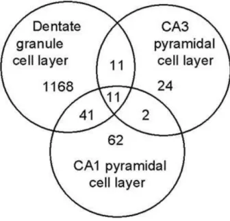
Methods:In TrkB null mice, the first coding exon of the TrkB gene is flanked by lox P sites and TrkB expression is eliminated from neurons expressing synapsin 1 promoter-driven Cre recombinase. TrkB expres-sion in these animals is eliminated from the majority of dentate granule and CA3 pyramidal cells, and a subset of cortical, thalamic and amygdala neurons. To induce hippocampal sclerosis, 4 TrkB null and 6 wildtype littermates were treated with 340 mg/kg pilocarpine, allowed to seize for 3 hours, and killed 48 hours later. Cell death was assessed by Nissl staining and Fluorojade B staining, the latter labeling dead and dying neurons.
Results: Pilocarpine-treatment produced status epilepticus in both wildtype and TrkB null mice. Surprisingly, despite qualitatively
simi-lar seizures, dentate hisimi-lar cell loss was profoundlyreducedin TrkB nulls relative to wildtype mice (wildtype, 52±11 Fluorojade B positive neu-rons/hemisection; null, 8±6; P<0.01). Even more surprising, neurons in other regions, including cortex, amygdala and thalamus, were not protected in TrkB nulls (e.g., cingulate cortex; wildtype, 43±21; null, 91±29). Nissl staining of adjacent sections confirmed the presence of healthy hilar neurons in wildtype control, TrkB null control and TrkB null pilocarpine-treated animals. Consistent with the Fluorojade B findings, significant cell loss in the hilus was only observed in pilocarpine-treated wildtype mice with Nissl staining.
Conclusions:Dentate hilar neurons are extremely sensitive to seizure-induced cell death, and the preservation of these neurons in the context of extensive cell death in other regions of the brain is striking. Studies are underway to determine whether this finding reflects reduced invasion of seizure activity into the hippocampus, or whether hippocampal seizure activity is equivalent in these animals and neuroprotection is due to a reduction in toxic effects of TrkB activation. In summary, the present findings indicate that TrkB, either directly or indirectly, may exacerbate hippocampal sclerosis. (Supported by NIH grants NS07370, NS32334 and NINDS grant NS17771, the Epilepsy Foundation, and the Ruth K. Broad Foundation.)
3.003
ACUTE CHANGES IN GENE EXPRESSION OF GABA RECEP-TOR SUBUNITS IN DENTATE GYRUS AFTER PROLONGED SEIZURES ARE AGE DEPENDENT
1Guojun Zhang,1YogendraSinh H. Raol, and1,2Amy R. Brooks-Kayal
(1Neurology, Children’s Hospital of Philadelphia; and2Pediatrics and Neurology, University of Pennsylvania, Philadelphia, PA)
Rationale:GABAAreceptors are the most abundant inhibitory neu-rotransmitter receptors in forebrain, and many studies indicated that al-terations in these receptor may play a important role in epileptogenesis. Previous studies from our laboratory suggest that long-term alterations in GABA receptor function and gene expression in hippocampal dentate granule neurons (DGNs) during epileptogenesis differ between imma-ture and maimma-ture rats. For example, a decrease in a1-mRNA levels were found in the rats that had experienced SE in adulthood, whereas, adult rats that had experienced SE at 20 days postnatal age (P20) had higher a1 levels in single DGNs. In the present study we determined the acute effect of SE at P20 and adult on GABAR expression in the whole dentate gyrus.
Methods:P20 and adult rats were subjected to pilocarpine induced status epilepticus (SE). The antisense RNA amplification (aRNA) tech-nique was used to examine the expression of 16 GABAAR and two GABABR subunits mRNA in acutely dissected dentate gyrus 24 hours and 7 days after SE.
Results:24 hours after SE, total GABAAR subunit mRNA expression was decreased in both P20 and adult rats; Bothα1 andα2 subunit mRNA expression were decreased significantly in adult rats; whereas, there were no changes after SE in P20 rats. 7 days after SE, total GABAAR subunits mRNA expression was decreased 1.5 fold in adult rats and increased 3 fold in P20 rats;α1 subunit mRNA expression was decreased 2 fold in adult rats and increased 3 fold in P20 rats;α2 subunit mRNA expression was decreased 1.5 fold in adult rats and increased 2.5 fold in P20 rats; andα5 subunit mRNA expression was decreased 1.5 fold in adult rats and increased 3 fold in P20 rats. GABAB receptor subunits R1 mRNA expression was decreased 2 fold in adult rats and increased 2.5 fold in P20 rats at 7 days after SE.
Conclusions:Prolonged seizures at P20 and in adulthood lead to differential alterations in GABAAR and GABABR subunits mRNA ex-pression. These findings suggest that GABA receptor subunit mRNA changes after SE are dependent on the age at which SE occurs. (Supported by NIH NS38595 to A.B.K.)
3.004
KCNQ/M-CHANNEL DEFICIENCY DURING POSTNATAL DEVELOPMENT CAUSES PROGRESSIVE HIPPOCAMPAL NEURODEGENERATION AND EPILEPSY IN TRANSGENIC MICE
1Christian Peters,2Huah Hu,3Edward C. Cooper,1Axel Neu,2Johan
Germany;2Institute of Physiology and Centre for Molecular Biology and Neuroscience, University of Oslo, Oslo, Norway; and3Department of Neurology, University of Pennsylvania, Philadelphia, PA)
Rationale:In certain neurons, stimulation of muscarinergic receptors attenuates a repolarizing potassium current, the M-current. This leads to increase in action-potential firing frequencies and neuronal electri-cal activity. M-currents are mediated by voltage-gated potassium chan-nels of the KCNQ family. Mutations in the genes coding for KCNQ2 and KCNQ3 subunits are associated with inherited forms of generalized epilepsy and myokymia.
Methods:In order to investigate the physiological role of M-channels in neurons of the central nervous system of mice, we have generated transgenic mouse lines, which specifically express dominant-negative KCNQ2 subunits in brain. This strategy was chosen to avoid lethal phenotypes associated with a general loss of KCNQ2 gene function. Also, the expected assembly of dominant-negative KCNQ2 subunits with wild-type KCNQ2 and/or KCNQ3 subunits was supposed to inactivate M-currents mediated by respective homo- or heteromultimeric KCNQ channels.
Results:CA1 neurons in acute slice preparations of mutant brains, which expressed the dominant negative KCNQ2 transgene, showed at-tenuated M-current amplitudes, reduced mAHP amplitude, increased excitability, and altered subthreshold resonance behavior. Mutant mice exhibit behavioural hyperactivity and spontaneous epileptic seizures. Here, we show that suppression of M-currents during postnatal devel-opment induced progressive morphological changes in hippocampus, which were most pronounced in the CA1 field and dentate gyrus (DG). Both structures are known to express KCNQ2/3 subunits. The changes included loss of neurons and degeneration of mossy fibres. Electron microscopy revealed the presence of inclusion bodies in CA1 and DG neurons as early as in seven-days-old mice. Postnatal suppression of transgene expression during the first two weeks of life prevented the neurodegenerative alterations.
Conclusions:These data indicate that M-channel activity plays an important role in the developing brain of newborn and adolescent mice. (Supported by German Genome Research Net.)
3.005
ALTERED SYNAPTIC INHIBITION IN AN ANIMAL MODEL FOR CORTICAL MALFORMATION
1Stacey A. Trotter,1Kevin S. Lee, and2Jaideep Kapur (1Neuroscience
and2Neurology, University of Virginia, Charlottesville, VA)
Rationale:Cortical malformations are a common cause of epilepsy, although the mechanism behind this association remains poorly under-stood. As such, several animal models have been developed to explore this association. A genetic model, the Tish rat, displays bilateral subcorti-cal band heterotopia and recurrent spontaneous seizure activity. Previous work has demonstrated altered GABA-mediated synaptic inhibition, and in the work presented here, we focused on determining potential mech-anisms for this alteration.
Methods: Whole-cell voltage clamp recordings from brain slices obtained from 15 day old heterozygotic and homozygotic rats were performed using standard techniques to study GABA-mediated IPSCs. Layer V pyramidal neurons from heterozygous (control cells) and ho-mozygous cortices (normotopic cells) were selected. The heterotopia in homozygotes is not layered, so large pyramidal cells were selected for recording (heterotopic cells). Excitatory glutamatergic activity was blocked to facilitate recording of multi-synaptic spontaneous IPSCs (sIP-SCs) followed by addition of TTX thus permitting recording single vesicle-mediated miniature IPSCs. Immunohistochemistry was done to indicate the relative distribution of inhibitory interneurons by labeling GAD-67.
Results:Amplitude: sIPSC amplitude in normotopic cells (52.7± 1.9 pA) was reduced comared to control (58.0± 2.8 pA) and het-erotopic (58.4±3.4 pA) cells. In contrast, mIPSC amplitude of het-erotopic cells (40.8±1.5 pA) was significantly smaller than control (49.2+/− 2.2 pA) and normotopic (48.7±2.6 pA) cells (p=0.3, One-way ANOVA).
Frequency: sIPSC frequency recorded from control cells (4.3 ± 0.8 Hz) was higher than that recorded from heterotopic (3.4±0.5 Hz) and normotopic (2.1±0.7±Hz) cells. Similarly, mIPSCs frequency
recorded from control cells (2.0 ± 0.3 Hz) was significatly higher than heterotopic (1.1±0.3 Hz) and normotopic (1.2±0.2 Hz) cells (p=0.04, One-way ANOVA).
Immunohistochemistry: The heterotopia showed enhanced labeling of GAD-67 compared to control and normotopic cells. Moreover, normo-topic cortex showed reduced labeling of GAD-67 compared to control.
Conclusions:Heterotopic cells of the Tish brain showed postsynaptic alterations. Smaller mIPSC amplitude but not sIPSC amplitude sug-gest an alteration of the GABAA receptor, possibly from altered sub-unit expression. These cells also showed a change in the presynaptic properties, as mIPSC frequency was reduced when compared to con-trol. Futhermore, normotopic cells of the Tish brain show presynap-tic alterations: reduced sIPSC amplitude but not mIPSC amplitude, lower mIPSC frequency, and reduced GAD-67 labeling when com-pared to control. Overall, these results reveal mechanisms of altered synaptic inhibition in the Tish rat cortex, and may have implications for strategies to treat human epilepsy associated with cortical mal-formation. (Supported by NIH:NS34124, RO1:NS040281, NARSAD Foundation.)
3.006
GABAA RECEPTOR INTERNALIZATION DURING STATUS EPILEPTICUS
1Howard P. Goodkin,2Jwu-Lai Yeh,1Patrick S. Mangan, and1Jaideep
Kapur (1Neurology, University of Virginia, Charlottesville, VA; and 2Pharmacology, Kaohsiung Medical University, Kaohsiung, Taiwan)
Rationale:Status epilepticus (SE) is a progressive condition in which a reduction in GABA-mediated inhibition facilitates the self-sustaining nature of the seizure. An activity-dependent increase in the rate of inter-nalization of postsynaptic GABAAreceptors (GABARs) is an attractive mechanism to explain the reduction. However, it is not known whether the rate of GABAR internalization is activity-dependent. Electrophysi-ological and immunocytochemical techniques were used to examine the effect of SE on GABAergic synaptic transmission and the internalization of GABARs in a network of cultured hippocampal neurons.
Methods: Hippocampal pyramidal neurons were cultured per the methods of Goslin and Banker. Removing MgCl2from the extracellular media results in sustained epileptiform bursting (in vitroSE). Electro-physiology: Miniature inhibitory postsynaptic currents (mIPSCs) and whole-cell GABA currents were recorded using standard whole-cell patch clamp techniques. Internalization assay: The GABARs on liv-ing cultured neurons were tagged with an antibody directed against the GABARβ2/3 subunit. After tagging, the neurons were incubated in an antibody-free external media at 37C allowing antibody-tagged receptors to undergo endocytosis. Following fixation, the neurons were exposed to secondary antibodies before and after permeabilization permitting antibody-tagged surface and internalized receptors to be independently identified. The surface and internalized immunoreactivity was measured and used to determine the percentage of internalized tagged-receptors.
Results:Synaptic transmission: Compared to controls,in vitroSE of >2 hours resulted in a 30% reduction in mIPSC amplitude (53.8±1.6 vs. 36.2±1.5 pA) and an 80% reduction in the maximal whole-cell GABA current (3897±364 vs. 685±48 pA). Rate of internalization: The rate of GABAR internalization was assessed under the following conditions: (1) control external media, (2) neuronal activity inhibited with TTX, and (3)
in vitroSE. Under all conditions, surface immunoreactivity diminished and internalized immunoreactivity increased with time. At 30 minutes, in control external media, approximately 50% of the receptors were internalized. This fraction remained stable over the next 30 minutes. When neuronal activity was inhibited, the percentage of internalized receptors at all time points (10, 20, 30, and 60 minutes) was reduced reaching a plateau of 35%. In contrast, duringin vitroSE, the percentage of internalized receptors was increased at all time points, with a plateau of 60% internalization.
3.007
REDUCED INHIBITION AND EPILEPSY IN DLX1−/−MICE
1Maria Elisa Calcagnotto,2Inma Cobos,1John L.R. Rubenstein, and 2Scott C. Baraban (1Neurological Surgery Department, and2Department
of Psychiatry, University of California, San Francisco, San Francisco, CA)
Rationale:GABA-mediated synaptic inhibition is the most targeted pathway among known inherited epilepsies in humans. Recent progress has been made in identifying molecules that control the development of cortical GABAergic neurons. For example, the Dlx family of tran-scription factors is essential for differentiation of GABAergic neurons in the embryonic ganglionic eminences, and for tangential migration of GABAergic interneurons into cerebral cortex and hippocampus. Here, we use morphological and electrophysiological approaches to study al-terations in GABA-mediated inhibition in mice lacking Dlx1 and its implications for hyperexcitability.
Methods: For immunohistochemistry, staining was performed on 10-40µm brain sections using antibodies against GABA, somatostatin, parvalbumin, calretinin, calbindin and NPY. For electrophysiology, IR-DIC visualized whole-cell voltage-clamp technique was used to record spontaneous and evoked inhibitory postsynaptic currents (sIPSCs-eIPSCs) from neocortical and CA3 pyramidal cells in brain slices from Dlx1−/−and Dlx+/(control) at 2 different ages (1 and 2 m.o). To isolate
GABAergic synaptic currents, slices were perfused with nACSF contain-ing CNQX and APV. IPSCs were recorded at h.p. of 0 mV and evoked at 0.1 Hz using a concentric bipolar electrode. IPSCs were abolished by 10µM bicuculline.
Results:In Dlx1−/−mice, the number of GABAergic neurons
(pri-marily those immunoreactive for GABA, NPY and calretinin) gradually decreased in all cortical and hippocampal regions starting at 1 month of age. At 2 months of age, an approximately 30% decrease in the num-ber of GABA-IR was noted. Analysis of sIPSC kinetics in Dlx1−/−mice
(1 m.o) revealed a small decrease in frequency and amplitude when com-pared with controls (n=10). At 2 m.o., decreases in sIPSC frequency and amplitude reached statistical significance (n=20; p<0.05). No significant changes in IPSC decay time constant or 10–90% rise time were observed at either timepoint. Coincident with the loss of GABA in-terneurons and reductions in synaptic inhibition, seizures were observed in 14 out of 19 Dlx1−/−mice video monitored at 2 m.o.
Conclusions:Adult Dlx1−/−mice display a time-specific loss of
cor-tical and hippocampal GABAergic interneurons. This reduction in in-terneuron density leads to impairment of the inhibitory network in re-gions linked to epileptogenesis (e.g., neocortex and hippocampus) and spontaneous seizures. To our knowledge, this is the first evidence of a requirement for Dlx transcription factor expression in the proper func-tion of cortical interneurons in adult brain. Thus, Dlx mutants may serve as an important new model to study epileptogenesis in an ani-mal with “programmed” loss of interneurons. [Supported by NARSAD (National Alliance for Research on Schizophrenia and Depression) to J.L.R. and CURE (Citizens United for Research in Epilepsy) to S.C.B.]
3.008
PROLONGED EXPERIMENTAL FEBRILE SEIZURES IN IMMATURE RAT CAUSE SPONTANEOUS BEHAVIORAL AND ELECTROPHYSIOLOGICAL SEIZURES DURING ADULTHOOD
1,2Tallie Z. Baram,1Celine M. Dube,1Grace Chung, and2Farah Akhtar
(1Anatomy & Neurobiology and2Pediatrics, University of California at Irvine, Irvine, CA)
Rationale: Experimental prolonged febrile seizures lead to struc-tural and molecular changes that promote hippocampal hyperexcitabil-ity and reduce the threshold to further convulsants. However, whether these early-life ‘prolonged febrile’ seizures lead to spontaneous seizures (epilepsy) later in life has remained unclear. Previously, daytime video-EEG monitoring did not reveal spontaneous seizures in adult rats sub-jected to experimental prolonged febrile seizures during infancy. Because limbic seizures may vary diurnally and may be behaviorally subtle, we determined here the presence ofnocturnalspontaneous limbic seizures, using chronic nocturnal hippocampal EEGs combined with videos.
Methods:Experimental prolonged febrile seizures were induced on postnatal day (P)10. Digital video EEG monitoring was performed chron-ically at night in control (n=9) and experimental (n=17) rats carrying
unilateral bipolar hippocampal and cortical electrodes. Starting on P70, each rat was recorded for a total of 6 nights over 3 months. EEGs were analyzed blindly for the presence of limbic seizures, and correlated with concurrent videotaped behavior.
Results:EEGs were normal in all control rats, and none developed spontaneous seizures. Spontaneous behavioral and EEG seizures were found in 31% of experimental rats, and seizures averaged 7.1±0.8 sec-onds. An additional 9 rats (56%) did not become epileptic but demon-strated abnormalities on nocturnal EEGs, consisting interictal bursts of spikes. Three experimental rats (13%) had no evidence of EEG or be-havioral abnormalities.
Conclusions:Prolonged experimental febrile seizures in immature rats result in spontaneous seizures (limbic epilepsy) later in life in a significant proportion of subjects, and to abnormal EEGs in the ma-jority. Understanding how these experimental febrile seizures lead to epilepsy, i.e., the mechanisms of this epileptogenic process, should yield molecular targets for epilepsy prevention. [Supported by an NIH grant 35439 (T.Z.B.) and by an Epilepsy Foundation of America postdoctoral research fellowship (C.D.).]
3.009
MIDDLE CEREBRAL ARTERY OCCLUSION: A CLINICALLY RELEVANT ANIMAL MODEL OF POSTSTROKE EPILEPSY
1Peter I. Jukkola,1Kathy L. Schmitt,2Jaroslaw Aronowski, and1,3Kevin
M. Kelly (1Neurology, Allegheny General Hospital, Pittsburgh, PA; 2Neurology, The University of Texas Medical School at Houston,
Houston, TX; and3Neurology, Drexel University College of Medicine, Philadelphia, PA)
Rationale: Middle cerebral artery occlusion (MCAO) is a well-established model of stroke, yet no study using MCAO has demonstrated the development of poststroke epilepsy. We assessed the MCAO model for its potential to generate epileptogenesis by testing both young adult and aged rats to determine whether age was a critical variable.
Methods:Six 2.5 mo old Long Evans rats were lesioned by transient (3 hour) unilateral occlusion of the left middle cerebral and common carotid arteries (MCA/CCAO), and five animals were sham-operated. Animals were implanted with six skull screw electrodes, and were en-tered into a rotating weekly schedule of video-EEG monitoring for 6 months following lesioning. Additionally, four 4 mo old and five 20 mo old Fischer 344 rats were subjected to the same procedures.
Results:Our initial study using young adult Long Evans animals showed similarities, but also significant differences between lesioned and control EEG recordings. Both cohorts demonstrated brief, focal, 1–3 sec 7-Hz spike-wave discharges originating independently or syn-chronously from bilateral hemispheres without any observable change in normal behavior. More prolonged, generalized 7-Hz spike-wave dis-charges with prominent motor arrest were frequent in all five control animals (100%), but present in only two (33%) of the six lesioned ani-mals. These discharges in lesioned animals were shorter in duration and decreased in frequency of occurrence compared to those of the control group. However, no lesion-associated epileptic seizure was recorded dur-ing the six-month monitordur-ing period. Preliminary studies of four 4 mo old F344 lesioned animals have not demonstrated any evidence of con-vulsive seizure activity. In contrast, 5/5 (100%) 20 mo old animals have demonstrated spontaneous, recurrent convulsive seizures within the first month following lesioning. Seizures were characterized by ictal EEG patterns associated with Racine grade 4–5 convulsions. Interictal EEG records and animal behavior was otherwise normal.
Conclusions:These results indicate the ability of transient unilateral MCA/CCAO to generate poststroke epilepsy characterized by recurrent convulsive seizures when used in aged animals. These findings suggest a physiologically relevant animal model of poststroke epilepsy in the elderly and establish the groundwork for a working model of poststroke epileptogenesis so that translational studies can shift from the control of symptoms to potential prevention and cure. (Supported by an NIH-NIA pilot study award, and Pennsylvania Department of Health RFA 01-07-26 awarded to K.M.K.)
3.010
CHANGES IN KAINATE RECEPTOR SUBUNIT EXPRESSION AFTER PILOCARPINE-INDUCED STATUS EPILEPTICUS
Rationale:Kainate receptors have been implicated in the pathogen-esis of epilepsy and contribute to seizures in hippocampal area CA3. The epileptogenic effect of these receptors may result from their abil-ity to both reduce GABAergic inhibition and directly excite principal cells. Kainate receptors are comprised of a family of subunits, including GluR5-7 and KA1 and KA2, the combination of which determines the pharmacological and physiological properties of the receptor. The KA1 and KA2 subunits do not form functional channels by themselves but modify the properties of the channel-forming subunits. Recent work has indicated that kainate receptors containing KA subunits serve distinct functional roles in hippocampal circuits. Furthermore, these subunits confer upon kainate receptors a greatly heightened sensitivity to inhi-bition by zinc. Changes in the expression of kainate receptor subunits following pilocarpine-induced status epilepticus may contribute to the development of epilepsy.
Methods:Microdissected tissue from the principal cell layers of the dentate gyrus, area CA3 and area CA1 was obtained from young adult rats 3 days after pilocarpine-induced status epilepticus or sham treat-ment. Real time RT-PCR using selective primers was used to quantify the level of mRNA for each kainate receptor subunit in the different hip-pocampal subregions. The RNA level of each kainate receptor subunit was normalized relative to that of neuron specific enolase. The normal-ized mRNA levels in pilocarpine treated rats were then expressed relative to that in sham treated animals. Field potential recordings from area CA3 of pilocarpine and sham treated rats were then used to determine whether changes in RNA levels of the kainate receptor subunits altered the func-tion of synaptic kainate receptors. CA3 responses were evoked by mossy fiber stimulation.
Results:KA1 mRNA expression increased by 17 and 3 fold in the dentate and area CA3 respectively, following pilocarpine-induced sta-tus epilepticus. In contrast, KA2 mRNA levels in the dentate and CA3 decreased, being only 0.4 and 0.8 fold, respectively of control levels following pilocarpine treatment. KA2 mRNA levels in CA1 remained unchanged. GluR6 mRNA level in area CA3 doubled following pilo-carpine treatment. In field potential recordings from hippocampal slices of pilocarpine-treated rats, the zinc sensitivity of the synaptic kainate response in CA3 was markedly reduced, as expected if the KA2 subunit confers high zinc sensitivity upon these receptors.
Conclusions:These findings indicate dynamic changes in kainate re-ceptor subunit expression during the period of epileptogenesis and sug-gest that decreases in the expression of the KA2 subunit might contribute to the seizure-induced loss of zinc sensitivity of kainate receptors in the mossy fiber pathway. Loss of this zinc-mediated inhibitory control fol-lowing pilocarpine treatment could enhance excitability of neurons in area CA3. [Supported by NINDS (D.M., R.D.) and NARSAD (D.M.).]
3.011
EXCITOTOXIC LESION-INDUCED EPILEPTOGENESIS IN IMMATURE RATS
Hana Kubova, Adela Mateffyova, and Rastislav Druga (Developmental Epileptology, Institute of Physiology CAS, Prague, Czech Republic)
Rationale:The aim of this study was to test weather or not unilateral excitotoxic hippocampal lesion in immature rats could induce epilepto-genesis.
Methods: Experiments were performed in 12-day-old Wistar rats (P12; n=49). NMDA in doses of 50, 75 and/or 90 nmol (pH 7.4) was injected in a volume of 0.5µl into the dorsal hippocampus under halothane anesthesia. Controls received the same volume of solvent. Pat-tern and duration of NMDA-induced convulsions was registered. Regis-tration electrodes were implanted into the dorsal hippocampus and the sensorimotor cortex four months after NMDA application. Animals were video/EEG monitored for one week to detect seizures, then they were given an overdose of urethane and the brains were prepared for histol-ogy. Severity and extension of damage was evaluated from Nissl-stained sections. Timm staining was used to evaluate mossy fiber sprouting and the density of sprouting was scored from 0 (no sprouting) to 5 (confluent dense band of sprouting covering the entire inner molecular layer).
Results:All three doses of NMDA immediately induced motor sta-tus epilepticus lasting approximately 4 h. No dose-related differences were found in intensity or pattern of convulsions. There was also no dif-ference in mortality between controls and experimental groups (<10%
rats died in every group). Four months after SE, EEG analysis demon-strated presence of epileptiform graphoelements consisting of spikes or sharp waves in all animals receiving NMDA. Nonconvulsive seizures were formed by series of spikes in both hippocampi with a moderate spread to the neocortex. They were accompanied by behavioral arrest or automatisms were also recorded. Percentage of animals exhibiting seizures increased from 43% to 75% in a dose-dependent manner. Mor-phological evaluation revealed extensive unilateral lesions and gliosis on the site of injection. In addition to the hippocampus the thalamus was also involved. Extension of lesion was clearly related to the dose of NMDA. Mossy fiber sprouting was present in all experimental animals and it was significantly more intense on the side of injection (1.8+0.2, 2.6+0.3 and 3.0+0.1, respectively, for the three doses of NMDA) than contralaterally (1.0+0.1, 1.1+0.2 and 1.2+0.1, respectively). There was no significant difference among these individual doses in intensity of sprouting. Intrahippocampal injection of solvent never resulted in an increase of sprouting density (0.6+0.1 and 0.7+0.1 for the side of injection and contralateral hippocampus, respectively).
Conclusions:Excitotoxic damage combined with status epilepti-cus early in development resulted in morphological damage, mossy fiber sprouting and development of epilepsy later in the develop-ment. (Supported by a Center for Neuropsychiatric Studies, project No.LN00B122.)
3.012
KAINATE MODULATION OF EPSCS AND EPILEPTIFORM ACTIVITY IN RAT NEOCORTEX
Susan Campbell and John Hablitz (Department of Neurobiology, Uni-versity of Alabama at Birmingham, Birmingham, AL)
Rationale:Activation of kainate receptors (KARs) has been shown to modulate excitatory and inhibitory synaptic transmission. Kainate has a dose-dependent biphasic effect on excitatory postsynaptic currents (EPSCs) in hippocampus. Although kainate receptors are expressed in the prefrontal cortex, the effects of kainate on EPSCs in layer II/III pyramidal cells have not been studied. In addition, the role of presynaptic KARs in modulation of epileptiform activity has not been examined. This study has examined the effect of bath application of kainate on EPSCs, mEPSC, and epileptiform discharges in neocortex.
Methods:Neocortical brain slices were prepared from rats 18-23 days of age. Whole cell patch clamp recordings were obtained from layer II/III of prefrontal cortex. EPSCs were evoked by subthreshold stimulation in deeper cortical layers in the presence of bath-applied bicuculline. Epileptiform discharges were evoked by stronger stimulation. mEPSCs were recorded in presence of TTX. Kainate (50 nM–3µM) was bath applied.
Results:In the presence of bicuculline, low concentrations of KA (50–500 nM) increased the amplitude of evoked EPSCs, while higher concentration (1–3µM) cause a depression. Kainate had a biphasic ef-fect on the probability of evoking epileptiform discharges, causing an increase at lower concentration and a decrease at higher concentration. At 250 and 500 nM, kainate application increased the amplitude and area of epileptiform discharges. Application of kainate at a higher concentration (3µM) caused a transient increase in both the amplitude and response area of epileptiform discharges followed by a sustained decrease below control levels. Miniature EPSC frequency but not amplitude was also increased by kainate (250 nM).
Conclusions:Our results show that presynaptic facilitatory KARs are present on layer II/III pyramidal cells where they modulate excitatory transmission and epileptiform discharges in a dose-dependent manner. Activation of these receptors by synaptically released glutamate is pro-convulsant and may underlie the pro-convulsant action of kainate. (Supported by NS22373.)
3.013
STATUS EPILEPTICUS ALTERS AMPA AND KAINATE GLU-TAMATE RECEPTOR mRNA LEVELS IN MATURE BUT NOT IMMATURE DENTATE GRANULE NEURONS
Rationale:There is an increase in the birth of dentate granule neurons (DGNs) after status epilepticus (SE) and concurrent alterations in DGN neurotransmitter receptors that may contribute to the development of spontaneous seizures. In this study, we identify which populations of DGN’s: immature, mature or both, undergo changes in their glutamate receptor (AMPA and Kainate) subunit expression following SE.
Methods: Rats at postnatal day 19–20 (P19–20) were injected with lithium and pilocarpine to induce a prolonged episode of SE. Fourteen days later (P34) animals were sacrificed and perfused with 4% paraformaldehyde for immunohistochemistry. Individual immature, PSA-NCAM expressing, or mature, NeuN expressing DGNs were dis-sected from antibody labeled sections. Single cell RNA amplification was performed and a reverse northern was probed for neurotransmitter receptor subunits, AMPA (gluR1, 2, 3, 4) and kainate (gluR5, 6, 7, KA2).
Results:In control animals only a single difference in AMPA (glu R1) subunit mRNA levels was identified between the immature and mature DGN and no diffference in the kainate receptor subunit mRNA levels. Mature DGN after SE had multiple alterations in their AMPA (glu R1↓, R2↑, R3↓, and R4↓), and kainate (glu R5↑, R6↓, R7↓, and KA2↑) receptor subunit mRNA levels. After SE mature DGN had a 50% overall reduction in total AMPA receptor mRNA levels (sum of gluR1,2,3 and 4), and no change in the total kainate receptor subunit (sum glu R5,6,7 and KA2) levels. In contrast, SE had little impact on immature DGN. A decreased expression of the glu R6 subunit was the only difference in immature DGN AMPA and kainate receptor mRNA levels, after SE.
Conclusions:Alterations in glutamate receptor transcription after SE are predominantly in the mature population of DGN, those neurons present at the time of SE. Mature DGN had alterations in all AMPA and kainate receptor subunit mRNA levels measured. Specific changes include increases in GluR2 and decreases in GluR1, 3, and 4 suggesting a shift to a Ca ion impermeable AMPA receptor and an overall 50% reduction in AMPA receptor mRNA levels. SE has distinct effects on transcriptional regulation of neurotransmitter receptors in immature and mature population of DGN. Thus, each population of DGN may differ-entially contribute to DGN physiology during the latent period and to the eventual development of epilepsy. (Supported by NINDS K08 grant to B.E.P. and Child Neurology Foundation grant to A.B.K.)
3.014
A NOVEL GEFS+MUTATION IN THE SODIUM CHANNEL SCN1A IDENTIFIES A CYTOPLASMIC DOMAIN FORβ SUB-UNIT INTERACTION
1J. Spampanato, 2J. A. Kearney, 2G. de Haan, 3D. P. McEwen, 2A. Escayg,2B. T. MacDonald, 2S. I. Levin,4I. Aradi,4I. Soltesz, 5P. Benna,5E. Montalenti,3L. L. Isom, 1A. L. Goldin, and2M. H.
Meisler (1Microbiology & Molecular Genetics, U. California, Irvine, CA;2Human Genetics and3Pharmacology, U. Michigan, Ann Arbor, MI; 4Anatomy & Neurobiology, U. California, Irvine, CA; and 5Neurosciences, U. Torino, Torino, Italy)
Rationale:A mutation in the sodium channel SCN1A was identified in a small Italian family with dominantly inherited Generalized Epilepsy with Febrile Seizures Plus (GEFS+). The mutation alters an evolutionar-ily conserved aspartate residue in the C-terminal cytoplasmic domain of the sodium channelαsubunit. Characterization of this disease allele of SCN1A can contribute to our understanding of how changes in sodium channel function can cause spontaneous seizures and epilepsy.
Methods: The electrophysiological properties of the mutant chan-nel were determined in the absence and presence of theβ1 subunit in
Xenopusoocytes using the cut-open oocyte voltage clamp. Molecular interactions between theαsubunit C-terminal domain and theβ1 sub-unit were identified using yeast two hybrid and co-immunoprecipitation assays. Finally, a computational model was used to analyze how the bio-physical effects of the mutation on sodium channel function might alter action potential generation in a model neuron.
Results: Theβ1 subunit causes a negative shift in the voltage-dependence of inactivation for the wild-type channel. There is less of a shift with the mutant channel, resulting in a 10 mV difference between the wild-type and mutant channels in the presence ofβ1. Computational analysis suggests that neurons expressing the mutant channels will fire an action potential with a shorter onset delay in response to a threshold current injection, and multiple action potentials with a shorter spike to
spike interval at higher stimulus. Direct interaction between the cyto-plasmic C-terminal domains of the wild-typeαsubunit withβ1 orβ3 subunits was demonstrated by yeast two-hybrid analysis. Coimmuno-precipiation analysis showed that the C-terminal domains of Nav1.1 and β1 interact and that the strength of this interaction is decreased for the mutantαsubunit.
Conclusions: Biophysical and computational analyses suggest a causal relationship between a positive shift in sodium channel inacti-vation and spontaneous seizure activity. This is further supported by the findings that the mutation reduces interaction with theβ1 sub-unit, a novel molecular mechanism leading to seizure susceptibility. [Supported by NIH grants NS34509 (M.H.M.), NS26729 (A.L.G.), NS48336 (A.L.G.), MH59980 (L.L.I.), NS38580 (I.S.), and McKnight Award 34653 (A.L.G.). D. McEwen was supported by NRSA NS43067 and the U. Michigan Pharmacological Science Training Program (NIH GM07767). S. Levin was supported by the Michigan Program in Biomed-ical Research Training for Veterinary Scientists (NIH T32 RR07008).]
3.015
SODIUM CHANNEL ALPHA SUBUNIT TYPE I (SCN 1A) GENE MUTATION IN GEFS+IN INFANTS AND CHILDREN
Ching-Shiang Chi, Chi-Ren Tsai, Hsiu-Fen Lee, and Chao-Huei Chen (Department of Pediatrics, Taichung Veterans General Hospital, Taichung, Taiwan, Taiwan)
Rationale:Mutations on SCN1A, the gene encoding the brain voltage-gated sodium channel alpha 1 subunit, are associated with epilepsy in infants and children. So we conducted the SCN1A genetic study in Taiwanese’s patients for further understanding the role of these mutations in the epileptic syndromes.
Methods:Total 21 patients had been enrolled in this study, who were consented to receive genetic study by their parents and had permitted by institutional review board (IRB). Among them, one intractable childhood epilepsy with GTC (ICEGTC), eight severe myoclonic epilepsy in infants (SMEI, Dravet syndrome) and twelve generalized epilepsy with febrile seizure plus (GEFS+) were classified based on Seino & Higashi (1979), ILAE (1989) and Scheffer & Berkovic (1997), respectively. We had also done hot water bath test with temperature between 38◦C and 40◦C for
the patients with permission by their parents. We used ABI 3100 for molecular analysis.
Results:Total nine gene mutations of SCN1A were found, includ-ing two nonsense mutations, one deletion, and six missence mutations, which were located in four different domains. Among ten cases who had done hot water bath test, nine cases had electroencephalographic seizure pattern during the test. 8 out of 9 had gene mutations and all were SMEI.
Conclusions:Most of the SCN1A gene mutations were discovered in patients with SMEI in our study group. But the genetic studies of those parents revealed normal results. These findings are indicated of de novo gene mutations in their children. In addition, we find all 8 SMEI patients could be induced electroencephalographic seizure pattern by hot water bath test. Hot water bath test might be helpful as a screening method for choosing the candidate to perform SCN1A genetic study, especially in patient suspicious with SMEI.
3.016
GENETIC INTERACTION BETWEENSCN2AANDKCNQ2 EX-ACERBATES EPILEPSY INQ54-SZT1DOUBLE MUTANT MICE
Sarah K. Bergren and Jennifer A. Kearney (Department of Human Genetics, University of Michigan, Ann Arbor, MI)
Rationale:A dominant, gain-of-function mutation in the voltage-gated sodium channelScn2aresults in epilepsy inQ54transgenic mice. The mice have adult-onset, progressive epilepsy beginning with short du-ration partial seizures that originate in the hippocampus (Kearney et al. Neuroscience 2001;102:307). The hippocampus shows pathologic fea-tures of mesial temporal lobe epilepsy including mossy fiber sprouting and extensive loss of CA1, CA3 and hilar neurons. M current (IKM) is thought to play a critical role in controlling the excitability and limit-ing repetitive firlimit-ing of hippocampal neurons (Cooper et al.J Neurosci