BED NUCLEUS OF THE STRIA TERMINALIS
␣
1
- AND
␣
2
-ADRENOCEPTORS DIFFERENTIALLY MODULATE THE
CARDIOVASCULAR RESPONSES TO EXERCISE IN RATS
F. H. F. ALVES,aL. B. M. RESSTEL,aF. M. A. CORREAa AND C. C. CRESTANIb*
a
Department of Pharmacology, School of Medicine of Ribeirão Preto, University of São Paulo, Ribeirão Preto, SP, 14049-900, Brazil bDepartment of Natural Active Principles and Toxicology, School of Pharmaceutical Sciences of Araraquara, São Paulo State University, UNESP, Araraquara, SP, 14801-902, Brazil
Abstract—Dynamic exercise evokes sustained blood pres-sure and heart rate (HR) increases. Although it is well ac-cepted that there is a CNS mediation of cardiovascular ad-justments during dynamic exercise, information on the role of specific CNS structures is still limited. The bed nucleus of the stria terminalis (BST) is involved in exercise-evoked cardio-vascular responses in rats. However, the specific neurotrans-mitter involved in BST-related modulation of cardiovascular responses to dynamic exercise is still unclear. In the present study, we investigated the role of local BST adrenoceptors in the cardiovascular responses evoked when rats are submitted to an acute bout of exercise on a rodent treadmill. We observed that bilateral microinjection of the selective ␣1-adrenoceptor antagonist WB4101 into the BST enhanced the HR increase evoked by dynamic exercise without affecting the mean arterial pressure (MAP) increase. Bilateral microinjection of the selec-tive ␣2-adrenoceptor antagonist RX821002 reduced exercise-evoked pressor response without changing the tachycardiac response. BST pretreatment with the nonselective -adreno-ceptor antagonist propranolol did not affect exercise-related cardiovascular responses. BST treatment with either WB4101 or RX821002 did not affect motor performance in the open-field test, which indicates that effects of BST adrenoceptor antago-nism in exercise-evoked cardiovascular responses were not due to changes in motor activity. The present findings are the first evidence showing the involvement of CNS adrenoceptors in cardiovascular responses during dynamic exercise. Our re-sults indicate an inhibitory influence of BST␣1-adrenoceptor on the exercise-evoked HR response. Data also point to a facili-tatory role played by the activation of BST␣2-adrenoceptor on the pressor response to dynamic exercise. © 2011 IBRO. Pub-lished by Elsevier Ltd. All rights reserved.
Key words: central nervous system, noradrenergic neu-rotransmission, adrenoceptors, dynamic exercise, treadmill, open-field test.
Physical exercise requires a higher blood flow in working
muscles in order to match the increase in metabolic
de-mand (
Waldrop et al., 1996; Rowell, 1997
). Therefore,
heart rate (HR), cardiac output and arterial pressure
in-crease during exercise (
Waldrop et al., 1996; Rowell,
1997
). Moreover, sympathetic-mediated vasoconstriction
diverts blood away from the kidneys and splanchnic
re-gions, thus redistributing the blood flow to active muscles
(
Amaral and Michelini, 1997; Rowell, 1997; Miki et al.,
2003
). These cardiovascular adjustments are controlled by
the CNS through several neural mechanisms. The central
command is a feed-forward mechanism originating in
tel-encephalic regions that involves a parallel activation of
brain stem and spinal circuits responsible for the control of
locomotion as well as cardiovascular activity during
exer-cise (
Raven et al., 2002; Williamson et al., 2006
).
Cardio-vascular responses during exercise are also driven by
neural feedback from working muscles, which are
stimu-lated by mechanical changes or metabolic products
origi-nating in active muscles (
Kaufman and Forster, 1996;
Fisher and White, 2004
). Cardiovascular changes during
exercise are accompanied by a resetting of baroreflex
toward higher blood pressure values (
Rowell and O’Leary,
1990; Raven et al., 2006
). It has been proposed that the
increase in sympathetic nerve activity and arterial pressure
observed during exercise depends on an upward shift of
the operating point of the arterial baroreflex (
Ludbrook and
Potocnik, 1986; DiCarlo and Bishop, 1992
). Although the
neural mechanisms are well established, the specific
nu-clei and CNS neural pathways involved in cardiovascular
responses to exercise are still poorly understood.
The bed nucleus of the stria terminalis (BST) is localized
in the rostral prosencephalon, and is associated with
auto-nomic and neuroendocrine functions (
Dunn, 1987; Gelsema
and Calaresu, 1987; Hatam and Nasimi, 2007; Alves et al.,
2009; Crestani et al., 2009b
). Previous results from our
lab-oratory showed that bilateral microinjection of the unspecific
neurotransmitter blocker CoCl
2into the BST of rats reduced
both pressor and tachycardiac responses evoked by an
acute bout of exercise on a rodent treadmill (
Crestani et al.,
2010b
). These results indicated a role of the BST in
cardio-vascular adjustments during dynamic exercise in rats.
How-ever, due to the nonselective blockade of local
neurotrans-mission caused by CoCl
2(
Kretz, 1984; Lomber, 1999
), the
specific neurotransmitter involved in the BST-related
modu-lation of cardiovascular responses to exercise is yet
un-known.
Previous evidence has suggested an involvement of
the CNS noradrenergic pathway in adjustments observed
during exercise (
Lambert and Jonsdottir, 1998;
Higa-Tani-guchi et al., 2007
). It has been reported that running on a
treadmill markedly activates neurons in A1, A2 and A6
*Corresponding author. Tel:⫹55-16-3301-6982; fax:⫹55-16-3301-6980.E-mail address:[email protected](C. C. Crestani).
Abbreviations:ACSF, artificial cerebrospinal fluid; BST, bed nucleus of the stria terminalis; CVLM, caudal ventrolateral medulla; HR, heart rate; MAP, mean arterial pressure; NTS, nucleus of the tractus soli-tarius; PAP, pulsatile arterial pressure.
0306-4522/11 $ - see front matter © 2011 IBRO. Published by Elsevier Ltd. All rights reserved. doi:10.1016/j.neuroscience.2011.01.003
noradrenergic neurons in the brain stem of rats (
Timofeeva
et al., 2003; Ohiwa et al., 2006
). Exercise on the treadmill
also enhances noradrenaline release and turnover rate in the
CNS (
Pagliari and Peyrin, 1995; Kitaoka et al., 2010; Takatsu
et al., 2010
). Moreover, administration of the selective
␣
2-adrenoceptor agonist clonidine attenuates increases in
arte-rial pressure and HR elicited during static muscle contraction
when injected either intracisternally, intrathecally, into the
cerebral aqueduct or microdialyzed into the L7 dorsal horn of
the spinal cord of anesthetized cats (
Williams, 1985; Williams
et al., 1987; Hill and Kaufman, 1991; Ally et al., 1996
).
Al-though the above results suggest involvement of the central
noradrenergic pathway in cardiovascular adjustments during
exercise, its role in cardiovascular responses to dynamic
exercise has never been investigated.
Among the numerous neural inputs to the BST,
norad-renergic synaptic terminals are prominent in the BST (
Phe-lix et al., 1992
). In fact, the BST is one of the major targets
of noradrenergic innervation in the brain (
Swanson and
Hartman, 1975; Moore, 1978
). Noradrenergic terminals in
the BST originate from noradrenergic neurons in the A1,
A2 and A5 cell groups, as well as in the locus coeruleus
(
Moore, 1978; Byrum and Guyenet, 1987; Woulfe et al.,
1988
). Previous studies have pointed out the presence of
adrenoceptors and noradrenaline transporters in that
nu-cleus (
Matsui and Yamamoto, 1984; Egli et al., 2005;
Crestani et al., 2008b
). BST-noradrenergic
neurotransmis-sion has been suggested to modulate the cardiovascular
and baroreflex activity in resting animals (
Crestani et al.,
2007, 2008a
). Noradrenaline microinjection into the BST
has been reported to cause arterial pressure and HR
changes (
Crestani et al., 2007
). It has also been reported
that activation of BST
␣
1-adrenoceptors modulates
baro-reflex activity in a similar manner to that observed during
exercise (
Crestani et al., 2008a
).
Given the involvement of BST-noradrenergic
neu-rotransmission in cardiovascular control, we hypothesized
an involvement of BST adrenoceptors in the control of
exercise-related cardiovascular adjustments in rats. To
test this hypothesis, we investigated the effect of
microin-jection into the BST of selective adrenoceptor antagonists
in pressor and tachycardiac responses elicited by an acute
bout of exercise on a rodent treadmill. Moreover, in order
to address whether changes in exercise-evoked
cardio-vascular responses induced by pharmacological
manipu-lation of BST noradrenergic neurotransmission were not
due to an indirect effect caused by an alteration in motor
activity, we studied whether adrenoceptor blockade in the
BST affects the performance motor in the open-field test.
EXPERIMENTAL PROCEDURES
Ethical approval and animals
Experimental procedures were carried out following protocols ap-proved by the Ethical Review Committee of the School of Medicine of Ribeirão Preto, which complies with the Guiding Principles for Re-search Involving Animals and Human Beings of the American Phys-iological Society. Fifty-six male Wistar rats weighing 170 –190 g were used in the present experiments. Animals were housed in plastic cages in a temperature-controlled room at 25 °C in the Animal Care
Unit of the Department of Pharmacology, School of Medicine of Ribeirão Preto, University of São Paulo. They were kept under a 12:12 h light-dark cycle (lights on between 6:00AMand 6:00PM) and had free access to water and standard laboratory food.
Surgical preparation
When animal body weight reached 230 –250 g, and 5 days before the trial, rats were anesthetized with tribromoethanol (250 mg/kg, i.p.). After scalp anesthesia with 2% lidocaine the skull was ex-posed and stainless-steel guide cannulas (26 G) were bilaterally implanted into the BST at a position 1 mm above the site of injection, using a stereotaxic apparatus (Stoelting, Wood Dale, IL, USA). Stereotaxic coordinates for cannula implantation into the BST were: AP⫽⫹8.6 mm from interaural; L⫽4.0 mm from the medial suture, V⫽⫺5.8 mm from the skull with a lateral inclination of 23° (Paxinos and Watson, 1997). Cannulas were fixed to the skull with dental cement and one metal screw. After surgery, the animals received a poly-antibiotic (Pentabiotico®, Fort Dodge, Brazil), with streptomycins and penicillins, to prevent infection, and a nonsteroidal anti-inflammatory, flunixine meglumine (Bana-mine®, Schering Plough, Brazil), for post-operation analgesia.
One day before the trial (Braga et al., 2000; De Angelis et al., 2006; Higa-Taniguchi et al., 2009; Crestani et al., 2010b), rats were anesthetized with tribromoethanol (250 mg/kg, i.p.) and a catheter was inserted into the abdominal aorta through the fem-oral artery, for arterial pressure and HR recording. The catheter was tunneled under the skin and exteriorized on the animal’s dorsum. After surgery, the nonsteroidal anti-inflammatory flunixine meglumine (Banamine®, Schering Plough, Brazil) was adminis-tered for post-operation analgesia.
Measurement of cardiovascular responses
On the day of the experiment, the arterial cannulas were connected to a pressure transducer. The pulsatile arterial pressure was re-corded using an HP-7754A preamplifier (Hewlett–Packard, Palo Alto, CA, USA) and an acquisition board (MP100A, Biopac Systems Inc, Goleta, CA, USA) connected to a personal computer. Mean arterial pressure (MAP) and HR values were derived from pulsatile arterial pressure recordings and were processed online.
Drug microinjection into the BST
The needles (33 G, Small Parts, Miami Lakes, FL, USA) used for microinjection into the BST were 1 mm longer than the guide cannulas and were connected to a hand-driven 2l syringe (7002-KH, Hamilton Co., Reno, NV, USA) through PE-10 tubing. Nee-dles were carefully inserted into the guide cannulas without re-straining the animals and drugs were injected in a final volume of 100 nl (Alves et al., 2010; Crestani et al., 2010b). After a 30 s period, the needle was removed and inserted into the second guide cannula for microinjection into the contralateral BST. Drugs microinjected into the BST were dissolved in artificial cerebrospi-nal fluid (ACSF) (ACSF composition: 100 mM NaCl; 2 mM Na3PO4; 2.5 mM KCl; 1 mM MgCl2; 27 mM NaHCO3; 2.5 mM CaCl2; pH⫽7.4).
Experimental protocol
Effect of ACSF, WB4101, RX821002 or propranolol microin-jection into the BST on dynamic exercise-induced cardiovascular changes. Before surgical preparation, the animals ran daily on the rodent treadmill for at least 1 week at a speed of 0.3– 0.8 km/h and 0% grade for 10 min. This procedure aimed to select the animals for their ability to walk on the treadmill and to familiarize them to exercise on the treadmill. No electrical stimulation was used to induce the animals to run.
the conditions of the experimental room, such as sound and illumination, before starting MAP and HR recordings. The exper-imental room had controlled temperature (25 °C) and was acous-tically isolated from the main laboratory. Constant background noise was generated by an air exhauster to minimize sound interference within the experimental room. At least one additional 30 min period was allowed for baseline MAP and HR recording. In the sequence, the first group received bilateral microinjection of vehicle (ACSF, 100 nl,n⫽7) into the BST (Crestani et al., 2006, 2010b); the second group received bilateral microinjection of the selective␣1-adrenoceptor antagonist WB4101 (10 nmol/100 nl,n⫽7) into the BST (Crestani et al., 2008a,b); the third group received bilateral microinjection of the selective␣2-adrenoceptor antagonist RX821002 (10 nmol/100 nl, n⫽6) into the BST (Crestani et al., 2008a,b); and the fourth group received bilateral microinjection of the nonselective-adrenoceptor antagonist propranolol (10 nmol/100 nl,
n⫽5) into the BST (Crestani et al., 2008a,b). Ten minutes later, the animals were submitted to an acute exercise test on the rodent treadmill. The test consisted of exercise at 0.8 km/h for 6 min (Dufloth et al., 1997; Higa-Taniguchi et al., 2009; Crestani et al., 2010b), which corresponds to about 70% of the rats’ maximum running capacity on the treadmill. Each animal received only one microinjec-tion per brain side. An untreated group (n⫽5), with no guide cannula in the brain, was also included.
Effect of microinjection into the BST of ACSF, WB4101 or RX821002 in the open-field test. A different group of animals was submitted to the open-field test. This protocol aimed to study whether changes in cardiovascular responses to exercise induced by block-ade of BST adrenoceptors were not due to an unspecific change in locomotor activity. For this, animals were divided into three experi-mental groups: the first group received bilateral microinjection of vehicle (ACSF, 100 nl,n⫽6) into the BST; the second group received bilateral microinjection of the selective␣1-adrenoceptor antagonist WB4101 (10 nmol/100 nl,n⫽6) into the BST; and the third group received bilateral microinjection of the selective␣2-adrenoceptor an-tagonist RX821002 (10 nmol/100 nl,n⫽6) into the BST. Ten minutes after BST pharmacological treatment animals were individually placed on the centre of a circular arena (76.5 cm diameter sur-rounded by 49-cm-high walls) made of dark transparent plastic. The distance coursed by the animals was measured for 10 min. Motor activity in the open field was videotaped and later analyzed with AnyMaze software (Stoelting, Wood Dale, IL, USA), which detects and calculates the distance moved by the animals.
Histological determination of the microinjection sites
At the end of experiments, animals were anesthetized with ure-thane (1.25 g/kg, i.p.) and 100 nl of 1% Evan’s Blue dye was injected into the BST as a marker of injection sites. They were then submitted to intracardiac perfusion with 0.9% NaCl followed by 10% formalin. Brains were removed and postfixed for 48 h at 4 °C and serial 40 m-thick sections were cut with a cryostat (CM1900, Leica, Wetzlar, Germany). Sections were stained with 1% Neutral Red for light microscopy analysis. The actual place-ment of the microinjection needles was determined by analyzing serial sections and identified according to the rat brain atlas of Paxinos and Watson (1997).
Drugs
WB4101 (Tocris, Westwoods Business Park Ellisville, MO, USA), RX821002 (Tocris) and propranolol (Sigma, St. Louis, MO, USA) were dissolved in ACSF. Urethane (Sigma) and tribromoethanol (Sigma) were dissolved in saline (0.9% NaCl). Flunixine meglu-mine (Banameglu-mine®, Schering Plough, Brazil) and poly-antibiotic preparation of streptomycins and penicillins (Pentabiotico®, Fort Dodge, Brazil) were used as provided.
Statistical analysis
Data are presented as mean⫾SEM. Basal values of MAP and HR before and after BST pharmacological treatment were compared using paired Student’st-test. Time-course curves of MAP and HR changes and of distance traveled during the open-field test were compared using two-way ANOVA for repeated measurements (treatment vs. time), with repeated measures on the second fac-tor, followed by Bonferroni’s post test. Total distance travelled during the open-field test and MAP and HR baseline values of all experimental groups were compared using one-way ANOVA. Sig-nificance was set atP⬍0.05.
RESULTS
Determination of microinjection sites into the bed
nucleus of the stria terminalis
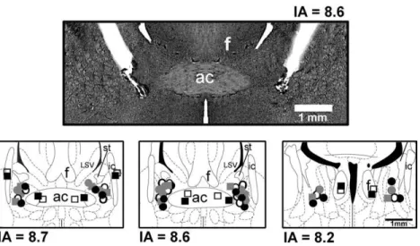
A representative photomicrograph of a coronal brain
sec-tion depicting bilateral microinjecsec-tion sites in the BST of
one representative animal is presented in
Fig. 1
.
Diagram-matic representation showing microinjection sites of
ACSF, WB4101, RX821002 and propranolol into the BST
and WB4101 and RX821002 into structures surrounding
the BST is also shown in
Fig. 1
.
Effect of microinjection into the BST of ACSF,
WB4101, RX821002 or propranolol on dynamic
exercise-induced cardiovascular changes
MAP (
F
⫽
0.4,
P
⬎
0.05) and HR (
F
⫽
0.9,
P
⬎
0.05) baseline
values were similar in all experimental groups.
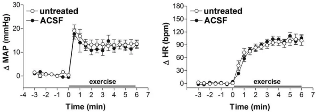
ACSF.
Bilateral microinjection of ACSF (
n
⫽
7) into
the BST did not affect either MAP (99
⫾
3 vs. 98
⫾
3 mmHg,
t
⫽
1.1,
P
⬎
0.05) or HR (349
⫾
10 vs. 352
⫾
8 bpm,
t
⫽
0.6,
P
⬎
0.05) baseline values. Exercise-evoked cardiovascular
responses in animals that received ACSF injected into the
BST were not significantly different from those of the
un-treated group (
n
⫽
5) (
Table 1
and
Fig. 2
).
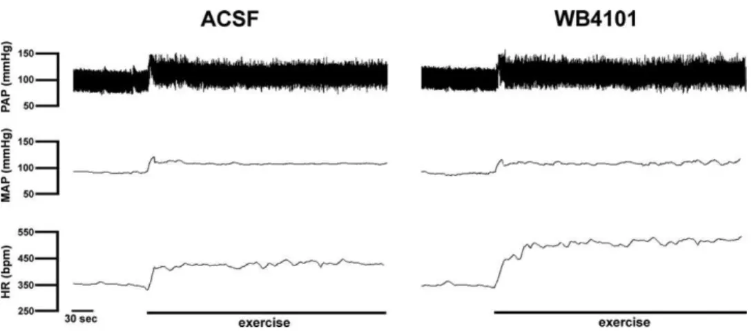
WB4101.
Bilateral microinjection of the selective
␣
1-adrenoceptor antagonist WB4101 (
n
⫽
7) into the BST did
not affect either MAP (98
⫾
2 vs. 96
⫾
3 mmHg,
t
⫽
1,
P
⬎
0.05) or HR (366
⫾
8 vs. 360
⫾
6 bpm,
t
⫽
0.5,
P
⬎
0.05)
baseline values. However, BST pretreatment with W4101
significantly increased the exercise-evoked tachycardiac
response without affecting the pressor response, when
compared with animals that received ACSF injected into
the BST (
Table 1
and
Fig. 3
). Microinjection of WB4101
into structures surrounding the BST (
n
⫽
4), such as the
anterior comissure, internal capsule or fornix did not affect
either MAP (
F
(1,171)⫽
0.3;
P
⬎
0.05) or HR (
F
(1,171)⫽
0.1;
P
⬎
0.05) responses to exercise. Representative
record-ings showing the cardiovascular responses to exercise on
the treadmill in animals treated with ACSF or WB4101
injected into the BST are presented in
Fig. 4
.
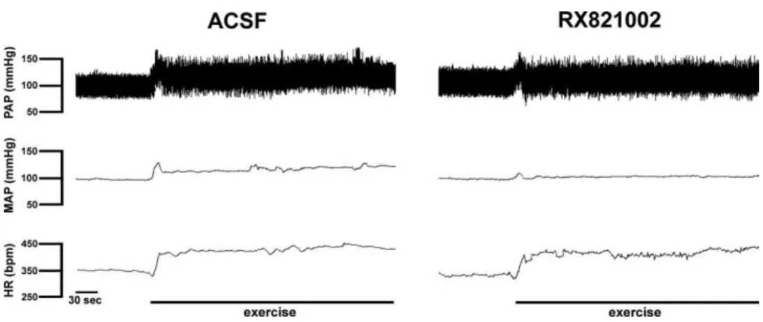
response without affecting the tachycardiac response,
when compared with animals that received ACSF injected
into the BST (
Table 1
and
Fig. 5
). Microinjection of
RX821002 into structures surrounding the BST (
n
⫽
4),
such as the anterior comissure, internal capsule or fornix
did not affect either MAP (
F
(1,171)⫽
0.5;
P
⬎
0.05) or HR
(
F
(1,171)⫽
0.3;
P
⬎
0.05) response to exercise.
Represen-tative recordings showing the cardiovascular responses
to exercise on the treadmill in animals treated with ACSF
or RX821002 injected into the BST are presented in
Fig. 6
.
Propranolol.
Bilateral microinjection of the
nonselec-tive

-adrenoceptor antagonist propranolol (
n
⫽
5) into the
BST did not affect either MAP (96
⫾
2 vs. 97
⫾
2 mmHg,
t
⫽
1.6,
P
⬎
0.05) or HR (348
⫾
13 vs. 355
⫾
9 bpm,
t
⫽
1.6,
P
⬎
0.05) baseline values. Propranolol microinjection into
the BST also did not affect cardiovascular responses to
exercise on the treadmill (
Fig. 7
).
Effect of microinjection into the BST of ACSF,
WB4101 or RX821002 in the open-field test
Bilateral microinjection of either WB4101 (
n
⫽
6) (12
⫾
3 vs.
14
⫾
3 m,
P
⬎
0.05) or RX821002 (
n
⫽
6) (12
⫾
3 vs. 13
⫾
2 m,
P
⬎
0.05) into the BST did not affect total distance travelled
during the open-field test (
F
(2,17)⫽
0.1,
P
⬎
0.05), when
compared with animals treated with ACSF (
n
⫽
6) (
Fig. 8
B).
Time-course analysis of distance traveled during the
open-field test also did not show a significant effect of BST
adrenoceptor antagonism (
F
(2,150)⫽
1.1,
P
⬎
0.05), but
indicated a significant effect over time (
F
(9,150)⫽
50,
P
⬍
0.0001) (
Fig. 8
A).
DISCUSSION
The present work brings the first direct evidence for the
involvement of CNS adrenoceptors in cardiovascular
re-sponses observed during dynamic exercise. We have
Fig. 1. Photomicrograph of a coronal brain section from one representative rat showing bilateral injection sites in the BST and a diagrammatic representation based on the rat brain atlas ofPaxinos and Watson (1997), indicating injection sites of ACSF (Œ), WB4101 (), RX821002 () and propranolol () into the BST, as well as WB4101 () and RX821002 (□) into structures surrounding the BST. ac, anterior commissure; IA, interaural coordinate; ic, internal capsule; LSV, lateral septal ventral; st, stria terminalis; f, fornix.Table 1.Statistical summary of time-course analysis of mean arterial pressure (⌬MAP) and heart rate (⌬HR) responses to dynamic exercise (0.8 km/h for 6 min). It was compared responses of groups vehicle (ACSF) (n⫽7) vs. untreatred (n⫽5), the selective␣1-adrenoceptor antagonist WB4101 (n⫽7) vs. ACSF, the selective␣2-adrenoceptor antagonist RX821002 (n⫽6) vs. ACSF and the nonselective-adrenoceptor antagonist propranolol (n⫽5) vs. ACSF
Treatment Time Interaction (treatment vs. time)
ACSF vs. untreated
⌬MAP F(1,190)⫽1 F(18,190)⫽25* F(18,190)⫽0.2
⌬HR F(1,190)⫽0.2 F(18,190)⫽110* F(18,190)⫽0.7
WB4101 vs. ACSF
⌬MAP F(1,228)⫽3 F(18,228)⫽36* F(18,228)⫽0.5
⌬HR F(1,228)⫽343* F(18,228)⫽315* F(18,228)⫽12*
RX821002 vs. ACSF
⌬MAP F(1,209)⫽177* F(18,209)⫽12* F(18,209)⫽6*
⌬HR F(1,209)⫽0.1 F(18,209)⫽182* F(18,209)⫽1
Propranolol vs. ACSF
⌬MAP F(1,190)⫽0.6 F(18,190)⫽24* F(18,190)⫽0.3
⌬HR F(1,190)⫽1 F(18,190)⫽130* F(18,190)⫽1.6
shown that bilateral microinjection of WB4101, a selective
␣
1-adrenoceptor antagonist, into the BST enhanced
exer-cise-evoked HR increase without affecting MAP response.
Moreover, BST treatment with RX821002, a selective
␣
2-adrenoceptor antagonist, reduced MAP increase observed
during dynamic exercise on the treadmill without changing
tachycardiac response. However, bilateral microinjection
into the BST of propranolol, a nonselective

-adrenoceptor
antagonist, did not affect cardiovascular responses to
ex-ercise on the treadmill.
Dynamic exercise causes cardiovascular responses,
which include increases in arterial pressure, HR and
car-diac output, associated with decreased venous
capaci-tance and redistribution of blood to different territories
(regional vasoconstriction or vasodilatation), via neural,
hormonal and local mechanisms (
Winder et al., 1978;
Wade, 1984; Waldrop et al., 1996; Michelini and Stern,
2009
). We have observed pressor and tachycardiac
re-sponse during dynamic exercise on the rodent treadmill.
Cardiovascular responses reported in the present study
were strictly related to exercise, and not due to exposure to
a novel environment, since we have previously reported no
fearful associations, including cardiovascular changes,
when the animals were kept at rest on the treadmill (
Cres-tani et al., 2010b
).
Recently, we have demonstrated that CoCl
2-induced
acute bilateral inhibition of BST neurotransmission greatly
attenuated both pressor and tachycardiac responses
evoked by exercise on the treadmill (
Crestani et al.,
2010b
). However, due to the nonselective blockade of
local neurotransmission caused by CoCl
2(
Kretz, 1984;
Lomber, 1999
), the possible neurotransmitter involved was
not identified. The present work has demonstrated that
blockade of
␣
2-adrenoceptors by bilateral microinjection of
RX821002 into the BST is able to reduce exercise-evoked
pressor response without changing HR response. This
result suggests that local
␣
2-adrenoceptor mediates, at
least in part, BST influence on MAP response during
ex-ercise. Although our results indicate a role of
␣
1-adreno-ceptor in modulation of the tachycardiac response evoked
by exercise, the blockade of
␣
1-adrenoceptor in the BST
affected HR response in an opposite manner to that
ob-served after BST treatment with CoCl
2. Therefore, further
experiments are necessary to clarify the neurotransmitter
and the receptors in the BST which are involved in its
influence on tachycardiac response to exercise. However,
the enhancement in exercise-evoked HR increase after the
blockade of BST
␣
1-adrenoceptors led to the interesting
observation of a reserve in the cardiac response to our
exercise protocol. Moreover, this result indicates an
impor-Fig. 2. Time-course of changes in mean arterial pressure (⌬MAP) and heart rate (⌬HR) during dynamic exercise on the treadmill (0.8 km/h for 6 min) of the untreated group (Œ,n⫽5), with no cannulas in the brain, and in rats that had received bilateral microinjection of vehicle (ACSF,,n⫽7) into the BST. The onset of exercise was at t⫽0. Circles represent the mean and bars the SEM. Microinjection of ACSF into the BST did not affect either MAP (P⬎0.05) or HR responses (P⬎0.05) to exercise on the treadmill.tant physiological meaning of BST
␣
1-adrenoceptor in the
control of cardiovascular activity during exercise, since
activation of this receptor counteracts excessive cardiac
activation. Thus, BST
␣
1-adrenoceptor plays an important
role in achieving fine tuning of the cardiac response during
exercise, thus ensuring the functional state stabilization of
the cardiac activity during exercise since the amplitude of
the response is reduced.
We have observed that animals treated with either
WB4101 or RX821002 injected into the BST behaved in a
similar manner in the open-field test as compared to rats
treated with vehicle, indicating that blockade of
adrenocep-tors in the BST did not influence motor performance.
Al-though connections between the BST and CNS locomotor
regions have been reported (
Dong et al., 2001; Dong and
Swanson, 2004
), previous studies from our group and
other laboratories also reported absence of effects in the
open-field test after BST electrolytic lesion or chemical
ablation in both male and female rats (
Schulz and
Can-beyli, 2000; Pezuk et al., 2006, 2008; Resstel et al., 2008;
Crestani et al., 2010a,b
). These results suggest that
changes in cardiovascular responses to dynamic exercise
observed in the present study after BST pharmacological
treatment is due to a direct interference in autonomic
con-trol, and not to an indirect effect caused by an alteration in
motor activity.
According to current theory, circulatory control during
exercise is governed by the CNS through several neural
mechanisms (
Raven et al., 2002; Fisher and White, 2004
).
Central command is a feed-forward mechanism originating
in higher brain centers that involves the parallel activation
of brainstem and spinal circuits responsible for the control
of locomotion as well as cardiovascular activity during
exercise (
Raven et al., 2002; Fisher and White, 2004;
Williamson et al., 2006
). Neuroimaging and
immunohisto-chemical studies have indicated that the neural pathway of
central command appears to encompass regions of the
cerebral cortex and hypothalamus involved in control of
autonomic functions, such as the insular cortex, medial
prefrontal cortex, paraventricular nucleus of the
hypothal-amus and lateral hypothalhypothal-amus (
Timofeeva et al., 2003;
Williamson et al., 2006; Williamson, 2010
), which interact
with other structures involved in locomotor and
cardiovas-cular integration during exercise (
Raven et al., 2002;
Wil-liamson et al., 2006
). Connections between the BST and
these cortical and hypothalamic structures were previously
described (
Yasui et al., 1991; Dong et al., 2001; Vertes,
2004
). In this way, it has been proposed that the BST could
Fig. 4. Recording from representative animals illustrating changes in pulsatile arterial pressure (PAP), mean arterial pressure (MAP) and heart rate (HR) observed during dynamic exercise on the treadmill after BST treatment with vehicle (ACSF) or the selective␣1-adrenoceptor antagonist WB4101. Note the increase in the HR response to exercise in the animal that received WB4101 injected into the BST.be a relay in the neural circuitry of cardiovascular control,
connecting telencephalic structures to autonomic regions
in the hypothalamus and brainstem (
Ulrich-Lai and
Her-man, 2009
). Therefore, control of exercise-evoked
cardio-vascular responses by BST adrenoceptors proposed in the
present study can occur through a modulation of signals
arising from cortical structures to the BST.
Cardiovascular adjustment during exercise is also
driven by type III and IV muscle afferent activity from
exercising muscles, which provide feedback regarding the
mechanical and metabolic conditions within those muscles
(
Kaufman and Forster, 1996; Fisher and White, 2004;
Potts, 2006
). Although medullary structures appear to be
the primary pathway involved in the feedback control from
active muscles, supramedullary nuclei may play a
modu-lating role that can affect the reflex control of autonomic
activity during exercise (
Waldrop and Stremel, 1989;
Kauf-man and Forster, 1996; Li, 2004
). Studies in the literature
have shown that static muscle contraction activates brain
stem regions consisting of noradrenergic cells (
Li et al.,
1998
), thus indicating that the reflex from active muscles
involves central noradrenergic pathways. Therefore, BST
noradrenergic neurotransmission could also be part of the
pathway of feedback control from active muscle receptors.
However, although the activation of
␣
2-adrenoceptor in the
CNS elicited by administration of clonidine, a selective
␣
2-adrenoceptor agonist, decreases the pressor and
tachycardiac response evoked by static muscle
contrac-tion (
Williams, 1985; Ally et al., 1996
), it has been
docu-mented that the selective
␣
2-adrenoceptor antagonist
yo-imbine does not affect the cardiovascular responses
evoked by static exercise when injected either into the
lateral ventricle or cerebral aqueduct (
Williams et al., 1987;
Ally et al., 1996
). These results indicate absence of a tonic
influence of
␣
2-adrenoceptor on the neurons regulating the
cardiovascular responses to static exercise. In this way, a
possible involvement of BST noradrenergic
neurotrans-mission in the pathway of feedback control from active
muscle receptors is not mediated by activation of local
␣
2-adrenoceptors.
It has been reported that the baroreflex
stimulus–re-sponse curve resets during exercise, with a vertical
up-ward shift on the response arm and a lateral rightup-ward shift
to higher operating pressures (
Rowell and O’Leary, 1990;
DiCarlo and Bishop, 2001; Raven et al., 2006; Dampney et
al., 2008
). It has been proposed that the central command
and reflex mechanism from active muscles may exert its
effects on cardiovascular parameters by changing baroreflex
activity (
Raven et al., 2002; Potts, 2006
). Previous results
from our laboratory indicated that BST noradrenergic
neu-Fig. 6. Recording from representative animals illustrating changes in pulsatile arterial pressure (PAP), mean arterial pressure (MAP) and heart rate (HR) observed during dynamic exercise on the treadmill after BST treatment with vehicle (ACSF) or the selective␣2-adrenoceptor antagonist RX821002. Note the decrease in the arterial pressure response to exercise in the animal that received RX821002 injected into the BST.rotransmission, through activation of
␣
1-adrenoceptor,
mod-ulates the baroreflex activity in a similar manner to that
ob-served during exercise (
Crestani et al., 2008a
). This result
suggests that activation of BST
␣
1-adrenoceptors could
facil-itate cardiovascular responses to dynamic exercise through
its modulation of baroreflex activity. However, results
re-ported in the present study indicate an inhibitory influence of
BST
␣
1-adrenoceptors on the exercise-induced HR increase.
This evidence suggests that BST noradrenergic
neurotrans-mission affects exercise-induced cardiovascular responses
by a mechanism independent of baroreflex modulation.
These results corroborate with previous data indicating that
BST stimulation evokes similar cardiovascular responses in
sham animals or those submitted to sinoaortic denervation
(i.e. baroreflex denervation) (
Dunn and Williams, 1998
).
Both the sympathetic and the parasympathetic branches
of the autonomic nervous system participate in the control
of cardiovascular activity during dynamic exercise.
Block-ade of parasympathetic control of HR reveals that most of
the initial response to exercise is attributable to the
with-drawal of tonic vagal activity, whereas

-adrenergic
block-ade reveals the importance of augmented cardiac
sympa-thetic activity during moderate and heavy exercise (
Over-ton, 1993; Goldsmith et al., 2000
). The BST sends direct
projections to medullary structures involved with
auto-nomic activity, such as the nucleus of the tractus solitarius
(NTS), dorsal motor nucleus of the vagus, nucleus
am-biguus and ventrolateral medulla (
Gray and Magnuson,
1987; Dong and Swanson, 2004
). In this way, it was
dem-onstrated that ablation of the caudal ventrolateral medulla
(CVLM) attenuated MAP and HR decreases elicited by
BST stimulation (
Giancola et al., 1993
). The CVLM
proj-ects to and inhibits sympathetic premotor neurons in the
rostral ventrolateral medulla, thus decreasing
sympa-thetic preganglionic neuronal outflow (
Sved et al., 2000
).
Previous evidence has also indicated an involvement of
the NTS in cardiovascular control during exercise (
Du-floth et al., 1997; Raven et al., 2006; Higa-Taniguchi et
al., 2009
). These results provide evidence of the neural
substrate for the influence of BST
␣
1-adrenoceptor on
exercise-related HR response. Thus, BST
␣
1-adreno-ceptors could modulate the cardiac response during
exercise by stimulating facilitatory inputs to vagal
neu-rons and/or by stimulating inhibitory inputs to
sympa-thetic medullary neurons. Connections from the BST to
the medulla could also be the neural substrate for the
facilitatory influence of BST
␣
2-adrenoceptors on the
pressor response to exercise.
The existence of specific neuronal pathways
control-ling autonomic activity to different organs provides the
structural substrate for differences between BST
␣
1- and
␣
2-adrenoceptors in modulating cardiovascular
adjust-ments during dynamic exercise (
Morrison, 2001
).
There-fore, subtypes of
␣
-adrenoceptors in the BST may
modu-late the activity of specific neural pathways in the CNS,
thus differentially affecting exercise-evoked pressor and
tachycardiac responses. Present results corroborate
with previous results that indicated specific actions of
BST
␣
1- or
␣
2-adrenoceptors on cardiovascular control
(
Crestani et al., 2008a, 2009a
). On the other hand, it
was reported that cardiovascular responses evoked by
microinjection of noradrenaline into the BST are
medi-ated by activation of both
␣
1- and
␣
2-adrenoceptors in
the BST (
Crestani et al., 2008b
). Because different
ex-perimental procedures were used in these studies with
different response parameters being analyzed, it is
pos-sible that BST
␣
1- and
␣
2-adrenoceptors have similar or
different roles depending on the stimulus.
BST treatment with adrenoceptor antagonists did not
affect either MAP or HR baseline values. Therefore,
although the present study supports the hypothesis that
BST noradrenergic neurotransmission plays an
impor-tant role in modulating the cardiovascular responses to
dynamic exercise, this neurotransmission is not involved
in the tonic maintenance of either arterial pressure or
HR. These results corroborate previous data in the
lit-erature indicating no changes in cardiovascular
param-eters after blockade of either glutamatergic, cholinergic
or adrenergic receptors in the BST (
Alves et al., 2007;
Hatam and Nasimi, 2007; Crestani et al., 2008a, 2009a
).
Fig. 8. (A) Time-course of the distance travelled when exposed to theCONCLUSION
In conclusion, the present results show that noradrenergic
neurotransmission in the BST modulates cardiovascular
ad-justments during dynamic exercise in a complex way. Our
data provide evidence of an inhibitory influence of BST
␣
1-adrenoceptors on exercise-evoked HR response. Moreover,
the results point to a facilitatory role played by the activation
of BST
␣
2-adrenoceptors on pressor response to dynamic
exercise. These results provide the first direct evidence for
the involvement of CNS adrenoceptors in cardiovascular
re-sponses observed during dynamic exercise.
Acknowledgments—The authors wish to thank I.A.C. Fortunato, I.I.B. Aguiar, S.S. Guilhaume and F.V. Gomes for technical help. Alves has a CNPq PhD fellowship (870307/1997-5). The present research was supported by grants from FAPESP (06/57670-4), CNPq (306381/2003-6, 505394/2003-0 and 480550/2007-7), FAEPA and PADC/FCF-UNESP.
REFERENCES
Ally A, Hand GA, Mitchell JH (1996) Cardiovascular responses to static exercise in conscious cats: effects of intracerebroventricular injection of clonidine. J Physiol 491 (Pt 2):519 –527.
Alves FH, Crestani CC, Gomes FV, Guimaraes FS, Correa FM, Ress-tel LB (2010) Cannabidiol injected into the bed nucleus of the stria terminalis modulates baroreflex activity through 5-HT1A receptors. Pharmacol Res 62:228 –236.
Alves FH, Crestani CC, Resstel LB, Correa FM (2007) Cardiovascular effects of carbachol microinjected into the bed nucleus of the stria terminalis of the rat brain. Brain Res 1143:161–168.
Alves FH, Crestani CC, Resstel LB, Correa FM (2009) Bed nucleus of the stria terminalis N-methyl-D-aspartate receptors and nitric oxide modulate the baroreflex cardiac component in unanesthetized rats. J Neurosci Res 87:1703–1711.
Amaral SL, Michelini LC (1997) Validation of transit-time flowmetry for chronic measurements of regional blood flow in resting and exer-cising rats. Braz J Med Biol Res 30:897–908.
Braga DC, Mori E, Higa KT, Morris M, Michelini LC (2000) Central oxytocin modulates exercise-induced tachycardia. Am J Physiol Regul Integr Comp Physiol 278:R1474 –R1482.
Byrum CE, Guyenet PG (1987) Afferent and efferent connections of the A5 noradrenergic cell group in the rat. J Comp Neurol 261: 529 –542.
Crestani CC, Alves FH, Correa FM, Guimaraes FS, Joca SR (2010a) Acute reversible inactivation of the bed nucleus of stria terminalis induces antidepressant-like effect in the rat forced swimming test. Behav Brain Funct 6:30.
Crestani CC, Alves FH, Resstel LB, Correa FM (2006) The bed nu-cleus of the stria terminalis modulates baroreflex in rats. Neurore-port 17:1531–1535.
Crestani CC, Alves FH, Resstel LB, Correa FM (2007) Cardiovascular effects of noradrenaline microinjection in the bed nucleus of the stria terminalis of the rat brain. J Neurosci Res 85:1592–1599. Crestani CC, Alves FH, Resstel LB, Correa FM (2008a) Bed nucleus of
the stria terminalis alpha(1)-adrenoceptor modulates baroreflex car-diac component in unanesthetized rats. Brain Res 1245:108 –115. Crestani CC, Alves FH, Resstel LB, Correa FM (2008b) Both alpha1
and alpha2-adrenoceptors mediate the cardiovascular responses to noradrenaline microinjected into the bed nucleus of the stria terminal of rats. Br J Pharmacol 153:583–590.
Crestani CC, Alves FH, Resstel LB, Correa FM (2010b) The bed nucleus of the stria terminalis modulates exercise-evoked cardio-vascular responses in rats. Exp Physiol 95:69 –79.
Crestani CC, Alves FH, Tavares RF, Correa FM (2009a) Role of the bed nucleus of the stria terminalis in the cardiovascular responses to acute restraint stress in rats. Stress 12:268 –278.
Crestani CC, Busnardo C, Tavares RF, Alves FH, Correa FM (2009b) Involvement of hypothalamic paraventricular nucleus non-N-methyl-D-aspartate receptors in the pressor response to noradrenaline mi-croinjected into the bed nucleus of the stria terminalis of unanesthe-tized rats. Eur J Neurosci 29:2166 –2176.
Dampney RA, Horiuchi J, McDowall LM (2008) Hypothalamic mecha-nisms coordinating cardiorespiratory function during exercise and defensive behaviour. Auton Neurosci 142:3–10.
De Angelis K, Ogawa T, Sanches IC, Rigatto KV, Krieger EM, Irigoyen MC (2006) Impairment on cardiac output and blood flow adjust-ments to exercise in L-NAME-induced hypertensive rats. J Cardio-vasc Pharmacol 47:371–376.
DiCarlo SE, Bishop VS (1992) Onset of exercise shifts operating point of arterial baroreflex to higher pressures. Am J Physiol 262: H303–H307.
DiCarlo SE, Bishop VS (2001) Central baroreflex resetting as a means of increasing and decreasing sympathetic outflow and arterial pres-sure. Ann N Y Acad Sci 940:324 –337.
Dong HW, Petrovich GD, Watts AG, Swanson LW (2001) Basic orga-nization of projections from the oval and fusiform nuclei of the bed nuclei of the stria terminalis in adult rat brain. J Comp Neurol 436:430 – 455.
Dong HW, Swanson LW (2004) Organization of axonal projections from the anterolateral area of the bed nuclei of the stria terminalis. J Comp Neurol 468:277–298.
Dufloth DL, Morris M, Michelini LC (1997) Modulation of exercise tachycardia by vasopressin in the nucleus tractus solitarii. Am J Physiol 273:R1271–R1282.
Dunn JD (1987) Plasma corticosterone responses to electrical stimu-lation of the bed nucleus of the stria terminalis. Brain Res 407:327–331.
Dunn JD, Williams TJ (1998) Effect of sinoaortic denervation on arte-rial pressure changes evoked by bed nucleus stimulation. Brain Res Bull 46:361–365.
Egli RE, Kash TL, Choo K, Savchenko V, Matthews RT, Blakely RD, Winder DG (2005) Norepinephrine modulates glutamatergic trans-mission in the bed nucleus of the stria terminalis. Neuropsychop-harmacology 30:657– 668.
Fisher JP, White MJ (2004) Muscle afferent contributions to the cardio-vascular response to isometric exercise. Exp Physiol 89:639 – 646. Gelsema AJ, Calaresu FR (1987) Chemical microstimulation of the
septal area lowers arterial pressure in the rat. Am J Physiol 252:R760 –R767.
Giancola SB, Roder S, Ciriello J (1993) Contribution of caudal ventro-lateral medulla to the cardiovascular responses elicited by activa-tion of bed nucleus of the stria terminalis. Brain Res 606:162–166. Goldsmith RL, Bloomfield DM, Rosenwinkel ET (2000) Exercise and
autonomic function. Coron Artery Dis 11:129 –135.
Gray TS, Magnuson DJ (1987) Neuropeptide neuronal efferents from the bed nucleus of the stria terminalis and central amygdaloid nucleus to the dorsal vagal complex in the rat. J Comp Neurol 262:365–374.
Hatam M, Nasimi A (2007) Glutamatergic systems in the bed nucleus of the stria terminalis, effects on cardiovascular system. Exp Brain Res 178:394 – 401.
Higa-Taniguchi KT, Felix JV, Michelini LC (2009) Brainstem oxytocin-ergic modulation of heart rate control in rats: effects of hyperten-sion and exercise training. Exp Physiol 94:1103–1113.
Higa-Taniguchi KT, Silva FC, Silva HM, Michelini LC, Stern JE (2007) Exercise training-induced remodeling of paraventricular nucleus (nor)adrenergic innervation in normotensive and hypertensive rats. Am J Physiol Regul Integr Comp Physiol 292:R1717–R1727. Hill JM, Kaufman MP (1991) Attenuating effects of intrathecal clonidine
Kaufman MP, Forster HV (1996) Reflexes controlling circulatory, ven-tilatory and airway response to exercise. In: Handbook of physiol-ogy, section 12, Exercise: regulation and integration of multiple systems (Rowell LB, Shepherd JT, eds), chap. 10, pp 381– 447. New York: Oxford University Press.
Kitaoka R, Fujikawa T, Miyaki T, Matsumura S, Fushiki T, Inoue K (2010) Increased noradrenergic activity in the ventromedial hypo-thalamus during treadmill running in rats. J Nutr Sci Vitaminol (Tokyo) 56:185–190.
Kretz R (1984) Local cobalt injection: a method to discriminate pre-synaptic axonal from postpre-synaptic neuronal activity. J Neurosci Methods 11:129 –135.
Lambert GW, Jonsdottir IH (1998) Influence of voluntary exercise on hypothalamic norepinephrine. J Appl Physiol 85:962–966. Li J (2004) Central integration of muscle reflex and arterial baroreflex
in midbrain periaqueductal gray: roles of GABA and NO. Am J Physiol Heart Circ Physiol 287:H1312–H1318.
Li J, Potts JT, Mitchell JH (1998) Effect of barodenervation on c-Fos expression in the medulla induced by static muscle contraction in cats. Am J Physiol 274:H901–H908.
Lomber SG (1999) The advantages and limitations of permanent or reversible deactivation techniques in the assessment of neural function. J Neurosci Methods 86:109 –117.
Ludbrook J, Potocnik SJ (1986) Circulatory changes during spontane-ous motor activity: role of arterial baroreflexes. Am J Physiol 250:H426 –H433.
Matsui H, Yamamoto C (1984) The possible involvement of adenylate cyclase inhibition in the field potential suppression through alpha-2 adrenergic receptors in the bed nucleus of the stria terminalis. Brain Res 293:187–190.
Michelini LC, Stern JE (2009) Exercise-induced neuronal plasticity in central autonomic networks: role in cardiovascular control. Exp Physiol 94:947–960.
Miki K, Yoshimoto M, Tanimizu M (2003) Acute shifts of baroreflex control of renal sympathetic nerve activity induced by treadmill exercise in rats. J Physiol 548:313–322.
Moore RY (1978) Catecholamin innervation of the basal forebrain. I. The septal area. J Comp Neurol 177:665– 684.
Morrison SF (2001) Differential control of sympathetic outflow. Am J Physiol Regul Integr Comp Physiol 281:R683–R698.
Ohiwa N, Saito T, Chang H, Omori T, Fujikawa T, Asada T, Soya H (2006) Activation of A1 and A2 noradrenergic neurons in response to running in the rat. Neurosci Lett 395:46 –50.
Overton JM (1993) Influence of autonomic blockade on cardiovascular responses to exercise in rats. J Appl Physiol 75:155–161. Pagliari R, Peyrin L (1995) Norepinephrine release in the rat frontal
cortex under treadmill exercise: a study with microdialysis. J Appl Physiol 78:2121–2130.
Paxinos G, Watson C (1997) The rat brain in stereotaxic coordinates. Sidney, Australia: Academic Press.
Pezuk P, Aydin E, Aksoy A, Canbeyli R (2008) Effects of BNST lesions in female rats on forced swimming and navigational learning. Brain Res 1228:199 –207.
Pezuk P, Goz D, Aksoy A, Canbeyli R (2006) BNST lesions aggravate behavioral despair but do not impair navigational learning in rats. Brain Res Bull 69:416 – 421.
Phelix CF, Liposits Z, Paull WK (1992) Monoamine innervation of bed nucleus of stria terminalis: an electron microscopic investigation. Brain Res Bull 28:949 –965.
Potts JT (2006) Inhibitory neurotransmission in the nucleus tractus solitarii: implications for baroreflex resetting during exercise. Exp Physiol 91:59 –72.
Raven PB, Fadel PJ, Ogoh S (2006) Arterial baroreflex resetting during exercise: a current perspective. Exp Physiol 91:37– 49.
Raven PB, Fadel PJ, Smith SA (2002) The influence of central com-mand on baroreflex resetting during exercise. Exerc Sport Sci Rev 30:39 – 44.
Resstel LB, Alves FH, Reis DG, Crestani CC, Correa FM, Guimaraes FS (2008) Anxiolytic-like effects induced by acute reversible inac-tivation of the bed nucleus of stria terminalis. Neuroscience 154:869 – 876.
Rowell LB (1997) Neural control of muscle blood flow: importance during dynamic exercise. Clin Exp Pharmacol Physiol 24:117–125. Rowell LB, O’Leary DS (1990) Reflex control of the circulation during exercise: chemoreflexes and mechanoreflexes. J Appl Physiol 69:407– 418.
Schulz D, Canbeyli RS (2000) Lesion of the bed nucleus of the stria terminalis enhances learned despair. Brain Res Bull 52:83– 87. Sved AF, Ito S, Madden CJ (2000) Baroreflex dependent and
inde-pendent roles of the caudal ventrolateral medulla in cardiovascular regulation. Brain Res Bull 51:129 –133.
Swanson LW, Hartman BK (1975) The central adrenergic system. An immunofluorescence study of the location of cell bodies and their efferent connections in the rat utilizing dopamine-beta-hydroxylase as a marker. J Comp Neurol 163:467–505.
Takatsu S, Ishiwata T, Meeusen R, Sarre S, Hasegawa H (2010) Serotonin release in the preoptic area and anterior hypothalamus is not involved in thermoregulation during low-intensity exercise in a warm environment. Neurosci Lett 482:7–11.
Timofeeva E, Huang Q, Richard D (2003) Effects of treadmill running on brain activation and the corticotropin-releasing hormone sys-tem. Neuroendocrinology 77:388 – 405.
Ulrich-Lai YM, Herman JP (2009) Neural regulation of endocrine and autonomic stress responses. Nat Rev Neurosci 10:397– 409. Vertes RP (2004) Differential projections of the infralimbic and
prelim-bic cortex in the rat. Synapse 51:32–58.
Wade CE (1984) Response, regulation, and actions of vasopressin during exercise: a review. Med Sci Sports Exerc 16:506 –511. Waldrop TG, Eldridge FL, Iwamoto GA, Mitchell JH (1996) Central
neural control of respiration and circulation during exercise. In: Handbook of physiology, section 12, Exercise: regulation and in-tegration of multiple systems (Rowell LB, Shepherd JT, Shepherd JT, eds), pp 333–380. New York: Oxford University Press. Waldrop TG, Stremel RW (1989) Muscular contraction stimulates
pos-terior hypothalamic neurons. Am J Physiol 256:R348 –R356. Williams CA (1985) Effect of clonidine and naloxone on the pressor
response during contraction of cat hind-limb muscles. Cardiovasc Res 19:474 – 482.
Williams CA, Blevins LS, Paul DJ (1987) Possible catecholaminergic-opioidergic control of blood pressure during muscular contraction. Cardiovasc Res 21:471– 480.
Williamson J (2010) The relevance of central command for the neural cardiovascular control of exercise. Exp Physiol 95:1043–1048. Williamson JW, Fadel PJ, Mitchell JH (2006) New insights into central
cardiovascular control during exercise in humans: a central com-mand update. Exp Physiol 91:51–58.
Winder WW, Hagberg JM, Hickson RC, Ehsani AA, McLane JA (1978) Time course of sympathoadrenal adaptation to endurance exer-cise training in man. J Appl Physiol 45:370 –374.
Woulfe JM, Hrycyshyn AW, Flumerfelt BA (1988) Collateral axonal projections from the A1 noradrenergic cell group to the paraven-tricular nucleus and bed nucleus of the stria terminalis in the rat. Exp Neurol 102:121–124.
Yasui Y, Breder CD, Saper CB, Cechetto DF (1991) Autonomic re-sponses and efferent pathways from the insular cortex in the rat. J Comp Neurol 303:355–374.