Anna A. Vanyushkina1, Gleb Y. Fisunov1, Alexey Y. Gorbachev1, Dmitri E. Kamashev1,2*, Vadim M. Govorun1,3,4
1Russian Institute of Physico-Chemical Medicine, Moscow, Russian Federation,2Russian Research Center Kurchatov Institute, Moscow, Russian Federation,3 Shemyakin-Ovchinnikov Institute of Bioorganic Chemistry, Russian Academy of Sciences, Moscow, Russian Federation,4Moscow Institute of Physics and Technology (State University), Dolgoprudny, Moscow Region, Russian Federation
Abstract
We present a systematic study of three bacterial species that belong to the class Mollicutes, the smallest and simplest bacteria,Spiroplasma melliferum,Mycoplasma gallisepticum, andAcholeplasma laidlawii. To understand the difference in the basic principles of metabolism regulation and adaptation to environmental conditions in the three species, we analyzed the metabolome of these bacteria. Metabolic pathways were reconstructed using the proteogenomic annotation data provided by our lab. The results of metabolome, proteome and genome profiling suggest a fundamental difference in the adaptation of the three closely related Mollicute species to stress conditions. As the transaldolase is not annotated in Mollicutes, we propose variants of the pentose phosphate pathway catalyzed by annotated enzymes for three species. For metabolite detection we employed high performance liquid chromatography coupled with mass spectrometry. We used liquid chromatography method - hydrophilic interaction chromatography with silica column - as it effectively separates highly polar cellular metabolites prior to their detection by mass spectrometer.
Citation:Vanyushkina AA, Fisunov GY, Gorbachev AY, Kamashev DE, Govorun VM (2014) Metabolomic Analysis of Three Mollicute Species. PLoS ONE 9(3): e89312. doi:10.1371/journal.pone.0089312
Editor:Glenn Francis Browning, The University of Melbourne, Australia
ReceivedMay 15, 2013;AcceptedJanuary 22, 2014;PublishedMarch 4, 2014
Copyright:ß2014 Vanyushkina et al. This is an open-access article distributed under the terms of the Creative Commons Attribution License, which permits unrestricted use, distribution, and reproduction in any medium, provided the original author and source are credited.
Funding:This work was supported by a Grant (12-04-31696) from Russian Foundation for Basic Research (RFBR). The funders had no role in study design, data collection and analysis, decision to publish, or preparation of the manuscript.
Competing Interests:The authors have declared that no competing interests exist.
* E-mail: dkamashev@gmail.com
Introduction
In contrast to the abundance of systems-oriented approaches describing changes on the transcriptome or proteome level, relatively few studies examined the metabolome. Metabolomics studies are necessary to shed light on the link between genotypes and phenotypes. The goal of the presented research is to identify as many intracellular metabolites as possible in bacteria belonging to three species, namelySpiroplasma melliferum,Mycoplasma gallisepti-cum, andAcholeplasma laidlawii. All these bacteria are members of the class Mollicute, a unique category of bacteria that share the absence of a cell wall, a reduced genome, and simplified metabolic pathways. Mollicutes are widespread organisms in nature engaged in a parasitic lifestyle.
The poultry pathogenM. gallisepticumis a small bacterium that causes chronic respiratory disease in chickens and turkeys. S. melliferum is an insect pathogen with the capability to infect the honey bee [1].A. laidlawiiis a common contaminant of cell culture and has a broad variety of hosts, including bovines, the human oral cavity, birds and plants; it was originally isolated from sewage [2,3,4,5,6].
The mycoplasmic metabolome is significantly reduced, possibly due to its parasitic nature. Like all Mycoplasma, the three bacterial species lack some general metabolic pathways, including the TCA, amino acids synthesis, as well as purine and pyrimidine synthesis [7]. Genome-reduced organisms have limited adaptability to external factors [8]. We believe that comparison of the metabolomes of the three Mollicutes may reveal the primary adaptive features of the simplest metabolomes.
Metabolite identification in our study was performed by flow injection time-of-flight mass spectrometry (Q-TOF). Metabolite identity was confirmed by fragmentation of previously detected ions using targeted mass spectrometry. The liquid phy approach applied here, hydrophilic interaction chromatogra-phy (HILIC) with silica columns, effectively separates highly polar cellular metabolites for their subsequent detection using a high accuracy mass spectrometer in positive and negative acquisition modes [9].
Here we present reliable measurements of a hundred metab-olites, including components of sugar, amino acid, and nucleotide metabolisms. We have identified about a third of all possible intracellular metabolites predicted by genome annotation and compared the metabolite content of three Mollicute species. Metabolomic data analysis is based on a previous proteogenomic study performed in our lab for all three bacterial species [3,10,11,12]. The high-throughput-omics analysis, including transcriptomics, proteomics, and complemented with the meta-bolomic study performed in this work, will improve our comprehensive understanding how the three closely-related Mollicute species are regulated under different conditions [13].
Materials and Methods
Bacteria strains and growth conditions
Petersburg, Russian Academy of Science.S. melliferumwas grown in SP4 medum without any specific aeration and with mixing at 30uC for 20 hours after 1:10 dilution, as described earlier [14].
Acholeplasma laidlawii PG-8A and Mycoplasma gallisepticum S6 were grown in a modified Edward’s medium (Tryptose 20 g/L, NaCl 5 g/l, NaOAc 5 g/l, KCl 1.3 g/l, Tris 3 g/l, yeast dialysate 5%, horse serum 6%, glucose 0.5%, pH 7.6) at 37uC for 20 hours after 1:100 dilution [15]. Bacteria were cultured in covered flasks, and the time of growth and the pH of the solution were controlled [11]. Bacteria were harvested by centrifugation (10 min, 16,000g) in the log phase, which was determined by pH values (6.5–6.6 for both,
S. melliferum and M. gallisepticum). The metabolic profiles were obtained from cultures grown from a single clone for less than five passages. Each metabolome experiment was independently repeated at least seven times; 50 ml cultures from the same strain and with the same sample preparation procedure were used for each experiment.
Determination of pH tolerance
We used the ‘‘color test’’ method to determine whether Mollicutes survive in acidic conditions and to estimate the critical pH value for each species. Aliquots ofM. gallisepticum,A. laidlawii
and S. melliferum culture were exposed to decreased pH (hydro-chloric acid was added) in steps ofDpH = 0.1 for 1 hour starting at pH 7.5. Each aliquot was then subjected to a series of dilutions (up to 109) in medium (pH 7.5) containing pH indicator (Phenol Red), and the presence of living cells was detected by the medium color change. The tolerated acidity was defined as the minimum acidity at which 10% of cells survived.
Reagents
The following chemicals were used as standards: sodium pyruvate (100 mg/ml, disodium salt hydrate), D-fructose 6-phosphate, hydrate of sodium phospho(enol)pyruvate (97% purity, enzyme quality), dehydrated disodium salt of D-ribose 5-phosphate, di-glyceraldehyde 3-phosphate (46.1 mg/ml). Purified (98%) amino acids, nucleotides, nucleosides (adenosine, deoxya-denosine, inosine, cytosine monophosphate, and thymidine) from Sigma-Aldrich (USA) were used as standards as well. The following reagents were used for extraction and solution prepa-ration: absolute methanol (HPLC grade) from Biosolve (The Netherlands), ammonium acetate (ultra clean grade) from Helicon (Russia), formic acid (98–100%) from Riedel-de Haen (Germany), ammonium hydroxide solution (29.73%) from Fisher Scientific (USA), water (HPLC-MS) and acetonitrile (HPLC-MS) from Panreac (Spain).
Extraction of metabolites
For metabolite analysis, S. melliferum, M. gallisepticum, or A. laidlawii (50 ml) were grown in a liquid medium. Cells were harvested by centrifugation at 16,000 g at 20uC for 10 min and washed with the washing solution (150 mM NaCl/50 mM Tris). The pH of the washing solution was 6.5 (the acidity in the logarithmic phase of Mycoplasma growth), while for acid stress studies the pH of washing solution was about 5.5 (depending on the type of culture and the selected conditions of acid stress). Thus, the culture was under the influence of selected acidity conditions during the washing procedure. The cell pellet was resuspended in 3 ml of washing solution (the pellet volume was 30ml) and precipitated again. Then cells were washed twice with 150ml of washing solution.
The metabolism was rapidly quenched by cold methanol extraction. A cold methanol extraction method was developed on the basis of a previously reported cold methanol extraction
protocol [16,17] as described below. The metabolites were extracted by adding 1000ml of methanol (277uC) to 75 ul of the resuspended cell pellet; the sample was vigorously shaken (1 min) and then kept at 277uC for 15 min. The sample was warmed for 3 min and then thoroughly shaken again. The resulting sample was centrifuged for 30 min at 16,000 g at 4uC. The supernatant was separated into aliquots and lyophilized. The dry extract was kept no longer than 4 days prior to analysis. The dry extract was dissolved in a mixture consisting of 20% acetonitrile and 80% water and analyzed immediately.
Until recently there was no published record of an LC-MS method of metabolome analysis of the studied bacteria (S. melliferum,M. gallisepticum, andA. laidlawii). Pre-testing a few other options (such as extraction by acetonitrile, methanol/water, acetonitrile/water/acid and others), we have chosen the cold methanol extraction method, as it has been demonstrated to be the most effective. The cold methanol extraction protocol described in [16] is the basis of the protocol used here. A similar modification has been successfully used recently forM. pneumoniaestudies [18].
Apparatus and HPLC/MS method
Mass spectrometry analysis was performed on a Q-TOF 6520 series time-of-flight mass spectrometer (Agilent Technologies). The flow from the analytical column was introduced directly into the electrospray ion source of the mass spectrometer. Prior to the experiment, the mass spectrometer was calibrated to 2 ppm accuracy of them/zvalue. The ionizing spray voltage was 3500 V in both positive and negative ionization modes. Nitrogen of various degrees of purity was used as a dry gas at a pressure of 20 psi with a flow rate of 6 l/min and as a gas in the collision cell. The temperature of the quartz capillary was 325uC. HPLC-MS and HPLC-MS/MS analyses were carried out using the series 1200 high performance liquid chromatograph (Agilent Technol-ogies), coupled with the mass spectrometer. The following chromatographic analytical column was used in the study: Zorbax RX-SIL Narrow-Bore (150 mm62.1 mm65mm) from Agilent Technologies. To protect the separating phase of these highly effective analytical columns from chemical damage, as a safety cartridge we used the Zorbax RX-SIL 4-Pack analytical guard column (4.6 mm612.5 mm65mm) purchased from Agilent Tech-nologies.
Chromatographic analysis was performed with the following parameters: auto-sampling temperature, 8uC; analytical column temperature, 18uC; injection volume, 2ml; solvent flow rate, 50ml/min. The following solvents were used as eluting solutions: eluent A was 20 mM ammonium acetate/0.25 mM ammonium hydroxide in water/acetonitrile mixture of 95:5 ratio, pH 8.02; eluent B was pure acetonitrile. The gradient of the solvent transition was as follows: for positive ionization modet= 0, 100% B; t= 30 min, 0% B; t= 32 min, 0% B; t= 35 min, 100% B;
t= 60 min, 100% B. In negative ionization modet= 0, 100% B;
t= 30 min, 0% B; t= 32 min, 0% B; t= 35 min, 100% B;
t= 60 min, 100% B. Metabolite identification was confirmed by fragmentation spectra of the detected ions. The collision energy was fixed at 20 eV, and MS/MS spectra were recorded in the range of 30–1000m/zwith the minimal width of the ion isolation window at 1.3m/z.
mzXML spectra results are available at: https://dl. dropboxusercontent.com/u/73633783/Spectra.rar
Identification of metabolites and data processing software
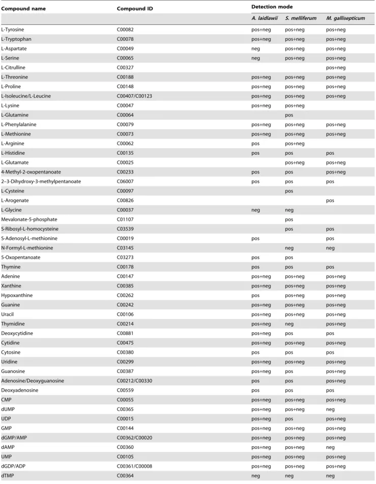
Table 1.Identified metabolites ofS. melliferum,M. gallisepticumandA. laidlawii.
Compound name Compound ID Detection mode
A. laidlawii S. melliferum M. gallisepticum
L-Tyrosine C00082 pos+neg pos+neg pos+neg
L-Tryptophan C00078 pos+neg pos+neg pos+neg
L-Aspartate C00049 neg pos+neg pos+neg
L-Serine C00065 neg pos+neg pos+neg
L-Citrulline C00327 pos+neg
L-Threonine C00188 pos+neg pos+neg pos+neg
L-Proline C00148 pos+neg pos+neg pos+neg
L-Isoleucine/L-Leucine C00407/C00123 pos+neg pos+neg pos+neg
L-Lysine C00047 pos+neg pos+neg
L-Glutamine C00064 pos
L-Phenylalanine C00079 pos+neg pos+neg pos+neg
L-Methionine C00073 pos+neg pos+neg pos+neg
L-Arginine C00062 pos pos+neg
L-Histidine C00135 pos pos pos
L-Glutamate C00025 pos+neg pos+neg
4-Methyl-2-oxopentanoate C00233 pos pos pos+neg
2–3-Dihydroxy-3-methylpentanoate C06007 pos pos pos
L-Cysteine C00097 pos
L-Arogenate C00826 pos
L-Glycine C00037 neg neg
Mevalonate-5-phosphate C01107 pos
S-Ribosyl-L-homocysteine C03539 pos pos
S-Adenosyl-L-methionine C00019 pos pos
N-Formyl-L-methionine C03145 neg neg
5-Oxopentanoate C03273 pos pos
Thymine C00178 pos pos pos
Adenine C00147 pos+neg pos+neg pos+neg
Xanthine C00385 pos+neg pos+neg pos+neg
Hypoxanthine C00262 pos pos+neg pos+neg
Guanine C00242 pos+neg pos+neg pos+neg
Uracil C00106 pos+neg pos+neg pos+neg
Thymidine C00214 pos+neg neg pos+neg
Deoxycytidine C00881 pos+neg pos pos
Cytidine C00475 pos+neg pos+neg pos+neg
Cytosine C00380 pos pos pos
Uridine C00299 pos+neg pos+neg pos+neg
Guanosine C00387 pos+neg pos pos+neg
Adenosine/Deoxyguanosine C00212/C00330 pos pos pos+neg
Deoxyadenosine C00559 pos pos pos
CMP C00055 pos+neg pos+neg pos+neg
dUMP C00365 pos+neg pos+neg neg
UDP C00015 pos+neg pos pos+neg
GMP C00144 pos+neg pos+neg pos+neg
dGMP/AMP C00362/C00020 pos+neg pos+neg pos+neg
dAMP C00360 pos+neg pos+neg neg
UMP C00105 pos+neg pos+neg pos+neg
dGDP/ADP C00361/C00008 pos+neg pos+neg pos+neg
Table 1.Cont.
Compound name Compound ID Detection mode
A. laidlawii S. melliferum M. gallisepticum
ITP C00081 pos pos pos
dUTP C00460 pos pos pos
2-Deoxyinosine 5-phosphate C06196 pos pos
Xanthosine C01762 pos neg
Inosine C00294 neg neg
XTP C00700 neg neg
dTDP C00363 neg neg
dUDP C01346 neg
Deoxyuridine C00526 pos+neg neg
dTTP C00459 neg neg
dCMP C00239 neg
S-Adenosyl-L-homocysteine C00021 pos
a-a-Trehalose/Maltose/Sucrose/Lactose C01083/C00208/C00185/C00089/C00243 pos+neg
D-Mannitol/D-Sorbitol C00392/C00794/C01697 pos+neg pos+neg pos+neg
Sedoheptulose 7-phosphate C05382 neg neg neg
D-Ribose 5-phosphate C00117 neg neg
GDP-6-deoxy-D-mannose C03117 neg pos
D-Mannitol 1-phosphate/D-Sorbitol-1-phosphate C00644/C01096 pos+neg
3-Phospho-D-glycerate C00197 pos
Phosphoenolpyruvate C00074 neg neg
N-Acetyl-D-galactosamine 6-phosphate/n-acetyl-D-
glucosamine-1-phosphate/N-Acetyl-D-mannosamine 6-phosphat
C06376/C04501/C04257 neg
Glycerone phosphate/glyceraldehydes-3-phosphateC00111/C00118 neg neg
beta-D-Fructose 1–6-bisphosphate C05378 pos
Glycerone C00184 neg neg
5-Phospho-alpha-D-ribose 1-diphosphate C00119 pos pos pos
Thiamin diphosphate C00068 pos pos+neg pos+neg
Formate C00058 neg neg neg
Geranylgeranyl diphosphate C00353 neg neg
2-C-Methyl-D-erythritol 2–4-cyclodiphosphate C11453 neg neg neg
5–10-Methenyltetrahydrofolate C00445 pos pos pos
5-Amino-6-(5-phospho-ribosylamino)uracil C01268 neg neg neg
5-Amino-6-(5-phospho-D-ribitylamino)uracil C04454 neg neg
sn-Glycerol 3-phosphate C00093 pos neg pos+neg
2-C-Methyl-D-erythritol 4-phosphate C11434 neg
5–10-Methylenetetrahydrofolate C00143 pos+neg neg
NADH C00004 neg pos
FMN C00061 pos+neg pos+neg
FAD C00016 neg
Nicotinate D-ribonucleoside C05841 pos+neg pos+neg pos+neg
NAD C00003 pos+neg pos+neg
Deamino-NAD+ C00857 pos+neg pos+neg pos+neg
Nicotinurate C05380 pos pos pos
2-Acetolactate C00900 pos pos
4-Methyl-3-oxoadipate C18312 pos pos
Nitrobenzene C06813 pos
UDP-N-acetyl-D-galactosaminuronic acid C13952 neg
UDP-N-acetyl-D-mannosamine C01170 neg pos+neg pos
[19] software and the Trans-Proteomic Pipeline (TPP) resource available online, where the data were converted into the mzXML format (MAVEN compatible).S. melliferum,M. gallisepticum, andA. laidlawii metabolites were identified using the list of all possible metabolites for these bacteria, garnered from all the protein annotation data that were previously reported [3,11]. A list of all theoretically possible metabolites ofS. melliferum, M. gallisepticum, and A. laidlawii was prepared in accordance with the KEGG database [20] and contained all the metabolites associated with the proteins that were annotated for these bacteria. The following parameters were used for the search: range of m/z values (extraction window), 15 ppm m/z; minimum intensity of peak, 1000 a.u.; minimum value of baseline signal intensity ratio, 10. If an identified metabolite was detected in at least five MS runs out of seven in specie one, while in specie two it was not detected, or it was detected only once, we attributed this metabolite to be present in the first species and to be absent in the second one.
Comparative analysis of theS. melliferum,M. gallisepticum, andA. laidlawiimetabolomes
A pairwise sample comparison of metabolomic data for three species was performed using the XCMS online service, providing a direct comparison of two sample groups [21]. From the resulting features of every compared pair of oganisms, we selected only those with a p value#0.05 in order to state that compound concentration is different in two species. Additionally these compounds were analyzed manually by counting the metabolite identifications among the seven repetitions of every species for both acquisition modes. The quantification of an identified metabolite is possible only if it was detected at least three times in each species. In the case a compound was detected less than three times of seven, only a qualitative comparison in the pair was possible, based on the detection of MS counts for this compound in the pair.
Results
MS spectra acquisition
Bacteria were cultivated in liquid media to more easily determine the growth phase. Bacteria were harvested in the logarithmic growth phase, which was determined based on the culture duplication time (see ‘‘Bacteria strain and growth conditions’’). Cold methanol extraction was used to instantly quench the metabolism [16]. The high effectiveness of this method and its low loss of compounds has been demonstrated previously [22]. Metabolite detection was carried out using HPLC analysis in combination with electrospray ionization quadrupole time-of-flight mass spectrometry (HPLC-MS Q-TOF). The parameters of the chromatographic method, as well as the mass spectrometric peak recording, were optimized using standards to obtain the most reliable data (see ‘‘Materials and Methods’’ section). Metabolites were initially identified using the Metabolomic Analysis and Visualization Engine (MAVEN) software [19]. The metabolite identity was then confirmed by analysis of the detected parent ions fragmentation spectra using targeted mass spectrometry [23].
There were seven independent repeats for metabolite detection for each of the three studied bacterial species. Usually one LC-MS analysis run produces ,120 peaks in which the m/z values are close to the exact metabolite m/z values (deviating by less than 10 ppm). However, in MS acquisition, some compounds show unstable signals and display low reproducibility; their peak intensities are comparable to the noise level. Each compound was considered to be present in bacteria if it was observed at least three times (in seven runs) in any of the ionization modes (positive
or negative) and the intensity of the corresponding peak was significantly higher than that of the background. List of identified metabolites is shown in Table 1.
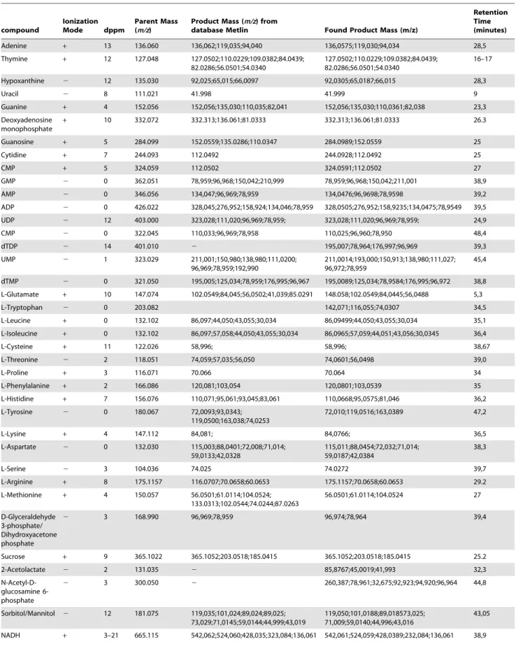
Fragmentation spectra were obtained for most of the detected metabolites using targeted mass spectrometry. The fragmentation spectra were compared with standard fragmentation spectra, which were obtained under similar ionization conditions (polarity of ionization and collision energy values were taken into account) and provided by the Metlin MS/MS Spectrum Match service [24,25,26]. Figure S1 shows an example of the metabolite identification as provided by the MS/MS. Interestingly, all fragmentation spectra confirmed the primary identification of the compounds based on the m/z values alone. There were no compounds whose fragmentation spectra contradicted the identi-fication based on the m/z values. The provided m/z values usually did not deviate from the theoretical value by more than 10 ppm (see Table 2). Among the detected metabolites, there were several compounds that presented a relatively large deviation from their theoretical ion m/z values (up to 15 ppm m/z). Nevertheless, MS/ MS fragmentation spectra render the unambiguous identification of such compounds possible.
Metabolite content ofS. melliferum,M. gallisepticum, and A. laidlawii
The strategy for metabolome analysis includes the following steps: 1) Metabolic map reconstruction; 2) Prediction of all possible metabolism intermediates; 3) Comparison of metabolic enzymes of Mollicute species; 4) Experimental MS detection and mapping of the detected compounds on the metabolic map for each specie; 5) Juxtaposition of the metabolome data to enzyme differences between three Mollicute species.
We manually reconstructed the metabolic network of M. gallisepticum, S. melliferum, and A. laidlawii in order to fill the network with the detected intermediates. To build a comprehen-sive metabolic network for each species, we used the Kyoto Encyclopedia of Genes and Genomes database and UniprotKB proteogenomic annotation data provided earlier by our lab [3,11,27]. For enzymes that could carry out more than one reaction, we removed the reactions that were decoupled from pathways and those for which the substrate was unavailable (i.e.
there is no way for substrate synthesis and transport into the cell according to the proteogenomic annotation). The final result is a map without gaps, isolated reactions, or open metabolic loops for each species (Figures 1–3, see text S1 and Figures S2, S3).
For each species a list of predicted metabolism intermediates was formed. The map includes all compounds, which serve as substrates and products for all enzymes and transporters predicted by the proteogenomic annotation. The metabolic database for raw MS data analysis by MAVEN includes all metabolism interme-diates predicted for the three species. This database contains 500 items and is suitable for the analysis of each species. The database used is wider than the list of possible metablolites for each species. It therefore permits detection of metabolites produced by putative uncharacterized enzymes.
Metabolic pathways were reconstructed with all the identified metabolites of S. melliferum, M. gallisepticum and A. laidlawii
Table 2.Fragmentation spectra results for the detected metabolites ofS. melliferum,M. gallisepticumandA. laidlawii, and fragmentation spectra of the analyzed ion standards from the Metlin database [23] in the same experimental conditions (fixed collision energy of 20 eV).
compound
Ionization
Mode dppm
Parent Mass (m/z)
Product Mass (m/z) from
database Metlin Found Product Mass (m/z)
Retention Time (minutes)
Adenine + 13 136.060 136,062;119,035;94,040 136,0575;119,030;94,034 28,5
Thymine + 12 127.048 127.0502;110.0229;109.0382;84.0439; 82.0286;56.0501;54.0340
127.0502;110.0229;109.0382;84.0439; 82.0286;56.0501;54.0340
16–17
Hypoxanthine 2 12 135.030 92,025;65,015;66,0097 92,0305;65,0187;66,015 28,3
Uracil 2 8 111.021 41.998 41.999 9
Guanine + 4 152.056 152,056;135,030;110,035;82,041 152,056;135,030;110,0361;82,038 23,3
Deoxyadenosine monophosphate
+ 10 332.072 332.313;136.061;81.0333 332.313;136.061;81.0333 26.3
Guanosine + 5 284.099 152.0559;135.0286;110.0347 284.0989;152.0559 25
Cytidine + 7 244.093 112.0492 244.0928;112.0492 25
CMP + 5 324.059 112.0502 324.0591;112.0502 27
GMP 2 0 362.051 78,959;96,968;150,042;210,999 78,959;96,968;150,042;211,001 38,9
AMP 2 0 346.056 134,047;96,969;78,959 134,0476;96,9698;78,9598 39,2
ADP 2 0 426.022 328,045;276,952;158,924;134,046;78,959 328,0505;276,952;158,9235;134,0475;78,9549 39,5
UDP 2 12 403.000 323,028;111,020;96,969;78,959; 323,028;111,020;96,969;78,959; 24,9
CMP 2 0 322.045 110,033;96,969;78,958 110,025;96,960;78,950 48,4
dTDP 2 14 401.010 2 195,007;78,964;176,997;96,969 39,3
UMP 2 1 323.029 211,001;150,980;138,980;111,0200; 96,969;78,959;192,990
211,0014;193,000;150,913;138,980;111,027; 96,972;78,959
45,4
dTMP 2 0 321.050 195,005;125,034;78,959;176,995;96,967 195,0089;125,034;78,9584;176,995;96,972 38,8
L-Glutamate + 10 147.074 102.0549;84,045;56,0502;41,039;85.0291 148.058;102.0549;84,0445;56,0488 5,3
L-Tryptophan 2 0 203.082 142,071;116,055;74,0307 34,5
L-Leucine + 0 132.102 86,097;44,050;43,055;30,034 86,09499;44,050;43,055;30,034 35,1
L-Isoleucine + 0 132.102 86,097;57,058;44,050;43,055;30,034 86,0965;57,059;44,051;43,056;30,0345 36,4
L-Cysteine + 11 122.026 58,996; 58,996; 38,67
L-Threonine 2 2 118.051 74,059;57,035;56,050 74,0601;56,0498 39,0
L-Proline + 3 116.071 70.066 70.064 34
L-Phenylalanine + 2 166.086 120,081;103,054 120,0801;103,0539 35
L-Histidine + 7 156.076 110,071;95,061;93,045;83,061 110,0668;95,0575;81,046 36,2
L-Tyrosine 2 0 180.067 72,0093;93,0343; 119,0500;163,038;74,0253
72,010;119,0516;163,0389 47,2
L-Lysine + 4 147.112 84,081; 84,0766; 36,5
L-Aspartate 2 0 132.030 115,003;88,0401;72,008;71,014; 59,0133;42,0328
115,011;88,0454;72,032;71,014; 59,0187;42,0384
38,3
L-Serine 2 3 104.036 74.025 74.0272 39,7
L-Arginine + 8 175.1157 116.0707;70.0658;60.0653 175.1157;70.0658;60.0653 29.2
L-Methionine + 4 150.057 56.0501;61.0114;104.0524; 133.0313;102.0544;74.0244;87.0263
56.0501;61.0114;104.0524 27
D-Glyceraldehyde 3-phosphate/ Dihydroxyacetone phosphate
2 3 168.990 96,969;78,959 96,974;78,964 39,4
Sucrose + 9 365.1022 365.1052;203.0518;185.0415 365.1052;203.0518;185.0415 25.2
2-Acetolactate 2 2 131.035 2 85,8767;45,0019;41,993 32,3
N-Acetyl-D-glucosamine 6-phosphate
2 3 300.050 2 260,387;78,961;32,675;92,923;94,920;96,964 44,8
Sorbitol/Mannitol 2 12 181.075 119,035;101,024;89,024;89,025; 73,029;71,0145;59,0144;44,999;43,019
119,050;101,0188;89,018573,025; 71,009;59,0140;44,996;43,016
43,05
Reconstruction of the three species’ metabolic maps reveals many pathways with enzymatic gaps in the middle. For pathways in which only one enzyme is missing, the gap can be filled by adding an unassigned reaction as performed earlier for another Mycoplasma species, M. pneumoniae [23]. Alternatively, for a missing enzymatic reaction, a bypass can be found by the manual analysis of the known enzyme’s side activities.
One example is the link between pentoses and hexoses (the pentose phosphate pathway) in Mollicutes. This link is omnipres-ent in living cells, although in many organisms, including mycoplasmas, only its non-oxidative branch is annotated. Two enzymes, transketolase (tktA) and transaldolase (tal), act in this branch of the pentose phosphate pathway (Figure 5). Transketol-ase transforms two pentoses, xylulose-5-phosphate and ribose-5-phosphate, into the seven-carbon product sedoheptulose-7-phos-phate and the three-carbon glyceraldehyde-3-phossedoheptulose-7-phos-phate. To produce the hexose, fructose 6-phosphate, and glyceraldehyde-3-phosphate and to finish the pentose glyceraldehyde-3-phosphate pathway, the same enzyme uses the four-carbon erythrose-4-phosphate and the pentose xylulose-5-phosphate. A key enzyme, transaldolase, acts to link these two transketolase reactions; it removes three-carbon fragment from sedoheptulose-7-phosphate and condenses it with glyceraldehyde-3-phosphate, forming the fructose 6-phosphate and the erythrose-4-phosphate [28].
Annotation of genome sequence shows that Mollicutes lack transaldolase, and this obviously creates a gap in the metabolic branch. Nevertheless, we detected a unique intermediate of transketolase reaction, sedoheptulose-7-phosphate, in all three species studied (along with less specific intermediates). It suggests that the pentose phosphate pathway is functional. The connection of the glycolytic pathway with a functional pentose phosphate pathway was earlier demonstrated for M.pneumoniae as 13C-6 glucose carbons were detected in ribose 5-phosphate using mass spectrometry [23].
Database search for transaldolase-like proteins in all other Mollicute genomes produces a negative result. Therefore we believe that transaldolase activity is absent in this genus. To fill the gap, it is possible to postulate the presence of aldolase activity by an unknown mycoplasma protein. The other way is to find a bypass of this reaction performed by other known mycoplasma enzymes. The literature shows that sedoheptulose-7-phosphate can act as acceptor for 6-phosphofructokinase; fructose-bisphosphate aldolase can bind with sedoheptulose-1,7-bisphosphate and decompose it to glycerone phosphate and erythrose-4-phosphate (see Figure 5) [29]. Glycerone phosphate can be isomerized to glyceraldehyde-3-phosphate by triosephosphate isomerase [28].
The proposed modifications in the pentose phosphate pathway make it possible to utilize ribose as an energy source despite the
lack of transaldolases. The described pathway is reversible inS. melliferum, andA. laidlawii. The first reaction, phosphorylation of sedoheptulose-7-phosphate, catalyzed by 6-phosphofructokinase, is irreversible; however, dephosphorylation of sedoheptulose-1,7-bisphosphate can be carried out by fructose -1,6-bisphosphatase [30], annotated inS. melliferum[11].
Fructose 1,6-bisphosphatase is not annotated inA. laidlawii, and eritrose-4-phosphate, formed as a result of the transketolase reaction between fructose-6- phosphate with glyceraldehyde-3-phosphate, must be utilized in some other way. Eritrose-4-phosphate may be utilized in phenylalanine synthesis, as all enzymes of phenylalanine biosynthesis are annotated inA. laidlawii
[3]. A lack of fructose-1,6-bisphosphatase, as well as the absence of transaldolase activity inM. gallisepticum, was experimentally shown by J.D. Pollack and M.V. Williams [31]. We assume that the resultant eritrose-4-phosphate inM. gallisepticum is utilized by D-fructose-6-phosphate D-erythrose-4-phosphate-lyase; this enzyme is not annotated forM. gallisepticum, but it is annotated for another Mycoplasma species (M. fermentans). We believe that this activity should be assigned forM. gallisepticum. This is a way to implement the nonoxidative reactions of the pentose phosphate pathway for
M. gallisepticum.
Among the detected metabolites we found compounds that participate in the main metabolic pathways, such as glycolysis, amino acid, sugar, and amino sugar metabolism, synthesis of terpenoids, riboflavines, purine and pyrimidine metabolism, etc. We used the LC-MS/MS method to analyze 95 compounds (see Table 1), representing a substantial fraction of the .500 compounds of the total metabolome of three Mollicute species,
M. gallisepticum, S. melliferum, and A. laidlawii. These compound were predicted by protein annotation, and performed by our lab in previous studies [3,11]. In particular, the purine and pyrimidine metabolism maps were the most uniformly and densely filled maps with the identified metabolites (see Figures 1–3). We detected a variety of purine and pyrimidine metabolism products: nitroge-nous bases- adenine, thymine, guanine, xanthine, hypoxanthine, uracil, cytosine; their nucleosides and deoxynucleosides and some of their mono-, di-, triphosphate nucleotides and deoxyribonucle-otides. It should be noted that we did not consider inosine mono-and dinucleotide, as according to V.M. Boeret al., this compound is not detectable, as it can be confused with peak interference of ADP and AMP [32]. Expression of the annotated proteins that are functionally associated with the metabolism of purine and pyrimidine was demonstrated in our previous studies [3,11]. In the S. melliferum metabolome, we did not detect deoxyuridine, dTDP, XTP, xanthosine, 2-deoxyinosine 5-phosphate; however, these compounds have been detected in M. gallisepticum and A. Table 2.Cont.
compound
Ionization
Mode dppm
Parent Mass (m/z)
Product Mass (m/z) from
database Metlin Found Product Mass (m/z)
Retention Time (minutes)
Betaine 118.086 118.086;59.0739;58.0660 118.086;59.0739;58.0660 28–30.7
Cytosine + 11 112.0489 112.0505;95.0242;94.0401; 69.0452;68.0136;67.0296;52.0189
112.0505;95.0242 26.3
NAD + 3 664.1164 542.0622;524.060;
428.035;232.0836;136.0603
664.1164;542.0622;
524.060;428.035;232.0836;136.0603
27
dGMP + 9 386.0227 152.0544;81.033 386.0227;152.0544 24.5
Figure 1. Reconstructed metabolic map ofM. gallisepticum.The pathways common for three Mollicute species are represented. Metabolites are shown as circles; compounds identified by LC-MS are marked in green, other predicted compounds are marked in red. Proteins that catalyze metabolic reactions are shown as diamonds, and their ID numbers are indicated. Enzymatic activities, which are not associated with annotated proteins, are indicated in italics. Abbreviations: F-6-P - fructose-6-phosphate; F-1,6-PP - fructose-1,6-bisphosphate; S-1,7-PP - D-sedoheptulose-1,7-bisphosphate; D-S-7-P - D-sedoheptulose-7-phosphate; GAP - glyceraldehyde-3-P).
laidlawii. AmongM. gallisepticumandA. laidlawiimetabolites, we did not identify inosine and dTTP, respectively.
There are many nutrient carriers in the Mycoplasma proteome, so the description of membrane fluxes and the discovery of exogenous metabolites acquired by the membrane transport is an important issue in metabolome research. Carbohydrates and their derivatives (such as glycolysis metabolites; see Figures 1–3) including sedoheptulose-7-phosphate, ribose-5-phosphate, D-mannitol 1-phosphate/D-sorbitol 1-phosphate, fructose-1,6-bi-sphosphate, deoxy-D-ribose-1-phosphate, 5-phospho-alpha-D-ri-bose-1-diphosphate, 3-phospho-D-glycerate, phosphoenolpyr-uvate, and n-acetyl-D-galactosamine-6-phosphate/n-acetyl-D-glucosamine-6-phosphate/N-Acetyl-D-mannosamine 6-phosphate were detected in all three species. We detected deoxy-D-ribose-1-phosphate inM. gallisepticumandA. laidlawiimetabolome, while the peak of this compound was considered to be peak interference according to V.M. Boer et al. [32]. We detected n-acetyl-D-galactosamine-6-phosphate and beta-D-fructose-1–6-bisphosphate in theA. laidlawii metabolome, while 3-phospho-D-glycerate is a characteristic feature of theM. gallisepticummetabolome. The latter compound was also detected in M. pneumoniae[18]. Detection of phosphorylated sugar products (mannitol 1-phosphate) is in good agreement with proteogenomic annotation [3,11] (PTS system mannitol-specific (MtlA)-like IIB domain protein MGA_1283) and with the identification of phosphorylated sugars inM. pneumoniae
[18].
In all three bacteria, using both positive and negative ionization modes, we detected non-phosphorylated forms of mannitol/ sorbitol/galactitol (sugars and some amino acids like leucine/ isoleucine isomers cannot be differentiated using the HPLC-MS method, since they have particularly identical fragmentation spectra and retention times) and alpha-alpha-trehalose/maltose/ cellobiose/sucrose/lactose.
Sugars are the most important energy source for the Mollicutes; their import in the cytoplasm is usually coupled with phosphor-ylation [11]. Here we directly demonstrate that mycoplasmas contain phosphorylated sugars. It was assumed that non-phosphorylated forms of carbohydrates can be accumulated from media or adsorbed to the cell surface [1,33,34,35,36]. Sugar ABC transporter systems which are not associated with phosphorylation were also annotated (MGA_1076, MGA_1077, MGA_1078 for
M. gallisepticum, SPM_2645S. melliferum). It cannot be ruled out, but it does seem unlikely that mycoplasmas store sugars to consume them during possible carbohydrate starvation.
We detected 17 of the 20 amino acids in the three Mollicute species. Their presence in the metabolome can be attributed to their exogenous nature and the fact that they are actively imported into the cells through specific transporters (permeases), since no amino acid synthesis enzymes have been annotated in these organisms [11]. Peaks in the MS spectra that corresponded to amino acids have the highest intensity. Only asparagine, alanine, and valine were not detected. In experiments employing the same equipment and another bacteria (not shown), we were able to detect these three amino acids. This suggests that the concentra-tion of the three amino acids, which were not found inS. melliferum,
M. gallisepticumandA. laidlawiitotal metabolome, was significantly lower than the concentration of the seventeen detected amino acids. Of note, in M. pneumoniae these three amino acids were detected [18]. Alanine is the most abundant amino acid in proteins and its absence in the metabolome could be explained by its rapid involvement into protein synthesis through aminoacyl-tRNA synthetase.
InM. pneumoniaeriboflavin was detected by LC-MS [18]. We have detected intermediates of riboflavin biosynthesis,
5-amino-6-(5-phospho-D-ribitylamino)uracil, 5-amino-6-(5-phosphoribosyla-mino)uracil, FMN and FAD. Among the explored three species, only A. laidlawii proteogenomic annotation contains proteins responsible for the diaminohydroxyphosphoribosylaminopyrimi-dines conversions in addition to the bifunctional FAD synthetase/ riboflavin kinase. However, in theS. melliferumandM. gallisepticum
metabolomes 5-amino-6-(5-phosphoribosylamino) uracil mass-spectral peaks were reliably detected with a high intensity. This finding can indicate activity of a metabolic branch responsible for FMN biosynthesis from GTP inS. melliferumandM. gallisepticum.
Discussion
The obtained list of metabolism intermediates taken together with our previous studies on the protein annotation of the same cells makes it possible to understand the exact mechanisms of the biochemical reactions in three Mollicute species (M. gallisepticum,S. melliferum, and A. laidlawii). Metabolome studies of the simplest bacteria reveal compounds and pathways that are important for the basic functions of bacteria maintenance. There were some metabolites that were not detected in our experiment but were present in the list of metabolites based on the bacterial proteome [3,11]. Some metabolites cannot be detected by LC-MS, because, for example, they are unstable under ionization conditions. Metabolites that are non-soluble in methanol cannot be detected either. Another reason why some metabolite intermediates were not detected is their possible consumption during cell wash in poor medium or even due to the residual enzymatic activity during the methanol extraction [22]. We detected several glycolysis interme-diates (3-phospho-D-glycerate, phosphoenolpyruvate, glyceralde-hyde-3-phosphate, beta-D-fructose 1–6-bisphosphate), so we conclude that the glycolysis rate is significantly decreased under the quenching conditions that we use. Similar sample preparation procedures revealed a similar glycolysis intermediate content inM. pneumoniae[18]. Several items that we did not expect to be revealed as metabolism components were detected. For example, in S. melliferum, S-ribosyl-L-homocysteine and 5-oxopentanoate were detected. As no enzymes responsible for their utilization or synthesis were annotated, we believe that such compounds are the products of amino acid degradation.
The differences that we identified primarily concerned amino acids and lipid biosynthesis and mechanism of carbohydrate assimilation (owing to Mollicutes’ parasitic nature, many nutrients are received from the host), and the biosynthesis of terpenoids and riboflavins. We found that citrulline concentration is higher inM. gallisepticumcompared toA. laidlawiiandS. melliferumwhile arginine concentration is lower inS. melliferumas compared toA. laidlawii
(Table 3). Citrulline is an intermediate in the arginine deiminase (ADI) pathway. The ADI path converts arginine to citrulline and ammonia, ornithine transcarbamylase subsequently converts citrulline to ornithine and carbamoyl phosphate, and finally carbamate kinase cleaves carbamyl phosphate to ammonia and CO2, generating an ATP in that process (see Figure 6). The system also requires an arginine/ornithine antiporter to rid the cell of the end-product, ornithine, while importing a new arginine substrate [27]. Ammonia that is generated by arginine deamination can alkalinize the environment [37], and helps the survival of cultures during acid stress. Some Mycoplasmas (M. hominis,M. arthritidis,M. gallinarum and others) use the arginine pathway only as an alternative energy source [38].The central enzyme of this pathway, arginine deiminase, was identified in M. gallisepticum
and S. melliferum genome and proteome in our previous studies, while it is absent fromA. laidlawii [3,11]. Only the S. melliferum
Figure 2. Reconstructed metabolic map ofA. laidlawii.The pathways common for three Mollicute species are represented. Abbreviations and symbols are given in figure 1.
complete ADI cycle, including arginine/ornithine antiporter [11], whileM. gallisepticumlacks ornithine carbamoyltransferase [39].
Another way that microbes use to respond to acidification is by producing enzymes that can convert acidic metabolites to neutral
products or neutral metabolites to alkaline products. Good examples of this type of enzymes are glutamate decarboxylase, lysine decarboxylase, and arginine decarboxylase ofEscherichia coli, all of which exhibit increased expression at external acidic pH Figure 3. Reconstructed metabolism ofS. melliferum.The pathways common for three Mollicute species are represented. Abbreviations and symbols are given in figure 1.
[40]. Ammonia can also be produced by the degradation of L-histidine, such as by the activity of L-histidine ammonialyase detected in ureaplasmas [41]. Glutamate decarboxylase is absent from all Mycoplasma species. The A. laidlawii annotation lacks arginine deiminase, glutamate decarboxylase or arginine decar-boxylase, while the lysine decarboxylase annotated forA. laidlawii
is unique among all Mollicutes. Lysine is a basic amino acid and possesses two amino groups. We found that lysine concentration is decreased in A. laidlawii compared to S. melliferum. Lysine decarboxylation can possibly serve as a mechanism to aid the survival of cultures in acid extracellular conditions.
Host infection caused by bacteria of both species,S. melliferum
andM. gallisepticum, necessitates acid adaptation of the pathogens. When infected, chickens and turkeys mycoplasmosis bacteria M. gallisepticum should adapt to acidic environment conditions. The clinical manifestation following the infection is called chronic respiratory disease in chickens and infectious sinusitis in turkeys [26]. As the infection begins with the colonization of the respiratory tract, tracheitis and airsacculitis are the predominant
symptoms of a localized infection in chickens [42]. In turn, bees are the main reservoir for spiroplasmas, where they primarily invade the gut lumen. Some species expanded their habitat range to include hemolymph, ovaries, fat bodies, hypodermis, and salivary glands. Spiroplasma infection of bees occurs with nectar consumption. The invertase of the pharyngeal gland secretions inverts sucrose into glucose and fructose, and then glucose oxidase converts glucose into gluconic acid to acidify honey (pH 4–5). To survive, spiroplasmas need to overcome this pH-barrier. The balance ofcitrulline/arginineduring the growth of Mycoplasmas will be investigated in future research.
We suggest thatA. laidlawiihas adaptation mechanisms different fromM. gallisepticumandS. melliferumand uses decarboxylation of lysine since A. laidlawii is widely distributed in nature and has infects a wide range of hosts [43]. Members of the Acholeplasma
genus do not have specific host associations and have been found in almost every type of living organism. They are one of the five common reasons of cell culture contamination, and a causative agent of some plant diseases [44,45,46,47].
To check whether M. gallisepticum,A. laidlawii and S. melliferum
are able to survive in acidic environment, we used the ‘‘color test’’ method to estimate a fraction of the surviving cells and found that
M. gallisepticum, A. laidlawii and S. melliferum can survive at low values of pH (,5.0–5.3); this type of adaptation can be necessary during an infection.
We detected intermediates of purine metabolism (Figures 1–3). It should be noted that a direct transition between purine nitrogen base and monophosphate nucleotide is catalyzed by AMP pyrophosphorylase, which is found in all three species. Sequential conversion of nucleotides- to nucleosides and then to nitrogen bases is performed by the protein 59-nucleotidase. This protein is not annotated for S. melliferum (in A. laidlawii, this protein is annotated, whileM. gallisepticum contains the protein thymidine kinase, which is capable to catalyzing this reaction). Purine nucleoside phosphorylase is available in all three species. 59-nucleotidase makes it possible to 1) consume AMP and use it as an energy source; 2) to use ribose from AMP as a building block for other nucleotide synthesis. However, we do not believe that 59-nucleotidase should be assigned forS. melliferum, as was suggested for M. pneumoniae [23]. We believe that the purine metabolism pathway inS. melliferumis directed from phosphoribosyl-pyrophos-Figure 4. Overlap of the detected metabolites for three
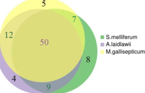
Mollicute species. Yellow circle represents metabolites of M. gallisepticum, violet circle represents metabolites ofS. melliferum, and green circle represents metabolites ofA. laidlawii. The list of identified metabolites of S. melliferum, M. gallisepticum and A. laidlawii is presented in Table 1.
doi:10.1371/journal.pone.0089312.g004
Figure 5. Reactions of the pentose phosphate pathway.Metabolites involved in the pathway are shown as circles; enzymes are shown as diamonds; and the path that we propose to convert sedoheptulose-7-phosphate is marked in green.
phate to DNA and RNA synthesis while the reverse direction is absent (S. melliferumdoes not utilize nucleotides).
We detected a high level of glycerol-3-phosphate inA. laidlawii
and a decreased CMP level compared to S. melliferum and M. gallisepticum (see Table 3). The composition of the A. laidlawii
membrane differs from that of other Mollicutes, since the main
lipids of its cytoplasmic membrane are the neutral glycolipids monoglucosyl diacylglycerol (MGDG) and diglucosyl diacylgly-cerol (DGDG), the phosphoglycolipid glycerophosphoryl digluco-syl diacylglycerol (GPDGDG), and the phospholipid phosphati-dylglycerol (PG), whereas cholesterol is a major membrane component of other mycoplasmas [8]. An important reaction in Figure 6. Arginine deiminase (ADI) pathway.In this pathway, metabolites are shown as circles; enzymes are shown as diamonds; and compounds that we detected are marked in green.
doi:10.1371/journal.pone.0089312.g006
Table 3.Metabolite peaks counting and pairwise comparison of three Mollicute species.
Compound Compound ID Spectral counts
Normalized peak
intensities ratio Conclusion
M.gal. A.laid. S.melli.
L-Lysine C00047 - 6 10 S.mellif./A.laid.= 3.0 S.mellif..A.laid..M.gal. = 0
L-Glutamate C00025 5 - 4 S.mellif./M.gal. = 1.2 S.mellif.=M.gal..A.laid.= 0
L-Glutamine C00064 - - 4 - InS.mellif.only
CMP C00055 8 7 12 S.mellif./M.gal.= 3.7
S.mellif./A.laid.= 1.6 A.laid./M.gal.= 1.8
S.mellif..A.laid..M.gal.
sn-Glycerol 3-phosphate C00093 11 7 4 A.laid./S.mellif./ = 1.4
M.gal./A.laid.= 24 M.gal./S.mellif.= 53
M.gal..A.laid.$S.mellif.
5-Amino-6-(5-phospho-D-ribitylamino)uracil
C04454 - 6 - - InA.laid.only
5-Amino-6-(5-phosphoribosylamino)uracil C01268 3 7 7 A.laid./M.gal.= 5.4 S.mellif/A.laid= 2.6 S.mellif/M.gal.= 30
S.mellif..A.laid..M.gal.
2-C-Methyl-D-erythritol 4-phosphate C11434 - 6 - - InA.laid.only
2-C-Methyl-D-erythritol 2–4-cyclodiphosphate
C11453 7 6 4 M.gal./A.laid.= 9.1
M.gal./S.mellif.= 20 A.laid./S.mellif= 1.3
M.gal..A.laid.$S.mellif.
L-Arginine C00062 - 5 4 A.laid./S.mellif.= 2.2 A.laid..S.mellif..M.gal = 0
L-Citrulline C00327 11 - - - InM.gal.only
Geranylgeranyl diphosphate C00353 0 7 5 A.laid./S.mellif.= 3.3 A.laid..S.mellif..M.gal = 0
a-a-Trehalose/Maltose/Sucrose/Lactose /C00208/C00185/C00089/C00243 - - 5 - InS.mellif.only
phospholipid synthesis is the replacement of the cytosine unit by glycerol-3-phosphate to form phosphatidylglycerol (PG) and CMP. PG is a precursor of lipoglycans, the main component of the
Acholeplasma membrane. Glycerol kinase is absent from the A. laidlawii genome annotation, while glycerol-3-phosphate (G3P) dehydrogenase is present [3]. G-3-P dehydrogenase catalyzes the reversible redox conversion of dihydroxyacetone phosphate to sn-glycerol 3-phosphate and serves as a major link between carbohydrate metabolism and lipid metabolism. A decreased level of glycerol-3-phosphate and a high content of CMP indicate an active synthesis of the main structural component of biological membranes - glycerophospholipids inA. laidlawii.
There are two metabolic pathways of terpenoid synthesis: the mevalonate acid pathway (MVA) and nonmevalonate pathway (2-C-methyl-D-erythritol 4-phosphate/1-deoxy-D-xylulose-5-phos-phate pathway (MEP/DOXP pathway)) (Figure 7). Although both pathways, MVA and MEP/DOXP, are mutually exclusive in most organisms, interactions between these pathways have been reported in plants and several bacterial species [48,49]. Many organisms manufacture terpenoids through the mevalonate acid pathway (MVA), that also produces cholesterol. These reactions,
directed to carotenoid biosynthesis, take place in theA. laidlawii
[3]. We have detected a high level of 2-C-methyl-D-erythritol 4-phosphate inA. laidlawii, we have not detected any metabolites involved the MVA path.
The alternative nonmevalonate pathway or 2-C-methyl-D-erythritol 4-phosphate/1-deoxy-D-xylulose-5-phosphate pathway (MEP/DOXP pathway) of isoprenoid biosynthesis is an alternative metabolic pathway leading to the formation of isopentenyl pyrophosphate (IPP) and dimethylallyl pyrophosphate (DMAPP). For numerous microbial pathogens such asEnterobacteria, Mycobac-terium tuberculosis, M. gallisepticumandS. melliferum, the nonmevalo-nate pathway is the exclusive source of terpenoids [49]. Because the enzymes of the nonmevalonate pathway have no orthologs in mammalian hosts, they are attractive targets for the development of novel antibiotic or antiprotozoal agents. InM. gallisepticum and S. melliferum metabolome, we detected the presence of substantial amounts of 2-C-methyl-D-erythritol-2,4-cyclodiphosphate (com-ponent of the MEP/DOXP branches of metabolism), and found geranylgeranyl diphosphate (an important precursor in the biosynthesis of all higher terpenoids and carotenoid biosynthesis) only in S. melliferum. In M. gallisepticum and S. melliferum
Figure 7. Terpenoid backbone biosynthesis pathway.Metabolites involved in this pathway are shown as circles; enzymes are shown as diamonds; and compounds that we detected are marked in green.
metabolisms, the presence of MEP/DOXP pathway proteins can be found, but the path terminated at the 1-hydroxy-2-methyl-2-butenyl 4-diphosphate [11] (Whole Genome Shotgun ID: AFFR00000000). Nevertheless, the identification of MEP/DOXP pathway components in the metabolome suggests active expres-sion of annotated proteins and terpenoid backbone biosynthesis in
M. gallisepticum and S. melliferum. Interestingly, we have also detected mevalonate-5-phosphate in S. melliferum metabolome; however, no proteins of the mevalonate acid pathway are noted in these bacteria.
We have systematically studied three bacterial species that belong to the class Mollicutes: the smallest and simplest bacteria,
Spiroplasma melliferum, Mycoplasma gallisepticum, and Acholeplasma laidlawii. We described the basic difference in the principles of three species’ metabolic adaptation to acidic environmental conditions and analyzed three species’ their metabolomes to confirm our assumptions. The metabolic pathways of three species were reconstructed and analyzed using the proteogenomic annotation data provided by our lab in previous studies [3,11].
Supporting Information
Text S1 Metabolome reconstruction. (DOCX)
Figure S1 MS/MS Spectrum match of Uracil fragmen-tation spectrum obtained for theS. melliferumsample.
Uracil fragmentation spectrum obtained for the S. melliferum
sample is above the OX axis and standard Uracil fragmentation spectrum in positive ionization mode collision energy 20 eV from Metlin Metabolites database is under the OX axis [30]; Uracil M+ H: m/z = 113.03344,D= 10 ppm.
(TIF)
Figure S2 Reconstructed pathways characteristic forA. laidlawii which are absent in M. gallisepticum. For description see legend to Figure 1.
(TIF)
Figure S3 Reconstructed pathways characteristic forS. melliferum which are absent in M. gallisepticum. For description see legend to Figure 1.
(TIF)
Acknowledgments
We thank G.B. Smirnov for the critical review of the manuscript and A.A.Vassilevski for his help during the preparation of the manuscript.
Author Contributions
Conceived and designed the experiments: VMG DEK. Performed the experiments: AAV. Analyzed the data: AAV DEK AYG GYF. Wrote the paper: AAV DEK AYG VMG.
References
1. Razin S (1973) Physiology of mycoplasmas. Adv Microb Physiol 10: 1–80. 2. Razin S, Michmann J, Shimshoni Z (1964) The Occurrence of Mycoplasma
(Pleuropneumonia-Like Organisms, Pplo) in the Oral Cavity of Dentulous and Edentulous Subjects. J Dent Res 43: 402–405.
3. Lazarev VN, Levitskii SA, Basovskii YI, Chukin MM, Akopian TA, et al. (2011) Complete genome and proteome of Acholeplasma laidlawii. J Bacteriol 193: 4943–4953.
4. Laidlaw PP, Elford,W J (1936) A new group of filterable organisms. B Biol Sci: 292–303.
5. Leach RH (1967) Comparative studies of mycoplasma of bovine origin. Ann N Y Acad Sci 143: 305–316.
6. Stipkovits L (1979) The pathogenicity of avian mycoplasmas. Zentralbl Bakteriol Orig A 245: 171–183.
7. Pollack JD, Tryon VV, Beaman KD (1983) The metabolic pathways of Acholeplasma and Mycoplasma: an overview. Yale J Biol Med 56: 709–716. 8. Cocaign-Bousquet M, Even S, Lindley ND, Loubiere P (2002) Anaerobic sugar
catabolism in Lactococcus lactis: genetic regulation and enzyme control over pathway flux. Appl Microbiol Biotechnol 60: 24–32.
9. van der Greef J, Stroobant P, van der Heijden R (2004) The role of analytical sciences in medical systems biology. Curr Opin Chem Biol 8: 559–565. 10. Levitskiy SA, Sycheva AM, Kharlampieva DD, Oberto J, Kamashev DE, et al.
(2011) Purification and functional analysis of recombinant Acholeplasma laidlawii histone-like HU protein. Biochimie 93: 1102–1109.
11. Alexeev D, Kostrjukova E, Aliper A, Popenko A, Bazaleev N, et al. (2012) Application of Spiroplasma melliferum proteogenomic profiling for the discovery of virulence factors and pathogenicity mechanisms in host-associated spiroplas-mas. J Proteome Res 11: 224–236.
12. Kamashev D, Oberto J, Serebryakova M, Gorbachev A, Zhukova Y, et al. (2011) Mycoplasma gallisepticum produces a histone-like protein that recognizes base mismatches in DNA. Biochemistry 50: 8692–8702.
13. Urano K, Kurihara Y, Seki M, Shinozaki K (2010) ‘Omics’ analyses of regulatory networks in plant abiotic stress responses. Curr Opin Plant Biol 13: 132–138.
14. Tully JG, Whitcomb RF, Clark HF, Williamson DL (1977) Pathogenic mycoplasmas: cultivation and vertebrate pathogenicity of a new spiroplasma. Science 195: 892–894.
15. Gorbachev AY, Fisunov GY, Izraelson M, Evsyutina DV, Mazin PV, et al. (2013) DNA repair in Mycoplasma gallisepticum. BMC Genomics 14: 726. 16. Maharjan RP, Ferenci T (2003) Global metabolite analysis: the influence of
extraction methodology on metabolome profiles of Escherichia coli. Anal Biochem 313: 145–154.
17. Bolten CJ, Kiefer P, Letisse F, Portais JC, Wittmann C (2007) Sampling for metabolome analysis of microorganisms. Anal Chem 79: 3843–3849. 18. Maier T, Marcos J, Wodke JA, Paetzold B, Liebeke M, et al. (2013) Large-scale
metabolome analysis and quantitative integration with genomics and proteomics data in Mycoplasma pneumoniae. Mol Biosyst 9: 1743–1755.
19. Melamud E, Vastag L, Rabinowitz JD (2010) Metabolomic analysis and visualization engine for LC-MS data. Anal Chem 82: 9818–9826.
20. Kanehisa M (1997) A database for post-genome analysis. Trends Genet 13: 375– 376.
21. Smith CA, Want EJ, O’Maille G, Abagyan R, Siuzdak G (2006) XCMS: processing mass spectrometry data for metabolite profiling using nonlinear peak alignment, matching, and identification. Anal Chem 78: 779–787.
22. Villas-Boas SG, Hojer-Pedersen J, Akesson M, Smedsgaard J, Nielsen J (2005) Global metabolite analysis of yeast: evaluation of sample preparation methods. Yeast 22: 1155–1169.
23. Yus E, Maier T, Michalodimitrakis K, van Noort V, Yamada T, et al. (2009) Impact of genome reduction on bacterial metabolism and its regulation. Science 326: 1263–1268.
24. Smith CA, O’Maille G, Want EJ, Qin C, Trauger SA, et al. (2005) METLIN: a metabolite mass spectral database. Ther Drug Monit 27: 747–751.
25. Tautenhahn R, Cho K, Uritboonthai W, Zhu Z, Patti GJ, et al. (2012) An accelerated workflow for untargeted metabolomics using the METLIN database. Nat Biotechnol 30: 826–828.
26. Vogl G, Plaickner A, Szathmary S, Stipkovits L, Rosengarten R, et al. (2008) Mycoplasma gallisepticum invades chicken erythrocytes during infection. Infect Immun 76: 71–77.
27. Moat A, Foster J, Spector M (2002) Microbial Physiology.: Wiley-Liss,Inc. 28. Lehninger A (2008) Princioals of biology.
29. Karadsheh NS, Tejwani GA, Ramaiah A (1973) Sedoheptulose-7-phosphate kinase activity of phosphofructokinase from the different tissues of rabbit. Biochim Biophys Acta 327: 66–81.
30. Pontremoli S, Traniello S, Luppis B, Wood WA (1965) Fructose diphosphatase from rabbit liver. I. Purification and properties. J Biol Chem 240: 3459–3463. 31. Pollack JD, Williams MV (1986) PPi-dependent phosphofructotransferase
(phosphofructokinase) activity in the mollicutes (mycoplasma) Acholeplasma laidlawii. J Bacteriol 165: 53–60.
32. Boer VM, Crutchfield CA, Bradley PH, Botstein D, Rabinowitz JD (2010) Growth-limiting intracellular metabolites in yeast growing under diverse nutrient limitations. Mol Biol Cell 21: 198–211.
33. Terry TM, Zupnik JS (1973) Weak association of glucosamine-containing polymer with the Acholeplasma laidlawii membrane. Biochim Biophys Acta 291: 144–148.
34. Cole RM, Tully JG, Popkin TJ, Bove JM (1973) Morphology, ultrastructure, and bacteriophage infection of the helical mycoplasma-like organism (Spiroplasma citri gen. nov., sp. nov.) cultured from ‘‘stubborn’’ disease of citrus. J Bacteriol 115: 367–384.
36. Schiefer HG, Gerhardt U, Brunner H, Krupe M (1974) Studies with lectins on the surface carbohydrate structures of mycoplasma membranes. J Bacteriol 120: 81–88.
37. Pereyre S, Sirand-Pugnet P, Beven L, Charron A, Renaudin H, et al. (2009) Life on arginine for Mycoplasma hominis: clues from its minimal genome and comparison with other human urogenital mycoplasmas. PLoS Genet 5: e1000677.
38. Hahn RG, Kenny GE (1974) Differences in arginine requirement for growth among arginine-utilizing Mycoplasma species. J Bacteriol 117: 611–618. 39. Papazisi L, Gorton TS, Kutish G, Markham PF, Browning GF, et al. (2003) The
complete genome sequence of the avian pathogen Mycoplasma gallisepticum strain R(low). Microbiology 149: 2307–2316.
40. Castanie-Cornet MP, Foster JW (2001) Escherichia coli acid resistance: cAMP receptor protein and a 20 bp cis-acting sequence control pH and stationary phase expression of the gadA and gadBC glutamate decarboxylase genes. Microbiology 147: 709–715.
41. Ajello F, Romano N, Massenti MF (1977) L-histidine ammonia-lyase from a T-strain Mycoplasma (Ureaplasma urealyticum). Boll Ist Sieroter Milan 56: 343– 350.
42. Ley D (2003) Mycoplasma gallisepticum infection; M. SY, editor. Ames.: Iowa State Press.
43. Lee IM, Davis RE, Gundersen-Rindal DE (2000) Phytoplasma: phytopathogenic mollicutes. Annu Rev Microbiol 54: 221–255.
44. Scripal IG, Bilay VI (1988) Microorganisms - agents of the plant diseases. Naukova Dumka.
45. Chernov VM, Mukhametshina NE, Gogolev YV, Nesterova TN, Chernova OA (2007) Mycoplasma adaptation to adverse growth conditions: nanotransforma-tion and phytopathogenicity of Acholeplasma laidlawii PG8. Dokl Biochem Biophys 413: 57–60.
46. David SA, Volokhov DV, Ye Z, Chizhikov V (2010) Evaluation of Mycoplasma inactivation during production of biologics: egg-based viral vaccines as a model. Appl Environ Microbiol 76: 2718–2728.
47. Folmsbee M, Howard G, McAlister M (2010) Nutritional effects of culture media on mycoplasma cell size and removal by filtration. Biologicals 38: 214–217. 48. Lichtenthaler HK (1999) The 1-Deoxy-D-Xylulose-5-Phosphate Pathway of
Isoprenoid Biosynthesis in Plants. Annu Rev Plant Physiol Plant Mol Biol 50: 47–65.