J. Anat. (2003) 202, pp431– 444
Blackwell Science, Ltd
Structure of the pelvic and penile urethra – relationship
with the ducts of the sex accessory glands of the
Mongolian gerbil (
Meriones unguiculatus
)
P. F. F. Pinheiro,
1C. C. D. Almeida,
1T. M. Segatelli,
1M. Martinez,
2C. R. Padovani
3and F. E. Martinez
11
Department of Anatomy, and 3
Department of Biostatistics, Institute of Biosciences, UNESP, Botucatu, SP, Brazil
2
Department of Morphology and Pathology, UFSCar, São Carlos, SP, Brazil
Abstract
The urethra is the main place of entry for sexually transmitted pathogens. However, there is little literature on the morphology of the urogenital system, principally the urethra and ducts of the sex accessory glands. The Mongolian gerbil is an insectivorous, herbivorous and monogamous rodent with nocturnal habits; it has been used successfully as a laboratory animal since the 1960s. Therefore, the objective of the present paper was to describe the structure and ultrastructure of the urethra and its relations to the ducts of the accessory sex glands of the Mongolian gerbil (Meriones unguiculatus), contributing to the understanding of the reproductive biology of the rodent and aiming to provide data for future experimental studies. Conventional techniques of light and scanning electron microscopy were utilized. The urethra and ducts of the accessory sex glands are similar to those of the albino rat and the mouse. However, there is variation in drainage type among accessory sex glands for the inner urethra. The ducts of the seminal vesicle, the ductus deferens, drain their contents independently into the ampullary duct that opens in the urethra. The ducts of the prostate, coagulating and bulbourethral glands drain their contents independently into the urethra.
Key words male urogenital system; morphology; rodents.
Introduction
The male urethra constitutes the place of entry for sexually transmitted pathogens (Quayle et al. 1994). In the rat and the mouse, the urethra is divided into pelvic and penile portions (Hayes, 1965; Hummel et al. 1966; Hebel & Stromberg, 1976).
The origin of the distal part of the male urethra has been disputed by researchers, although there is general agreement on the development of the proximal part (Kanagasuntheram & Anandarajara, 1960). Embryolo-gists have stated that the urethra within the navicular fossa was formed by an ectodermal surface, while the remainder of the urethra was formed by an endodermal
surface. Despite the embryological and histological interest described above, the urethra has been given little attention (Nakano & Muto, 1989), in contrast to the abundant studies on the urinary bladder, ureter and kidney (Fulker et al. 1971; Kumagai, 1975; Kjaer et al. 1976; Newman & Hicks, 1977; Tannenbaum et al. 1978; Hayakawa, 1979; Nelson et al. 1979).
To date, the study of the structure of the sex ducts in mammals (including man and laboratory rodents), the accessory glands, has been reviewed only partially. The Mongolian gerbil (Meriones unguiculatus) is a rodent known for its suitability for laboratory use. By nature, it is a curious and friendly rodent, almost without odour, with monogamous behaviour and physiological mechanisms of corporal-water conservation, present-ing spontaneous attacks of epilepsy and relative absence of natural illnesses. The purpose of the present paper was to describe the structure of the pelvic and penile urethra and their relationship to the ducts of the accessory sex glands, and to provide data for future experimental studies.
Correspondence
Dr Patricia Fernanda Felipe Pinheiro, Caixa Postal 510, Department of Anatomy, Biosciences Institute, UNESP, Campus of Botucatu, Distrito de Rubião Júnior s/n, Botucatu-SP, Brazil, CEP:18618–000. Tel.: (5502114) 6802–6040; fax: (5502114) 6821–3744; e-mail: pffp@uol.com.br; pinheiro_patricia@hotmail.com
Morphology of male gerbil urogenital system, P. F. F. Pinheiro et al.
432
Materials and methods
Animals
Thirteen adult male gerbils (Meriones unguiculatus), aged 3 months and with an average weight of 90 g, were used. Gerbils were maintained under controlled temperature (23 °C) and lighting conditions (12 : 12 h light–dark photoperiod, lights-out at 07:00 h). The experimental protocol followed the ethical principles in animal research adopted by the Brazilian College of Animal Experimentation.
Light microscopy (LM)
Nine adult male gerbils (Meriones unguiculatus), aged 3 months and with an average weight of 90 g, were used. The animals were anaesthetized with ether and perfused with Bouin′s solution through the left ventricle according to the method of Sprando (1990). After the organs of the urogenital system, including the prostate, seminal vesicles, coagulating glands, urethra, urinary bladder, part of the ureters, ductus deferens and crus penis, had been isolated, collected and fixed in Bouin′s solution for 24 h, they were washed in a solution of 70% alcohol, dehydrated in a graded ethanol series, embedded in paraplast and cut into 5–7-µm-thick sections. The paraplast blocks were cut so that the sections were either parallel or perpendicular to the urethra. The sections were mounted on glass slides, stained with Hematoxylin-Eosin (HE), Mallory′s thricrome (MlT), Masson′s thricrome (MaT), Calleja (C) and Azan-Heidenhain-Mallory (A). The sections were observed and photographed in a Zeiss Axiophot 2 light microscope.
Scanning electron microscopy (SEM)
Four adult male gerbils (Meriones unguiculatus), aged 3 months and with an average weight of 90 g, were used. The animals were anaesthetized with ether and perfused with glutaraldehyde solution through the left ventricle according to the method of Sprando (1990). Fragments of the pelvic and penile urethra were fixed in 2.5% glutaraldehyde in 0.1 M phosphate buffer
(pH 7.2) for 3 h. Next, the fragments were post-fixed in 1% osmium tetroxide in 0.1 M phosphate buffer
(pH 7.2) for 2 h in darkness. Subsequently, the frag-ments were dehydrated in graded ethanol. They were then submitted to critical point drying in liquid CO2
(Balzers CPD-010), coated with gold ions in SEM coating
(Balzers MED 010) and examined under a Phillips FEM 515 SEM at 15 kV.
Results
Urethra
The urethra of the gerbil presented two portions: pelvic and penile. The pelvic urethra extended from an internal ostium in the urinary bladder to the level of the penile bulb. The penile urethra extended from the penile bulb to the external ostium of the urethra (Figs 1A,E,F and 4A). The pelvic and penile urethra were shown to be connected through the isthmus (Fig. 5B). The structure of the urethra varied in its different segments.
Next to the neck of the urinary bladder, the pelvic urethra presented transitional epithelium with basal, intermediate and superficial cells. The basal presented cubic or prismatic forms, whereas the intermediate were polyhedral forms of various sizes and the super-ficial irregular polygonal forms (Fig. 6A). The pelvic urethra presented a thick layer of urethral muscle, surrounding it like a belt and finishing between the ischiocavernosum muscle and penile bulb (Figs 2A,B and 5A). In the stromal tissue thin blood vessels and muscle fibres were observed.
The portion of the pelvic urethra next to the penile bulb presented stratified cylindrical epithelium, with occa-sional areas of pseudostratified cylindrical epithelium. The proper lamina and the muscle layer (urethral muscle) were shown to be similar to the origin (Fig. 7A,B).
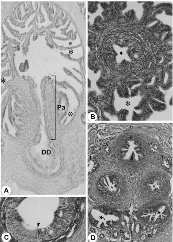
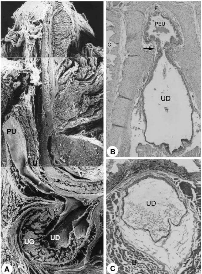
At the ischiadic arch the pelvic urethra enters in the urethral diverticle, an expansion of the penile urethra with a heart-shaped form and bilateral symmetry. The diverticle mucosa, with longitudinal folds allowing its dilation, presented stratified columnar epithelium and rich proper lamina in elastic, collagenous and smooth muscle fibres and blood vessels. Adenomeres of the urethral gland were connected to the diverticle, unloading its secretions in the lumen (Fig. 6B–D).
Morphology of male gerbil urogenital system, P. F. F. Pinheiro et al. 433
Fig. 1 (A)Diagram of the ventral view of the sex accessory glands. (B)Male reproductive organs in situ. ×1.5. (C)Ventral view of
Morphology of male gerbil urogenital system, P. F. F. Pinheiro et al.
434
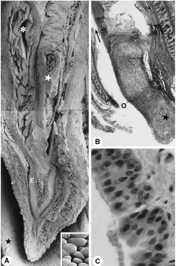
The epithelium presented stratified columns (Fig. 8C) with a varying number of cellular layers.
Ducts of the sex accessory glands
The glandular portion of the urogenital system is formed by the seminal vesicles, coagulating glands, ductus deferens, bulbourethral and urethral glands and also prostate involving the urethra (Fig. 1A–D,F). Surrounding the mucosa of the pelvic urethra are the ducts of the accessory sex glands: ventral and proximal to the pelvic urethra are the ventral prostatic ducts (Figs 2D and 3D); dorsal are the coagulating gland ducts, preceding the opening of the ampullary duct. The ampullary duct received the ducts of the seminal vesicle, of the ductus deferens gland and the papilla of the ductus deferens, later to open up in the dorsal surface of the pelvic urethra. The dorsolateral prostatic ducts drained into the dorsal surface of the pelvic urethra simultaneously to the ampullary duct (Fig. 3D). The bulbourethral gland drained directly into the penile bulb that contains the urethral diverticle (Fig. 4A).
The seminal vesicle duct comprises a simple cylin-drical epithelium, with nucleus located in the basal third of the cell with nucleolus evident and with a granular cytoplasm. Delicate folds of the mucosa and a single smooth muscle cell layer (myoepithelium) were observed involving the entirety of the duct. The seminal vesicle duct was located dorsally with respect to the ductus deferens en route to its opening into the ampullary duct. Next to the roof of the pelvic urethra, the seminal vesicle duct was connected to several dorso-lateral prostatic ducts and fibromuscular tissue (Fig. 3D). The coagulating gland duct is located laterally and ventrally to the ductus deferens, opening craniolater-ally to the ostium of the ampullary duct in the roof of the pelvic urethra (Fig. 3D). The coagulating gland duct presented a simple cylindrical epithelium (Fig. 4C).
The ventral prostatic ducts presented simple cylindrical epithelium (Fig. 4E) with folds in the mucosa. The dorsal prostatic ducts opened in the roof of the pelvic urethra, connected by smooth muscle fibres (Fig. 4B). The pros-tatic ducts, close to the urethra, presented a thick layer of smooth muscle cells, guiding the ducts until the ure-thral mucosa, forming paths between ureure-thral muscles.
The ductus deferens gland ducts presented simple cylindrical epithelium, distributed in radial form in the muscle layer of the ductus deferens toward the ampul-lary duct to drain the glandular secretion (Fig. 3B).
The distal (terminal) portion of the ductus deferens was characterized by the transition of the epithelium from a pseudostratified type with stereocilia to a stratified cylindrical type (Fig. 3A,C). Next to the pelvic urethra we observed the formation of the ductus deferens papilla opening the ampullary duct. This duct resulted from confluence of the ducts of the ductus deferens gland and the origin of the papilla of the ductus defe-rens. The papilla presented a star-shaped format, con-sisting of the stratified cylindrical epithelium (Fig. 3B). At its origin, next to the roof of the pelvic urethra, the ampullary duct appeared to be flanked by the dorsal prostate, seminal vesicle and coagulating gland ducts. The ampullary duct opened in the roof of the pelvic urethra (Fig. 4A).
The bulbourethral gland duct was lined with epithe-lium that varied from cylindrical to paving-stone shaped. The ducts congregated into only one duct, accompanied by glandular tissue, surrounded by a single smooth-muscle-cell layer (myoepithelium), muscular fibres, adipose tissue and a serosa layer. The duct extended close to the bulb of the penis and opened into the wall of the diverticle of the urethra (Fig. 6B).
Discussion
Dellmann (1993) considered three distinct portions in the urethra: pelvic, from the neck of the urinary blad-der to the penile bulb; bulbar contained in the penile bulb; and penile, from the bulbous extremity to the external ostium of the urethra. Getty (1986) and Dyce et al. (1997) described two portions of the male urethra in domestic animals: pelvic, from the internal ostium of the urethra to the seminal colliculus; and penile, from the seminal colliculus to the external ostium of the urethra. According to Nakano & Muto (1989), the urethra of various mammals including man is divided into three parts: prostatic, membranous and spongy (penile). The prostatic urethra is lined with the transitional epithe-lium, while the membranous and spongy parts are lined with stratified (or pseudostratified) columnar
Fig. 2 (A)Sagittal section through urinary bladder. Stars, glandular tissue; arrow, pelvic urethra. ×20. (B)Cross-section through
Morphology of male gerbil urogenital system, P. F. F. Pinheiro et al.
436
Fig. 3 (A)Ducts of the ductus deferens gland (asterisk), Pa. HE stain. ×733. DD, ductus deferens. (B)Cross-section through the
Morphology of male gerbil urogenital system, P. F. F. Pinheiro et al. 437
epithelium. According to Carrol & Dixon (1992), ana-tomically the human male urethra can be divided into posterior and anterior. The posterior urethra is com-posed of the membranous and prostatic segments. The
anterior urethra is composed of the penile urethra with its bulbar and pendulous segments. Williams et al. (1989) decsribe four regional parts in the human male urethra: preprostatic, prostatic, membranous and spongiose. In
Fig. 4 (A)Diagram of the ventral view of the sex accessory gland, ducts and urethra. Star, urethral diverticle. (B)Dorsal prostatic
Morphology of male gerbil urogenital system, P. F. F. Pinheiro et al.
438
Fig. 5 Sagittal section through the penile bulb. ×25. (B)Transition between pelvic and penile urethra (region of the penile bulb).
Morphology of male gerbil urogenital system, P. F. F. Pinheiro et al. 439
Fig. 6 Pelvic urethra. F, floor; R, roof. MlT stain. ×1000. (B)Duct (d) of the urethral gland (UG); arrow, duct of the bulbourethral
Morphology of male gerbil urogenital system, P. F. F. Pinheiro et al.
440
the rat and mouse, according to Hayes (1965), the urethra presents two portions: pelvic and penile. The pelvic urethra initiates in the internal ostium of the urethra and finishes in the penile bulb. The penile bulb contains a large urethral diverticle, an expansion of the penile urethra. The penile urethra initiates in the penile bulb and extends to the external ostium of the urethra. In the gerbil, the urethra was divided into pelvic and penile, similar to that seen in the rat, mouse and domestic animals.
The penile bulb of the gerbil shelters the urethral diverticle that receives the duct of the bulbourethral gland. The diverticle was situated between the pelvic and penile urethra, matching the point of urethral transition, the isthmus. The topographical anatomical position of the urethral diverticle in the gerbil is com-parable to that of the rat and the mouse (Hayes, 1965; Çiner et al. 1996) and with the suburethral diverticle in the sow and the cow. In these animals the urethra forms a small pouch immediately before entering the vagina (Çiner et al. 1996). Among domestic mammals, the male urethra of small ruminants extends beyond the tip of the penis, forming the urethral process (processus urethrae) (Ghoshal & Bal, 1976), and is not a homologue of the urethral diverticle in the gerbil, rat or mouse. Goats, sheep, cattle and swine present a ure-thral recess projecting dorsocaudally in the region of the junction between the pelvic and penile urethra. The urethral recess constitutes the urethral diverticle that, like that in the Muridae, receives the duct of the bulbourethral gland (Garrett, 1987).
The urethral diverticle of the gerbil is heart-shaped, presenting bilateral symmetry. In accord with Çiner et al. (1996), the bifid organization of the diverticle, also heart-shaped, can be considered a remnant from an original paired system, which is fused incompletely in the Muridae.
Next, the pelvic urethra of the gerbil presents its transition epithelium origin, changing to stratified (or pseudostratified) cylindrical, being similar therefore to those of man (Leeson & Leeson, 1977) and mouse (Nakano & Muto, 1989). Already, the penile urethra presents a lesser diameter than the pelvic urethra. The stratified cylindrical epithelium and rich propria lamina in the elastic, collagenous and smooth muscle fibres
determine the folds in the mucosa (Leeson & Leeson, 1977; Nakano & Muto, 1989; Dellmann, 1993).
Throughout the extension of the male gerbil urethra, in the mucosa, urethral glands were observed, related to those of female rodents (Shehata, 1974), but rarely identified in humans (Newman & Hicks, 1981). The appearance of the female urethral glands only differs from the male in the openings of the ducts.
The glandular portions of the seminal vesicle are constituted by a simple cylindrical epithelium. The duct is located dorsally to the ductus deferens and its opening occurs in the ampullary duct, as in the albino rat (Hebel & Stromberg, 1976). In the gerbil, there is no formation of the ejaculatory duct as in primates, and in this way the gerbil is similar to the chinchilla (Martinez et al. 1995). The duct of the coagulating gland opens inde-pendently in the roof of the urethra, as occurs in the albino rat (Jesik et al. 1992). In the gerbil, a larger number of ventral ducts drain the dorsolateral lobe of the prostate. The ducts drain independently in the pelvic urethra. According to Jesik et al. (1992), the ventral lobes are drained by four or five ducts that originate in the central portion of the lobe and follow a straight-forward course to the ventrolateral muscle layers of the urethra wall. Small ducts are occasionally observed that drain the acini and enter the main channels, which in turn empty into the urethra. In the lateral lobe, each duct drains only three or four acini, and the acini and their ducts are wrapped in connective tissue. There are seven or eight ducts draining each lateral lobe. The por-tions of the lateral lobe closest to ventral lobe area are drained by two ducts that enter the urethral lumen ventrolaterally. Four ducts that enter the urethral wall medially drain the middle portion of the lateral lobes. The most anterior parts of the lateral lobes are drained by one or two ducts that enter the dorsal urethra. The dorsal lobes are each drained by 10 –14 long and slen-der ducts that enter the dorsal roof of the urethra. In the gerbil, it was not possible to quantify the number of ducts. However, in the albino rat it was shown that the biggest prostatic lobe (dorsolateral) possesses the greatest number of ducts. The heights of the epithe-lium in the ventral and dorsolateral prostatic lobes and in the ductus deferens gland do not present differences among themselves.
Fig. 7 (A) Cross-section through pelvic urethra (asterisk). U, urethral muscle. ×40. (B)Cross-section through roof (R) and floor
Morphology of male gerbil urogenital system, P. F. F. Pinheiro et al.
442
Fig. 8 (A) Sagittal section through body of the penis. Asterisk, penile urethra; white star, penile bone; E, corpus spongiosum; black
star, prepuce. ×60. Inset: detail of the surface of the epithelium of the penile urethra. The cells show a paving-stone appearance.
Morphology of male gerbil urogenital system, P. F. F. Pinheiro et al. 443
The ducts of the ductus deferens glands form diver-ticles, surrounding the papilla of the ductus deferens, constituting the ampullary duct, similar to those described in the albino rat (Hebel & Stromberg, 1976). They are presented firstly as next to the muscular layer of the ductus deferens and, subsequently, constitute the ampullary duct.
The organization of the parenchyma, stroma and distribution of the ducts of the bulbourethral gland of the gerbil was shown to be similar to that in the albino rat (Hebel & Stromberg, 1976) and man (Junqueira & Carneiro, 1995).
The ducts of the seminal vesicle, the ductus deferens gland and ductus deferens, drain their contents inde-pendently through the ampullary duct that opens into the pelvic urethra. The prostatic ducts and both coagu-lating and bulbourethral glands drain their contents independently into the pelvic urethra. It has been con-cluded that the structures of the urethra and of the ducts are similar to those of the albino rat (Hebel & Stromberg, 1976), the mouse (Greene, 1966) and the golden hamster (Silva et al. 1995). However, there are variations in the disposition of the opening of the ducts of the sex accessory glands toward the interior of the urethra.
Abbreviations
Acknowledgments
We gratefully acknowledge the collaboration of Pro-fessor Francisco Eduardo Martinez of the University of the State of São Paulo, Botucatu, as well as the support given by FAPESP (98/11142–9).
References
Carrol PR, Dixon CM (1992) Surgical anatomy of the male and
female urethra. Urologic Clinics North Am. 19, 339 –346.
Çiner M, Van Vorstenbosch CJAHV, Dijkstra G (1996) Penile
bulb and its relationship with the pelvic urethra and the penile urethra in the rat: Light and scanning electron micro-scopical observations. Anat. Rec. 244, 452– 469.
Dellmann HD (1993) Textbook of Veterinary Histology,
4th edn. Philadelphia: Lea & Febiger.
Dyce KM, Sack WO, Wensing CLG (1997) Tratado De Anatomia
Veterinária, 2nd edn. Rio de Janeiro: Guanabara Koogan.
Fulker MJ, Cooper EH, Tanaka T (1971) Proliferation and
ultrastructure of papillary transitional. Cell of the human bladder. Cancer27, 71.
Garrett PD (1987) Urethral recess in male goats, sheep, cattle,
and swine. JAVMA191, 689 – 691.
Getty R (1986) Anatomia Dos Animais Domésticos, 2nd edn.
Rio de Janeiro: Guanabara Koogan.
Ghoshal NG, Bal HS (1976) Histomorphology of the urethral
process of the goat (Capra hircus). Acta Anat. 94, 567–573.
Greene EL (1966) Biology of the Laboratory Mouse, 2nd edn.
New York: McGraw-Hill Co.
Hayakawa M (1979) The clinical and cytological studies on
urothelial tumours of the upper urinary tract. Jpn. J. Urol.
69, 1439.
Hayes KJ (1965) The so-called levator ani of the rat. Acta
Endo-crinol. 48, 337–347.
Hebel R, Stromberg MW (1976) Anatomy of the Laboratory
Rat. Baltimore: Williams & Wilkins Co.
Hummel KP, Richardson FL, Fekete E (1966) Anatomy. In
Bio-logy of the Laboratory Mouse (ed. Greene EL), pp. 247–307. New York: McGraw-Hill.
Jesik CJ, Holland JM, Lee C (1992) An anatomic and histologic
study of the rat prostate. Prostate3, 81– 97.
Junqueira LC, Carneiro J (1995) Histologia Básica, 8th edn. Rio
de Janeiro: Guanabara Koogan.
Kanagasuntheram R, Anandarajara S (1960) Development of
the terminal urethra and prepuce in the dog. J. Anat. 94, 124 –129.
Kjaer TB, Carlson SD, Nilsson T, Madsen PO (1976) Scanning
electron microscopy of the normal and malignant human urothelium. Urology8, 59.
Kumagai I (1975) A scanning electron microscopic study on the
urothelium. I. Ultrastructure of the urothelial surface of the bladder. Jpn. J. Urol. 66, 131.
Leeson RC, Leeson TS (1977) Histologia, 3rd edn. Rio de
Janeiro: Interamericana.
Martinez FE, Martinez M, Garcia PJ, Cagnon VHA, Gralhoz G,
Dos Santos CXC (1995) Morfologia da vesícula seminal da
chinchila (Chinchilla laniger). Rev. Ciênc. Bioméd. 16, 17–23. A ampullary duct
B bulbospongiosus muscle BG bulbourethral gland C corpus cavernosum
CG coagulating gland/duct of the coagulating gland D duct of the bulbourethral gland
DD ductus deferens
DL dorsal prostatic lobe/dorsal prostatic duct E corpus spongiosum
F floor of the urethra GDD ductus deferens gland
L lateral part of the bulboespongiosus muscle M medial part of the bulboespongiosus muscle
P penis
Pa papilla of the ductus deferens PEU penile urethra
PU pelvic urethra R roof of the urethra
SV seminal vesicle/duct of the seminal vesicle
T testis
U urethral muscle UB urinary bladder UD urethral diverticle UG urethral gland
Morphology of male gerbil urogenital system, P. F. F. Pinheiro et al.
444
Nakano T, Muto H (1989) Scanning electron microscopic
observation on the distal part of the male urethra of the mouse. Z. Mikrosk. Anat. Forsch. Leip. 103, 28 –35.
Nelson CE, Croft WA, Nilsson T (1979) Characterization of
differ-ent regions of the normal human urinary bladder by means of scanning electron microscopy. Scand. J. Urol. Nephrol. 1, 23.
Newman J, Hicks RM (1977) Detection of the neoplastic and
preneoplastic urothelia by combined scanning and trans-mission electron microscopy of the urinary surface of human and rat bladders. Histopathology1, 125.
Newman J, Hicks RM (1981) Surface ultrastructure of the
epithelia lining the normal human lower urinary tract. Br.
J. Exp. Pathol. 62, 232 –251.
Quayle AJ, Pudney J, Muñoz DE, Anderson DJ (1994)
Charac-terization of the T limphocytes antigen-presenting cells in the murine male urethra. Biol. Reprod. 51, 809 – 820.
Shehata R (1974) Urethral glands in the wall of the female
urethra of rats, mice and closely related rodents. Acta Anat.
90, 381–387.
Silva TP, Orsi AM, Goes ERC (1995) Características morfológicas
do ducto deferente do hamster dourado (Mesocricetus auratus, W). Rev. Ciênc. Bioméd. 16, 37– 45.
Sprando RL (1990) Perfusion of the rat testis through the heart
using heparin. In Histological and Histopathological Evalu-ation of the Testis (eds Russell LD, Ettlin RA, Hikkim APS, Clegg ED), pp. 277–280. Clearwater: Cache River.
Tannenbaum M, Tannenbaum S, Carter HW (1978) SEM, BEL
and TEM ultrastructural characteristics of the normal pre-neoplastic and pre-neoplastic human transitional epithelial.
Scanning Electron Microscop. 2, 949.
Williams PL, Warwick R, Dyson M, Bannister LH (1989) Gray’s