Inhibition of Advanced Glycation End
Products (AGEs) Accumulation by
Pyridoxamine Modulates Glomerular and
Mesangial Cell Estrogen Receptor
α
Expression in Aged Female Mice
Simone Pereira-Simon1☯, Gustavo A. Rubio1☯, Xiaomei Xia1, Weijing Cai2, Rhea Choi3, Gary E. Striker2, Sharon J. Elliot1*
1Department of Surgery, University of Miami Miller School of Medicine, Miami, Florida, United States of America,2Division of Experimental Diabetes and Aging, Department of Geriatrics and Palliative Care, Mount Sinai School of Medicine, 1 Gustave Levy Place, New York, New York, United States of America, 3Department of Medicine, University of Miami Miller School of Medicine, Miami, Florida, United States of America
☯These authors contributed equally to this work.
Abstract
Age-related increases in oxidant stress (OS) play a role in regulation of estrogen receptor (ER) expression in the kidneys. In this study, we establish thatin vivo17β-estradiol (E2)
replacement can no longer upregulate glomerular ER expression by 21 months of age in female mice (anestrous). We hypothesized that advanced glycation end product (AGE) accumulation, an important source of oxidant stress, contributes to these glomerular ER expression alterations. We treated 19-month old ovariectomized female mice with pyridox-amine (Pyr), a potent AGE inhibitor, in the presence or absence of E2replacement.
Glomer-ular ERαmRNA expression was upregulated in mice treated with both Pyr and E2
replacement and TGFβmRNA expression decreased compared to controls. Histological sections of kidneys demonstrated decreased type IV collagen deposition in mice receiving Pyr and E2compared to placebo control mice. In addition, anti-AGE defenses Sirtuin1
(SIRT1) and advanced glycation receptor 1 (AGER1) were also upregulated in glomeruli fol-lowing treatment with Pyr and E2. Mesangial cells isolated from all groups of mice
demon-strated similar ERα, SIRT1, and AGER1 expression changes to those of whole glomeruli. To demonstrate that AGE accumulation contributes to the observed age-related changes in the glomeruli of aged female mice, we treated mesangial cells from young female mice with AGE-BSA and found similar downregulation of ERα, SIRT1, and AGER1 expression. These results suggest that inhibition of intracellular AGE accumulation with pyridoxamine may protect glomeruli against age-related oxidant stress by preventing an increase of TGFβ
production and by regulation of the estrogen receptor. a11111
OPEN ACCESS
Citation:Pereira-Simon S, Rubio GA, Xia X, Cai W, Choi R, Striker GE, et al. (2016) Inhibition of Advanced Glycation End Products (AGEs) Accumulation by Pyridoxamine Modulates Glomerular and Mesangial Cell Estrogen Receptorα
Expression in Aged Female Mice. PLoS ONE 11(7): e0159666. doi:10.1371/journal.pone.0159666
Editor:Prasun K Datta, Temple University, UNITED STATES
Received:February 23, 2016
Accepted:July 5, 2016
Published:July 18, 2016
Copyright:© 2016 Pereira-Simon et al. This is an open access article distributed under the terms of the
Creative Commons Attribution License, which permits unrestricted use, distribution, and reproduction in any medium, provided the original author and source are credited.
Data Availability Statement:All relevant data are within the paper.
Funding:Author SJE was supported by the National Institute of Aging grant RR01 AG17170-13 (https:// www.nia.nih.gov). The funders had no role in study design, data collection and analysis, decision to publish, or preparation of the manuscript.
Introduction
Normal aging is associated with an increase in oxidant stress in multiple organs including the kidneys [1,2]. This effect is observed in both sexes, however, young men have higher levels of oxidant stress markers compared with pre-menopausal age-matched women [3,4]. These parameters of oxidant stress increase in women after menopause [5]. We previously reported that an age-related increase in oxidant stress mediates a decrease in estrogen receptor alpha (ERα) expression and function in the kidneys [6]. However, the consequences of differences in oxidant stress in the kidneys between pre-and post-menopausal women have not been well-studied.
Advanced glycation end products (AGEs) are a well-known cause of chronic renal oxidant stress and inflammation [7]. Their source is thought to be the high-AGE modern diet [4,7–9]. Circulating levels of AGEs correlate with the AGE content of common foods, especially those of animal origin [10]. Food AGEs are accumulated by routine methods of industrial and/or home food processing, especially dry heat [11–14]. The amount of orally-absorbed AGEs that interact with tissues is estimated to be 2 to 3-fold greater than the amount in the circulation, an amount that far exceeds the kidney’s excretion capacity [15–17]. Chronic ingestion of excess AGEs is associated with a marked down-regulation of important anti-oxidant defense mecha-nisms. These include Sirtuin 1 (SIRT1), an NAD+-dependent histone deacetylase, advanced glycation receptor 1 (AGER1), and other anti-oxidant systems such as nuclear factor erythroid 2-related factor 2 (Nrf2) [10,18]. Reduction of renal SIRT1 results in multiple downstream effects including inhibition of ER signaling and reduction of mitochondrial biogenesis and function [19]. In addition, SIRT1 plays a role in preventing NF-kB (nuclear factor kappa-light-chain-enhancer of activated B cells) activation, which may also regulate ER expression [20,21]. In this study, we investigated the potential role of AGEs as a mechanism of glomerular ER regulation. First, we determined the time course of age-related loss of 17β-estradiol (E2
)-stimu-lated ER expression regulation in the glomerulus of aged female mice. This phenomenon was observed in 21-month old female mice that were ovariectomized at 19 months at the advent of their anestrous period and prolonged exposure to oxidant stress. We therefore selected 21-month old ovariectomized female mice and fed them a regular mouse diet (high in AGEs) with or without pyridoxamine (Pyr), which is a potent anti-AGE that is currently used in patients with kidney disease. Additional mice were administered E2alone or E2in addition to
pyridoxamine. We found thatin vivotreatment with Pyr and E2increased glomerular ERα
expression, while administration of E2alone did not. The combination of Pyr and E2also
low-ered the glomerular mRNA expression of transforming growth factor beta (TGFβ), a profibro-tic cytokine. Moreover, this combination treatment prevented type IV collagen accumulation, which is associated with age-related glomerulosclerosis [22,23]. SIRT1 and AGER1, important anti-AGE defenses, were upregulated in the Pyr and E2group. Finally, we demonstrate a
decrease in ERαand SIRT1 expression in response to AGEsin vitrousing mesangial cells iso-lated from young female kidneys, suggesting that AGE accumulation is involved in oxidant stress-related changes in the aged kidney.
Materials and Methods
Mice
[24]. The mice were divided into 2 groups and received either placebo or 17β-estradiol (E2)
90-day release pellets (Innovative Research of America, Sarasota FL) as previously described [25]. The 19-month group was further divided and were provided water with or without pyri-doxamine (200 mg/kg per day in 10 ml H2O; Biostratum). Mice were euthanized by
intraperi-toneal injection of ketamine and xylazine as approved by protocol.
Mouse Sacrifice. Mice were housed under pathogen-free conditions with food and water
ad libitum. Mice were sacrificed 2 months after treatment (at 14 or 21 months of age). Left kid-neys were perfused with a buffered solution containing collagenase and RNAse inhibitors for micro dissection of glomeruli, as previously described [25]. Right kidneys were perfusedin situ with 6 ml of phosphate-buffered saline and 3 ml of 4% paraformaldehyde, post-fixed in 4% paraformaldehyde solution for at least 12 hours and embedded in methacrylate. 4μm thick
sec-tions were stained with periodic acid–Schiff stain. Other kidney fragments were immediately frozen in OCT [26]. Glomeruli were microdissected to isolate mesangial cells from each group.
Measurements of Urinary Albumin and Creatinine
Spot urine samples were collected at the same hour on a weekly basis and at time of sacrifice. Urine albumin was measured by ELISA following manufacturer’s instructions (Bethyl, Hous-ton, TX) and was corrected for the concentration of urine creatinine. This was expressed as the urinary albumin/creatinine excretion ratio (UAE).
Kidney tissue histological analysis of type collagen IV
Deparaffinized kidney sections (4μm) were blocked for endogenous peroxidases. Sections
were stained with either rabbit anti-mouse (Biodesign, Saco, ME) or rabbit anti-mouse collage IV. After 1 h, the slides were washed and incubated for 30 min at room temperature with bioti-nylated-labeled goat anti-rabbit, followed by Vectastain ABC reagent (Vector Labs, Burlin-game, CA) and 3,3'-diamino-benzidine chromogen solution (Sigma, St. Louis, MO). The sections were examined and graded on a scale of 0 to 4, as previously described [26], by a renal pathologist (GS) who was blinded to the treatment group.
Real time PCR
Amplification and measurement of target RNA was performed on the Step 1 Real Time-PCR System, as previously described [25]. The mRNA sequence was obtained from the National Center for Biotechnology Information (Bethesda, MD) to acquire the copy number for each ER subtype, as previously described [27]. The number of occurrences of each of the four nucleobases was counted and multiplied by its respective molecular weight. These four num-bers were then summed together to obtain the mass of 1 mol of each subtype of the ER. The mass of the purified plasmid of each subtype and the unknown samples was calculated by the A260 method on a Molecular Devices SpectraMax PLUS (Ramsey, MI, USA) [27]. TGFβ, SIRT1 and AGER1 primers were purchased from Life Technologies (Carlsbad, CA). Specific primer sequences used were as previously described for ER [28], TGF-β[23], SIRT1 and AGER1 [8].
Isolation of Mesangial Cells
Mesangial cells were isolated from each group of mice treated with and without Pyr in the pres-ence and abspres-ence of E2pellets, as previously described [29]. Mesangial cells previously isolated
expression [30]. Once the effective dose was established at 100μg/ml of AGE-BSA, cells were
treated with AGE-BSA for 24 hours. This treatment time frame was determined by exposing cells to increasing time intervals (2–48 hours) of AGE-BSA and determining its effect on ERα
protein expression.
Western Blot Analysis
For protein analyses, cell lysates were extracted and protein quantity assessed using the Pierce BCA protein assay kit (Rockford, IL). Equal amounts of protein were applied to precast SDS polyacrylamide gels (Life Technologies, Grand Island, NY) and analyzed as previously described for ERα, AGER1, SIRT1, andβ-actin [31]. In some experiments, cells were treated overnight with AGE-BSA (100μg/ml for 18 hours). Western blots were also exposed toβ-actin
(Sigma Chemical, St. Louis MO.) to control for protein loading. Human recombinant ERαwas used as a control (PanVera, Madison, WI). Immunoreactive bands were determined by expos-ing nitrocellulose blots to a chemiluminescent solution (Denville Scientific Inc., Metuchen, NJ) followed by exposure to Amersham Hyperfilm ECL (GE Healthcare Limited, Buchingham-shire, UK). Relative amounts of protein were determined by densitometry using ImageJ soft-ware version 1.48 (National Institutes of Health, Bethesda, MD).
Statistical analysis
All values are expressed as mean ± standard error of the mean (SEM). Significance of overall differences within experimental groups was determined by analysis of variance (ANOVA) in combination of Tukey’s multiple comparison test. Student’s t-test was used to determine differ-ences between groups, using Welch’s correction as appropriate. P values<0.05 were
consid-ered significant.
Results
Glomerular ER
α
mRNA upregulation by 17
β
-estradiol replacement is
lost by anestrous period (21 months of age)
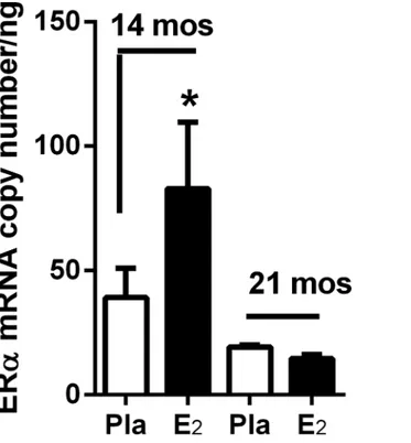
To determine the time course of age-related loss of 17β-estradiol (E2)-stimulated glomerular
ER expression regulation, we replaced E2for 2 months in 12-month old (pre-anestrous) and
19-month old (anestrous) female mice. All mice were ovariectomized two weeks prior to E2
administration to ensure equivalent replacement. E2replacement was only effective in up
regu-lating glomerular ERαmRNA expression in 12-month old mice prior to entering the anestrous period (at approximately 18 months of age), and thus correlating with a shorter exposure to endogenous oxidant stress (Fig 1). By 21 months of age (anestrous) E2replacement failed to
upregulate ERαmRNA expression (Fig 1).
Effect of pyridoxamine and 17
β
-estradiol replacement on body, kidney,
uterine weight and albumin/creatinine ratio
Treatment of mice did not alter body weight, however, kidney weight increased in Pyr+E2
treatment compared to placebo and Pyr alone (p<0.05). Uterine weight as a marker of
estro-gen replacement was increased in all mice receiving E2regardless of whether they were also
Inhibition of AGE accumulation with pyridoxamine and 17
β
-estradiol
increases glomerular ER
α
mRNA expression and reduces TGF
β
mRNA
expression
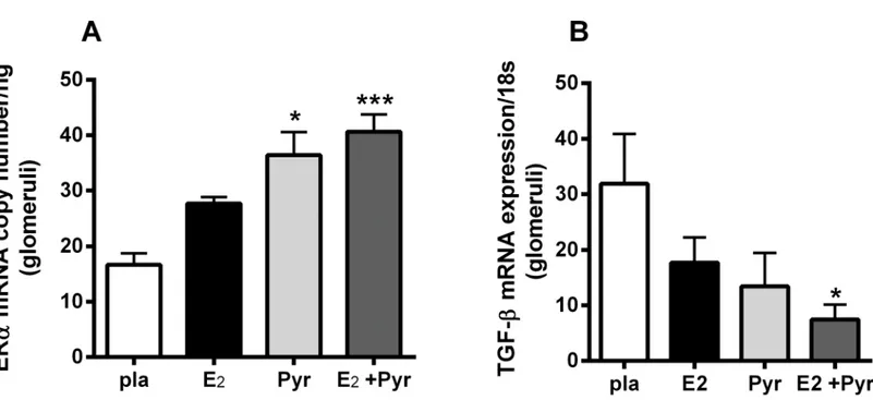
Our previous study showed an oxidant stress-related glomerular ERαdownregulation associ-ated with aging [6]. In this study,in vivoinhibition of AGEs, a source of oxidant stress, with Pyr and E2administration increased ERαmRNA expression (Fig 2A) in 21 month-old
ovariec-tomized female mice. TGFβ, a profibrotic cytokine, was decreased in an inverse manner to ERα
mRNA expression in the group receiving Pyr and E2(Fig 2B). There was no significant
Fig 1. Glomerular ERαmRNA upregulation by 17β-estradiol replacement is lost by anestrous period (21 months of age).ERαmRNA copy number was determined by real-time-PCR of glomeruli isolated from female C57BL6 mice ovariectomized at 12-, and 19-months of age (n = 5/group). E2was administered within 2 weeks of ovariectomy and mice sacrificed at 14 and 21 months. Data are graphed as mean±SEM.
*p<0.05, compared with placebo.
doi:10.1371/journal.pone.0159666.g001
Table 1. Effect of pyridoxamine and 17β-estradiol replacement on body, kidney, uterine weight and albumin/creatinine ratio.
Pla (n = 10) E2(n = 12) Pyr (n = 10) Pyr+E2(n = 6)
Body weight (g) 31±1.3 30±0.9 31±1.5 30±1.4
Kidney weight (g) 0.28±0.01 0.29±0.01 0.27±0.009 0.34±0.2a
Uterine weight (g) 0.02±0.00b 0.14±0.02c 0.02±0.001d 0.15±0.02
Albumin/Creatinine ratio 0.43±0.32 0.26±0.06 0.34±0.06 0.25±0.05
a*p<0.05 compared to placebo (pla) and pyridoxamine (Pyr) b***p<0.005 compared to 17β-estradiol (E2) and Pyr+E2 c***p<0.005 compared to Pyr
d***p<0.005 compared to Pyr+E2.
difference in ERαor TGFβmRNA expression between placebo group and mice receiving either Pyr or E2alone.
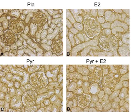
Type IV collagen deposition decreases with pyridoxamine and 17
β
-estradiol treatment in aged estrogen-deficient female mice
Type IV collagen, one of the hallmarks of glomerulosclerosis, increased in placebo-treated glo-meruli of ovariectomized aged female mice as expected (3+ staining;Fig 3A). Treatment with the antioxidant pyridoxamine decreased the accumulation of type IV collagen in glomeruli and tubules (1 and 2+ staining;Fig 3C). E2replacement, with or without pyridoxamine, also
pre-vented accumulation of type IV collagen in estrogen-deficient (ovariectomized) aged female mice (1 and 2+ staining;Fig 3B–3D).
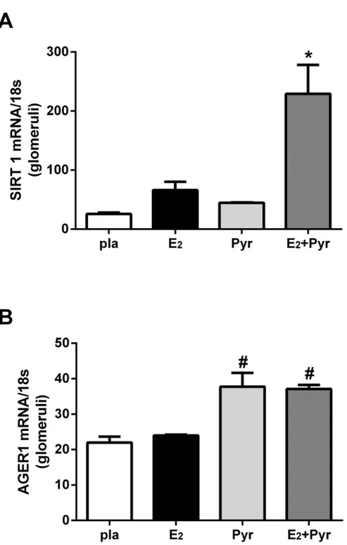
Prevention of AGE accumulation with pyridoxamine in the presence of
E2 replacement increases glomerular SIRT1 and AGER1 mRNA
AGE accumulation down-regulates anti-oxidant stress defenses such as SIRT1 and AGER1 [8,
18]. Therefore, we measured glomerular SIRT1 and AGER1 mRNA expression in our 4 groups of ovariectomized 21-month old female mice. Glomerular expression of SIRT1 mRNA was increased in mice treated with pyridoxamine and E2replacement compared to all other groups
(Fig 4A,p<0.05). Similarly, AGER1 mRNA expression was increased in the glomeruli of
mice receiving pyridoxamine and E2replacement compared to placebo or E2alone groups (Fig
4B, #p<0.05). AGER1 expression also increased in mice treated with pyridoxamine alone
(Pyr) versus placebo or E2alone (Fig 4B, #p<0.05).
Fig 2. 17β-estradiol (E2) combined with anti-AGE treatment pyridoxamine (Pyr) increases ER and decreases TGF mRNA in glomeruli of aged
ovariectomized female mice.21 month-old ovariectomized female C57/B6 mice were treated with either placebo (pla), 17-estradiol (E2), pyridoxamine (Pyr) or E2+ Pyr for 2 months. Real time-PCR for ER and TGF were performed as described in methods. Data are graphed as mean±SEM.*p<0.05 compared to pla,***p<0.005 compared to pla, # p<0.05 compared to pla. n = 5–7 mice/group.
Mesangial cells isolated from aged female mice treated with Pyr + E2
maintain a phenotypic switch with increased ER
α
, SIRT1 and AGER1
mRNA expression
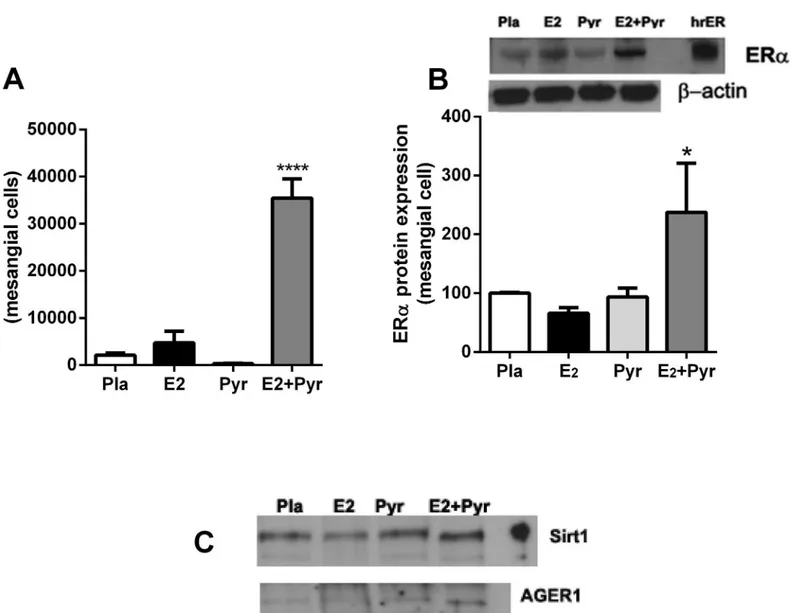
At the time of sacrifice, glomeruli were isolated and cells propagated from the four groups of mice described above. ERαmRNA copy number and protein expression was increased only in mesangial cells isolated from mice that were treated with both Pyr and E2(Fig 5A and 5B).
Sim-ilarly, we found an increase in SIRT1 and AGER1 protein expression in cells derived from mice treated with Pyr + E2(Fig 5C).
AGEs reduce glomerular ER
α
protein expression
in vitro
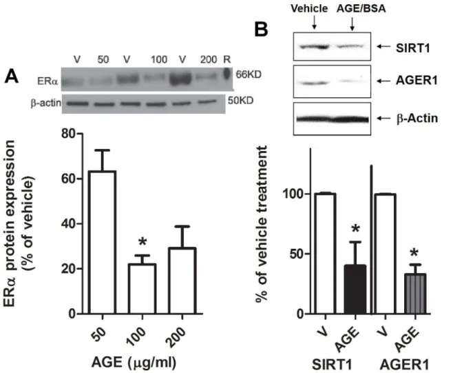
To further confirm that AGEs reduce glomerular ERαexpression, mesangial cells isolated from young female mice were treated with AGEsin vitro. ERαprotein expression was decreased after treatment with AGE-BSA (Fig 6A). There was also a decrease in SIRT1 and AGER1 pro-tein expression in these cells (Fig 6B).
Discussion
We have previously shown that E2upregulates glomerular ERαmRNA and protein expression
in young mice [28], but during aging there is a steady decline in both [6]. In the present study, we demonstrate that timing of estrogen replacement in relation to reproductive age is critical for regulation of glomerular ER expression. E2replacement at 14 months (before anestrus) was
effective in upregulating ERα. This effect, however, was lost by 21 months of age coinciding with the anestrus period and prolonged exposure to oxidant stress. These data derived in
Fig 3. Age-related glomerular collagen deposition is reduced after pyridoxamine (Pyr) and 17β -estradiol treatment.Collagen type IV deposition is increased in ovariectomized old female C57/B6 mice (placebo control, panel A). 17β-estradiol (E2) treatment (panels B and D) prevented collagen accumulation, particularly in combination with pyridoxamine (Pyr) treatment (panel D). All mice were rendered estrogen deficient by ovariectomy at 19 months of age. Images are representative of staining that was performed on at least five mice per group. Magnification, 40x.
experimental animals may provide insight into the findings of the Women’s Health Initiative (WHI) and Heart and Estrogen/Progestin Replacement Study (HERS). In those trials, women that received estrogen replacement up to 10 years after menopause exhibited some adverse clinical outcomes. The KEEPs trial, on the other hand, studied women not more than three
Fig 4. Glomerular AGER1 and SIRT-1 mRNA are upregulated by reduction of AGEsin vivo.Glomeruli were isolated from 4 groups of mice; placebo (pla), 17β-estradiol (E2), pyridoxamine (Pyr) or E2+ Pyr. SIRT1, AGER1 and 18s were measured by RT-PCR as described in Methods. Data are graphed as mean±SEM of ratio of SIRT1/18s (*p<0.05 compared to all groups) or AGER1/18s (#p<05 compared to placebo and E2 treatments). n = 5/group.
years post menopause and suggested that this window of time for initiation of hormone replacement may lead to a beneficial effect for disease prevention [32]. Our previous data showed that increased oxidant stress is associated with reduced ERαexpression in the kidney of aging mice [6]. Therefore, it is possible that administration of estrogen during this time of increased age-related oxidant stress leading to decrease in ER expression and action may exac-erbate downstream deleterious events.
Based on our previous findings, we designed the current study to further investigate the role of oxidant stress and regulation of glomerular ERαexpressionin vivo. We examined the effect on glomerular ERαexpression of pyrydoxamine, a derivative of vitamin B6that prevents
intra-cellular accumulation of AGEs and scavenges reactive oxygen species [33]. Pyridoxamine treat-ment coupled with E2replacement increased glomerular ERαexpression, while E2replacement
alone did not. Furthermore, ERαexpression in mesangial cells isolated fromin vivotreated
Fig 5. Mesangial cells isolated from aged female mice treated with pyridoxamine and 17β-estradiol maintain a phenotypic switch with increased ERα, SIRT1 and AGER1 mRNA expression. A)Mesangial cell ERαmRNA copy number was measured on isolates by RT-PCR as described in Methods.
****p<0.0001 compared to all groups.B + C)Mesangial cell protein expression of ERα(Fig 5B), SIRT-1 and AGER1 (Fig 5C) was measured by western blot from each group of treated mice. Shown is a representative Western blot andβ-actin loading control. For ERαdata are graphed as mean±SEM % of placebo control.*p<0.05 compared to all groups. n = 2 isolates/group.
mice followed a similar expression pattern as in the glomeruli. This was expected, as we have previously reported that a phenotypic switch in glomerular ERαexpression occurringin vivois maintainedin vitro[25,34].
Aged female mice (24 months of age and older) have increased urinary albumin excretion and collagen types I and IV deposition leading to glomerulosclerosis [23]. This increase in glo-merulosclerosis markers associated with age can be observed in experimental models and humans [23] [35,36]. Although baseline urinary albumin excretion was higher in our aged female mice compared to young female mice (data not shown), this was not affected by treat-ment with pyridoxamine and/or E2in aged ovariectomized female mice. It is possible that
pro-longed treatment period and sacrifice at an older age may have revealed an effect. In contrast, all treatment combinations prevented glomerular type IV collagen deposition in aged females. Of note, despite the effectiveness of oral pyridoxamine in preserving kidney function in type 1 and 2 diabetic rat and mouse models [37–39], recent clinical trials in patients with type 1 and type 2 diabetes produced mixed results [40,41]. Williams et al. [40] showed a reduction of baseline serum creatinine without a change in urine albumin excretion. A larger study failed to
Fig 6. Advanced glycation end products (AGEs) suppress ER protein levels in mesangial cells: A)Western analysis of ERα
protein expression in mesangial cells isolated from young female mice treated with increasing doses of AGE-BSA. Density data show % of control BSA (or vehicle, V) and graphed as mean±SEM,*p<0.05 compared to 50μg/ml. B) Western blot of SIRT1, AGER1 and -actin in mesangial cells from young female mice treated with 100μg/ml of AGE-BSA for 24 hours. Density data graphed as
mean±SEM % of BSA (vehicle or V).*p<0.05 compared to vehicle, n = 6.
show any change in renal function after 1 year, although the authors suggested that patients with less severe renal damage may respond to the drug [41].
In the present study,in vivopyridoxamine treatment along with E2replacement decreased TGFβmRNA expression in kidneys of aged ovariectomized female mice. Accumulation of gene expression of growth factors and cytokines such as TGFβand vascular endothelial growth factor (VEGF) are associated with the formation of AGEs [42]. We and others have shown that kidney disease in mice and humans is often associated with increased TGFβexpression [26,
43–45]. In fact, TGFβsignaling can be initiated by reactive oxygen species, which could ulti-mately increase extracellular matrix protein (ECM) accumulation through direct upregulation of collagen synthesis and/or decreased matrix metalloproteinase activity. In addition, TGF-β1 contributes to glomerulosclerosis by stimulating podocyte apoptosis [44,46]. Finally, TGF-β
receptor 2 is increased in isolated mesangial cells and in glomeruli of diabetic mice, suggesting an increased sensitivity due to the effects of endogenous TGF-β1 [47,48]. Interestingly, there was an inverse relationship in our study between the NAD+-dependent deacetylase SIRT1 and TGFβexpression. Negative cross-talk between TGFβsignaling and SIRT has been previously demonstrated in the kidney, liver, and lung [49–51]. SIRTs have been shown to downregulate TGFβeither by degradation or inhibition of transcriptional activity and further studies are ongoing in our laboratory to understand these findings.
SIRT1 and ERαexpression were positively correlated in both glomeruli and mesangial cells. We postulate that SIRT may have a direct effect on ER regulation. Estrogen receptors are dynamically modulated by post-translational modification, i.e. phosphorylation, methylation, acetylation, ubiquitination, or sumoylation [52]. For instance, hyperactivation of ERK/MAPK (Extracellular-signal-regulated kinases/Mitogen-activated protein kinases) causes functional repression of ER transcription through NFkB activation [53,54], which we have shown to be increased in 28-month old female mice [22]. In contrast, SIRT1 prevents undue activation of NFkB [55,56]. AGEs promote NFkB activation [57] but suppress SIRT1 and its deacetylase activity on NFkB-p65 [58]. This could influence ER transcription, given that decreased SIRT1 expression can disrupt the basal transcription factor complex of ERαpromoter in some cells [20]. These studies are currently under investigation.
The concentration of AGEs and their cross-linked products increases with aging and leads to higher basal levels of oxidant stress [10,59]. Importantly, levels of AGEs are elevated in post-menopausal women compared to healthy young women. This increase is more pro-nounced in diabetic post-menopausal women [3–5]. These data correlate with the higher female to male ratio in patients with diabetic end-stage renal disease, which increases sharply in the postmenopausal age groups [60]. To confirm ourin vivodata suggesting an important role for AGEs in regulation of glomerular ERαexpression in aged females, we examined the direct effects of AGEsin vitro. Mesangial cells isolated from young (estrogen replete) female mice were treated with increasing concentrations of AGEs. We observed a dose- and time-dependent reduction in ERαexpression in response to AGEs. Similarly, levels of the major cel-lular anti-AGE/oxidant stress defenses, anti-AGE receptor AGER1 and SIRT1 protein expres-sion were decreased in response to AGE. This correlates with the inverse relationship between SIRT1/AGER1 and AGEs both in the current study and other experimental and human studies [2,8,61].
Acknowledgments
The authors would like to thank Dr. Helen Vlassara for her guidance and support in this study. All authors have no financial conflicts of interest to disclose.
Author Contributions
Conceived and designed the experiments: SJE GES. Performed the experiments: SP XX WC RC. Analyzed the data: SP GR SJE. Contributed reagents/materials/analysis tools: SJE GES. Wrote the paper: SJE GR.
References
1. Lee HC, Wei YH. Oxidative stress, mitochondrial DNA mutation, and apoptosis in aging. ExpBiolMed (Maywood). 2007; 232(5):592–606.
2. Cai W, He JC, Zhu L, Chen X, Wallenstein S, Striker GE, et al. Reduced oxidant stress and extended lifespan in mice exposed to a low glycotoxin diet: association with increased AGER1 expression. AmJ-Pathol. 2007; 170(6):1893–902.
3. Loft S, Vistisen K, Ewertz M, Tjonneland A, Overvad K, Poulsen HE. Oxidative DNA damage estimated by 8-hydroxydeoxyguanosine excretion in humans: influence of smoking, gender and body mass index. Carcinogenesis. 1992; 13(12):2241–7. PMID:1473230
4. Uribarri J, Cai W, Peppa M, Goodman S, Ferrucci L, Striker G, et al. Circulating glycotoxins and dietary advanced glycation endproducts: two links to inflammatory response, oxidative stress, and aging. J GerontolA BiolSci MedSci. 2007; 62(4):427–33.
5. Helmersson J, Mattsson P, Basu S. Prostaglandin F(2alpha) metabolite and F(2)-isoprostane excretion rates in migraine. ClinSci (Lond). 2002; 102(1):39–43.
6. Pereira-Simon S, Xia X, Catanuto P, Elliot S. Oxidant Stress and Mitochondrial Signaling Regulate Reversible Changes of ERa Expression and Apoptosis in Aging Mouse Glomeruli and Mesangial Cells. Endocrinology. 2012; 153((11)):5491–9. doi:10.1210/en.2012-1379PMID:23027807
7. Koschinsky T, He CJ, Mitsuhashi T, Bucala R, Liu C, Buenting C, et al. Orally absorbed reactive glyca-tion products (glycotoxins): an environmental risk factor in diabetic nephropathy. ProcNatlAcadSci USA. 1997; 94(12):6474–9.
8. Cai W, Ramdas M, Zhu L, Chen X, Striker GE, Vlassara H. Oral advanced glycation endproducts (AGEs) promote insulin resistance and diabetes by depleting the antioxidant defenses AGE receptor-1 and sirtuin 1. Proc NatlAcadSciUSA. 2012.
9. Vlassara H, Cai W, Goodman S, Pyzik R, Yong A, Chen X, et al. Protection against loss of innate defenses in adulthood by low advanced glycation end products (AGE) intake: role of the antiinflamma-tory AGE receptor-1. J Clin Endocrinol Metab. 2009; 94(11):4483–91. doi:10.1210/jc.2009-0089 PMID:19820033
10. Vlassara H, Striker GE. AGE restriction in diabetes mellitus: a paradigm shift. Nat Rev Endocrinol. 2011; 7(9):526–39. doi:10.1038/nrendo.2011.74PMID:21610689
11. Cai W, Gao QD, Zhu L, Peppa M, He C, Vlassara H. Oxidative stress-inducing carbonyl compounds from common foods: novel mediators of cellular dysfunction. Mol Med. 2002; 8(7):337–46. PMID: 12393931
12. Brands CM, Alink GM, van Boekel MA, Jongen WM. Mutagenicity of heated sugar-casein systems: effect of the Maillard reaction. J AgricFood Chem. 2000; 48(6):2271–5.
13. Finot PA. Historical perspective of the Maillard reaction in food science. AnnN YAcadSci. 2005; 1043:1–8.
14. Pouillart P, Mauprivez H, it-Ameur L, Cayzeele A, Lecerf JM, Tessier FJ, et al. Strategy for the study of the health impact of dietary Maillard products in clinical studies: the example of the ICARE clinical study on healthy adults. AnnN YAcadSci. 2008; 1126:173–6.
15. Goldberg T, Cai W, Peppa M, Dardaine V, Baliga BS, Uribarri J, et al. Advanced glycoxidation end prod-ucts in commonly consumed foods. J Am DietAssoc. 2004; 104(8):1287–91.
16. Uribarri J, Tuttle KR. Advanced glycation end products and nephrotoxicity of high-protein diets. ClinJ Am Soc Nephrol. 2006; 1(6):1293–9.
18. Uribarri J, Cai W, Ramdas M, Goodman S, Pyzik R, Chen X, et al. Restriction of advanced glycation end products improves insulin resistance in human type 2 diabetes: potential role of AGER1 and SIRT1. Diabetes Care. 2011; 34(7):1610–6. doi:10.2337/dc11-0091PMID:21709297
19. Nogueiras R, Habegger KM, Chaudhary N, Finan B, Banks AS, Dietrich MO, et al. Sirtuin 1 and Sirtuin 3: Physiological Modulators of Metabolism. Physiological Reviews. 2012; 92(3):1479–514. doi:10. 1152/physrev.00022.2011PMID:22811431
20. Yao Y, Li H, Gu Y, Davidson NE, Zhou Q. Inhibition of SIRT1 deacetylase suppresses estrogen recep-tor signaling. Carcinogenesis. 2010; 31(3):382–7. doi:10.1093/carcin/bgp308PMID:19995796 21. Moore RL, Dai Y, Faller DV. Sirtuin 1 (SIRT1) and steroid hormone receptor activity in cancer. Journal
of Endocrinology. 2012; 213(1):37–48. doi:10.1530/JOE-11-0217PMID:22159506
22. Zheng F, Cheng QL, Plati AR, Ye SQ, Berho M, Banerjee A, et al. The glomerulosclerosis of aging in females: contribution of the proinflammatory mesangial cell phenotype to macrophage infiltration. AmJ-Pathol. 2004; 165(5):1789–98.
23. Zheng F, Plati AR, Potier M, Schulman Y, Berho M, Banerjee A, et al. Resistance to glomerulosclerosis in B6 mice disappears after menopause. AmJPathol. 2003; 162(4):1339–48.
24. Elliot SJ, Berho M, Korach K, Doublier S, Lupia E, Striker GE, et al. Gender-specific effects of endoge-nous testosterone: Female alpha-estrogen receptor-deficient C57Bl/6J mice develop glomerulosclero-sis. Kidney Int. 2007; 72(4):464–72. PMID:17495854
25. Karl M, Berho M, Pignac-Kobinger J, Striker GE, Elliot SJ. Differential effects of continuous and intermit-tent 17beta-estradiol replacement and tamoxifen therapy on the prevention of glomerulosclerosis: mod-ulation of the mesangial cell phenotype in vivo. AmJPathol. 2006; 169(2):351–61.
26. Elliot SJ, Karl M, Berho M, Xia X, Pereria-Simon S, Espinosa-Heidmann D, et al. Smoking induces glo-merulosclerosis in aging estrogen-deficient mice through cross-talk between TGF-beta1 and IGF-I sig-naling pathways. JAmSocNephrol. 2006; 17(12):3315–24.
27. C P., Doublier S, Fornoni A, Lupia E, Berho M, Striker GE, et al. 17b-estradiol and Tamoxifen upregu-late estrogen receptor â and reguupregu-late podocyte signaling pathways in a model of type 2 diabetes. Kid-ney International. 2009; 75:1194–201. doi:10.1038/ki.2009.69PMID:19279558
28. Potier M, Elliot SJ, Tack I, Lenz O, Striker GE, Striker LJ, et al. Expression and regulation of estrogen receptors in mesangial cells: influence on matrix metalloproteinase-9. JAmSocNephrol. 2001; 12 (2):241–51.
29. MacKay K, Striker LJ, Elliot S, Pinkert CA, Brinster RL, Striker GE. Glomerular epithelial, mesangial, and endothelial cell lines from transgenic mice. Kidney Int. 1988; 33(3):677–84. PMID:2835539. 30. Cai W, He JC, Zhu L, Lu C, Vlassara H. Advanced glycation end product (AGE) receptor 1 suppresses
cell oxidant stress and activation signaling via EGF receptor. Proc NatlAcadSci USA. 2006; 103 (37):13801–6.
31. Potier M, Karl M, Zheng F, Elliot SJ, Striker GE, Striker LJ. Estrogen-Related Abnormalities in Glomeru-losclerosis-Prone Mice:Reduced Mesangial Cell Estrogen Receptor Expression and Prosclerotic Response to Estrogens. Am J Path. 2002; 160:1877–85. PMID:12000739
32. Wharton W, Gleason CE, Miller VM, Asthana S. Rationalle and design of the Kronos Early Estrogen Prevention Study (KEEPS) and the KEEPS cognitive and affective sub study (KEEPS Cog). Brain Research. (0: ).
33. Kang Z, Li H, Li G, Yin D. Reaction of pyridoxamine with malondialdehyde: mechanism of inhibition of formation of advanced lipoxidation end-products. AminoAcids. 2006; 30(1):55–61.
34. Elliot SJ, Striker LJ, Hattori M, Yang CW, He CJ, Peten EP, et al. Mesangial cells from diabetic NOD mice constitutively secrete increased amounts of insulin-like growth factor-I. Endocrinology. 1993; 133 (4):1783–8. PMID:7691581
35. Eikmans M, Baelde HJ, de Heer E, Bruijn JA. Effect of age and biopsy site on extracellular matrix mRNA and protein levels in human kidney biopsies. Kidney Int. 2001; 60(3):974–81. PMID:11532092 36. Kalant N, Satomi S, White R, Tel E. Changes in renal glomerular basement membrane with age and
nephritis. CanJ Biochem. 1977; 55(12):1197–206.
37. Alderson NL, Chachich ME, Frizzell N, Canning P, Metz TO, Januszewski AS, et al. Effect of antioxi-dants and ACE inhibition on chemical modification of proteins and progression of nephropathy in the streptozotocin diabetic rat. Diabetologia. 2004; 47(8):1385–95. PMID:15309289
38. Alderson NL, Chachich ME, Youssef NN, Beattie RJ, Nachtigal M, Thorpe SR, et al. The AGE inhibitor pyridoxamine inhibits lipemia and development of renal and vascular disease in Zucker obese rats. Kid-ney Int. 2003; 63(6):2123–33. PMID:12753299
40. Williams ME, Bolton WK, Khalifah RG, Degenhardt TP, Schotzinger RJ, McGill JB. Effects of pyridox-amine in combined phase 2 studies of patients with type 1 and type 2 diabetes and overt nephropathy. Am J Nephrol. 2007; 27(6):605–14. PMID:17823506
41. Lewis EJ, Hunsicker LG, Rodby RA. A clinical trial in type 2 diabetic nephropathy. Am J Kidney Dis. 2001; 38(4 Suppl 1):S191–S4.
42. Kovacic P, Somanathan R. Cell signaling and receptors in toxicity of advanced glycation end products (AGEs): alpha-dicarbonyls, radicals, oxidative stress and antioxidants. J ReceptSignalTransductRes. 2011; 31(5):332–9.
43. Blush J, Lei J, Ju W, Silbiger S, Pullman J, Neugarten J. Estradiol reverses renal injury in Alb/TGF-beta1 transgenic mice. Kidney Int. 2004; 66(6):2148–54. PMID:15569304
44. Schiffer M, Bitzer M, Roberts IS, Kopp JB, ten Dijke P, Mundel P, et al. Apoptosis in podocytes induced by TGF-beta and Smad7. JClinInvest. 2001; 108(6):807–16.
45. Schnaper HW, Jandeska S, Runyan CE, Hubchak SC, Basu RK, Curley JF, et al. TGF-beta signal transduction in chronic kidney disease. Front Biosci. 2009; 14:2448–65.
46. Poncelet AC, Schnaper HW. Regulation of human mesangial cell collagen expression by transforming growth factor-beta1. AmJPhysiol. 1998; 275(3 Pt 2):F458–66.
47. Cohen MP, Sharma K, Guo J, Eltayeb BO, Ziyadeh FN. The renal TGF-beta system in the db/db mouse model of diabetic nephropathy. ExpNephrol. 1998; 6(3):226–33.
48. Goldfarb S, Ziyadeh FN. TGF-beta: a crucial component of the pathogenesis of diabetic nephropathy. TransAmClinClimatolAssoc. 2001; 112:27–32.
49. Casalena G, Daehn I, Bottinger E. Transforming Growth FactorB, Bioenergetics, and Mitochondria in Renal Disease. Seminars in Nephrology. 2012; 32(3):295–303. doi:10.1016/j.semnephrol.2012.04. 009PMID:22835461
50. Kume S, Haneda M, Kanasaki K, Sugimoto T, Araki S, Isshiki K, et al. SIRT1 inhibits transforming growth factor beta-induced apoptosis in glomerular mesangial cells via Smad7 deacetylation. The Jour-nal of biological chemistry. 2007; 282(1):151–8. doi:10.1074/jbc.M605904200PMID:17098745. 51. Minagawa S, Araya J, Numata T, Nojiri S, Hara H, Yumino Y, et al. Accelerated epithelial cell
senes-cence in IPF and the inhibitory role of SIRT6 in TGF-beta-induced senessenes-cence of human bronchial epi-thelial cells. American journal of physiology Lung cellular and molecular physiology. 2011; 300(3): L391–401. doi:10.1152/ajplung.00097.2010PMID:21224216; PubMed Central PMCID: PMC3284316.
52. Yang XJ, Seto E. Lysine Acetylation: Codified Crosstalk with Other Posttranslational Modifications. Molecular Cell. 2008; 31(4):449–61. doi:10.1016/j.molcel.2008.07.002PMID:18722172
53. Oh AS, Lorant LA, Holloway JN, Miller DL, Kern FG, El Ashry D. Hyperactivation of MAPK induces loss of ERalpha expression in breast cancer cells. MolEndocrinol. 2001; 15(8):1344–59.
54. Holloway JN, Murthy S, El-Ashry D. A cytoplasmic substrate of mitogen-activated protein kinase is responsible for estrogen receptor-alpha down-regulation in breast cancer cells: the role of nuclear fac-tor-kappaB. Mol Endocrinol. 2004; 18(6):1396–410. PMID:15056731
55. Kawahara TL, Michishita E, Adler AS, Damian M, Berber E, Lin M, et al. SIRT6 links histone H3 lysine 9 deacetylation to NF-kappaB-dependent gene expression and organismal life span. Cell. 2009; 136 (1):62–74. doi:10.1016/j.cell.2008.10.052PMID:19135889
56. Yeung F, Hoberg JE, Ramsey CS, Keller MD, Jones DR, Frye RA, et al. Modulation of NF-kappaB-dependent transcription and cell survival by the SIRT1 deacetylase. EMBO J. 2004; 23(12):2369–80. PMID:15152190
57. Lu C, He JC, Cai W, Liu H, Zhu L, Vlassara H. Advanced glycation endproduct (AGE) receptor 1 is a negative regulator of the inflammatory response to AGE in mesangial cells. Proceedings of the National Academy of Sciences of the United States of America. 2004; 101(32):11767–72. PMID:15289604 58. Guarente L. Sirtuins, Aging, and Medicine. New England Journal of Medicine. 2011; 364(23):2235–44.
doi:10.1056/NEJMra1100831PMID:21651395
59. Turgut F, Bolton WK. Potential new therapeutic agents for diabetic kidney disease. American journal of kidney diseases: the official journal of the National Kidney Foundation. 2010; 55(5):928–40. doi:10. 1053/j.ajkd.2009.11.021PMID:20138415.
60. Yu M. Gender differences in chronic kidney disease and progression in type 2 diabetes. Journal of American Society of Nephrology. 2011.