Amyotrophic lateral sclerosis
Considerations on diagnostic criteria
Marco A. Chieia, Acary S.B. Oliveira, Helga C.A. Silva, Alberto Alain Gabbai
ABSTRACT
Amyotrophic lateral sclerosis (ALS) is a neurodegenerative disorder, compromising the motor neuron, characterized by progressive muscle weakness, with reserved prognosis. The diagnosis is based on inclusion and exclusion clinical criteria, since there is no specific confirmation test. The objective of this research is to critically examine the main diagnosis instrument - El Escorial revisited, from the World Federation of Neurology (1998). Of the 540 patients with initial ALS diagnosis, either probable or definite, seen at UNIFESP-EPM, 190 underwent thorough investigation, following regular clinical and therapeutic treatment for over two years. Thirty patients (15.78%) had their diagnosis completely changed. The false-positive diagnoses were related to: early age, clinical presentation of symmetry, weakness greater than atrophy, symptomatic exacerbation. In addition, three patients with myasthenia gravis developed framework for ALS, suggesting the post-synaptic disability as a sign of early disease.
Key words: amyotrophic lateral sclerosis, correct diagnosis, El Escorial revisited.
Esclerose lateral amiotrófica: considerações sobre critérios diagnósticos
RESUMO
Esclerose lateral amiotrófica (ELA) é uma doença neurodegenerativa, que compromete o neurônio motor, caracterizada por fraqueza muscular progressiva, com prognóstico reservado. O diagnóstico é baseado na inclusão e exclusão de critérios clínicos, uma vez que não existe um teste de confirmação específica. O objetivo desta pesquisa é analisar criticamente o instrumento de diagnóstico principal - El Escorial revisited, da Federação Mundial de Neurologia (1998). Dos 540 pacientes com diagnóstico inicial de ELA, seja provável ou definitiva, vistos pela UNIFESP-EPM, 190 foram submetidos a investigação aprofundada, após tratamento clínico e terapêutico regular há mais de dois anos. Trinta pacientes (15,78%) tiveram seu diagnóstico mudado completamente. Os diagnósticos falso-positivos foram relacionados à idade precoce, a apresentação clínica da simetria, a fraqueza superior a atrofia, exacerbação sintomática. Além disso, três pacientes com miastenia gravis desenvolveram quadro de ELA, sugerindo a lesão pós-sináptica como um sinal precoce da doença.
Palavras-chave: esclerose lateral amiotrófica, diagnóstico correto, El Escorial revisited.
Correspondence Marco Antonio Troccoli Chieia Rua Mairinque 267
04037-020 - São Paulo SP - Brasil E-mail: [email protected]
Received 27 January 2010 Received in final form 11 May 2010 Accepted 18 May 2010
Neuromuscular Disorders Unit, Department of Neurology and Neurosurgery, Escola Paulista de Medicina, Federal University of São Paulo, São Paulo SP, Brazil.
Motor neuron diseases (MND) con-stitute a group of neurodegenerative dis-orders characterized by loss of neurons in the motor cortex, brainstem, and ven-tral horn of the spinal cord. The clinical presentation depends on the involvement of upper motor neurons – UMN (weak-ness, spasticity and hyperrelexia) or low-er motor neurons – LMN (weakness,
The disorders include: progressive muscle atrophy (PMA), represented by a pure involvement of LMN; pri-mary lateral sclerosis, characterized by progressive in-volvement of pyramidal tract; progressive bulbar palsy (PBP), deined as the onset in the bulbar region; amyo-trophic lateral sclerosis (ALS), as described by Charcot in 1874, showing deterioration of UMN and LMN.
he latter accounts for the most frequent clinical pre-sentation (80% of total cases) and as a result many cen-ters use the name ALS for the diseases with impairment of motor neuron2.
he causes of motor neuron injury in MND/ALS re-main incompletely understood. here are several mech-anisms that may be involved in its development and a multifactorial disease is considered. he mechanisms re-lated to ALS include the toxic efects caused by the mu-tation of superoxide desmutase13, inclusion of abnormal protein aggregation, intermediary ilaments disorganiza-tion, anterograde and retrograde axonal transport change, microglial activation, excitotoxicity mediated by gluta-mate, abnormalities in regulation of intracellular calci-um and others.
he diagnosis is based on clinical aspects. herefore, we do not have a real biomarker for MND diagnosis.
Diagnosis
For many years, the only published criteria for the recognition of MND / ALS were done by Lambert, es-tablished through electroneuromyography (ENMG)4. In 1990, guidelines set by diferent researchers have been in-corporated within the diagnostic criteria formulated by a Subcommittee on ALS of the World Federation of Neu-rology (WFN), which culminated in the meeting and edit-ing of the diagnostic criteria in El Escorial, Spain, in 19945. ALS diagnosis is deined within the evidence of signs of impairment of lower motor neuron, by means of clin-ical examination, electrophysiologclin-ical or neuropatholo-gycal changes, associated with clinically proven impair-ment of upper motor neuron, with chronic and progres-sive development. It is still necessary, for diagnosis, the absence of electrophysiological and pathological indings characteristic of other diseases that explain the degenera-tion of motor neurons, as well as changes in neuroimag-ing to justify the electrophysiological signals.
here has been, however, consensus among research-ers, since certain clinical pictures and, above all, certain electrophysiological indings left doubt about the com-pletion of diagnosis in some speciic situations. Gener-al neurologists and speciGener-alists in neuromuscular diseas-es claimed additional diicultidiseas-es for the necdiseas-essary ALS early diagnosis.
he diagnostic criteria in El Escorial were reformu-lated in 1998, at the World Federation of Neurology ALS
meeting in Airlie House, Warrenton, Virginia, U.S. his revised document, known as El Escorial Revisited, was published by the WFN-ALS on the Web, with an aim at reining the diagnosis6.
New methods of electrophysiology, neuroimaging, immunohistochemistry and genome analysis were add-ed for accuracy of diagnosis. he document El Escorial Revisited has been regarded as an important step to alle-viate the diiculties in producing the diagnosis.
In a recent meeting, held in Japan, researchers im-proved the diagnostic criteria and formulated “Awaji criteria”7. here was a reformulation of electromyogra-phy, adding fasciculation with signal of neuronal damage, and inclusion new methods of diagnosis with transcrani-al magnetic stimulation, voxel based morphometry and difusium tensor imaging.
Despite the diagnostic criteria established by the WFN, aided by more reined laboratory tests for diseas-es that mimic ALS, accompanied by recent advancdiseas-es in imaging techniques, incorrect diagnosis is not infrequent. In previous studies, 45% of ALS patients presented ini-tial wrong diagnosis and 25% of these were performed by neurologists.
In addition, there is a long waiting period between the outset of symptoms and the diagnosis. In an internation-al multicenter study named ISIS Survey, with 201 ELA-diagnosed patients, median time required to conirm di-agnosis was 14 months, with a 2-month waiting period for the irst appointment. It took 8 months for the irst appointment with the neurologist and it took another 4 months for neurologist’s diagnosis observation and re-evaluation8.
Systematic reviews of ALS patients, in the last de-cade, have shown 9 to 10% of false-positive ALS diag-nosis. hese, primarily included in the group of ALS at El Escorial Revisited, have unusual developments, with a break of the progression of chronic and progressive motor involvement and the appearance of signs and symptoms uncommon in clinical course, leading to a careful review diagnostic, pointing to other diseases such as multifocal motor neuropathy (MMN) and Kennedy disease9,10.
he objective of this study is a critical analysis of the ALS diagnostic criteria based on clinical presentations of patients with atypical presentation of motor neuron disease.
METHOD
he outpatient monitoring is carried out under reg-ular (at least quarterly) periodic consultations encom-passing multidisciplinary orientation therapy. To the best characterization of the goals, these patients’ medical re-cords were analyzed in retrospect, with an aim at espe-cially identifying the following medical conditions: pa-tients with ALS initial clinical diagnosis, with an illness history longer than two years, classiied as probable or de-ined ALS, with diferent clinical course from the initial diagnosis, then ruling out MND / ALS diagnosis. Patients characterized, initially, with other neuromuscular diseas-es, evolving, later on, to classic aspects of MND / ALS.
RESULTS
Of the 540 patients registered in the Clinic, 190
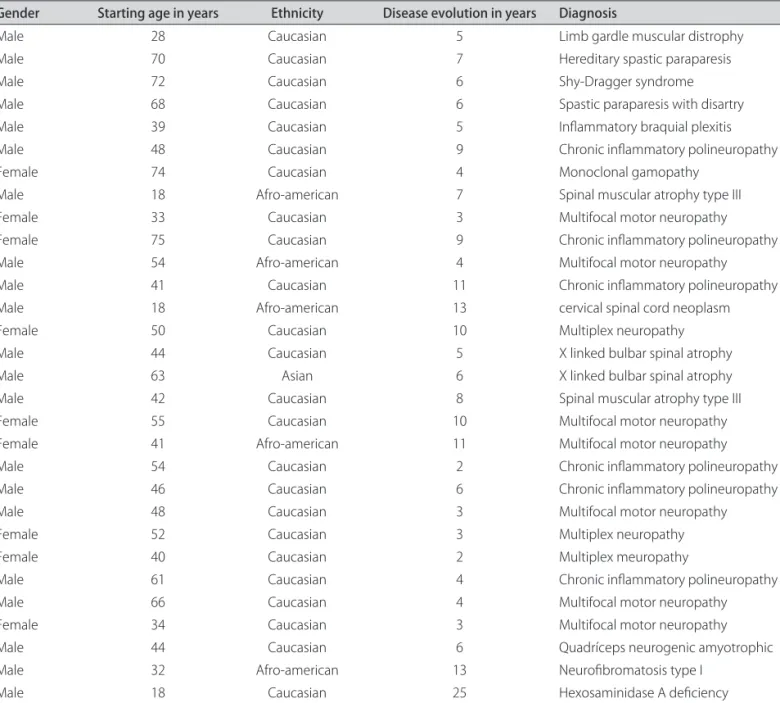
pa-tients met the MND / ALS diagnostic criteria, comple-mented with laboratory research in compliance with both research protocols and regular monitoring. hirty of these patients (15.78%) had their diagnosis complete-ly changed, during the clinical observation development period (Table 1).
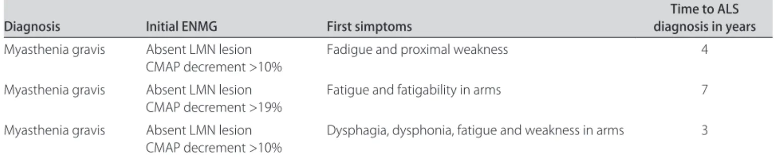
On the other hand, three patients with initial symp-toms and diagnosis of other neuromuscular diseases de-veloped features typical of MND / ALS during the clinical evolution. hey are male, aged between 55 and 58 years old, whose characteristics are described in Table 2.
he Motor Neuron Disease Unit is a reference ser-vice, and last months, has been receiving a great number of patients with deined ALS, which reduces the number of false negative diagnosis in this work.
Table 1. False-positive diagnosis: diagnosis review in patients who have amyotrophic lateral sclerosis with irst diagnosis in tandem with El Escorial revisited criteria.
Gender Starting age in years Ethnicity Disease evolution in years Diagnosis
Male 28 Caucasian 5 Limb gardle muscular distrophy
Male 70 Caucasian 7 Hereditary spastic paraparesis
Male 72 Caucasian 6 Shy-Dragger syndrome
Male 68 Caucasian 6 Spastic paraparesis with disartry
Male 39 Caucasian 5 Inlammatory braquial plexitis
Male 48 Caucasian 9 Chronic inlammatory polineuropathy
Female 74 Caucasian 4 Monoclonal gamopathy
Male 18 Afro-american 7 Spinal muscular atrophy type III
Female 33 Caucasian 3 Multifocal motor neuropathy
Female 75 Caucasian 9 Chronic inlammatory polineuropathy
Male 54 Afro-american 4 Multifocal motor neuropathy
Male 41 Caucasian 11 Chronic inlammatory polineuropathy
Male 18 Afro-american 13 cervical spinal cord neoplasm
Female 50 Caucasian 10 Multiplex neuropathy
Male 44 Caucasian 5 X linked bulbar spinal atrophy
Male 63 Asian 6 X linked bulbar spinal atrophy
Male 42 Caucasian 8 Spinal muscular atrophy type III
Female 55 Caucasian 10 Multifocal motor neuropathy
Female 41 Afro-american 11 Multifocal motor neuropathy
Male 54 Caucasian 2 Chronic inlammatory polineuropathy
Male 46 Caucasian 6 Chronic inlammatory polineuropathy
Male 48 Caucasian 3 Multifocal motor neuropathy
Female 52 Caucasian 3 Multiplex neuropathy
Female 40 Caucasian 2 Multiplex meuropathy
Male 61 Caucasian 4 Chronic inlammatory polineuropathy
Male 66 Caucasian 4 Multifocal motor neuropathy
Female 34 Caucasian 3 Multifocal motor neuropathy
Male 44 Caucasian 6 Quadríceps neurogenic amyotrophic
Male 32 Afro-american 13 Neuroibromatosis type I
DISCUSSION
Despite the recent reinement of the World Federa-tion of Neurology Consensus for ALS diagnosis, errone-ous diagnosis is not infrequent.
Even in an experienced ALS diagnostic service at the MND / ALS Clinic at UNIFESP-EPM, thirty patients out of a group of 190 with regular monitoring and laborato-ry research for complete characterization of ELA, have had their initial diagnosis changed over time. hese pa-tients’ clinical presentations display features that can help in ALS diagnosis, especially as to what regards a deini-tive diagnosis of this disease11.
Age
he age of onset of irst symptoms, usually at less than 30, increases the proportion of the diferential MND / ALS diagnosis. he early onset, represented by 5 of the 30 patients with false positive diagnosis, displayed genetic abnormalities, including deiciency of the enzyme hexo-saminidase A and B12, lack of expression of proteins of the cytoskeleton of cortical spinal tract in hereditary spas-tic paraplegia13, amendment of the suppressor gene ex-pression in the formation of neurinomas in the peripher-al nervous system in neuroibromatosis type I, or abnor-malities of muscle protein constituents of the cytoskele-ton such as disferlina.
Clinical forms of presentation
Unilateral/Bilateral – MND/ALS has, characteristi-cally, unilateral initial presentation, involving most of the time limb distal regions, where roots C8-T1 and L5-S1 are the most commonly afected, with ipsilateral or con-tralateral progression to the other roots, in a progressive and cumulative manner.
Forms with peculiar clinical presentation as the monomelic atrophy of Hirayama14, attended with atrophy of a limb, predominantly in the upper limbs, in youths, with self-progression or, in some cases, slow progres-sion15. In 12 (40%) of the 30 false-positive cases, we found unilateral atrophy, due to the speciic motor involvement linked to immunoglobulins, in the associated cases of the
multifocal motor neuropathy or monoclonal gammopa-thies16 related to autoimmune dyscrasies.
he involvement of bilateral and symmetrical limbs, at the beginning of the clinical picture, makes ALS diag-nosis less likely, suggesting a multirradicular pattern more often found in polirradiculopathies17 with inlammatory motor predominance.
Proximal/Distal – Predominance of involvement of MND / ALS is distal, then, beginning with proximal limb forms, leaves room for doubt in diagnosis, especially if there are no signs of pyramid release. he forms called “lail in arms” or brachial paraplegia and the legs paraplegia, as early symptoms, have predominance of proximal involve-ment and presents blurry forms of spinal muscular atrophy of the adult form, which have variability progression and diferent prognosis19. Kennedy’s disease, also called spinal bulbar atrophy, linked to the X chromosome20,21, with typ-ical symptoms of proximal atrophy associated with bulbar involvement, was found in two patients with false positive diagnosis in our study, and two other patients were diag-nosed with an adult form of spinal muscular atrophy, type III, all of whom required more follow-up, since they have, until now, 6 years of tracking with localized forms of in-volvement. One patient with chronic neurogenic quad-riceps amyotrophy22 and another one with spastic para-paresis with dysarthria13 may later develop MND / ALS.
he motor and distal involvement presents other dif-ferential diagnoses such as multifocal motor neuropathy, characterized by MMSS asymmetric distal involvement, distal myopathies, hereditary motor neuropathies, which can provide the clinical motor aspects in a given moment, similar to MND / ALS.
Acute/Subacute/Chronic – he evolutionary pattern that characterizes the MND/ALS presents, in most of its forms, a chronic progressive decrease of motor function, bodily involvement with sum of the territories, ranging, on average, between three and ive years.
he changes to this pattern, with acute or subacute evolution of motor deicits and stabilization of the dis-ease with acute relapses are characteristic of autoim-mune-driven diseases. Moreover, part of the slower
evo-Table 2. False-negative diagnosis (three patients).
Diagnosis Initial ENMG First simptoms diagnosis in years Time to ALS
Myasthenia gravis Absent LMN lesion CMAP decrement >10%
Fadigue and proximal weakness 4
Myasthenia gravis Absent LMN lesion CMAP decrement >19%
Fatigue and fatigability in arms 7
Myasthenia gravis Absent LMN lesion CMAP decrement >10%
Dysphagia, dysphonia, fatigue and weakness in arms 3
lution, with onset of signs and symptoms over years of evolution, attests to other forms involving the degenera-tive motor neuron.
he presentation of typical clinical aspects of spinal muscular atrophy can display a wide variability in its evo-lution as well as in its clinical presentation and are ex-cluded from their diagnosis, often for long periods with follow-up monitoring, and they may show signs of prog-ress and bulbar involvement in 20 to 30 years of the on-set of symptoms23.
More accentuated weakness than atrophy – ALS di-agnosis depends on clear evidence of both compromised lower motor neuron and upper motor neuron. he com-mitment of the LMN is clinically more easily identiiable through the presence of atrophy and fasciculations as well as electromyography analysis. We have evidence that UMN commitment is more unusual. Signs of pyramid re-lease are not always marked and, only recently, non-inva-sive tests to reveal cortical spinal tract commitment have been incorporated into the medical practice.
he MRI examination with sequences ST1 with MTC and transcranial magnetic stimulation (TMS) has iden-tiied abnormalities in the cortical spinal tract, thus be-coming a useful tool in ALS diagnosis26. In patients where there is no clinically deined involvement of motor tract, image and TMS analysis must be requested. In the ab-sence of abnormalities, ALS diagnosis should be placed in suspicion. Moreover, the atrophy of the motor cor-tex, with severe loss of grey substance (cell body) and its brain connections are highlighted by recent advances in radiology and proton emission through tomography (PET), which quantiies the metabolism of regional areas of motor cortex, associated with quantifying the volume by voxel, making early diagnoses possible27. Transcrani-al magnetic stimulation has become an auxiliary method for early diagnosis through the quantiication of the de-crease in speed within the motor central nervous system in patients with MND / ALS28.
Pyramidal signs and muscle atrophy – Character-istically in ALS, the signals of pyramidal release must be present at sites above the sites where the muscular at-rophy is evident. The presence of muscle atat-rophy and signs of pyramid release below the region of common cord nerve in the diagnosis, refer to a myelopathy, thus one should consider various causes, including spinal cord compression, tumor, syringomyelia, syringobulbia.
Temporal development – he abnormal temporal evolution, with a challenge in the chronic and progres-sive course of disease that involves the motor neuron can characterize changes in their etiology, and thus open ther-apeutic possibilities.
Time becomes a factor in the diagnosis in both false-positive and false-negative cases and should be seen in
serial appointments, as well as abrupt changes in clini-cal signs, complaints of uncharacteristic symptoms typi-cal of a motor neuron degenerative disease and the evo-lutionary course of the disease. hus, the erroneous ear-ly diagnosis or lack of introduction of a feasible therapeu-tic measure would be deployed17,25.
Electroneuromyography and nerve conduction studies (ENMG)
Due to the lack of a deinitive marker for MND / ALS diagnosis, the ENMG study is a key element not only to detect abnormalities consistent with the disease diagno-sis, showing activity of acute neuropathy damage and re-innervation30, but also to rule out possible syndromes that mimic MND / ALS.
he examination must encompass all the spinal terri-tories, that is, the four limbs plus the tongue (hypoglossal nerve), including the paravertebral, abdominal and tho-racic muscles. In addition, there is search for conduc-tion motor blocking, both distal and proximal, in upper and lower limbs. In the light of new indings, such as the emergence of sensory changes or sudden changes in the course of evolution of the clinical situation, the need for the repetition of ENMG examinations is not infrequent. Considering clinical experience in outpatient clinic of motor neuron disease, attention must be drawn to these clinical features, which were identiied and valued, with-out which many of our patients would have been attrib-uted erroneous ALS diagnosis.
Diferential diagnoses that mimic the MND/ALS pres-ent, as predominant syndromes, lower motor neuron in-volvement, in which the major diseases are multifocal motor neuropathy, spinal muscular atrophy, or diseas-es that afect the cortical spinal cord tract in its portion, such as neuroibromatosis type I and neoplasms of cer-vical spine, degenerative involvement associated with en-zyme deiciencies, as hexosaminidase A and B and spastin (hereditary spastic paraparesis), among others.
he onset of unusual signs and symptoms in the clini-cal course, in the follow up also give way to other diagno-ses, as well as long-time survival. Systemic involvement associated with endocrinal abnormalities, gynecomastia, insulin resistance and high levels of testosterone have led us to diagnose Kennedy disease.
com-mon cord nerve, variation of symptoms, new worsening symptoms; [4] Longer clinical evolution; [5[ ENMG: done incompletely, lack of information on motor conduction block investigation.
Albeit not speciic, fatigue and fatigability can be early symptoms of MND/ALS, as seen in three patients.
REFERENCES
1. Rowland LP, Shneider NA. Amyotrophic lateral sclerosis. N Engl J Med 2001; 344:1688-1700.
2. Swash M, Desai J. Motor neuron disease: classiication and nomenclature. ALS 2000;1:105-112.
3. Andersen PM, Nilsson P, Keranen ML, et al. Phenotypic heterogeneity in mo-tor neuron disease patients with CuZn-superoxide dismutase mutations in Scandinavia. Brain 1997;120:1723-1737.
4. Lambert EH, Mulder DW. Electromyographic studies in amyotrophic lateral sclerosis. Mayo Clinic Proc 1957;32:441.
5. Brooks BR. El Escorial World Federation of Neurology criteria for the diagno-sis of amyotrophic lateral sclerodiagno-sis. Subcommittee on motor neuron diseas-es/amyotrophic lateral sclerosis of the World Federation of Neurology Re-search Group on Neuromuscular Diseases and the El Escorial “clinical lim-its of amyotrophic lateral sclerosis” workshop contributors. J Neurol Sci 1994;124(Suppl):S96-S107.
6. Brooks B R, Miller R G, Swash M, et al. El Escorial Revisited: revised criteria for the diagnosis of amyotrophic lateral sclerosis. Amyotroph Lateral Scler Oth-er Motor Neuron Disord 2000;1:293-299.
7. Carvalho M, Reinhard D, Andrew E, et al. New diagnostic criteria of ALS: Awa-ji criteria. Shinkei Kenkyu No Shinpo 2007;59:1023-1029.
8. Survey: an international study on the diagnostic process and its implica-tions in amyotrophic lateral sclerosis. Chio A, et al; Europe, North América and South América ALS. ALS and Other MND 2001;1:59-511. http://www.in-formaworld.com/informahealth
9. Traynor BJ, Codd MB, Corr B, et al. Amyotrophic Lateral Sclerosis mimic syn-dromes: a population-based study. Arch Neurol 2000;57:109-113. 10. Belsh JM, Schifman PL. Misdiagnosis in patients with amyotrophic lateral
sclerosis. Arch Intern Med 1990;150:2301-2305.
11. Davenport RJ, Swingler RJ, Cchancellor AM, et al. Avoiding false positive di-agnoses of motor neuron disease: lessons from the scottish motor neuron disease register. J Neurol Neurosurg Psychiatry 1996;60:147-151. 12. Mitsumoto H, Sliman RJ, Schafer IA, et al. Motor neuron disease and adult
hexosaminidase A deiciency in two families: evidence for multisystem de-generation. Ann Neurol 1985;17:378-385.
13. Fink IK, Heiman-Patterson T, Bird T, et al. Hereditary spastic paraplegia: ad-vances in genetic research. Neurology 1996;46:1507-1514.
14. Hirayama K. Juvenile nonprogressive muscular atrophy localised in hand and forearm observation in 38 cases. Rinsho Shinkeigaku 1972;12:313-324. 15. Gourie-Devi M, Nalini A. Long-term follow-up of 44 patients with brachial
monomelic amyotrophy. Acta Neurol Scand 2003;107:215-220.
16. Rowland LP, Defendini R, Sherman WH, et al. Macroglobulinemia with pe-ripheral neuropathy simulating motor neuron disease. Ann Neurol 1981;11: 532-536.
17. Van Schaik IN, Bossuyt PM, Brand A, Vermeulen M. Diagnostic value of GM1 antibodies in motor neuron disorders and neuropathies: a meta-analysis. Neurology 1995;45:1570-1577.
18. Tartaglia MC, Rowe A, Findlater K, Oorange JB, Grace G, Strong MJ. Diferen-tiation between primary lateral sclerosis and amyotrophic lateral sclerosis. Arch Neurol 2007;64:232-236.
19. Vucic S, Kiernan MC. Abnormalities in cortical and peripheral excitability in lail arm variant amyotrophic lateral sclerosis. J Neurol Neurosurg Psychia-try 2007;78:849-852.
20. Kennedy WR. Kennedy disease: a historical note. J Clin Neuromuscul Disord 2000;2:3-5.
21. Parboosingh JS, Figlewicz DA, Krizus A, et al. Spinobulbar muscular atrophy can mimic ALS: the importance of genetic testing in male patients with atyp-ical ALS. Neurology 1997;49:568-572.
22. Serratrice G, Pou-Serradel A, Pellisier JF, Roux H, Lamarco-Civro J, Pouget J. Chronic neurogenic quadriceps amiotrophies. J Neurol 1985; 232:150-153. 23. Dubowitz V. Disorders of the lower motor neurone: the spinal muscular
at-rophies. In: Dubowitz V (Ed). Muscle disorders in childhood. 2nd Edition
Lon-don: Saunders 1995:325-369.
24. La Spada AR, Wilson EM, Lubahn DB, Harding AE, Fischbeck KH. Androgen receptor gene mutations in X-linked spinal and bulbar muscular atrophy. Nature 1991;352:77-79.
25. Van Den Berg LH, Kerkhof H, Oey PL, et al. Treatment of multifocal motor neuropathy with high dose intravenous immunoglobulins: a double blind, placebo controlled study. J Neurol Neurosurg Psychiatry 1995;59:248-252. 26. Garcia LN, Silva, AV, Carrete HIR, et al. Relação entre degeneração do trato
córtico-espinhal através de ressonância magnética e escala funcional (ALS-FRS) em pacientes com esclerose lateral amiotróica. Arq Neuropsiquiatr 2007;65:869-874.
27. Lloyd CM, Richardson MP, Brooks DJ, et al. Extramotor involvement in ALS: PET studies with the gaba(a) ligand [(11)c]lumazenil. Brain 2000;123: 2289-2296.
28. Pouget J, Trefouret S, Attarian S. Transcranial magnetic stimulation (TMS): compared sensitivity of diferent motor response parameters in ALS. Amyo-troph Lateral Scler Other Motor Neuron Disord 2000;1(Suppl 2):S45-S49. 29. Beghi E, Balzarini C, Bogliun G, et al. Reliability of the el escorial
diagnos-tic criteria for amyotrophic lateral sclerosis. Neuroepidemiology 2002;21: 265-270.