Jebmh.com
Original Article
J. Evid. Based Med. Healthc., pISSN- 2349-2562, eISSN- 2349-2570/ Vol. 3/Issue 50/June 23, 2016 Page 2540
REVISION MASTOIDECTOMY AND ITS GOAL
Sampath Kumar Singh Katewad1, Rajesh Kumar Avuluri2, Mahendra Kumar Maimal3
1AssociateProfessor, Department of ENT, Government Medical College, Anantapuram, Andhra Pradesh. 2Assistant Professor, Department of ENT, Government Medical College, Anantapuram, Andhra Pradesh. 3AssociateProfessor, Department of ENT, Government Medical College, Anantapuram, Andhra Pradesh.
ABSTRACT BACKGROUND
The main aims in doing surgery for chronic otitis media are: 1. Complete clearance of progressive disease from its site and form dry and well-epithelialised cavity. 2. Prevention of recurrent and residual cholesteatoma achieved by modifying the anatomy of tympanomastoid compartments. 3. Hearing improvement by reconstructing the ossicles and tympanic membrane. The main indication for revision surgery is failure to achieve above said aims by previous surgeon. The aim of our study was to identify the causes of recurrent disease and the factors that helps in chronic otitis media surgery to minimise the revisions & report the results of revision mastoidectomy.
MATERIALS AND METHODS
In this study, thirty patients are selected and operated for revision mastoidectomy surgery at our institute during the period from May 2013 – Dec 2015. These cases were analysed retrospectively, patients who had discharging ear with the history of previous intact canal wall and canal down mastoidectomy surgeries were selected for this study.
OBSERVATION AND RESULTS
In this study, the common age group of patients who underwent revision surgery - 8-46 yrs. (mean 19 yrs.). Majority of patients are female, 16 cases (53.33%); and males 14 cases (46.66%). Revision mastoidectomies were applied to 12 cases (40%) of previous canal wall up mastoidectomies and 18 cases (60%) of prior canal down mastoidectomies. 60% of cases had residual/recurrent cholesteatoma which was the most common finding seen. While in 33.3% cases patient had only chronic granulations. The most frequent site of cholesteatoma was mastoid antrum/mastoid cavity seen in 73% followed by attic 42.3% and mesotympanum in 40% of cases. The common failure in primary surgery was inadequate clearance of diseased mastoid air cells - 48%, high facial bridge - 48%, stenotic meatoplasty - 40%, incomplete removal of buttress - 30%. Tympanic membrane perforation - 6.66% of cases with poor architecture of mastoidectomy were seen.
CONCLUSION
Residual/recurrent cholesteatoma, high facial bridge, improper removal of mastoid air cells, incomplete removal of buttresses, improper saucerisation of mastoid cavity and narrow meatoplasty are causes for discharging cavity. Thorough knowing of the anatomy & behaviour of disease, identify the causes of primary surgical failure and experience of the surgeon are the key factors in outcome of results. Revision mastoidectomy surgery has high success rate in obtaining dry and epithelialised ear by experienced surgeon.
KEYWORDS
Mastoidectomy, Type 3 Tympanoplasty, Recurrent Cholesteatoma, Narrow Meatoplasty.
HOW TO CITE THIS ARTICLE: Katewad SKS, Avuluri RK, Maimal MK. Revision mastoidectomy and its goal. J. Evid. Based Med. Healthc. 2016; 3(50), 2540-2543. DOI: 10.18410/jebmh/2016/559
INTRODUCTION: The main aims in doing surgery for chronic otitis media are: 1. Complete clearance of progressive disease from its site and form dry and well-epithelialised cavity. 2. Prevention of recurrent and residual cholesteatoma achieved by modifying the anatomy of tympanomastoid compartments. 3. Hearing improvement by reconstructing the ossicles and tympanic membrane.[1] The
main indication for revision surgery is failure to achieve above said aims by previous surgeon.
A successful canal down mastoidectomy requires removal of all diseased mastoid air cells, lowering the facial bridge up to the mastoid segment of the facial nerve, complete removal of lateral epitympanic wall and amputation of the mastoid tip. Additionally, posterior canal wall should be lowered up to floor of external auditory canal and expose the hypotympanum. Adequate wide meatoplasty is important to inspect the cavity and cleaning the cavity postoperatively. Revision mastoidectomy has high success rate in obtaining dry and epithelialised cavity with acceptable hearing.[2] in majority of cases.
OBJECTIVES: This study reports results of revision mastoidectomy, causes of failure for previous surgery and factors that helps in chronic otitis media surgery to minimise the revisions.
Financial or Other, Competing Interest: None. Submission 26-05-2016, Peer Review 01-06-2016, Acceptance 10-06-2016, Published 22-06-2016. Corresponding Author:
Dr. Rajesh Kumar Avuluri,
S2/ Block 1, Govt. Medical College Teaching Staff Quarters, Jesusnagar, Beside Power Office, Anantapuram-515001. E-mail: [email protected]
Jebmh.com
Original Article
J. Evid. Based Med. Healthc., pISSN- 2349-2562, eISSN- 2349-2570/ Vol. 3/Issue 50/June 23, 2016 Page 2541
MATERIALS AND METHODS: In this study, thirty patients are selected and revision mastoidectomy surgery done at our institute during the period from May 2013 to Dec 2015. These cases were analysed retrospectively. Patients who had discharge from ear with the history of previous intact canal wall and canal down mastoidectomy surgeries were included in this study. All the patients who presented in ENT OPD of our hospital having ear complaints with the history of previous mastoidectomy in spite of vigorous medical treatment for the first year postoperative were considered as surgical failure. These patients’ previous records are reviewed and age, sex, social status, clinical presentation information obtained and were serially examined by otoscopy and otomicroscopy done to know the cause of discharging cavity in spite of previous surgery.
Radiological investigations, plain X-ray mastoid (Schuller’s view) and CT scan temporal bone done for each and every case to know the anatomy of tympanomastoid area, ossicular status, anatomy of facial nerve, semi-circular canal and disease extension. Preoperative hearing assessment done by pure tone audiometry in each and every case, previous surgical notes were available only with few cases and they are analysed.
After explanation, thorough clinical examination and complete investigations under general anaesthesia, these cases are posted for revision mastoidectomy by post-auricular approach. Standard canal down mastoidectomy and clearing the disease was done in all 30 cases after identifying the cause of failure. In 19 cases, canal down mastoidectomy with reconstruction of ossicular chain and tympanic membrane reconstruction was done. The ossicular reconstruction done by using autoincus, conchal cartilage and PORP as a graft. In 11 cases due to extensive disease, only reconstruction of tympanic membrane was done. The post-aural incision sutured in layers by 3[0] Vicryl for soft issue and 3[0] silk for skin.
Suture removal done on 8th postoperative day.
Antibiotics prescribed for two weeks postoperatively. These patients were reviewed weekly for the first month every fortnight for three months and every month till first postoperative year. A repeat audiogram done in all 26 followed cases once the cavity becomes dry. The dry cavity usually achieved in 6 to 9 weeks of postoperative period.
RESULTS: In this study, the common age group of patients who underwent revision surgery - 8-46 yrs. (Mean 19 yrs.). Majority of patients are female, 16 cases (53.33%); and males 14 cases (46.66%). All were from rural areas belonging to poor socioeconomic status. The symptomatology given by our patients - listed in Table 1.
Symptoms No. of Cases Percentage (%)
Otorrhoea 30 100
Deafness 24 86
Vertigo 8 29
Otalgia 7 26
Post auricular fistula 2 6.6
Table 1: Showing Symptomatology and their Proportions
In our cases, predominant symptoms are otorrhoea (100%), followed by deafness, vertigo, otalgia and post-auricular fistula. The two cases of post-post-auricular fistula were of children. The duration between the primary and revision surgery was between 1-6 years. In our study, preoperative mean hearing threshold level was 41 dB, (range from 15 dB to 50 dB) and mean airborne gap was 33.8 dB. By previous surgeon, 12 cases (40%) underwent canal wall up mastoidectomy and 18 cases (60%) underwent canal down mastoidectomies. 60% of cases had residual cholesteatoma/recurrent cholesteatoma which was the most common finding seen.
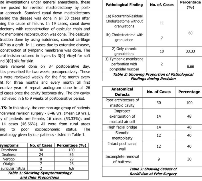
While 33.3% cases had only chronic granulations, 2 patients had tympanic membrane perforation with extensive polypoidal mucosa involving the middle ear cavity and mastoid antrum. The most frequent site of cholesteatoma was mastoid antrum/mastoid cavity seen in 73% followed by attic 42.3% and mesotympanum in 40% of cases. The common failure in primary surgery was inadequate clearance of diseased mastoid air cells-48%, high facial bridge-48%, stenotic meatoplasty-40%, incomplete removal of buttress-30%. Tympanic membrane perforation-6.66% of cases with poor architecture of mastoidectomy were seen.
Pathological Finding No. of. Cases Percentage (%)
1a) Recurrent/Residual Cholesteatoma without
granulations
1b) Cholesteatoma with granulation
11
7
60
2) Only chronic
granulations 10 33.33
3) Tympanic membrane perforation with polypoidal mucosa
2
6.66
Table 2: Showing Proportion of Pathological Findings during Revision
Anatomical
Defects No. of Cases Percentage
Poor architecture of
mastoid cavity 30 100 Improper
exenteration of mastoid air cell
14 48
High facial bridge 14 48 Stenotic
meatoplasty 12 40 Intact post canal
wall 12 40
Incomplete removal
of buttress 9 30
Jebmh.com
Original Article
J. Evid. Based Med. Healthc., pISSN- 2349-2562, eISSN- 2349-2570/ Vol. 3/Issue 50/June 23, 2016 Page 2542 The common ossicle affected is incus which was absent
in 86.66% of the cases, eroded in 4 cases (13.33%), malleus erosion seen in 20 cases (66.66%) and absence of stapes suprastructure in 16 cases (53.33%) observed respectively. In 19 cases (63.33%), canal down mastoidectomy with tympanoplasty (III and IV type) were done. In 11 cases (36.66%), canal down mastoidectomy without reconstruction of ossicles done due to extension of disease. Temporalis fascia was used for all cases to repair the tympanic membrane and also cover the bony cavity. Silastic sheet kept between the promontory and the graft for prevention of adhesions. In 63.33% cases, ossiculoplasty done. In 4 cases (21.05%), ossicular reconstruction done by auto incus transposition. In 13 cases (68.42%), myringostapediopexy done. In 2 cases (10.52%) myringo palatopexy was done by using PORP material.
Fig. 1: Pie Chart Showing Ossiculoplasty Types
In all cases, wide meatoplasty done. We achieved dry ear in 23 cases (88.46%), wet ear in 3 cases (11.53%) and 4 cases are not followed in our study. In followed 26 cases, the postoperative functional hearing results are as shown below in the Table 4. 19 cases were satisfactory with their outcome of hearing result. (73.07%). In one case, there was extrusion of PORP material.
AB Gap Closure No. of Cases Percentage
0-10 dB 6 23.07 10-20 dB 9 34.61 20-30 dB 4 15.38 No improvement 5 19.23
Worsen 2 7.69
Table 4: Post-Operative Hearing Results
DISCUSSION: The common age group of patients who underwent revision mastoid surgery - 8-46 yrs. In this study, male to female ratio was 7:8, i.e. there was no much difference between males and females for recurrence of disease. The duration between primary and revision surgery was between 1-6 yrs., ear discharge was the commonest chief complaint given by all patients in this series and 2 cases were present with post-auricular fistula. Vertigo complaints seen in 29% (8 cases) cases. Ajalloueyan et al[3] reported a
staggering 31% of patients having vertigo in his study.
In most series, the common causes of failure in mastoid cases are inadequate primary surgery, high facial ridge, incomplete removal of posterior canal wall, presence of anterior and posterior buttresses, deep cavity at the mastoid tip, incomplete saucerisation of cortical bony edges and inadequate meatoplasty which leads to mechanical hindrance to the field of the debris from the mastoid into ear canal in turn causing discharging cavity.[4] Incomplete
exenteration of diseased cellular tracts like sinodural angle cells, perilabyrinthine cells, retrofacial cells, tip cells, squamotegmen cells, cells at root of zygoma and incomplete clearance of diseased tissue from sinus tympani, facial recess, oval and round windows, hypotympanum, eustachian tube leads to recurrence/residual pathology.
Exposed middle ear and eustachian tube orifice with perforation of tympanic membrane also causes a discharging cavity.[5] Canal wall down mastoidectomy is the most widely
used surgical method worldwide. It is supposed to be easier, of shorter duration and necessitate less surgical experience than the CWU procedures. The anatomic and functional outcome is satisfactory, and the rate of complication is acceptably low.[6] The primary advantage of a CWD
procedure is increased visibility and adequate access to the mesotympanum and epitympanum. This increased exposure accounts for reduced rates of recurrence versus intact canal wall procedures.[7] CWD procedure is supposed to be the
unique solution for cholesteatoma in certain cases, such as the only hearing ear, hence there is a labyrinthine fistula, etc. Hulka and Mc Elveen claimed that CWD procedures do permit visualisation of the anterior epitympanum and sinus tympani region (the latter is not fully visualised with any technique).[8]
Another indication for CWD mastoidectomy is failure of previous CWU procedures with recurrent cholesteatoma. However, postoperative care is more intense in the CWD surgery, both in the immediate postoperative period and longterm. Cholesteatoma recurrence in open cavity procedures occurs in 4 to 28% of cases.[9] and is usually
caused by inaccessible disease or a remnant of matrix that is amputated at the time of surgery. Through routine follow-up, these “pearls” of recurrent cholesteatoma can be readily identified and removed in the office. Extensive recurrent disease, with its attendant complications, is more commonly found behind an intact canal wall rather than in an open cavity.
The primary aetiological factors for cavity problems in our series was inadequate primary surgery-48%, inadequate exenteration of air cells-48%, high facial bridge-48%, incomplete removal of buttress – 30%, narrow meatoplasty-40% were observed. The commonest pathology we encountered was cholesteatoma 36.66% with granulation tissue- 33.33%, only chronic granulation tissue in mastoid cavity antrum and middle ear cavity – 23.33%. In 6.66% cases (2), we observed tympanic perforation with extensive polypoidal middle ear mucosa as a basic pathological feature. The above said anatomical defects and pathological features are main causes for discharge in cavity.[2] and these
Jebmh.com
Original Article
J. Evid. Based Med. Healthc., pISSN- 2349-2562, eISSN- 2349-2570/ Vol. 3/Issue 50/June 23, 2016 Page 2543 In addition to the removal of all the factors mentioned
above, by removing anterior and posterior buttresses, saucerisation of cavity, lowering the facial bridge up to the level of facial nerve, sloping of mastoid cavity, levelling the floor of middle ear and mastoid cavity, reconstruction of tympanic membrane and ossicles (11 cases) by autoincus, conchal cartilage and using of biomaterial PORP are being done after the complete clearance of the disease from the site. The anatomical defects prevents the self-cleaning of cavity and promotes the disease process by accumulation of debris leading to discharge from cavity. Hence lowering of facial ridge and wide meatoplasty is very important to overcome above said problem.[10] A large meatoplasty is
necessary for epithelialisation of the cavity and easier postoperative care.
Generally, the meatus initially should be made about the size of the mastoid cavity because it will undergo about 25% contraction over time. A good approximation of the size is the surgeon’s thumb. Postoperative vertigo in CWD revision surgery has been clearly diminished due to the mastoid obliteration.[11] CWR procedure can be used for all patients
with acquired cholesteatoma, including children.[12]
26 cases were followed regularly up to one year and postoperative hearing assessment done once the cavity becomes dry. We achieved complete dry ear in 88.46% (23) of cases. In 3 (11.53 %) cases, mastoid cavity was still wet in spite of vigorous antiseptic and anti-allergic medication. The probable cause in these cases may be eustachian tube malfunction and allergy. The postoperative audiogram was done after 8 weeks, 6 months and one year regularly; during the followup period hearing results were fairly accepted in 73.06% of the patients. In 5 cases (19.23%), there is no improvement of hearing seen, in 2 cases (7.69%) hearing worsened. The above results were comparable with various studies.[5,13,14]
CONCLUSION: By this study, we concluded that
Residual/recurrent cholesteatoma, high facial bridge, improper removal of mastoid air cells, incomplete removal of buttresses, improper saucerisation of mastoid cavity and narrow meatoplasty are the prime causes for discharge from cavity.
Knowledge of thorough anatomy, behaviour of the disease, identifying the cause for primary surgical failure and experience of the surgeon are key factors in outcome of results.
Choosing correct surgical procedure initially and executing meticulously can prevent the revision surgeries.
REFERENCES
1. Nadol JB. Revision mastoidectomy. Otalryngol Clin North Amm 2006;39(4):723-40.
2. Bercin S, Ktuluhan A, Bozdemir K. Results of revision mastoidectomy. Actaotolaryngol 2009;129(2):138-141.
3. Ajalloueyanm M. Modified mastoidectomy techniques to decrease failure. Medical J of ISL Rep of Iran 1999;13(3):179-183.
4. Ayubi S, Ahmad S, Ali A. Revision mastoidectomy and oto endoscopy. JUMDC 2014;5(1):61-67.
5. Weiss MH, Parsier SC, Han JC. Surgery for recurrent and residual cholesteatoma. Laryngoscope. 1992;102(2):145-151.
6. Grewal DS, Hathiram BT, Saraiya SV. Canal wall down tympanomastoidectomy: the on-disease approach for retraction pockets and cholesteatoma. J Laryngol Otol 2007;121(9):832-839.
7. Dornhoffer JL. Retrograde mastoidectomy with canal wall reconstruction: a follow-up report. Otol Neurotol 2004;25(5):653-660.
8. Hulka GF, Mc Elveen JT. A randomized, blinded study of canal wall up versus canal wall down mastoidectomy determining the differences in viewing middle ear anatomy and pathology. Am J Otol 1998;19(5):574-578.
9. Hirsch BE, Kamerer DB, Doshi S. Single-stage management of cholesteatoma. Otolaryngol Head Neck Surg 1992;106(4):351-354.
10. Bhatiya S, Karmarkar S, DeDonato G, et al. Canal down mastoidectomy: causes of failure, pitfalls and their management. The Journal of Laryngology and Otology 1995;109(7):583-589.
11. Beutner D, stumpf R, Zahnert T, et al. Long-term results following mastoid obliteration in canal wall down tympano-mastoidectomy. Laryngorhinootologie 2007;86(12):861-866.
12. Gantz BJ, Wilkinson EP, Hansen MR. Canal wall reconstruction tympanomastoidectomy with mastoid obliteration. Laryngoscope 2005;115(10):1734-1740. 13. Sheehy JL. Recurrent and residual disease in cholesteatoma surgery. Clin Otolaryngol: 1978;3(4):393-403.