UDC: 616.348/.35-006.6-056.7(497.7) Original scientiic paper
Molecular proile of the Lynch Syndrome in the Republic of
Macedonia
Marija Hiljadnikova-Bajro
1, Toni Josifovski
2, Milco Panovski
2,
Aleksandar J. Dimovski
3*
1
Institute of Applied Biochemistry, Faculty of Pharmacy, University “Ss Cyril and Methodius”, Skopje, Republic of Macedonia
2
University Clinic for Digestive Surgery, Medical Faculty, University “Ss Cyril and Methodius”, Skopje, Republic of Macedonia
3*
Center for Biomolecular Pharmaceutical Analyses, Faculty of Pharmacy, University “Ss Cyril and Methodius”, Skopje, Republic of Macedonia
Received: November 2012; Accepted: December 2012
Abstract
The most frequent type of hereditary colorectal cancer, the one occurring in the setting of the Lynch syndrome (LS) is considered a phenotypic manifestation of a germline defect in the mismatch repair mechanism i.e. in the MLH1, MSH2, MSH6 or PMS2 gene. Aiming
towards establishment of a standardized protocol involving molecular analyses for diagnosis of this syndrome and developing a unique na-tional register of families with hereditary colorectal cancer syndromes in the Republic of Macedonia, we began a prospective study to re-veal the genetic defects among Macedonian patients with colorectal cancer (CRC) and identifying families with hereditary CRC.
A total of 53 patients fulilling the revised Bethesda criteria for MSI-genetic testing were compared to 350 patients with sporadic CRC. The results reveal signiicant differences in age at diagnosis (p=0.03), involvement of microsatellite instability (p<0.0001) and localization of the tumor in respect to lexura lienalis (p=0.009) and suggest afiliation of the majority of the “Bethesda+” CRCs to the so called Famil-ial Colorectal cancer Type X group. The molecular characterization of LS suspects identiied the novel MLH1 c.392C>G nonsense
muta-tion with a possible founder effect in the Macedonian populamuta-tion, the MLH1 ex.3-12 deletion, as well as the c.244A>G mutation,
IVS14-19A>G and IVS4+65A>C changes in MLH1 without conirmed pathological signiicance. The observed high frequency (87.5%) of the
Il-e219Val (c.655A>G) variant in MLH1 among the LS suspects prompts further analyses to evaluate its involvement in the development of
hereditary CRC by itself or as a risk modifying factor among the patients from the Republic of Macedonia.
Keywords: Colorectal cancer, Lynch syndrome, MLH1, microsatelite instability
Introduction
The most frequent hereditary form of colorectal cancer (CRC), responsible for 2-4% of all CRC cases (Aaltonen, 1998; Jasperson, 2010) is the hereditary nonpolyposis col-orectal cancer-HNPCC, later renamed to Lynch syndrome (LS). The autosomal dominant pattern of inheritance and
The molecular deinition of the Lynch Syndrome im-plies germline mutations in any of the mismatch repair (MMR) genes responsible for the basepair mismatch rep-aration during DNA replication as: MLH1 and MSH2 car-rying almost 90% of the mutations, MSH6 holding 10% of the genetic defects and rare mutations in EPCAM and PMS2.
The initial criteria for the establishment of a clinical diagnosis of LS, the so called Amsterdam I and II crite-ria (Vasen et al., 1991 and 1994) are based on the familial cancer history, but the revised Bethesda criteria for genet-ic testing for LS (Umar et al., 2004) are more sensitive and enable identiication of LS cases resulting from de novo MMR genetic defects.
Approximately ten to ifteen percent of individuals with CRC and/or colorectal adenomas have diseased rel-atives (National Cancer Institute, 2012), but don’t meet the criteria for clinical diagnosis of neither LS nor the syn-drome of familial adenomatous polyposis. They are consid-ered as cases with familial colorectal cancer (FCC), which might be caused by unidentiied genetic factors, exposition of the family members to common environmental risk fac-tors or by chance. Furthermore, families fulilling the Am-sterdam criteria for LS but lacking genetic MMR-defects i.e microsatellite instable tumors, are designated as famil-ial colorectal cancer type X (FCC-X) (Jass, 2006; Muel-ler-Koch et al., 2005; Valle et al., 2007). The risk for de-velopment of CRC is lower among these patients (Lindor et al., 2005) but if a malignant transformation eventually occurs, the diagnosis is usually established at an older age (55 years on average) in comparison to the LS. Further in-vestigations are needed for their precise characterization, but it is quite obvious that tumors arising in FCC-X fami-lies feature a speciic pathological phenotype with less tu-mor-iniltrating leukocytes versus typical LS tumors. (Llor et al., 2005).
Currently, there aren’t any oficially published data on the incidence/prevalence of LS in the Republic of Mace-donia and there isn’t a unique national register of patients/ families with any of the hereditary colon cancer syndromes, which seriously obstruct the investigation and character-ization of this type of hereditary cancer in our country and disable appropriate prevention/treatment. Hence, we began a prospective study to aid the establishment of standard-ized protocols involving molecular analyses for clinical di-agnosis of the Lynch syndrome in our country.
Materials and methods
From a total of 403 randomly selected patients under-going colectomy for resection of histopathologically con-irmed CRC at the University Clinic for Digestive Surgery in Skopje, a cohort of 53 cases was selected based on ful-illment of the Bethesda clinical criteria for MSI testing and compared to a group of 350 cases with sporadic CRC. Detailed personal and familial history as well as relevant
information on dietary and life-style habits were obtained by the responsible clinicians.
The molecular analyses were performed on samples of peripheral blood and fresh tumor tissues obtained by the surgical resection of the cancer. The DNA was isolated us-ing Proteinase K digestion/ phenol- chloroform-isoamyla-lcohol extraction/ethanol precipitation, according to stan-dard procedures (Sambrook et al., 1989).
The evaluation of the microsatellite instability was performed by a luorescent multiplex PCR reaction em-ploying the Bethesda ive marker panel (Rodrigues-Bigas et al., 1997; Boland et al., 1998), followed by capillary gel electrophoresis and fragment analysis on the Applied Bio-systems’ genetic analyzer AbiPrism310.
All microsatellite instable cancers were evaluated for their MLH1-promoter methylation status using methyla-tion-speciic quantitative PCR reaction following a bisulite DNA-conversion (Xiong and Laird, 1997) and tested for the BRAF V600E mutation (Davies et al., 2002), while the corresponding blood DNA samples were screened for large genomic deletions/duplications using the Multiplex Liga-tion- Dependant Probe Ampliication (MLPA) kit reagents from MRC-Holland (Amsterdam, Netherlands) according to the procedure irst described by Schouten et al. (2002)
Patients having microsatellite instable cancers with-out hypermethylated MLH1-promoters and BRAFV600E mutation, lacking large genomic defects but fulilling the Bethesda criteria for MSI testing, were subjected to bidi-rectional sequencing of the MLH1 and MSH2 genes using the Applied Biosystems’ BigDye Terminator v1.1 Cycle sequencing kit and subsequent sequencing analysis on the AbiPRism310.
For detection of an aberrant mRNA transcript we per-formed a Reverse transcription-polymerase chain reaction (RT-PCR) using the GeneAmp RNA PCR kit (Life technol-ogies, Califormia, USA) on peripheral mononuclear RNA obtained by the acidic guanidine-phenol-chlorophorm ex-traction (Chomczynski and Sacchi, 1987).
The detailed conditions for the above reactions and the oligonucleotide sequences of the employed primers are available upon request.
Results and Discussion
The studied group was comprised of 53 younger pa-tients diagnosed with “Bethesda +”colorectal cancer at an average age of 50 years and progressed mainly to clinical stage B or C. Ovarial, gastic, brain but also breast, hepat-ic and gall bladder malignancies were identiied among the patients or their relatives. Three of these patients were pre-viously diagnosed with endometrial cancer, which is con-sidered one of the commonest extracolonic malignancies in LS, apart from the ovarial cancer (Cruz-Correa and Gi-ardiello, 2002).
instabili-ty (30%). The comparison with the sporadic CRC cas-es (Table 1) reveals signiicant differenccas-es in age at di-agnosis (p=0.04), involvement of microsatellite instabili-ty (p<0.0001) and tumor localization in respect to lexu-ra lienalis (p=0.009) so that, MSI is 3.99 times more fre-quently present than MSS in cancers of LS suspects and they develop 2.19 times more frequently in proximal than distal colon, compared to cancers of sporadic cases. These indings comply with the common deinitions of the Lynch Sy (Watson and Lynch, 1994; Aarnio et al., 1999), but the published data state an MSI-involvement in 90% of these cancers. The lower frequency (30%) of MSI detected in our cohort, suggests that a signiicant proportion of these patients are not actually LS-cases but cases with FCC-X. These patients might be having the hereditary syndrome recently proposed by Valle et al. (2007), which is consid-ered a separate entity from LS, featuring MSS tumors, de-velopment of colorectal but not other cancers and absence of multiple primary tumors. The vast majority of the hered-itary cancers in our group suggest an existence of a yet un-identiied gene, being responsible for the development of
syndrome, but lacking the MMR activity.
Aiming towards an establishment of a national register of families with hereditary CRC syndromes in our country, we created an interactive computer database of CRC cases, available for access and use by the involved clinicians and laboratory investigators. Additionally, we designed a pro-tocol for molecular conirmation of the LS among clinical suspects for the disease.
The algorithm for detection of MMR defects em-ployed in our study was based on unselective MSI- testing of all CRCs, which had been proven as a cost effective ap-proach in identiication of individuals at an increased risk for LS (Palomaki et al., 2009), with a 93% sensitivity for identiication of tumors resulting from MMR-mutations (Shia, 2008). This type of genetic instability was identi-ied among 11% of all CRCs. A total of 53 patients with “Bethesda+”tumors were subjected to MSI testing. Somat-ic changes as MMR-promoter hypermethylation and the BRAFV600E mutation are considered as strong negative predictors of MMR-deiciency (Parsons et al., 2012; Bou-zourene et al., 2010; Bellizzi and Frankel, 2009) and were
Table 1. Comparison between colorectal cancers of patients fulilling the Bethesda criteria for LS and sporadic cases.
“Bethesda +” CRCs
(n=53) Sporadic CRCs(n=350) M2-M1 p
Age
Number (n), Аrithmetical mean
(Ar) ± standard deviation Ar=50.27±13.16 Ar=60.39±10.78 10.12 0.04
Number (frequency) OR 95%CI p
Gender Male
Female 28 (0.52)25 (0.48) 204 (0.58)146 (0.42) 0.80 0.45-1.43 0.45 Clinical Tumor Stage
A B C D
2 (0.06) 18 (0.56)
9 (0.28) 3 (0.09)
19 (0.06) 120 (0.41) 128 (0.43) 27 (0.09)
1.87 0.88-3.96 0.09 a
Genetic instability MSI
MSS 14 (0.30)33 (0.70) 282(0.9)30 (0.1) 3.99 1.92-8.27 <0.0001
BRAF V600E +
BRAF V600E - 40(0.98)1 (0.02) 188 (0.96)8 (0.04) 0.59 0.07-4.83 0.61 Localization
Proximal b
Distal c 23 (0.44)29 (0.56) 213 (0.74)77 (0.26) 2.19 1.19-4.02 < 0.01
Colon
Rectum 35 (0.67)17 (0.33) 188 (0.63)109 (0.37) 1.19 0.64-2.23 0.58 a comparison between CRCs of lower (A+B) and higher stage (C+D)
evaluated in all MSI CRCs prior to the MMR-genetic anal-ysis. Patients complying with the revised Bethesda crite-ria and carrying microsatellite instable tumors without ev-idence of neither somatic MLH1-promotor hypemethyla-tion nor BRAF-V600E mutahypemethyla-tion, were selected for genet-ic testing whgenet-ich included MLPA-analysis for detection of large genomic rearrangements in MLH1, MSH2, PMS2 and MSH6 genes, followed by bidirectional DNA-sequencing of the 19 and 16 exons and lanking intronic regions of MLH1 and MSH2 respectively. Using this approach, we narrowed the selected group to 10 patients highly suspi-cious and suitable for LS-genetic testing. Mutations in the MSH2 gene were not detected but several genetic defects in MLH1 were identiied as most probable causative de-fects for LS.
We reported previously (Hiljadnikova-Bajro, 2012) the irst molecular characterization of the Lynch Syndrome in the Republic of Macedonia by the identiication of the nonsense MLH1 c.392C>G mutation in two seemingly un-related patients, and suspecting its founder effect suggest-ed screening of all LS suspects from the region for this mu-tation by a Restriction Fragment Length Polymorphism - test. Using this approach, we later identiied another pa-tient with the same mutation, who has distant familial re-lationship with one of the abovementioned index patients but wasn’t included in the genetic counseling of the pa-tient’s family. This case highlights the necessity of present-ing a thorough familial history by the patients and in case of identiication of a LS patient, genetic counseling of all
relatives at risk of cancer development in order to prevent or timely treat it.
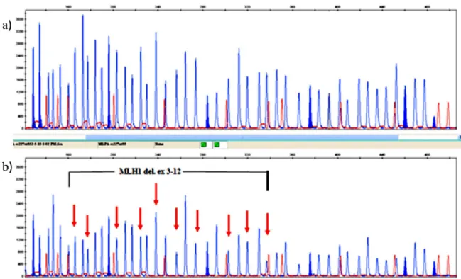
Using the MLPA analysis, we detected quantitative changes at either single or multiple nonsuccessive exons in several patients, but these isolated changes probably re-sult from variations within the probes’s hybridization loci on the tested DNA leading to impaired hybridization and consequently decreased ampliication of the correspond-ing fragments. But, in one pediatric patient, we detected a large deletion in MLH1 targeting exons 3 through 12 (Fig. 1). The patient is an adolescent boy, diagnosed with two metasynchronous cancers of the transversal and sigmoid colon of stage IIa at the age of 15 and 16 years respectively. The molecular analyses revealed a microsatellite instable character of the cancers, but only one of them was suitable for BRAF and MLH1-hypermethylation testing and scored negative for the BRAF V600E mutation but exerted a high degree of MLH1-promoter methylation. Exploring the fa-milial history of the disease, we concluded that the MLH1 -genetic defect is most probably inherited from the mother, who was diagnosed with endometrial cancer few years ago. Genetic testing of the mother was offered and if performed in future, it should conirm or exclude this hypothesis. On the other hand, somatic MLH1 promoter hypermethylation is associated with sporadic cases of MSI CRCs, opening a possibility though minor for different etiologies of the two cancers diagnosed in this patient: the irst which is current-ly unavailable for promoter hypermethylaton-testing might be having a hereditary basis, and the other one with
methylated MLH1 promoter could be a sporadic one. But, if MSI and MLH1 promoter hypermethylation are present in both tumors and the genetic testing of the mother con-irms the hereditary character of the disease, we might get a strong argument against the hypothesis that somatic pro-moter hypermethylation of MLH1-gene promoter excludes the Lynch Syndrome diagnosis.
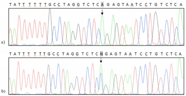
The DNA sequencing-analysis identiied a patient with a point DNA-change on the fourteenth intron of MLH1: IVS14-19A>G (Fig.2), located in the splicing branch site region. This change hasn’t been reported in the Human Ge-nome Mutation database (HGMD) as a cause for the Lynch Sy so far. In order to determine the potential impact of this alteration on the posttranscriptional mRNA process-ing and the existence of an alternative splicprocess-ing mechanism that would cause production of an aberrant transcript dif-fering in size from the wild type, we performed a RT-PCR reaction on peripheral leukocytes’ RNA to achieve reverse transcription and ampliication of the investigated region. The obtained DNA-fragment was analyzed by electropho-resis on a 10% polyacrilamide gel. According to the re-sults presented on the electrophoregram (Fig. 3), we didn’t detect an aberrant mRNA-transcript differing in size from the 192bp-long fragments identiied among the two wild-type RNA samples. Still, this result doesn’t completely ex-clude the hypothesis for existence of an alternative splicing mechanism due to the IVS14-19A>G mutation. Namely, aberrant mRNA transcripts longer by 17, 19 or 20bp than the wild type ones, might be degraded by the mechanism of nonsense mediated decay and hense become unidentii-able by this test. Therefore, additional functional tests are warranted to conirm/exclude the deleterious effect of this alteration.
In one patient diagnosed with two metachronous CRCs by the age of 40, burdened with a heavy familial history of cancer, we identiied a germline genetic alteration on the fourth MLH1 intron: IVS4+65A>C (Fig.4) which hasn’t been previously reported to be involved in the LS, and con-sidering its location within the gene doesn’t appear to have a pathological impact. Further genetic testing of MSH6, EPCAM and PMS2 might conirm the LS-association of this cancer or classify it among the FCC-X type CRCs.
Fig. 3. PAGE electrophoregram of RT-PCR amplicons from: RNA samples corresponding to the patient with theIVS14-19A>G mutation (lane 2) and two control samples (lanes 3 and 4) in comparison to a blank (lane 5) and the molecular size marker StepLadder 50 (lane 1).
We detected the novel missense mutation MLH1 c.244A>G, p.T82A in one patient diagnosed with a can-cer of the ascending colon at an age of 40, whose moth-er was also diseased with an early onset CRC. This genetic change leads to a substitution of the polar aminoacid Thre-onine with the hydrophobic aminoacid Alanine, probably causing а pathogenic effect similar to the c.245C>T muta-tion already reported in the HGMD.
In 7/8 patients (87.5%), we detected the c.655A>G point mutation in the eighth exon of MLH1 (Fig.5) caus-ing synthesis of a polypeptide with a substitution of Iseleu-cine with Valine at position 219 (p.Ile219Val). One of these patients holds this genetic variant in a homozygous form, the remaining are being heterozygous. This genetic varia-tion has been previously reported as a polymorphism in the general population (Liu, 1995), but Tomlinson and his col-Fig. 2.Electrophoregram corresponding to the sequenced PCR-amplicon of the MLH1 gene in the patient having the
laborators (Tomlinson et al., 1997) suggest that this change with a frequency of 1% should be treated as a low pen-etrant mutation for development of CRC. The Ile219Val change is located in a conserved region of the eighth MLH1 exon, hence it is quite rational to expect its impact on de-velopment of pathological consequences. According to
the functional analyses performed so far, the products of the wild-type and polymorphic allele poses similar DNA-reparative activity (Trojan et al., 2002; Raevaara et al., 2005), but these tests do not address the stability of the proteins which also might be a cause for their impaired ac-tivity. Evidence indicating that this change is not totally be-Fig. 5. Detection of the Ile219Val genetic variant in a homozygous (a) and heterozygous (b) form in comparison to a
con-trol sample (c).
nign exist. Some of them even suggesting it to be a predic-tor of a high mutation frequency haplotype (Hutter et al., 2000, 2002). Furthermore, homozygosity for this variation is associated with reduced expression of the MLH1 protein in sporadic CRCs in Korea (Kim et al., 2004). According to several published studies (Berndt et al., 2007; Yu et al., 2006), Ile219Val is not associated with colonic cancers it-self, but the western diet modiies this association in a way that 219 Val/Val genotype in patients on a western diet (red overroasted meat, fast food, eggs, full-fat dairy products, reined cereals and sugar) is associated with a two-fold in-creased risk for CRC development in comparison to indi-viduals of the Ile/Ile genotype avoiding the western diet (Campbell, 2009). Additionally, the Ile219Val variant is as-sociated with 5 times higher risk of ulcerative colitis (Ba-gnoli et al., 2004) which is a risk factor for CRC develop-ment itself.
Conclusions
The molecular proiling of LS among patients with CRC from the Republic of Macedonia has identiied the causative deletion MLH1 del.ex3-12 and the novel, pos-sibly founder, nonsense mutation MLH1 c.392C>G and suggests plausible involvement of the c.244A>G mis-sense mutation, the intronic changes: IVS14-19A>G and IVS4+65A>C as well as the Ile219Val genetic variant with-in MLH1 in the development of this syndrome. The vast majority of the clinically suspected LS patients are most probably cases with the hereditary syndrome recently pro-posed by Valle, or cases with a familial cancer of unknown etiology. Focusing the further genetic analyses on these pa-tients might reveal new genes with high or low penetrance for development of colorectal cancer.
References
Aaltonen, L.A., Salovaara, R., Kristo, P., Canzian, F., Hemminki, A., Peltomaki, P., Chadwick, RB., Kaariainen, H., Eskelinen, M., Jarvinen, H., Mecklin, JP., de la Chapelle, A., 1998. Incidence of hereditary nonpolyposis colorectal cancer and the feasibility of molecular screening for the disease. N. Engl. J. Med. 338(21);1481-1487.
Aarnio, M., Sankila, R., Pukkala, E., Salovaara, R., Aaltonen, LA., de la Chapelle, A., Peltomaki, P., Mecklin, JP., Jarvinen HJ., 1999. Cancer risk in mutation carriers of DNA-mismatch-repair genes. Int. J. Cancer. 81(2):214-218.
Bagnoli, S., Putignano, AL., Melean, G., Baglioni, S., Sestini, R
.
, Milla, M.
, d’Albasio, G.
, Genuardi, M.
, Pacini, F.
, Trallori, G.
, Papi, L., 2004. Susceptibility to refractory ulcerative colitis is associated with polymorphism in the hMLH1 mismatch repair gene. Inlamm Bowel Dis. 10(6):705-708. Bellizzi, AM., Frankel, WL., 2009. Colorectal cancer due todeiciency in DNA mismatch repair function: a review. Adv Anat Pathol. 16:405–417.
Berndt, SI., Platz, EA., Fallin, MD., Thuita, LW., Hoffman, SC., Helzlsouer, KJ., 2007. Mismatch repair polymorphisms and
the risk of colorectal cancer. Int. J. Cancer. 120(7):1548-1554.
Boland, CR., Thibodeau, SN., Hamilton, SR., Sidransky, D., Eshleman, JR., Burt, RW., Meltzer, SJ., Rodriguez-Bigas, MA., Fodde, R., Ranzani, GN., Srivastava, S., 1998. A National Cancer Institute Workshop on Microsatellite Instability for cancer detection and familial predisposition: development of international criteria for the determination of microsatellite instability in colorectal cancer. Cancer Res. 15; 58(22):5248-5257.
Bouzourene, H., Hutter, P., Losi, L., Martin, P., Benhattar, J., 2010. Selection of patients with germline MLH1 mutated Lynch syndrome by determination of MLH1 methylation and BRAF mutation. Fam Cancer. 9:167–172.
Cruz-Correa, M., Giardiello, FM., 2002. Diagnosis and management of hereditary colon cancer. Gastroenterol. Clin. North. Am. 31(2): 537-549.
Campbell, PT., Curtin, K., Ulrich, CM., Samowitz, WS., Bigler, J., Velicer, CM., Caan, B., Potter, JD., Slattery, ML., 2009. Mismatch repair polymorphisms and risk of colon cancer, tumour microsatellite instability and interactions with lifestyle factors. Gut. 58(5): 661-667.
Hiljadnikova-Bajro, M., Josifovski, T., Panovski, M., Dimovski, AJ., 2012. A novel germline MLH1 mutation causing Lynch Syndrome in patients from the Republic of Macedonia. Croat Med J. 53 (5):496-501. http://www.hgmd.cf.ac.uk/ac/index. php assessed at 18.05.2012
Jasperson, KW., Tuohy, TM., Neklason, DW., Burt, RW., 2010. Hereditary and familial colon cancer. Gastroenterology. 138(6):2044-2058
Jass, JR., 2006. Hereditary Non-Polyposis Colorectal Cancer: the rise and fall of a confusing term. World J. Gastroenterol. 12(31):4943-4950.
Kim, JC., Roh, SA., Koo, KH., Ka, IH., Kim, HC., Yu, CS., Lee, KH., Kim, JS., Lee, HI., Bodmer, WF., 2004. Genotyping possible polymorphic variants of human mismatch repair genes in healthy Korean individuals and sporadic colorectal cancer patients. Fam Cancer. 3(2):129-137.
Lindor, NM., Rabe, K., Petersen, GM., Haile, R., Casey, G., Baron, J., Gallinger, S., Bapat, B., Aronson, M., Hopper, J., Jass, J., LeMarchand, L., Grove, J., Potter, J., Newcomb, P., Terdiman, JP., Conrad, P., Moslein, G., Goldberg, R., Ziogas, A., Anton-Culver, H., de Andrade, M., Siegmund, K.., Thibodeau, SN., Boardman, LA., Seminara, D., 2005. Lower cancer incidence in Amsterdam-I criteria families without mismatch repair deiciency: familial colorectal cancer type X. JAMA293(16):1979-1985.
Liu, B., Nicolaides, NC., Markowitz, S., Willson, JK., Parsons, RE., Jen, J., Papadopolous, N., Peltomäki, P., de la Chapelle, A., Hamilton, SR., Kinzler, KW., Vogelstein, B., 1995. Mismatch repair gene defects in sporadic colorectal cancers with microsatellite instability. Nat. Genet. 9(1): 48-55. Llor, X., Pons, E., Xicola, RM., Castells, A., Alenda, C., Piñol,
V., Andreu, M., Castellví-Bel, S., Payá, A., Jover, R., Bessa, X., Girós, A., Roca, A., Gassull, MA., 2005. Gastrointestinal Oncology Group of the Spanish Gastroenterological Association. Differential features of colorectal cancers fulilling Amsterdam criteria without involvement of the mutator pathway. Clin. Cancer. Res.11(20):7304-7310. Mueller-Koch, Y., Vogelsang, H., Kopp, R., Lohse, P., Keller, G.,
colorectal cancer: clinical and molecular evidence for a new entity of hereditary colorectal cancer. Gut. 54(12):1733-1740.
National Cancer Institute, Genetics of Colorectal Cancer (PDQ), Health Professional Version, last modiied 2012, assessed at 18.05.2011; http://www.cancer.gov/cancertopics/pdq/ genetics/colorectal/HealthProfessional
Palomaki, GE., McClain, MR., Melillo, S., Hampel, HL., Thibodeau, SN., 2009. EGAPP supplementary evidence review: DNA testing strategies aimed at reducing morbidity and mortality from Lynch syndrome. Genet Med. 11(1):42-65.
Parsons, MT., Buchanan, DD., Thompson, B., Young, JP., Spurdle, AB., 2012. Correlation of tumour BRAF mutations and MLH1 methylation with germline mismatch repair (MMR) gene mutation status: a literature review assessing utility of tumour features for MMR variant classiication. J. Med. Genet. 49(3):151-157
Raevaara, TE., Korhonen, MK., Lohi, H., Hampel, H., Lynch, E., Lönnqvist, KE., Holinski-Feder, E., Sutter, C., McKinnon, W., Duraisamy, S., Gerdes, AM., Peltomäki, P., Kohonen-Ccorish, M., Mangold, E., Macrae, F., Greenblatt, M., de la Chapelle, A., Nyström, M., 2005. Functional signiicance and clinical phenotype of nontruncating mismatch repair variants of MLH1. Gastroenterology 129(2):537-549. Risinger, J.I., Barrett, J.C., Watson, P., Lynch, H.T., Boyd, J.,
1996. Molecular genetic evidence of the occurrence of breast cancer as an integral tumor in patients with the hereditary nonpolyposis colorectal carcinoma syndrome. Cancer. 77(9):1836-1843.
Rodriguez-Bigas, MA., Boland, CR., Hamilton, SR., Henson, DE., Jass, JR., Khan, PM., Lynch, H., Perucho, M., Smyrk, T., Sobin, L., Srivastava, S., 1997. A National Cancer Institute Workshop on Hereditary Nonpolyposis Colorectal Cancer Syndrome: meeting highlights and Bethesda guidelines. J. Natl. Cancer Inst. 89(23):1758-1762.
Sambrook, J., Fritsch, EF., Maniatis, T., 1989. Molecular Cloning - A Laboratory Manual, 2nd Edition. Cold Spring Habour Laboratory Press, New York.
Schouten, JP., McElgunn, CJ., Waaijer, R., Zwijnenburg, D., Diepvens, F., Pals, G., 2002. Relative quantiication of 40 nucleic acid sequences by multiplex ligation-dependent probe ampliication. Nucleic Acids Res. 30(12):e57.
Shia J., 2008. Immunohistochemistry versus microsatellite instability testing for screening colorectal cancer patients at risk for hereditary nonpolyposis colorectal cancer syndrome. Part 1: The utility of immunohistochemistry. J. Mol. Diagn. 10: 293–300.
Tomlinson, IP., Beck, NE., Homfray, T., Harocopos, CJ., Bodmer, WF., 1997. Germline HNPCC gene variants have little inluence on the risk for sporadic colorectal cancer. J. Med. Genet. 34(1):39-42.
Trojan, J., Zeuzem, S., Randolph, A., Hemmerle, C., Brieger, A., Raedle, J., Plotz, G., Jiricny, J., Marra, G., 2002. Functional analysis of hMLH1 variants and HNPCC-related mutations using a human expression system. Gastroenterology 122(1): 211-219.
Umar, A., Boland, CR., Terdiman, JP., Syngal, S., de la Chapelle, A., Ruschoff, J., Fishel, R., Lindor, NM., Burgart, LJ., Hamelin, R., Hamilton, SR., Hiatt, RA., Jass, J., Lindblom, A., Lynch, HT., Peltomaki, P., Ramsey, SD., Rodriguez-Bigas, MA., Vasen, HF., Hawk, ET., Barrett, JC., Freedman, AN., Srivastava, S., 2004. Revised Bethesda Guidelines for hereditary nonpolyposis colorectal cancer (Lynch syndrome) and microsatellite instability. J. Natl. Cancer Inst. 96(4):261-268.
Valle, L., Perea, J., Carbonell, P., Fernandez, V., Dotor, AM., Benitez, J., Urioste, M., 2007. Clinicopathologic and pedigree differences in Amsterdam I-positive hereditary nonpolyposis colorectal cancer families according to tumor microsatellite instability status. J. Clin. Oncol. 25(7):781-786.
Vasen, HF., Mecklin, JP., Khan, PM., Lynch, HT., 1991. The International Collaborative Group on Hereditary Non-Polyposis Colorectal Cancer (ICG-HNPCC). Dis Colon Rectum. 34(5):424-425.
Vasen, HF., Mecklin, JP., Khan, PM., Lynch, HT., 1994. The International Collaborative Group on HNPCC. Anticancer Res. 14(4B):1661-1664
Watson, P., Lynch, HT., 1994. The tumor spectrum in HNPCC. Anticancer Res. 14(4B):1635-1639. Xiong, Z., Laird, PW., 1997. COBRA: a sensitive and quantitative DNA methylation assay. Nucleic Acids Res. 25(12):2532-2534.
Резиме
Молекуларен профил на Линч Синдромот во Република
Македонија
Марија Хиљадникова-Бајро
1, Тони Јосифовски
2, Милчо Пановски
2,
Александар Ј. Димовски
3*
1Институт за применета биохемија, Фармацевтски факултет, Универзитет „Св. Кирил и Методиј“, Скопје, Република Македонија
2Универзитетска клиника за дигестивна хирургија, Медицински факултет, Универзитет „Св. Кирил и Методиј“, Скопје, Република Македонија
3*Центар за биомолекуларни фармацевтски анализи, Фармацевтски факултет, Универзитет „Св. Кирил и Методиј“, Скопје, Република Македонија
Клучни зборови: Колоректален карцином, Линч синдром, MLH1, микросателитска нестабилност
Најзастапениот тип на наследен колоректален карцином, карактеристичен за Линч синдромот (ЛС), се смета за фенотипска манифестација на генетски дефект во механизмот за репарација на несоодветно спарување на бази при ДНК репликација, одно-сно во MLH1, MSH2, MSH6 или PMS2 генот. Во рамките на нашите напори за воспоставување на стандардизиран протокол што ќе
вклучува молекуларни анализи за дијагноза на овој синдром и формирање на единствен национален регистер на семејства со на-следни синдроми на колоректален карцином во Република Mакедонија, отпочнавме проспективна студија за разоткривање на ге-Mакедонија, отпочнавме проспективна студија за разоткривање на ге-акедонија, отпочнавме проспективна студија за разоткривање на ге-нетските дефекти кај Македонските пациенти со колоректален карцином (КРК) и идентификација на семеjства со наследен КРК. Вкупно 53 пациенти кои ги исполнуваат ревидираните Бетезда критериуми за генетско тестирање за Линч синдромот беа спо-редени со 350 пациенти со спорадичен КРК. Резултатите покажуваат сигнификанти разлики во однос на возраста при дијагноза (р=0,03), инволвираност на микросателитската нестабилност (р<0,001) и локализација на туморот во однос на lexura lienalis (р=0,009) и сугерираат припадност на повеќето од „Бетезда+“ КРКи кон групата позната како Фамилијарен Колоректален карци-ном од тип „Х“. Со молекуларната карактеризација на ЛС-суспектите идентификувавме нова MLH1 c.392C>G nonsense мутација
со потенцијален „founder“ ефект кај Македонската популација и MLH1 ex.3-12 делецијата, како и c.244 A>G мутацијата,
IVS14-19A>G и IVS4+65A>C промените без потврдено патолошко значење. Високата фреквенција (87.5%) на Ile219Val (c.655A>G)
варијантата во MLH1 забележана кај ЛС суспектите ја наложува потребата од дополнителни аналзи за евалуирање на нејзината