American Journal of Infectious Diseases 3 (4): 208-216, 2007 ISSN 1553-6203
© 2007 Science Publications
Corresponding Author: José W. Rodríguez, Department of Microbiology and Immunology, Universidad Central del Caribe School of Medicine, P.O. Box 60327, Bayamón, Puerto Rico 00960
Co-expression of Apoptosis-Related Molecules on Activated CD8
+CD38
+T-cells
is Associated with HIV-1 Disease Progression
1
José W. Rodríguez,
1Luis A. Rodríguez,
1Griselle Font,
1Luis A. Cubano,
Nawal M. Boukli,
1
Miguel Otero,
2Robert Hunter,
3Madhavan P. Nair, and
1Eddy Ríos-Olivares
1
Department of Microbiology and Immunology and
2Internal Medicine, Universidad Central del Caribe
School of Medicine, Bayamón, Puerto Rico
3
Department of Immunology, Florida International University College of Medicine, Miami, Florida
Abstract: CD8+ T cells play a major role in controlling HIV-1 infection through the release of soluble lytic and non-lytic antiviral factors. Their decrease or defective function contributes to the HIV-1 disease progression. HIV-1 disease progression has been associated with a remarkable increase of CD38 expression on CD8+ T-cells. It has been also documented that a significant distribution of HIV-specific CD8+T-cells resides in the CD8+CD38+ T-cell sub-population. The failure of HIV-specific CD8+CD38+ T-cells to control HIV-1 infection has been attributed to several mechanisms including apoptosis. However, the relationship between the CD38 expression and molecular events involved in CD8+ T-cell apoptosis is not well understood. Using four-color flow cytometric analysis, the present cross-sectional study we evaluated the expression of four membrane-associated apoptosis-related molecules (TNFR-1, Annexin-V, CXCR4, and CD95) and two cytoplasm-associated apoptosis-related molecules (Bcl-2 and the active form caspase-3) in 41 HIV-1 positive patients and 15 HIV-1 negative individuals. Flow cytometric analysis made on freshly isolated PBMC showed that HIV-1 infection alters the level of expression of CD38, CD95, CXCR4, Bcl-2 and active caspase-3. No significant change in the expression of Annexin V or TNFR-1 was found. A positive correlation was established between CD95, CXCR4, and active caspase-3 expression with low CD4 count and high plasma viremia and CD38 expression. Data suggest that the majority of activated CD8+CD38+ T-cells were apoptotic because they expressed active caspase-3 and the rest of these cells were highly susceptible to become apoptotic since they co-expressed CD95 and CXCR4. Results also suggest that one of the most likely HIV-mediated apoptosis mechanisms is via CD95 and CXCR4 induction through the caspase cascade despite the expression of Bcl-2. All these observations may provide an additional explanation of why HIV-1 infection is not fully contained by HIV-specific CD8+CD38+ T-cells leading to HIV-1 disease progression.
Key words: Human Immunodeficiency virus-1 (HIV-1); CD8+CD38+ T-cells;
apoptosis-related molecules; four-color flow cytometry
INTRODUCTION
It has been demonstrated that the surface molecule CD38 is expressed at higher levels on activated CD8+ T-cells during HIV-1 infection and this has been well accepted as a prognostic activation marker for HIV-1 disease progression in infected individuals who do not control viral replication [1, 2]. CD38 is a 45-kDa type II membrane glycoprotein that functions as a calcium-dependent transmembrane signaling receptor and it can transmit either positive or negative signals regulating T and B lymphocyte activation, proliferation, and differentiation [3].
Several studies have revealed that most of HIV-specific CD8+ T-cells express CD38 [4]. These cells are generated during the acute phase of HIV-1 infection, continue to stay in control of viremia during the chronic phase and are depleted during the AIDS phase [5-7]. It has been shown also that the capacity of HIV-specific CD8+CD38+ T cells to produce antiviral factors diminishes as the disease progresses and the loss of their antiviral activity is critically dependent on the excessive levels of cellular activation leading to cell-programmed death or apoptosis [4, 8-9].
HIV-1 infection is characterized by a persistent immune activation leading to a subsequent decline in
SCI-PUBLICATION
Author Manuscrip
t
SCI-PUBLICATIONS
Author Manuscrip
t
both CD4+ and CD8+ T-lymphocytes in the early stages of the disease [10]. Apoptosis is seen in peripheral blood CD4+ and CD8+ T-lymphocytes of HIV-infected patients [11-12] and it has been proposed as an important event that contributes to the HIV-1 immunopathogenesis [13-14]. It has been shown that the degree of apoptosis observed in CD8+ T-cells is significantly higher in infected patients than in uninfected individuals and that it strongly correlates with disease progression and severity [12, 15].
Previous studies demonstrated that enhanced levels of Fas (CD95)-mediated apoptosis occur in HIV-specific CD8+CD38+ T-cells [4]. In addition, HIV-specific CD8+ T-cells from infected patients were susceptible to pro-apoptotic signaling through the tumor necrosis factor receptors-1 (TNFR-1) and this result was associated with the expression of active caspase-3 and a lack of protection of Bcl-2 [13-14,16].
Another molecule that is closely related to HIV-specific CD8+ T-cell apoptosis is the surface receptor CXCR4 [17]. It has been demonstrated that the increased expression of this molecule on the surface of HIV-specific CD8+ T-cells after HIV-induced activation plays an important role in these cells’ apoptosis [17]. Furthermore, HIV-1 infection has the capability to disrupt cell death through the release of pro-apoptotic proteins that kill infected and uninfected cells creating a state of chronic immune activation that is responsible for the exacerbation of clonal deletion [12, 18-19]. Thus, apoptosis has been considered to be a central mechanism of HIV-specific CD8+ T-cell loss and a contributor of HIV-1 disease progression.
Based on these previous studies, a comprehensive analysis of HIV-mediated CD8+CD38+ T-cell apoptosis during HIV-1 infection was needed. In the present study, a correlation was established between the levels of expression of apoptosis-related molecules in these cells with well-accepted progression markers such as CD4 count and viral load. We believe that the survival or turnover of HIV-specific CD8+CD38+ T-cells could have a great impact in the ability to clear HIV-1 infection. These findings provide further information for a possible explanation of the failure of containment of HIV-1 infection.
MATERIALS AND METHODS
Study Participants: This investigation was conducted at the Department of Microbiology and Immunology of the Universidad Central del Caribe School of Medicine, Bayamón, Puerto Rico. It was a cross-sectional study, conducted from October 2005 through April 2006. The recruitment of 41 HIV-1 positive participants for this
study was achieved through a single Recruitment and Assessment Unit that admits and collects research data on all participants for other research projects located at the Retrovirus Research Center (RRC) at the Universidad Central del Caribe School of Medicine/Ramón Ruiz Arnau University Hospital in Bayamón, Puerto Rico. This study was performed following the approval of the Institutional Review Board (IRB) and met all the confidentiality and privacy criteria.
On admission, all subjects that enter the RRC require an HIV-ELISA followed by a Western blot confirmatory assay. Those participants, that were injecting drug users, receiving antiviral therapy, and older than 50 years of age, were excluded from the study. All the individuals were enrolled according to their most recent CD4 and CD8 T-cell counts. Demographic characteristics of the individuals were also recorded, which included: age, gender, and HIV-1 viral load. From the total of 56 participants, 15 HIV-negative individuals with proven seronegativity served as healthy controls.
Apoptosis analysis: Using a FACSCalibur flow cytometer (BD Biosciences, San Jose, CA), a four-color cytometric analysis was performed on freshly isolated peripheral blood mononuclear cells (PBMC). The cells were incubated for 15 minutes with matching combinations of 10 µl of monoclonal antibodies directly conjugated with fluorochromes, and then washed twice with phosphate buffer saline (PBS). The cells were resuspended and fixed with 1% paraformaldehyde for flow cytometric analysis. Stained cells with PE-Cy5-conjugated anti-CD3, APC-conjugated anti-CD8, and PE-APC-conjugated anti-CD38 monoclonal antibody (mab) identified the activated CD8+ T-cells. Apoptotic cells were analyzed by staining cells with either FITC-conjugated anti- TNFR-1 (R&D Systems, Minneapolis MN), FITC-conjugated anti-Annexin-V, conjugated anti-CD95, or FITC-conjugated anti-CXCR4 (BD Biosciences, San Diego CA). In addition, the expression of Bcl-2 and active caspase-3 molecules were also analyzed on activated CD8+CD38+ T-cells by intracellular staining. Briefly, cells were stained with PE-Cy5-conjugated anti-CD3, APC-conjugated CD8, and PE-conjugated anti-CD38 mabs (BD Biosciences, San Diego, CA). After
Author Manuscrip
the surface staining, the cells were resuspended in 250 ml of BD Cytofix/Cytoperm solution (BD Biosciences, San Diego, CA) for 30 min. at 4oC in the dark. The permeabilized cells were washed twice with 1 ml (1 ml/wash) of 1 x BD Perm/Wash solution (BD Biosciences, San Diego, CA). The remaining pellet (fixed/permeabilized cells) was resuspended in 50µl of BD Perm/Wash solution and incubated with 10µl at 4oC for 15 min. in the dark with either FITC-conjugated anti-Bcl-2 or FITC-FITC-conjugated anti-active caspase-3 monoclonal antibodies for the corresponding tube. The cells were washed twice with 1 x BD Perm/Wash solution (1 ml/wash). The cells were then resuspended and fixed with 1% paraformaldehyde in order to analyze the samples by a FACSCalibur flow cytometer.
Statistical analysis: The Shapiro-Wilk test was performed to assess the normality assumption of the data. If the data was normally distributed, the two-sample Student’s t-tests were performed. For the data that was not normally distributed, the Wilcoxon two-sample test was performed. The correlation between variables was determined by Spearman’s (nonparametric) and Pearson (parametric) rank correlation. All measurements are presented as means ± standard deviation and medians. The level of statistical significance was set at value of p< 0.05 (two-tailed). All the statistical analyses were performed using SPSS statistical software version 14 (SPSS, Inc., Chicago, IL).
RESULTS
A cross-sectional study was conducted in which the level of expression of apoptosis-related molecules on activated CD8+CD38+ T lymphocytes in 15 HIV-negative individuals and 41 HIV-positive patients at different stages of infection based on their CD4+ T-cell count and plasma viremia were analyzed (Table 1). The assessment was performed by four-color flow cytometric analysis, using monoclonal antibodies against CXCR4, CD95, TNFR-1, Annexin V, Bcl-2 and the active form of caspase-3 on freshly isolated PBMC. To determine the frequency of the apoptosis-related molecules, a proper gating scheme was made on the second quadrant of the histogram containing the markers CD8 and CD38, which is interpreted as a double positive for their presence.
Table 1: Immunovirological profiles of study participants Sample Plasma
viremia (copies/ml)*
CD4+ T cell (count/µl)
CD8+ T cell (count/µl)
CD8+CD38+ T cells, % CO1 N/A 965 400 7 CO2 N/A 951 399 7 CO3 N/A 991 412 2 CO4 N/A 940 405 6 CO5 N/A 986 400 9 CO6 N/A 990 412 7 CO7 N/A 955 384 12 CO8 N/A 1033 510 11 CO9 N/A 1116 449 7 CO10 N/A 1434 636 11 CO11 N/A 1143 722 4 CO12 N/A 907 331 3 CO13 N/A 796 548 11 CO14 N/A 820 229 3 CO15 N/A 952 436 5 P01 9,650 667 366 32 P02 400 874 1,050 11 P03 400 219 527 21 P04 54,800 245 777 31 P05 2,720 196 721 65 P06 63,100 297 782 59 P07 1,980 460 665 38 P08 1,730 492 529 42 P09 296,000 406 1,450 69 P10 66,800 668 1,537 54 P11 400 532 1,458 44 P12 29,200 200 1,028 46 P13 400 468 771 30 P14 27,000 515 816 48 P15 54,900 759 1,752 48 P16 910 247 312 30 P17 40,400 257 972 31 P18 2,250 612 595 20 P19 2,600 165 879 57 P20 12,600 211 1,271 37 P21 502,000 65 327 57 P22 385,000 57 827 63 P23 21,500 400 709 25 P24 471,000 161 396 85 P25 640 527 1,158 27 P26 400 393 252 52 P27 14,000 589 700 32 P28 400 855 812 18 P29 38,300 328 1418 26 P30 16,833 113 95 10 P31 750,000 35 1419 61 P32 28,700 798 1050 26 P33 156,000 92 1410 55 P34 750,000 44 1372 75 P35 144,700 151 1152 36 P36 8,475 566 660 8 P37 21,400 506 328 10 P38 142,000 154 1245 28 P39 21,100 556 559 24 P40 407,000 317 1708 66 P41 275,000 197 1052 62 *Measured by Amplicor PCR (Roche, IN) with a low detection limit of 400 RNA copies per ml of plasma.
Relationship between CD38 expression on CD8+ T-cells and immunovirological status based on CD4
Author Manuscrip
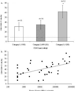
count and plasma viremia: Establishing a relationship between CD8+ T-cells expressing CD38 and CD4+ T-cell count and plasma viremia in the 41 HIV-1 infected individuals was first attempted. The HIV-infected individuals were divided according to the categorical classification of the Centers for Disease Control and Prevention (CDC) [20]. Figures 1A shows a strong association between the decreased number of CD4+ T-cells and the increased expression of CD38 on CD8+ T-cells. As expected, Figure 1B shows a strong positive correlation between CD38 expression on CD8+ T-cells and the levels of plasma viremia (r=0.780, p<0.001). These results confirm that the expression of CD38 on CD8+ T-cells is a strong cellular marker of HIV-1 disease progression.
Fig. 1: Relationship between levels of CD38 expression on CD8+ T-cells and CD4 count and plasma viremia. The level of CD38 expression was determined by flow cytometry by measuring the expression of CD38 on CD8+ T-lymphocytes. The HIV-infected population was divided according to 1993 CDC categorical classification. (A) CD4 count was determined by flow cytometry and (B) plasma viremia was determined by RT-PCR. The p-values were determined by Student’s t -test and Spearman’s rank correlation.
Relationship between levels of expression of apoptosis-related molecules on CD8+CD38+ T-cells and immunological status based on CD4+ T-cell count: To explain the gradual loss of anti-HIV activity during HIV-1 disease progression despite a significant increase of CD8+CD38+ T-cells in HIV-infected individuals, the levels of expression of apoptosis-related molecules on CD8+CD38+ T-cells were determined. The level of expression of two apoptosis-related molecules, TNFR-1 and Annexin V on CD8+CD38+ T-cells had no significant difference between HIV-negative and HIV-positive individuals in any of the three immunological categories (data not shown).
Fig. 2: Relationship between levels of expression of apoptosis-related molecules on CD8+CD38+ T-cells and immunological status based on CD4+ T-cell count. The level of apoptosis-related molecules was determined by flow cytometry by measuring the expression of CD95, Bcl-2, activated form of caspase-3, and CXCR4 on activated CD8+CD38+ T-lymphocytes. The HIV-infected population was divided according to 1993 CDC categorical classification. The p-values were determined by Student’s t-test.
Author Manuscrip
However, the level of expression of CD95, Bcl-2, CXCR4, and the active form of caspase-3 on CD8+CD38+ T-cells was examined, and an increase in the expression of these apoptosis-related molecules was observed depending on the immunological status.
Figure 2A shows that there was a significant increase in the expression of each apoptosis-related molecule on freshly isolated CD8+CD38+ T-cells from individuals falling into Category 1 and Category 2, but the most significant increment was observed in the late stage of disease or Category 3 as compared to HIV-negative group. The levels of apoptosis in CD8+CD38+ T-cells were not significantly different between Category 1 and Category 2 when the expression of one apoptosis-related molecule was analyzed. However, when the levels of expression of two apoptosis-related molecules were analyzed simultaneously on CD8+CD38+ T-cells shown in Figure 2B- and Figure 2C, the levels of apoptosis were lower in Category 1 than in Category 2 or Category 3. CD8+CD38+ T-cells from HIV-infected individuals in Category 3 showed the highest level of apoptosis among the three categories.
These results suggest that apoptosis analyses should be performed by evaluating the expression of more than one apoptosis-related molecule simultaneously to observe a difference between categories. In addition, these results confirmed that CD8+CD38+ T-cells are undergoing apoptosis at early stages of the infection and becoming more susceptible at the late stage of the disease, which could lead to a loss of anti-HIV effector function despite the high number of this CD8+ T-cell subpopulation.
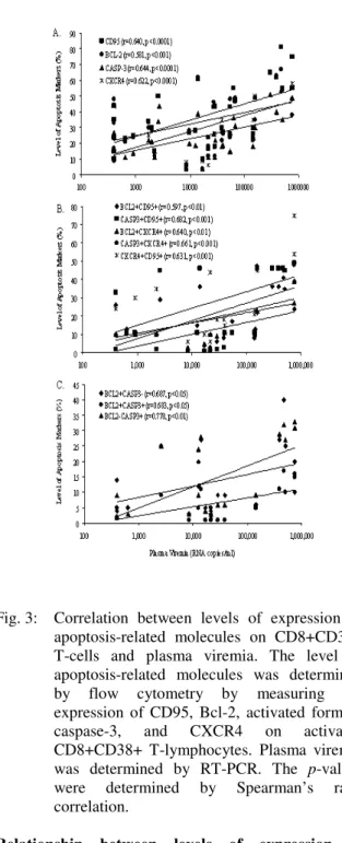
Correlation between levels of expression of apoptosis-related molecules on CD8+CD38+ T-cells and plasma viremia: The relationship between the expression of apoptosis-related molecules on CD8+CD38+ T-cells and the levels of plasma viremia was determined. A strong positive correlation was observed between these two parameters (Figure 3), suggesting that active HIV replication has a profound impact on the expression of apoptosis-related molecules on activated CD8+CD38+ T-cells. Figure 3 shows that there was a significant increase in the expression of apoptosis-related molecules on freshly isolated CD8+CD38+ T-cells from individuals with high levels of plasma viremia especially in those HIV-infected patients with more than 500,000 RNA copies/ml. These results confirmed that CD8+CD38+ T-cells are undergoing apoptosis in HIV-infected patients with low plasma viremia and becoming more susceptible at the
late stage of the disease possibly leading to immune dysfunction.
Fig. 3: Correlation between levels of expression of apoptosis-related molecules on CD8+CD38+ T-cells and plasma viremia. The level of apoptosis-related molecules was determined by flow cytometry by measuring the expression of CD95, Bcl-2, activated form of caspase-3, and CXCR4 on activated CD8+CD38+ T-lymphocytes. Plasma viremia was determined by RT-PCR. The p-values were determined by Spearman’s rank correlation.
Relationship between levels of expression of apoptosis-related molecules and levels of CD38 expression on CD8+ T-cells: To further delineate possible explanations for the lack of containment of
Author Manuscrip
viral replication despite an abundance of CD8+CD38+ T-cell compartment, the relationship between levels of expression of apoptosis-related molecules with the levels of CD38 expression on CD8+ T-cells from HIV-infected patients was determined.
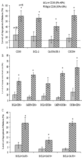
Fig. 4: Relationship between levels of expression of apoptosis-related molecules and levels of CD38 expression on CD8+ T-cells. The HIV-infected population was divided into low-CD38 and high-low-CD38 groups according to CD38 expression on their CD8+ T-cells. The levels of expression of CD95, Bcl-2, activated form of caspase-3, and CXCR4 on CD8+CD38+ T-cells was determined in both groups. The p-values were determined by Student’s t-test.
We divided the study subjects into two groups; a low-CD38 group (mean 32%, range 8% to 49%) and a high-CD38 group (mean 62%, range 50% to 85%) based on the mean value of CD8+CD38+ T-cells of all patients (43%).
In Figure 4A and Figure 4B, an increment for each of the four apoptosis-related molecules was observed in the low-CD38 group and was most prominent in the high-CD38 group as compared to the control group. Figure 4B shows that the portion of CD8+CD38+ T-cells that expressed CD95, also expressed Bcl-2 in high levels, with a significant increase in those CD8+ T-cells that expressed high levels of CD38 or at the late stage of HIV-1 infection. Similarly, it was also noted that CXCR4 and the active form of caspase-3 are highly co-expressed together with CD95 in the high-CD38 group. The expression of Bcl-2 shows no difference between the low-CD38 group and the control group; however, CD95, CXCR4, and the active form of caspase 3 show a significant difference between the low-CD38 group and the control group.
These results suggest that CD8+ T-cells are becoming highly susceptible to apoptosis following activation (CD38 expression) during the early stage of HIV-1 infection and the more CD8+ T-cells become activated the more they are susceptible to apoptosis at the late stage of the disease possibly leading to a poor effector function against HIV-1.
DISCUSSION
Inappropriate priming for apoptosis in lymphocytes from HIV-infected individuals could be a major factor contributing for the destruction of CD4+ and CD8+ T-cells in HIV-1 immunopathogenesis. The irregular apoptotic process seen in the infection is also the result of the state of chronic immune activation, which is characteristic of HIV-1 infection. This abnormal chronic stimulation leads to an aberrant up-regulation of the physiologic mechanisms that control T-cell apoptosis [16]. A recent study demonstrated a significant correlation between the frequency of HIV-specific CD8+ T-cells with the high-CD38 expression on these cells [4]. Another study showed the importance of CD8+CD38+ T-cells by demonstrating that these cells produce anti-HIV-1 soluble factors [21]. In addition, the presence of CD38 on CD8+ T-cells has been associated with HIV-1 disease progression, and its expression has been directly associated with the HIV-mediated immune activation that ultimately renders the CD8+ T-cells unable to control the viral replication [4]. The incapacity of CD8+ T-cells to control viral replication
Author Manuscrip
could be the result of an irregular and accelerated apoptotic process, due to immune hyperactivation, which is characteristic of HIV-1 infection. To understand the apoptotic process of CD8+CD38+ T-cells and their role in HIV-1 immunopathogenesis, the relationship between the expression of several apoptosis-related molecules on CD8+CD38+ T-cells with CD4+ T-cell count and plasma viremia was demonstrated.
A gradual augment in CD8+CD38+ T-cells during HIV-1 disease progression was shown in Figure 1. This increment was seen from the early stages of HIV-1 infection and was most notable at late stages of the disease. As the CD4+ T-cell count decreased during HIV-1 infection, the expression level of the CD38 marker on CD8+ T-cells incremented, as shown in Figure 1A. Similarly to other investigators, viral load was also demonstrated to positively correlate with CD38 expression on CD8+ T-cells. These findings are in accordance with previous studies that have shown that CD38 is associated with HIV-1 disease progression
[1, 2]
.
Based on the fact that there is limited information on the association between HIV-induced CD8+CD38+ apoptosis with CD4+ T-cell count and plasma viremia, a comprehensive analysis of HIV-mediated CD8+CD38+ T-cell apoptosis during 1 infection in 41 HIV-infected patients who were not injecting drug users and not receiving antiviral therapy was needed. When the samples were divided into three immunological categories according to their CD4+ T-cell count, the expression of the CXCR4, Bcl-2, CD95, and active caspase-3 was compared among three immunological categories according to the CD4+ T-cell count. The expression level of these apoptosis-related markers was found to incremented in Category 2 and the increment was most remarkably in Category 3 which is represents the late stage of HIV-1 infection (Figure 2). A significant positive correlation was also found between the levels of apoptosis-related molecules on CD8+CD38+ T-cells and plasma viremia (Figure 3).
The patients studied were also divided into two groups a group for low-CD38 expression and a group for high-CD38 expression. When the expression of the CXCR4, Bcl-2, CD95, and active caspase-3 in the low-CD38 and high-low-CD38 groups was compared, the expression level of apoptosis-related markers was found to increment in the low-CD38 group and increment was most remarkably in the high-CD38 group (Figure 4). These results suggest that the expression levels of CXCR4, CD95, Bcl-2 and active caspase-3 augments with the increase in activation and in the pool of CD8+ T-cells expressing high (greater than 50%) CD38.
The co-expression of apoptosis-related molecules on CD8+CD38+ T-cells was investigated and the majority of the CD8+CD38+ T-cells express CD95 and CXCR4 molecules at the late stage of the disease. All of the CD8+CD38+ T-cells that expressed active caspase-3 also expressed CD95 and CXCR4, suggesting that the most likely apoptosis mechanism is via the induction of these molecules. The co-expression of active caspase-3 (pro-apoptosis) and Bcl-2 (anti-apoptosis) on CD8+CD38+ T-cells was also investigated. A small percentage of CD8+CD38+ T-cells co-expressed both Bcl-2 and active caspase-3 and increased during HIV-1 infection. From this analysis, we also found that the majority of CD8+CD38+ T-cells at the late stage of the disease also expressed either Bcl-2 or active caspase-3. A majority of these cells are apoptotic because they expressed active caspase-3, and a minority of these cells are not apoptotic because they expressed Bcl-2 and not active caspase-3, however, they are highly prone to undergo apoptosis because these cells co-expressed both CD95 and CXCR4.
Based on these results, one question arises: Can apoptosis take place in a cell that co-expressed both Bcl-2 and active caspase-3? The answer lies in the mechanisms of the apoptotic process. Results indicate that CD95, CXCR4, and active caspase-3 are co-expressed on the majority of the CD8+CD38+ T-cells and increment from the early to the late stage of HIV-1 infection. When CD95 is bound by its ligand CD95L, a chain of processes toward apoptosis takes place in the cell. During this process, caspase-8 undergoes autocatalytic activation. Activated caspase-8 activates caspase-3, then active caspase-3 cleaves the DNA fragmentation factor 45 (DFF45) into a heterodimeric factor of DFF40 and DFF45. The DFF40 oligomer causes the internucleosomal DNA fragmentation, which is an apoptotic hallmark indicative of chromatin condensation [22]. Alternatively, Bcl-2 is an anti-apoptosis intracellular molecule that acts by inhibiting the cytochrome c-dependent apoptosis pathway [23]. However, Bcl-2 does not inhibit the action of active caspase-3 and the apoptotic process can take place in the cell. This provides a logical explanation in the sense that even in the presence of Bcl-2 in the cell caspase-8 can exert its function via another pathway which is the direct activation of procaspase-3 into active caspase-3 and once this is achieved, the cell undergoes apoptosis. Another question arises based on these results: Why is the number of CD8+CD38+ T-cells consistently elevated in HIV- infected patients despite the fact of the displayed high level of apoptosis? Although the levels of apoptotic-related molecules are elevated, the CD8+CD38+ T-cell count was not reduced. This
Author Manuscrip
apparent paradox may be resolved by observations in SIV-infected primates receiving total body irradiation, in which it was observed that significant CD8+ T-cell recovery preceded the recovery of CD4+ T-cells. A similar delay was also observed in humans receiving high dose chemotherapy. These data demonstrate that CD8+ T-cell rebound occurs prior to the CD4+ T-cell number rising again [24]. In HIV-infected patients, even though the number of both CD4+ and CD8+ T-cells is being reduced, the number of CD8+ T-cells may be greater than the CD4+ T-cell number because of the faster CD8+ T-cell recovery time [24].
Other therapeutic strategies in combination with HAART should aim at preventing HIV-induced CD8+ T-cell apoptosis. Recently, the presence of a surface molecule on CD8+ T-cells called PD-1, a member of CD28 family, has been demonstrated. This receptor is associated with CD8+ T-cell unresponsiveness, apoptosis, and disease progression [25]. The blockade of this receptor has been demonstrated to enhance antiviral immunity by rescuing antiviral CD8+ T-cells from apoptosis [26-27]. Thus, the blockade of PD-1 may prevent these cells from apoptosis and consequently should extend the life of HIV-specific CD8+ T-cells, resulting in a prolonged and more efficient containment of viral replication in HIV-infected patients.
In summary, these data suggest that activated CD8+CD38+ T-cells are undergoing apoptosis during HIV-1 infection, and one most likely mechanism is via CD95 and CXCR4 induction through the caspase cascade regardless of the presence of the anti-apoptotic molecule Bcl-2. All these observations may provide another explanation of why HIV-1 infection is not fully contained by HIV-specific CD8+CD38+ T-cells during HIV-1 disease progression. Future studies should aim at understanding the mechanisms by which HIV-specific CD8+CD38+ T-cells up-regulate CD95 and CXCR4 expression, for instance, by identifying soluble factors that can induce these molecules on CD8+CD38+ T-cells and once identified, these soluble factors could be blocked with neutralizing antibodies, assuming that these soluble factors are not integral part of natural physiological processes required by the cell.
ACKNOWLEDGEMENT
This work was supported by NIH/RCMI Grant # G12-RR03035 and #P20-RR016470 and by NIDA grants RO1-DA015628, RO1-DA012366, RO1-DA014218, and RO1-DA021537.
REFERENCES
1. Giorgi, J.V., Z. Liu, L.E. Hultin, W.G. Cumberland, K. Hennessey and R. Detels, 1993. Elevated levels of CD38+ CD8+ T cells in HIV infection add to the prognostic value of low CD4+ T cell levels: results of 6 years of follow-up. The Los Angeles Center, Multicenter AIDS Cohort Study. J. Acquir. Immune. Defic. Syndr., 6(8): 904-912.
2. Liu Z., W.G. Cumberland, L.E. Hultin, H.E. Prince, R. Detels and J.V. Giorgi, 1993. Elevated CD38 antigen expression on CD8+ T cells is a stronger marker for the risk of chronic HIV disease progression to AIDS and death in the Multicenter AIDS Cohort Study than CD4+ cell count, soluble immune activation markers, or combinations of HLA-DR and CD38 expression. J. Acquir. Immune. Defic. Syndr., 6(8): 904-912.
3. Ferrero, E and F. Malavasi, 1997. Human CD38, a leukocyte receptor and ectoenzyme, is a member of a novel eukaryotic gene family of nicotinamide adenine dinucleotide-converting enzymes: extensive structural homology with the genes for murine bone marrow stromal cell antigen 1 and aplysian ADP-ribosyl cyclase. J. Immunol., 159(8): 3858-3865.
4. Chun, T.W., J.S Justement, C. Sanford, C.W. Hallahan, M.A. Planta, M. Loutfy, S. Kottilil, S. Moir, C. Kovacs and A.S. Fauci, 2004. Relationship between the frequency of HIV-specific CD8+ T cells and the level of CD38+CD8+ T cells in untreated HIV-infected individuals. Proc. Natl. Acad. Sci., 101(8): 2464-2469.
5. Phillips, A.N.1996. Reduction of HIV concentration during acute infection: independence from a specific immune response. Science, 271(5248): 497-499.
6. Borrow, P., H. Lewicki, X. Wei, M.S. Horwitz, N. Peffer, H. Meyers, J.A. Nelson, J.E. Gairin, B.H. Hahn, M.B. Oldstone and G.M. Shaw, 1997. Antiviral pressure exerted by HIV-1-specific cytotoxic T lymphocytes (CTLs) during primary infection demonstrated by rapid selection of CTL escape virus. Nat. Med., 3(2): 205-211.
7. Schmitz, J.E., M.J. Kuroda, S. Santra, V.G. Sasseville, M.A. Simon, M.A. Lifton, P. Racz, K. Tenner-Racz, M. Dalesandro, B.J. Scallon, J. Ghrayeb, M.A. Forman, D.C. Montefiori, E.P. Rieber, N.L. Letvin and K.A.Reimann, 1999. Control of viremia in simian immunodeficiency virus infection by CD8+ lymphocytes. Science, 283(5403): 857-860.
Author Manuscrip
8. Stranford, S.A., J. Skurnick, D. Louria, D. Osmond, S.Y. Chang, J. Sninsky, G. Ferrari, K. Weinhold, C. Lindquist and J.A. Levy, 1999. Lack of infection in HIV-exposed individuals is associated with a strong CD8+ cell noncytotoxic anti-HIV response. Proc. Natl. Acad. Sci. USA, 96(3):1030-1035.
9. Papagno, L., C.A. Spina, A. Marchant, M. Salio, N. Rufer, S. Little, T. Dong, G. Chesney, A. Waters, P. Easterbrook, P.R. Dunbar, D. Shepherd, V. Cerundolo, V. Emery, P. Griffiths, C. Conlon, A.J. Mc Michael, D.D. Richman, S.L. Rowland-Jones and V. Appay, 2004. Immune activation and CD8+ T-cell differentiation towards senescence in HIV-1 infection. Plos. Biol., 2(2): E20.
10. Roederer M, J.G.Dubs, M.T. Anderson, P.A. Raju, L.A. Herzemberg, 1995. CD8 naïve T cell counts decrease progressively in HIV-infected adults. J. Clin. Invest., 95:2061-2066.
11. Cotton, M.F., D.N. Ikle, E.L. Rapaport, S. Marschner, P.O. Tseng, R. Kurrie and T.H. Finkel, 1997. Apoptosis of CD4+ and CD8+ T cells isolated immediately ex vivo correlates with disease severity in human immunodeficiency virus type 1 infection. Pediatr. Res., 42(5): 656-664.
12. Gougeon, M.L, 2003. Apoptosis as an HIV strategy to escape immune attack. Nat. Rev. Immunol., 3(5): 392-404.
13. Petrovas, C., Y.M. Mueller, P.D. Katsikis, 2004. HIV-specific CD8+ T cells: serial killers condemned to die? Curr. HIV Res., 2(2): 153-162. 14. Petrovas, C., Y.M. Mueller and P.D. Katsikis,
2005. Apoptosis of HIV-specific CD8+ T cells: an HIV evasion strategy. Cell Death Differ. Suppl.1: 859-870.
15. Katsikis, P.D., E.S. Wunderlich, C.A, Smith and L.A. Herzenberg, 1995. Fas antigen stimulation induces marked apoptosis of T lymphocytes in human immunodeficiency virus-infected individuals. J. Exp. Med., 181(6):1030-1039. 16. De Oliveira Pinto, L.M., S. Garcia, H. Lecoeur, C.
Rapp and M.L. Gougeon, 2002. Increased sensitivity of T lymphocytes to tumor necrosis factor receptor I (TNRFI) - and TNRF2-mediated apoptosis in HIV infection: Relation to expression of Bcl-2 and active caspase-8 and caspase-3. Blood, 99(5): 1666-1675.
17. Decrion, A.Z., A.Varin, J.M. Estavoyer and G. Herbein, 2004. CXCR4-mediated T cell apoptosis in human immunodeficiency virus infection. J. Gen. Virol., 85(6):1471-1478.
18. Terai, C., R.S. Kornbluth, C.D. Pauza, D.D. Richman and D.A. Carson, 1991. Apoptosis as a mechanism of cell death in cultured T lymphoblasts acutely infected with HIV-1. J. Clin. Invest., 87(5): 1710-1715.
19. Holm, G.H and D. Gabuzda, 2005. Distinct mechanisms of CD4+ and CD8+ T-cell activation and bystander apoptosis induced by human immunodeficiency virus type 1 virions. J. Virol., 79(10): 6299-6311.
20. Center for Disease Control and Prevention, 1992. 1993 revised classification system for HIV infection and expanded surveillance case definition for AIDS among adolescents and adults. MMWR Recomm. Rep., 41(RR-17): 1-19.
21. Jiang, J.Q., S. Balasubramanian, N.C. Hawley-Foss, A.D. Badley, K.L. Rosenthal and K.F. Copeland, 2003. Production of CD8+ T cell nonlytic suppressive factors by CD28, CD38, and HLA-DR subpopulations. AIDS Res. Hum. Retroviruses, 19(6): 497-502.Widlak, P., 2000. The DFF40/CAD endonuclease and its role in apoptosis. Acta. Biochim. Pol., 47(4):1037-1044. 23. Hawkins, C.J. and D.L. Vaux DL, 2004. Analysis
of the role of bcl-2 in apoptosis. Immunol. Rev., 142:127-139.
24. Badley, A.D., A.A. Pilon, A. Landay and D.H. Lynch, 2000. Mechanisms of HIV- associated lymphocyte apoptosis. Blood, 96(9): 2951-2964. 25. Day, C.L., D.E. Kaufmann, P. Kiepiela, J.A.
Brown, E.S. Moodley, S. Reddy, E.W. Mackey, J.D. Miller, A.J. Leslie, C. DePierres, Z. Mncube, J. Duraiswamy, B. Zhu, Q. Eichbaum, M. Altfeld, E.J. Wherry, H.M. Coovadia, P.J. Goulder, P. Klenerman, R. Ahmed, G.J. Freeman and B.D. Walker, 2006. PD-1 expression on HIV-specific T cells is associated with T-cell exhaustion and disease progression. Nature, 443(7109): 350-354. 26. Petrovas, C., J.P. Casazza, J.M. Brenchley, D.A.
Price, E. Gostick, W.C. Adams, M.L. Precopio, T. Schacker, M. Roederer, D.C. Douek, and R.A. Koup, 2006. PD-1 is a regulator of virus-specific CD8+ T cell survival in HIV infection. J. Exp. Med., 203: 2281-2292.
27. Trautmann, L., L. Janbazian, N. Chomont, E.A. Said, S. Gimmig, B. Bessette, M.R. Boulassel, E. Delwart, H. Sepulveda, R.S. Balderas, J.P. Routy, E.K. Haddad and R.P. Sekaly, 2006. Upregulation of PD-1 expression on HIV-specific CD8+ T cells leads to reversible immune dysfunction. Nat. Med., 12(10): 1198-1202.