Original article
Etiological treatment during early chronic indeterminate
Chagas disease incites an activated status on innate and adaptive
immunity associated with a type 1-modulated cytokine pattern
Renato Sathler-Avelar
a,b,*
, Danielle Marquete Vitelli-Avelar
a, Rodrigo Lima Massara
a,
Marta de Lana
c, Jo
~
ao Carlos Pinto Dias
d, Andre´a Teixeira-Carvalho
a,
Silvana Maria Elo´i-Santos
a,b, Olindo Assis Martins-Filho
aaLaboratory of Chagas Disease, CPqRR-FIOCRUZ, Av. Augusto de Lima 1715, 30190-002 Belo Horizonte, MG, Brazil bPost-graduation Center, FM, UFMG Av. Alfredo Balena 190, 31130-100 Belo Horizonte, MG, Brazil cLaboratory of Parasitology and Pathology, UFOP, Campus Universita´rio-ICEB II, 35400-000 Ouro Preto, MG, Brazil
dLaboratory of Triatomines and Epidemiology of Chagas Disease, CPqRR-FIOCRUZ, Av. Augusto de Lima 1715, 30190-002 Belo Horizonte, MG, Brazil Received 28 August 2007; accepted 17 October 2007
Available online 22 October 2007
Abstract
Pro-inflammatory immune response is usually associated with Chagas disease pathogenesis, but is also relevant to treatment effectiveness. Cross-sectional studies have suggested that this activated state may persist for years after therapeutic intervention. However, short-term longi-tudinal investigation has suggested that the Benznidazole treatment (Bz-treatment) leads to decreased immunological activation. In order to elu-cidate this issue, we performed a longitudinal study to evaluate the immunological status following Bz-treatment during early indeterminate Chagas disease. Our results demonstrated that Bz-treatment led to higher activation status of circulating monocytes but was negatively associated with the number of IL-12þ
CD14þ
cells. Moreover, Bz-treatment triggered a high frequency of circulating CD3
CD16þ
CD56
NK cells, in addition to elevated activation status associated with a type 1-modulated cytokine pattern. Bz-treatment induced substantial T and B-cell activation status associated with an overall IL-10 modulated type 1 cytokine profile. In summary, these findings provide new information regarding immune activation status following the etiological treatment of Chagas disease. These results suggest that in addition to the increased number of activated leukocytes in the peripheral blood, Bz-treatment may also involve a qualitative change in their functional capacity that drives their activation state toward a modulated cytokine profile. These changes may account for the benefits of etiological treatment of Chagas disease.
Ó2007 Elsevier Masson SAS. All rights reserved.
Keywords:Chagas disease; Benznidazole; Immune response; Cytokines; Leukocytes subsets
1. Introduction
Trypanosoma cruzi is the etiological agent of American Trypanosomiasis or Chagas disease, which affects 16e18
million people in South and Central America [1]. Chagas
disease is a long-lasting infection with a short acute phase, which is usually clinically non-apparent, that progresses to a lifelong chronic phase characterized by distinct clinical forms known as indeterminate, cardiac and digestive [1].
Specific chemotherapy is recommended for the treatment of Chagas disease applying the general assumption that the ear-lier the specific treatment is initiated the greater the chance of parasitological cure[2]. At present, Chagas disease chemo-therapy in Brazil has been restricted to the use of Benznida-zole, which is recommended for the treatment of acute, * Corresponding author. Laboratory of Chagas Disease, CPqRR-FIOCRUZ,
Av. Augusto de Lima 1715, 30190-002 Belo Horizonte, MG, Brazil. Tel.:þ55
31 3349 7764; fax:þ55 31 3295 3115.
E-mail address:[email protected](R. Sathler-Avelar).
1286-4579/$ - see front matterÓ2007 Elsevier Masson SAS. All rights reserved.
doi:10.1016/j.micinf.2007.10.009
Microbes and Infection 10 (2008) 103e113
congenital and the initial stage of the indeterminate form, known as early-indeterminate disease (E-IND), usually seen in children and adolescents[2,3]. An effective treatment might lead to parasite clearance and prevent the progression of infec-tion to disease, heart-related pathology and its complicainfec-tions
[4e7].
It has been suggested that parasite clearance following che-motherapy in the chronic phase may contribute to better clin-ical outcome [4,5,8]. Indeed, Andrade et al. [8] have demonstrated regression of myocardial inflammatory lesions following parasite clearance archived by etiological treatment in mice.
Despite the well known role of the host immune response in the pathogenesis of Chagas disease, little has been reported about the impact of Bz-treatment on this response. It has been demonstrated that after specific treatment cured patients produce high levels of IFN-g[9]. Because an exacerbated pro-duction of IFN-gmay favor the development of a strong pro-inflammatory response, which is mainly observed in patients with cardiac disease[10], it is possible that a fine balance of pro- and anti-inflammatory cytokines could be the major key in controlling Chagas disease morbidity following treatment
[11]. We have previously reported that Bz-treatment leads to a type 1-modulated immune profile, with IL-10 as the putative key element for controlling the deleterious tissue damage that eventually might occur due to the IFN-g-mediated pro-inflam-matory response observed during Bz-treatment [11]. These findings suggest that in addition to the direct role in blocking parasite growthin vivo, Bz-treatment appears to affect host im-mune regulation[12]. Together, these parasitological and im-munological hypotheses have brought new perspectives to clinical investigations and have stimulated studies to establish the effect of specific anti-parasite therapy on the immune state and the evolution of Chagas disease.
In spite of these insights, little research has been carried out to address the impact of etiological Bz-treatment in the host immune response during early indeterminate Chagas disease. Therefore, our goal in this study was to evaluate in a longitudi-nal investigation, the impact of Bz-treatment on the ex vivo
phenotypic profile of peripheral blood leukocytes in associa-tion with their cytokine pattern during early indeterminate
T. cruzi infection, prior to, and one year after the end of the Bz-therapeutic intervention.
2. Patients, materials and methods
2.1. Study population
The patients included in this investigation consisted of thir-teen schoolchildren from Berilo and Jose´ Gonc¸alves de Minas. The early-indeterminate Chagas disease group (E-IND) con-sisted of six schoolchildren (9e14 years old), with antibodies
toT. cruzias detected by serology performed as recommended by the World Health Organization criteria[1]. The clinical and physical examination revealed that all children were asymp-tomatic, with normal conventional electrocardiograms and un-altered thoracic X-rays. Haemoculture was positive in all but
one (5/6) of the seropositive children, generally within the first month of blood cultivation in Liver Infusion Tryptose medium (LIT). Chagasic children were treated with benznidazole (RochaganÒ; Roche, SP, Brazil) following the protocol recom-mended by the Brazilian Health Ministry [3], consisting of 8 mg/kg per day for 60 consecutive days. Following etiologi-cal treatment, all children were re-evaluated one year after the end of Bz-treatment (E-INDT).
The non-infected control group (NI) consisted of seven age-matched schoolchildren with negative serology for anti-T. cruzi(9e14 years old).
Informed written consent was obtained from all through their parents or legal guardians. This work fulfilled resolution number 196/1996 from the Brazilian National Health Council for research involving humans.
2.2. Immune-staining for cell surface markers of peripheral blood
White blood cell phenotypes were analyzed following an immunofluorescence procedure recommended by Becton-Dickinson (Mountain View, CA, USA), modified as follows: 100ml peripheral blood, collected using Vacutainer tubes with EDTA as anticoagulant, was mixed with 5ml undiluted monoclonal antibodies (mAbs) specific for several cell surface markers labeled with, fluorescein isotiocyanate-FITC, phy-coerithrin-PE or tricolor dye-TC, all purchased from Becton Dickinson (San Diego, CA, USA) or Caltag (Burlingame, CA, USA), including anti-CD3-FITC and PE, clone UCHT1; anti-CD4-FITC and PE, clone RPA-T4; anti-CD5-FITC, clone L17F12; anti-CD8-FITC and TC, clone RPA-T8; anti-CD14-TC, clone Tu¨K4; anti-CD16-FITC and TC, clone 3G8; anti-CD18-FITC, clone YF118.3; anti-CD19-FITC and TC, clone 4G7; anti-CD23-PE, clone M-L233; anti-CD28-FITC, clone 15E8; CD54-PE, clone 15.2; CD56-PE, clone B159; anti-CD62L-FITC, clone DREG-56; anti-CD69-FITC, clone FN50 and anti-HLA-DR-PE, clone TU¨ 36. Following incubation in the dark for 30 min at room temperature, erythrocytes were lysed in FACS Lysing Solution (Becton Dickinson Biosciences Pharmigen, San Diego, CA, USA). The remaining cells were then washed in phosphate-buffered saline containing 0.01% sodium azide. Cell preparations were fixed in 200ml of FACS-FIX Solution (10 g/l paraformaldehyde, 1% sodium-cacodylate, 6.65 g/l sodium-chloride, 0.01% sodium azide). Data acquisition was performed with a Becton-Dickinson FACScalibur instrument. CELLQuestÔ software provided
by the manufacturer was used for data acquisition and analysis.
2.3. Analysis of intracellular cytokines in leukocytes after in vitro short-term culture of whole blood
For each blood sample collected using Vacutainer tubes with sodium heparin as anticoagulant, short-termin vitro cul-tures of whole blood were performed to reproduce theex vivo
NY, USA) plus Brefeldin A (BFA) (Sigma, St. Louis, MO, USA), at a final concentration of 10mg/ml, in 14 ml polypro-pylene tubes (FalconÒ, BD Pharmingen). The culture was maintained for 4 h at 37
C in a 5% CO2humidified incubator.
At the end of incubation, all cultures were treated with EDTA (Sigma) at a final concentration of 2 mM for 15 min at room temperature. EDTA-treated whole blood cultures were washed once with FACS buffer (PBS with 0.5% of bo-vine serum albumin, BSA, pH 7.4, Sigma), by centrifugation at 600g for 7 min at room temperature, and resuspended to half the original volume with FACS buffer. Samples of cell suspension (200ml) from cultures were incubated with 10ml diluted TC-labeled anti-cell surface marker mAbs pur-chased from Caltag (Burlingame, CA, USA) including anti-CD4, clone RPA-T4; anti-CD8, clone RPA-T8; anti-CD14, clone Tu¨K4; anti-CD16, clone 3G8 and anti-CD19, clone 4G7. After incubation for 30 min at room temperature in the dark, cell surface-stained samples were treated with 2 ml of FACS Lysing/fix Solution (BD Pharmingen), immediately vor-texed and re-incubated for an additional 10 min. After the lys-ing/fixation procedure, membrane-stained leukocytes were permeabilized for another 10 min with 2 ml of FACS perm-buffer (FACS perm-buffer supplemented with 0.5% saponin, Sigma), washed and resuspended into 200ml FACS perm-buffer. Fixed/ permeabilized membrane-stained leukocyte suspensions were distributed in 30ml aliquots in 96-well U-bottomed microtiter plates and incubated for 30 min at room temperature in the dark in the presence of 20ml diluted PE-labeled anti-cytokine mAbs purchased from BD-Pharmingen (San Diego, CA, USA) including anti- IL-12p40/p70, clone C11.5; anti-IFN-g, clone 4S.B3; anti-TNF-a, clone MAb11; anti-IL-4, clone BVD4-1D11 or anti-IL-10, clone JES3-19F1). TC and PE-labeled iso-typic control was included in each batch of experiments. After intracytoplasmatic staining, the cells were washed once with FACS perm-buffer, followed by one wash step with FACS buffer and were fixed in FACS FIX Solution.
Flow cytometric analyses were performed using a FACScaliburÒ
flow cytometer (BD Pharmingen, San Diego, CA, USA), which acquired a total of 30,000 events per tube.
2.4. Statistical analysis
Comparative analysis was performed by unpairedttest (NI versus E-IND or E-INDT) or paired t test (E-IND versus
E-INDT) using Prism software (version 4.03). Correlation
analysis was performed by Pearson’s test. Significance was defined in all cases atp<0.05.
3. Results
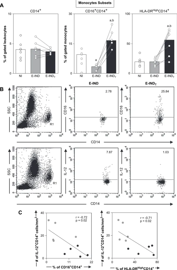
3.1. Higher activation status of circulating monocytes exhibiting negative association with IL-12producing CD14þ
cells following Chagas disease Bz-treatment
In order to verify whether etiological treatment induces variations in the percentage of monocyte subsets, including macrophage-like (CD16þ
CD14þ
) and pro-inflammatory
monocytes (HLA-DRHighCD14þ), we evaluated CD16 and HLA-DR expression on the surface of circulating CD14þ cells. Despite the fact that the frequency of CD14þmonocytes was similar in all groups (Fig. 1A), the percentage of macro-phage-like and pro-inflammatory monocytes (Fig. 1A) was significantly higher in the E-INDTgroup than in both E-IND
and NI groups.
We observed a negative correlation between the frequency of CD16þ
CD14þ
and HLA-DRHighCD14þ
with IL-12þ
CD14þ
monocytes (Fig. 1C). Additionally, no correlation was found between these monocyte subsets and the level of TNF-aþCD14þ monocytes (data not shown). Representative flow cytometry charts illustrate the enhanced frequency of macro-phage-like cells as well as lower IL-12þCD14þmonocytes in the E-INDTgroup as compared to the E-IND group (Fig. 1B).
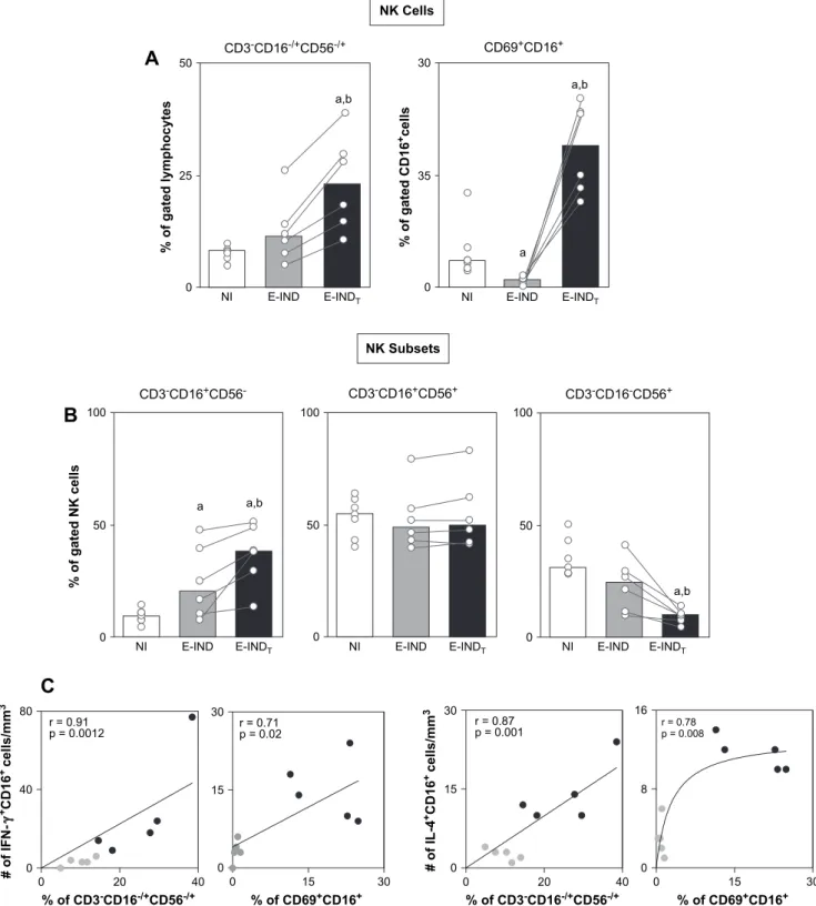
3.2. Bz-treatment during early indeterminate Chagas disease led to a high frequency of circulating total CD3CD16/þCD56/þNK cells that mainly exhibit the CD3CD16þCD56phenotype as well as elevated percentage of CD69þCD16þNK cells associated with a type 1-modulated cytokine pattern
Three major NK cell subsets have been previously evalu-ated in Chagas disease [13], including CD3
CD16þ
CD56
, CD3
CD16þ
CD56þ
and CD3
CD16
CD56þ
cells. Our find-ings showed a higher percentage of total CD3
CD16/ þ
CD56/þ
NK cells in the E-INDTgroup than in the E-IND
and NI groups (Fig. 2A, left panel), mainly due to the in-creased percentages of the CD3CD16þCD56 subset (Fig. 2B). No significant differences were observed in the mean percentages of CD3CD16þCD56þ subset (Fig. 2B) in the E-INDTgroup as compared to the E-IND and NI groups.
Despite the slight decrease in CD3CD16CD56þ (Fig. 2B), the frequency of CD16þcells expressing the early activation marker CD69þ
CD16þ
was considerably increased in the E-INDT group compared to the E-IND and NI groups
(Fig. 2A).
Additional analyses demonstrated a positive correlation be-tween the percentage of total CD3
CD16/þ
CD56/þ
NK cells as well as CD69þ
CD16þ
NK cells and the absolute num-ber of both IFN-gþand IL-4þNK-cells (Fig. 2C).
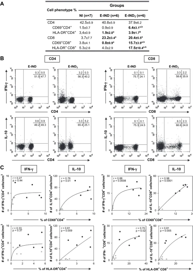
3.3. Bz-treatment led to substantial T-cell activation status, but was associated with an overall type 1 highly modulated T-cell-derived cytokine profile
Despite several reports of higher T-cell activation during chronic Chagas disease [14e16], we have previously
demon-strated that E-IND is characterized by a T-cell-independent immunity with no changes in the frequency of circulating HLA-DRþ
R1
SSC IL-12 IL-12
SSC
R1
CD16 CD16 E-IND
%
o
f
g
a
te
d
l
e
u
k
o
c
y
te
s
0 5 10
0 15 30
a,b
a
a,b
0 50 100
NI E-INDT NI E-IND E-INDT NI E-IND E-INDT
CD14+ CD16+CD14+ HLA-DRHighCD14+
Monocytes Subsets
0 11 22
0 20 40
0 40 80
0 20 40
A
r = -0.71 p = 0.02 r = -0.72
p = 0.02
#
o
f
IL
-1
2
+C
D
1
4
+ c
e
ll
s
/m
m
3
#
o
f
IL
-1
2
+C
D
1
4
+ c
e
ll
s
/m
m
3
% of HLA-DRHighCD14+
% of CD16+CD14+
C
CD14
E-IND E-INDT
CD14
%
o
f
g
a
te
d
m
o
n
o
c
y
te
s
B
2.78 25.847.87 1.03
Fig. 1. (A) Analysis of monocyte subsets in the peripheral blood from non-infected children (NIe ) and early indeterminateT. cruziinfected patients prior to
the Bz-treatment (E-INDe ) and one year after it (E-INDTe ). Phenotypic studies were performed by a triple-labeling protocol using anti-CD16 FITC,
anti-HLA-DR PE, and anti-CD14 TC to identify total monocytes CD14þ
, macrophage-like monocytes CD16þ
CD14þ
, and pro-inflammatory monocytes
HLA-DRHighCD14þ
. The results are expressed as scattering of individual values and median percentage. Significant differences atp<0.05 are identified by letters
‘‘a’’ and ‘‘b’’ in comparison to NI and E-IND groups, respectively. (B) Representative dot plots illustrating the higher frequency of CD16þ
CD14þ
monocytes
and a lower frequency of IL-12þ
CD14þ
monocytes in the E-INDTgroup compared to the E-IND group. Quadrant statistics were used for data analysis, and
the results are expressed as the percentage of positive cells within the selected gate. (C) Correlation analyses showed the negative correlation between
macro-phage-like or pro-inflammatory monocytes and IL-12þ
CD14þ
0 15 30 0
8 16
0 20 40
0 15 30
0 15 30
0 15 30
0 20 40
0 40 80
%
o
f
g
a
te
d
l
y
m
p
h
o
c
y
te
s a,b
0 25 50
NI E-IND E-INDT
NK Cells
0 35 30
NI E-IND E-INDT a,b CD3-CD16-/+CD56-/+ CD69+CD16+
CD3-CD16+CD56
-a a,b
0 50 100
NI E-IND E-INDT
CD3-CD16+CD56+
0 50 100
NI E-IND E-INDT
CD3-CD16-CD56+
NI E-IND E-INDT a,b
0 50 100 NK Subsets
%
o
f
g
a
te
d
N
K
c
e
ll
s
#
o
f
IF
N
+C
D
1
6
+ c
e
ll
s
/m
m
3
% of CD3-CD16-/+CD56-/+ % of CD69+CD16+
#
o
f
IL
-4
+C
D
1
6
+ c
e
ll
s
/m
m
3
% of CD3-CD16-/+CD56-/+ % of CD69+CD16+
A
B
C
r = 0.71 p = 0.02 r = 0.91
p = 0.0012
r = 0.78 p = 0.008 r = 0.87
p = 0.001 a
%
o
f
g
a
te
d
C
D
1
6
+c
e
ll
s
Fig. 2. (A) Analysis of total CD3
CD16/þ
CD56/þ
NK cells, CD69þ
CD16þ
NK cells and (B) its subsets in the peripheral blood from non-infected children
(NIe ) and early indeterminateT. cruzi-infected patients prior to the Bz-treatment (E-INDe ) and one year after it (E-INDTe ). Phenotypic studies
were performed by a triple-labeling protocol using anti-CD3 FITC, anti-CD56 PE, and anti-CD16 TC to identify CD3
CD16/þ
CD56/þ
, CD3
CD16þ
CD56
,
CD3
CD16þ
CD56þ
, and CD3
CD16
CD56þ
within total NK cells. Double-labeling protocol was performed to quantify the percentage of CD69þ
CD16þ
cells.
The results are expressed as scattering of individual values and median percentage. Significant differences atp<0.05 are identified by letters ‘‘a’’ and ‘‘b’’ in
comparison to the NI and E-IND groups, respectively. (C) Correlation analyses showed a positive correlation between CD3
CD16/þ
CD56/þ
NK cells or
CD69þCD16þNK cells with IFN-gþCD16þand IL-4þCD16þcells after Bz-treatment.
upon lymphocyte activation, not detected in resting cells. Al-though CD69 have been considered a transient cell surface molecule expressed following lymphocyte activation, it has been demonstrated that CD69 can be persistently expressed
in vivo by T-cells under certain conditions characterized by chronic inflammation [18]. Moreover, despite CD69 have been considered a typical activation markers associated with pro-inflammatory function, recent studies have also indicated that this receptor may act as a regulatory molecule, dow-reg-ulation the immune response through the production of pleio-tropic cytokines [19]. Herein, we have evaluated the expression of CD69 immediately after blood collection, re-ferred as ‘‘ex vivo’’ expression in the absence of in vitro stim-ulation with antigen or mitogen in culture. Therefore, the results reflect the activation in vivo status of circulation lymphocytes.
Despite the lower percentage of HLA-DRþ CD4þT-cells and the lower percentage of circulating CD69þCD8þT-cells observed in the E-IND group as compared with NI, which sup-ports our previous hypothesis that E-IND is characterized by a T-cell independent immunity our results reveal, for the first time, an overall high activation status in both innate and adaptive immune response following Bz-treatment during early indeterminate Chagas disease. Interestingly, increased percent-ages of HLA-DRþ
as well as CD69þ
cells within both T-cell subsets can be observed in the E-INDTgroup (Fig. 3A) in
com-parison to the E-IND group.Moreover, decreased percentage of CD62Lþ
CD8þ
T-cells was observed in the E-INDT group in
comparison to the E-IND group (Table 1). Increased levels of activated T-cells following Bz-treatment of chronic Chagas dis-ease have been already described[15].
We have further characterized this highly activated status by associating the T-cell activation phenotypes with their cytokine secretion pattern. Our data demonstrated a positive correlation between activated CD4þ
T-cells (both CD69þ
and HLA-DRþ
) and the number of IL-10þ
CD4þ
T-cells (Fig. 3C, left panel). A positive correlation was also observed between activated CD8þ
T-cells (both CD69þ
and HLA-DRþ
) and the absolute number of IL-10þ
CD8þ
cells. Additionally, a positive correlation between activated CD8þ
T-cells was observed with the IFN-gþCD8þcells.
Further analysis between have demonstrated positive correla-tion between activated CD8þT-cells and the absolute number of IL-4þCD8þcells (r¼0.84,p¼0.002 for CD69þCD8þcells andr¼0.79,p¼0.005 for HLA-DRþCD8þcells). Additional correlation analysis between cytokine producing cells revealed positive correlation between IFN-gþCD8þT-cells and IL-10-producing T-cells (r¼0.86,p¼0.005 for CD4þandr¼0.92,
p¼0.0003 for CD8þ
T-cells) as well as between IFN-gþCD8þT-cells and IL-4þCD8þcells (r¼0.84,p¼0.003).
In summary, our data suggest that Bz-treatment led to substantial T-cell activation status associated with an overall type 1 highly modulated T-cell-derived cytokine profile (Fig. 3C, right panel). Representative flow cytometry charts are provided to illustrate the type 1-modulated cytokine pattern synthesized by T-cell subsets after Bz-treatment (Fig. 3B).
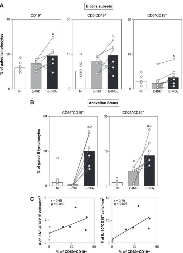
3.4. B-lymphocytes displayed an activated profile associated with a mixed type 1/type 2 cytokine pattern following Bz-treatment of early indeterminate Chagas disease
In order to further characterize the impact of Bz-treatment in the adaptive immunity, we have quantified the frequency of circulating B-cells and their subsets, as well as their activation status. The analysis on the B-cell compartment revealed that members of the E-INDT group have increased percentages
of circulating B-lymphocytes (Fig. 4A), owing to increased percentages of both B1 (CD5þ
CD19þ
) and B2 (CD5
CD19þ
) lymphocyte subsets (Fig. 4A, middle and right panel). Analy-sis of B-cell activation status revealed a markedly higher per-centage of activated CD69þCD19þand CD23þCD19þB-cells in the E-INDTgroup compared with the E-IND and NI groups
(Fig. 4B).
Additional analyses demonstrated a positive correlation be-tween activated CD69þCD19þB-lymphocytes and the number of both TNF-aþand IL-10þCD19þB-cells (Fig. 4C). As was later demonstrated for CD4þ
T-cells, a positive correlation was observed between IFN-gþCD8þT-cells and IL-10þCD 19þ
B-lymphocytes (data not shown), suggesting, again, an im-portant role of IL-10 synthesis by other cells in the control of inflammatory CD8þ
T-cell activity.
4. Discussion
The etiological treatment of Chagas disease has a beneficial impact on clinical status besides the parasite clearance, and may also affect the nature of the immune response of treated hosts [4,5,8e12,20]. Despite the ability of Bz-treatment to
postpone or prevent clinical progression in human and exper-imental models[2,5,7], there are still few studies focusing on the immune response following Bz-treatment for Chagas disease.
It has been proposed that Bz-treatment is able to affect the host immunity profile and contributes to reduce the clinical symptoms of Chagas disease, regardless of the parasitological cure. Indeed, the Bz-treatment in chronic experimental infec-tion prevents cardiomyopathy, despite the lack of complete parasite eradication [5], and long-term follow-up studies of Bz-treatment of human chronic infection demonstrated lower clinical progression to heart disease despite parasitological cure[4].
On the other hand, it has been postulated that etiological treatment per se does not seem to be responsible for the changes, since therapeutic failure, demonstrated by the pres-ence of circulating parasites, is not accompanied by significant alterations in immunological profile[15]. Indeed, Dutra et al.
Cell phenotype %
Groups
NI (n=7) E-IND (n=6)
CD69+CD4+ 1.5±0.7 0.9±0.9 6.4±3.1a,b HLA-DR+CD4+ 3,4±0.9 1.9±2.0a 3.9±1.7b
HLA-DR+ CD8+ 5.3
±2.6 4.0±2.9 17.5±10.4a,b
CD4 42.5±5.9 40.8±5.9 37.9±6.2
E-INDT (n=6)
CD8 3.7±7.7 23.2±3.4a 20.4±5.5a CD69+CD8+ 3.8±4.1 0.8±0.9a 15.7±3.9a,b
A
B
CD4 E-IND IF N -γγ IL -1 0 E-INDT CD4 # o f I L 1 0 +C D 4+ c
e ll s /m m 3
% of CD69+CD4+
# o f IL 1 0 +C D 4
+ c
e ll s /m m 3
0 5 10
0 8 16
0 5 10
0 40 80 120
0 2 4 6
0 8 16
0 3 6
0 40 80 120 r = 0.33
p = 0.37 r = 0.81p = 0.009
CD4
IFN-γ IL-10
C
r = 0.27
p = 0.44 p = 0.01 r = 0.76
# o f IF N -γ +C D 4
+ c
e ll s /m m 3 # o f IF N -γ +C D 4
+ c
e ll s /m m 3
% of HLA-DR+CD4+
0.3 3.2 40.2 56.3 0.5 0.3 47.7 51.5 0.9 0.6 49.3 49.2 5.2 3.3 25.1 65.4 IL -1 0
E-IND E-INDT
CD8 IF N -γ CD8 # o f IL -1 0 +C D 8
+ c
e ll s /m m 3 # o f IL -1 0 +C D 8
+ c
e ll s /m m 3
% of CD69+CD8+
0 12 24
0 15 30
0 12 24
0 30 60
0 20 40
0 15 30
0 20 40
0 30 60 r = 0.70
p = 0.02 r = 0.81 p = 0.005
CD8
IFN-γ IL-10
r = 0.88
p = 0.0008 p < 0.0001 r = 0.98
% of HLA-DR+ CD8+
# o f IF N -γ +C D 8
+ c
e ll s /m m 3 # o f IF N -γ +C D 8
+ c
e ll s /m m 3 0.1 0.1 24.1 75.7 2.2 3.3 35.9 58.6 0.1 0.1 24.5 75.3 2.3 6.3 24.8 66.6
Fig. 3. (A) Activation status of T-lymphocytes subsets (CD4þ
and CD8þ
) in the peripheral blood from non-infected children (NI) and early indeterminate T. cruzi-infected patients prior to the Bz-treatment (E-INDe ) and one year after it (E-INDTe ). A double-labeling protocol was performed to identify
percentage of CD69þ
and HLA-DRþ
in T-cell subsets. The results are expressed as scattering of individual values and median percentage. Significant differences at
p<0.05 are identified by letters ‘‘a’’ and ‘‘b’’ in comparison to the NI and E-IND groups, respectively. (B) Representative dot plots illustrating the increase on
IL-10 synthesis by CD4þ
T cells and a type 1-modulated immune profile by CD8þ
T-cells in the E-INDTgroup compared to the E-IND group. Quadrant statistics were
used for data analysis, and the results are expressed as the percentage of positive cells within the selected gate. (C) Correlation analyses showed the positive
cor-relation between activated CD4þ
T-lymphocytes subset and anti-inflammatory cytokine pattern (IL-10þ
CD4þ
) after Bz-treatment; however, a positive correlation
was observed between activated CD8þ
T-lymphocytes subset and a mixed cytokine pattern (IFN-gþ
CD8þ
and IL-10þ
CD8þ
Bz has a selective impact in host immune response as demon-strated by its ability to deregulate cytokine and nitric oxide synthesis[21]. It is possible that Bz-treatment may lead to se-lective time-dependent changes in the immunologic profile, which may also differ if therapeutic intervention is performed during acute or chronic disease.
In Chagas disease, analysis of immunity pre- and post-treat-ment is essential for both an understanding of the mechanisms of benznidazole action and the rational development of new trypanocidal agents[11,22,23]. In the last decades, the litera-ture has accumulated evidence that correlates immune re-sponse and chemotherapy efficacy [24]. Recent studies suggest that activation of the immune system enhances Bz-treatment efficacy during murine T. cruzi infection [24]. To further highlight this issue, we have performed a longitudinal follow-up investigation to evaluate the effect of etiological treatment on circulating leukocyte phenotypes ofT. cruzi- in-fected children and its correlation with cytokine pattern.
Our data demonstrated that despite an overall low immune activation observed during ongoing early indeterminate Cha-gas disease, an elevated activation of innate and adaptive im-munity was observed in peripheral blood of Bz-treated children. These findings are in agreement with those from Dutra et al.[15], demonstrating increased percentages of acti-vated T- and B-cells in peripheral blood of chagasic patients for at least 5 years after etiological specific treatment, inde-pendent of its success. Furthermore, Bahia-Oliveira et al.[9]
showed higher rates of proliferative responses of peripheral blood cells against parasite antigens seen in treated and cured patients, suggesting the presence of long-term memory T-cells. In this regard, immunological memory after parasitological cure was suggested to be sustained by the presence ofT. cruzi
antigens, but not intact parasites, at the surface of germinal center splenocytes and in heart inflammatory foci in benzoni-dazole-treated mice [8]. These results are in agreement with those reported by Olivieri et al. [12] demonstrating that Bz-treatment leads to increased levels of lymphocyte expansion in peripheral lymphoid organs of T. cruzi-infected mice,
mainly due to the expansion of effector and memory CD8þT-cell subsets. The selective migration of CD8þT-cells to lymphoid organs may explain the lower number of CD8þT-cells in addition to decreased frequency of CD62Lþ
CD8þ
and CD54þ
CD8þ
cell counts in peripheral blood of members of the E-INDT group. We hypothesize
that the massive antigen release triggered by Bz-treatment could lead to enhanced immune activation with consequent cy-tokine synthesis, which can be maintained by residual parasite antigens and/or idiotypic interactions.
One point of concern is the possibility that this large num-ber of activated cells may result in adverse outcome for treated patients, leading to a pro-inflammatory immune response and progressive tissue damage. In response, we have further char-acterized theex vivointracellular cytokine pattern of these cir-culating activated cells.
Our data indicate that Bz-treatment during early-indetermi-nate Chagas disease induced higher levels of macrophage-like (CD16þCD14þ) and pro-inflammatory monocytes (HLA-DRHighCD14þ
). Despite the higher percentage of circulating macrophage-like and pro-inflammatory monocytes, we did not observe a positive correlation between activated mono-cytes and pro-inflammatory cytokines synthesis by CD14þ
cells (TNF-aþCD14þ and IL-12þCD14þ). While these data may sound controversial, as the pro-inflammatory monocytes have been pointed out to be an important source of TNF-aand IL-12[25,26], it is possible that some immunomodula-tory mechanism controls the cytokine synthesis of these cells after Bz-treatment. We do not yet fully understand the down-regulation of monocyte IL-12 production. It is possible that Bz-treatment could modulate the cytokine synthesis by mono-cytes, since recent studies have reported the inhibitory effects of benznidazole on TNF-aand IL-12 cytokine synthesis by LPS-stimulated murine macrophage[21]. Another possibility is that the synthesis of modulatory cytokines from another cell source may underlie this phenomenon, since the autocrine immunomodulatory IL-10 loop seems not to be present fol-lowing Bz-treatment as demonstrated by Sathler-Avelar et al.
[11].
Additionally, our data revealed that the increased frequency of CD69þ
CD16þ
NK cells was correlated with a mixed cyto-kine pattern, here represented by increased levels of both IFN-gþand IL-4þNK cells. Studies have reported that trypo-mastigote antigens are able to stimulate IFN-g synthesis by NK cells[27]. Together, our data suggest that the expansion of pro-inflammatory monocytes as well as CD69þCD16þ NK cells might represent effective anti-T. cruzi mechanisms after Bz-treatment, since macrophages can be efficiently acti-vated by NK-derived IFN-g, which invokes nitric oxide pro-duction and controls parasite replication [27]. In this context, the NK-derived IL-4 may be relevant to control the IL-12 synthesis by monocytes or drive a modulatory pathway that ultimately allows the presence of the monocyte and NK effector mechanism in the absence of the harmful immune response that would result in tissue damage. These findings corroborate a previous report [11], suggesting that a type 1-modulated cytokine pattern is important to successful Table 1
Frequency of adhesion molecule expression by peripheral blood CD4þ
and
CD8þ
T-cell subsets from early-indeterminateT. cruziinfected children, before
and after Bz-treatment, and non-infected children
Phenotype* Groups
NI (n¼7) E-IND (n¼6) E-INDT(n¼6)
CD62LþCD4þ/CD4þ 83.1810.34 64.067.36 69.207.93
CD62Lþ
CD8þ
/CD8þ
52.1813.04 48.3012.03 35.509.55a,b
CD18þ
CD4þ
/CD4þ
23.559.69 25.7712.69 22.118.54
CD18þ
CD8þ
/CD8þ
65.4814.49 55.5613.10 59.8216.76
CD54þ
CD4þ
/CD4þ
4.6310.01 0.734.86 2.472.87
CD54þ
CD8þ
/CD8þ
33.2717.17 71.7715.03a 15.895.46b
*The results are expressed as proportion within a give leukocyte subsets, e.g.,
ration of CD4þ
CD62Lþ
within CD4þ
population, allowing the normalization
of data when percentage of a given subset may differ. NI¼non-infected
chil-dren, E-IND¼early-indeterminateT. cruziinfected children before treatment
and E-INDT¼early-indeterminateT. cruziinfected children after treatment.
Significant differences atp<0.05 are identified by letters ‘‘a’’ and ‘‘b’’ in
comparison to NI and E-IND, respectively.
0 20 40
a
NI E-IND E-INDT NI E-IND E-INDT NI E-IND E-INDT 0
15 30
b
0 15 30
b
CD19+ CD5-CD19+ CD5+CD19+
B cells subsets
%
o
f
g
a
te
d
l
y
m
p
h
o
c
y
te
s
A
NI E-IND E-INDT NI E-IND E-INDT
%
o
f
g
a
te
d
B
l
y
m
p
h
o
c
y
te
s
Activation Status
CD69+CD19+ CD23+CD19+
a
a,b
0 15 30
0 30 60
a,b
B
0 30 60
0 7 14
0 30 60
0 25 50 r = 0.65
p = 0.039
#
o
f
T
N
F
-α
+C
D
1
9
+ c
e
ll
s
/m
m
3
% of CD69+CD19+ % of CD69+CD19+
r = 0.79 p = 0.005
#
o
f
IL
-1
0
+C
D
1
9
+ c
e
ll
s
/m
m
3
C
Fig. 4. (A) Percentage of B-cells (CD19þ
), B-cell subsets (CD5
CD19þ
and CD5þ
CD19þ
) and (B) activation status (CD69þ
CD19þ
and C23þ
CD19þ
) in the
peripheral blood from non-infected children (NIe ) and early indeterminateT. cruzi-infected patients prior to the Bz-treatment (E-INDe ) and
1 year after it (E-INDTe ). To identify these cell populations phenotypic studies were performed using a triple-labeling protocol with anti-CD5 FITC,
anti-CD69 or CD23 PE, and anti-CD19 TC. The results are expressed as scattering of individual values and median percentage. Significant differences at
p<0.05 are identified by letters ‘‘a’’ and ‘‘b’’ in comparison to the NI and E-IND groups, respectively. (C) Correlation analyses showed the positive correlation
between activated CD19þ
B-cells (CD69þ
CD19þ
) and a mixed cytokine pattern (TNF-aþ
CD19þ
and IL-10þ
CD19þ
) after Bz-treatment.
therapeutic intervention in human Chagas disease, with IFN-g produced by NK cells acting synergistically with the benznida-zole, favoring the parasite clearance during Chagas disease treatment[11].
In the adaptive immune compartment, we have also ob-served that the activation status was closely related to an over-all type 1-modulated immunological profile within circulating T- and B-lymphocytes. Specifically, we have pointed out that activated CD4þ
T-cells exclusively produce IL-10, as a key el-ement to the control of deleterious tissue damage triggered by exacerbated inflammatory immune response during chemo-therapy. Moreover, the secretion of type 1 cytokines by CD8þ
T-cells (IFN-g) and CD19þB-cells (TNF-a) with simultaneous synthesis of IL-10 further illustrates the effective modulatory event following Bz-treatment.
Another cytokine that may also be involved in this modula-tory phenomenon is IL-4. Increased level of IL-4þT cells has already been reported following Bz-treatment exclusively pro-duced by CD8þT-cells[11]. Aiming to characterize whether IL-4-producing T-cells may contribute to modulate the in-creased levels of INF-g produced by CD8þT-cells we have further focus on additional correlation analysis between cyto-kine producing cells. Analysis between cytocyto-kine producing cells suggested that IL-10 from CD4þ
and CD8þ
T-cells and IL-4 derived from CD8þ
T-cells may represent putative regula-tory events to overcome the higher levels of IFN-gproduced by CD8þ
T-cells following the Bz-treatment, preventing dele-terious implications of the increase in IFN-gproduction, as in-creased levels of IFN-g are usually associated with severe chronic Chagas disease [10].
We believe that the large amount of parasite antigens re-leased by infected host cells following Bz-treatment probably mediates a strong activation of CD4þ and CD8þT lympho-cytes, thereby preventing tissue lesions that arise from the type 1-modulated immune profile incited by these antigens. In fact, we have previously demonstrated that the addition of
T. cruziantigens to peripheral blood leukocytes from untreated infected children have a marked impact on their cytokine profile, leading to a pattern like that observed in treated individuals [11].
It is important to mention that the use of uninfected Bz-treated hosts is critical to confirm the hypothesis suggested by our findings. What is the effect of Bz on the immune sys-tem in the absence of parasite infection? Are the effects on the immune response truly a result of parasite reduction and the subsequent impact of this action on the immune response, or simply the drug working on the immune system directly? Since T. cruzi has been shown by others to generally down-regulation of the immune responses at various times during in-fection, perhaps by eliminating parasites this restriction is overcome, allowing the innate and adaptive immunity to prog-ress at an optimal level. Therefore, the knowledge regarding effect of Bz-treatment in healthy hosts would help to know whether the impact of Bz-treatment observed here its direct impact on immunity or if it includes the pathogen in the resul-tant equation. However, it is imporresul-tant to keep in mind the limitations and conditions to work with human population
considering the ethical restrictions. Indeed, the studies in ex-perimental models should be an important alternative to an-swer to these queries. Even in experimental models it should be considered the fact that some models are not appropriate to mimicry the human infection since many event of the im-mune response does not reproduce the human disease. The ad-ministration of highly toxic compounds such as benznidazole into healthy children can not be performed nor justified for ethical reason. So the impact of Bz on immunological features of healthy individuals must be carried out in experimental models or even in vitro, using cells derived form healthy vol-unteers. It is possible that the drug causes a massive antigen release, but this would coincidentally be limiting the total amount of antigen available to the immune system by elimi-nating parasite replication and expansion.
Another important issue would be the measure of parasite antigen in serum under different treatment conditions. How-ever, it is important to observe that a massive antigen realize may represent a compartimentalized phenomenon not detect-able in the blood circulation. An important perspective that is under investigation by our group is the analysis of para-site-specific immune responses during treatment to assess the kinetic of changes in the immune response following Bz-treatment.
In summary, the major contribution of this investigation support the hypothesis that the presence of activated leuko-cytes in the peripheral blood is not sufficient to limit etiolog-ical treatment due to the findings that Bz-treatment alters the basal cytokine setting toward a mixed pro-/anti-inflammatory profile.
Acknowledgements
This work was supported by CPqRR/FIOCRUZ and Con-selho Nacional de Desenvolvimento Cientı´fico e Tecnolo´gico (CNPq). We thank Anna Carolina Lustosa Lima from Centro de Pesquisas Rene´ Rachou, Oswaldo Cruz Foundation for sta-tistical support. We also thank John VandeBerg, Jane Vande-Berg and April Hopstetter, from the Southwest Foundation for Biomedical Research, for critically reading the manuscript and making editorial suggestions and changes.
References
[1] WHO, Expert Committee, control of Chagas disease, World Health
Or-gan Tech. Rep. 905 (2002) 1e109.
[2] A.L. de Andrade, F. Zicker, R.M. de Oliveira, S. Almeida Silva, A. Luquetti, L.R. Travassos, I.C. Almeida, S.S. de Andrade, J.G. de An-drade, C.M. Martelli, Randomised trial of efficacy of benznidazole in
treatment of early Trypanosoma cruzi infection, Lancet 343 (1996)
1407e1413.
[3] Secretaria de Vigilaˆncia em Sau´de do Ministe´rio da Sau´de, Consenso brasileiro em doenc¸a de Chagas, Revista Soc Bras Med Trop 38
(2005) 1e29.
[4] R. Viotti, C. Vigliano, H. Armenti, E. Segura, Treatment of chronic Cha-gas’ disease with benznidazole: clinical and serologic evolution of
pa-tients with long-term follow-up, Am. Heart J. 127 (1994) 51e62.
Treatment with benznidazole during the chronic phase of experimental Chagas’ disease decreases cardiac alterations, Antimicrob. Agents
Che-mother. 49 (2005) 1521e1528.
[6] A. Rassi, A. Rassi Jr., Treatment of chronic Chagas’ disease. Is the
etio-logical treatment effective? Arq. Bras. Cardiol. 71 (1998) 643e646.
[7] S. Sosa Estani, E.L. Segura, A.M. Ruiz, E. Velazquez, B.M. Porcel, C. Yampotis, Efficacy of chemotherapy with benznidazole in children in the indeterminate phase of Chagas’ disease, Am. J. Trop. Med. Hyg.
59 (1998) 526e529.
[8] S.G. Andrade, L.A. Freitas, S. Peyrol, A.R. Pimentel, M. Sadigursky,
Ex-perimental chemotherapy ofTrypanosoma cruziinfection: persistence of
parasite antigens and positive serology in parasitologically cured mice,
Bull. World Health Organ. 69 (1991) 191e197.
[9] L.M. Bahia-Oliveira, J.A. Gomes, J.R. Canc¸ado, T.C. Ferrari, E.M. Lemos, Z.M. Luz, M.C. Moreira, G. Gazzinelli, R. Correa-Oliveira, Immunological and clinical evaluation of chagasic patients subjected to
chemotherapy during the acute phase ofTrypanosoma cruzi infection
14e30 years ago, J. Infect. Dis. 182 (2000) 634e638.
[10] J.A. Gomes, L.M. Bahia-Oliveira, M.O. Rocha, O.A. Martins-Filho, G. Gazzinelli, R. Correa-Oliveira, Evidence that development of severe cardiomyopathy in human Chagas’ disease is due to a Th1-specific
im-mune response, Infect. Immun. 71 (2003) 1185e1193.
[11] R. Sathler-Avelar, D.M. Vitelli-Avelar, R.L. Massara, J.D. Borges,
M. Lana, A. Teixeira-Carvalho, J.C. Dias, S.M. Eloi-Santos,
O.A. Martins-Filho, Benznidazole treatment during early-indeterminate Chagas’ disease shifted the cytokine expression by innate and adaptive immunity cells toward a type 1-modulated immune profile, Scand. J.
Im-munol. 64 (2006) 554e563.
[12] B.P. Olivieri, V. Cotta-De-Almeida, T. Araujo-Jorge, Benznidazole
treat-ment following acuteTrypanosoma cruziinfection triggers CD8þT-cell
expansion and promotes resistance to reinfection, Antimicrob. Agents
Chemother. 46 (2002) 3790e3796.
[13] D.M. Vitelli-Avelar, R. Sathler-Avelar, R.L. Massara, J.D. Borges, P.S. Lage, M. Lana, A. Teixeira-Carvalho, J.C. Dias, S.M. Eloi-Santos, O.A. Martins-Filho, Are increased frequency of macrophage-like and natural killer (NK) cells, together with high levels of NKT and
CD4þ
CD25highT cells balancing activated CD8þ
T cells, the key to
con-trol Chagas’ disease morbidity? Clin. Exp. Immunol. 145 (2006) 81e92.
[14] W.O. Dutra, O.A. Martins-Filho, J.R. Cancado, J.C. Pinto-Dias, Z. Brener, G.L. Freeman Jr., D.G. Colley, G. Gazzinelli, J.C. Parra, Ac-tivated T and B lymphocytes in peripheral blood of patients with Chagas’
disease, Int. Immunol. 6 (1994) 499e506.
[15] W.O. Dutra, Z.M. da Luz, J.R. Canc¸ado, M.E. Pereira, R.M.
Brigido-Nunes, L.M. Galv~ao, D.G. Colley, Z. Brener, G. Gazzinelli,
J.F. Carvalho-Parra, Influence of parasite presence on the immunologic profile of peripheral blood mononuclear cells from chagasic patients after
specific drug therapy, Parasite Immunol. 18 (1996) 579e585.
[16] D.M. Vitelli-Avelar, R. Sathler-Avelar, J.C.P. Dias, V.P. Pascoal, A. Teixeira-Carvalho, P.S. Lage, S.M. Eloi-Santos, R. Correa-Oliveira, O.A. Martins-Filho, Chagasic patients with indeterminate clinical form
of the disease have high frequencies of circulating CD3þ
CD16
CD56þ
natural killer T cells and CD4þ
CD25High regulatory T lymphocytes,
Scand. J. Immunol. 62 (2005) 297e308.
[17] R. Sathler-Avelar, E.M. Lemos, D.D. Reis, N. Medrano-Mercado, T.C. Araujo-Jorge, P.R. Antas, R. Correa-Oliveira, A. Teixeira-Carvalho, S.M. Eloi-Santos, D. Favato, O.A. Martins-Filho, Phenotypic features of peripheral blood leukocytes during early stages of human infection with Trypanosoma cruzi, Scand. J. Immunol. 58 (2003) 655e663.
[18] A. Laffon, R. Garcia-Vicuna, A. Humbria, A.A. Postigo, A.L. Corbi, M.O. de Landazuri, F. Sanchez-Madrid, Upregulated expression and function of VLA-4 fibronectin receptors on human activated T cells in
rheumatoid arthritis, J. Clin. Invest. 88 (1991) 546e552.
[19] D. Sancho, M. Gomez, F. Sanchez-Madrid, CD69 is an immunoregulatory
molecule induced following activation, Trends Immunol. 26 (2005) 136e140.
[20] E.D. Gontijo, L.M. Galv~ao, S. Eloi-Santos, Chagas disease: criteria of
cure and prognosis, Mem. Inst. Oswaldo Cruz 94 (1999) 357e362.
[21] M.F. Pascutti, M. Pitashny, A.L. Nocito, P. Guermonprez, S. Amigorena, J. Wietzerbin, E. Serra, O. Bottasso, S. Revelli, Benznidazole, a drug used in Chagas’ disease, ameliorates LPS-induced inflammatory response
in mice, Life Sci. 76 (2004) 685e697.
[22] J. Rodriques Coura, S.L. de Castro, A critical review on Chagas disease
chemotherapy, Mem. Inst. Oswaldo Cruz 97 (2002) 3e24.
[23] J.A. Urbina, Chemotherapy of Chagas’ disease: the how and the why, J.
Mol. Med. 77 (1999) 332e338.
[24] V. Michailowsky, S.M. Murta, L. Carvalho-Oliveira, M.E. Pereira, L.R. Ferreira, Z. Brener, A.J. Romanha, R.T. Gazzinelli, Interleukin-12
enhancesin vivoparasiticidal effect of benznidazole during acute
exper-imental infection with a naturally drug-resistant strain ofTrypanosoma
cruzi, Antimicrob. Agents Chemother. 42 (1998) 2549e2556.
[25] K.U. Belge, F. Dayyani, A. Horelt, M. Siedlar, M. Frankenberger, B. Frankenberger, T. Espevik, L. Ziegler-Heitbrock, The
pro-inflamma-tory CD14þCD16þDRþþmonocytes are a major source of TNF, J.
Im-munol. 168 (2002) 3536e3542.
[26] H.W. Ziegler-Heitbrock, Heterogeneity of human blood monocytes: the
CD14þ
CD16þ
subpopulation, Immunol. Today 17 (1996) 424e428.
[27] Z. Brener, R.T. Gazzinelli, Immunological control ofTrypanosoma cruzi
infection and pathogenesis of Chagas’ disease, Int. Arch. Allergy
Immu-nol. 114 (1997) 103e110.