Original article
Received: June 22, 2010; Accepted: Sep 30, 2010
The Effect of Linear PEI on Characteristics and Transfection Efficiency of
PEI-Based Cationic Nanoliposomes
1Mohammad Ramezani, * 2Bizhan Malaekeh-Nikouei, 2Tahreh Khakshoor, 3Mohammad Malaekeh-Nikouei
Abstract
Objective(s)
The development of efficient and safe carrier system to transfer DNA into cells is essential in non-viral gene therapy. The aim of the present study was to evaluate the effect of linear polyetheneimine (lPEI) (2500 Da) on the physicochemical and biological properties of lipopolyplexes constructed from liposomes and lPEI. Materials and Methods
Different lipopolymers were synthesized from lPEI and acrylate derivatives. Nanocarriers were composed of the lipids (DOPE, DPPE and DOTAP) and the synthesized lipopolymers. After characterization of the prepared vectors by determination of size and zeta potential, transfection activity was tested in Neuro2A cells. Ethidium bromide and MTT test were used to evaluate the DNA condensation ability and cytotoxicity of vectors, respectively.
Results
Vector’s size ranged from 95 to 337 nm and they had positive charge. The differences in DNA binding properties of lipopolyplexes were not significant. Among lipids, DOTAP showed better impact on transfection efficiency. The highest transfection activity was achieved by liposomal formulation consist of DOTAP and lipopolymer composed of lPEI and hexyl acrylate. The lipopolyplexes showed minimum cytotoxicity to the cultured cells in vitro.
Conclusion
The results of study confirmed that it is possible to improve gene expression using lipopolyplexes.
Keywords: Acrylates,Gene transfer, Linear polyethyleneimine, Liposomes
1- Pharmaceutical Research Centre, School of Pharmacy, Mashhad University of Medical Sciences, Mashhad, Iran 2- Nanotechnology Research Centre, School of Pharmacy, Mashhad University of Medical Sciences, Mashhad, Iran 3- Pharmaceutical Research Centre, Mashhad University of Medical Sciences, Mashhad, Iran
Introduction
Different research groups are working to develop a safe and efficient vector for gene delivery. Among gene delivery carriers, non-viral vectors were more considered because of lower safety risk, capability of carrying large sized DNA, lower immunotoxicity and easy modeling (1-3). The main disadvantage of non-viral vectors is their low transfection activity but other restrictions such as inhibition by serum, cell toxicity, immune responses should be considered as well (4).
Different barriers impact the biological activity of non-viral vectors including cell membrane, lysosomal degradation, and nuclear membrane (5-7). Endosomal escape is essential for efficient transfection. Cationic liposomes are quite effective for delivery of DNA into cytoplasm through endosomal pathway (8). They act by flip-flop process that the interaction between the anionic lipids and cationic liposomes causes the release of DNA to cytoplasm (9, 10). Cationic polymers like PEI can release the plasmid to the cytoplasm by proton sponge effect (11, 12).
The synergism effect of combination of PEI and cationic liposome on transfection efficiency were reported previously (3, 11, 13-15). The effect of high molecular weight PEIs (linear PEI 22 kDa and branched PEI 25 kDa) in combination with cationic liposomes was examined by Hanzlikova et al (10). They showed that combination of PEI improved the transfection activity and slightly higher gene expression was achieved for PEI 25 kDa than PEI 22 kDa. Yamazaki et al (11) prepared the novel polycation liposomes constructed from cetylated PEI. This polycation liposome showed remarkable transfection efficiency.
New generation of non-viral vectors that is a combination of cationic polymer, liposome and DNA, named lipopolyplex, has been developed. The aim of the present study was preparation of novel lipopolyplexes combining of cationic liposomes and lPEI. Some physicochemical and biological properties of this gene delivery system were evaluated.
Materials and Methods
Materials
1,2-dipalmitoyl-sn-glycero-3-phosphoethanolamine (DPPE),
1,2-Dioleoyl-3-trimethylammonium-propane (DOTAP) and 1,2-dioleoyl-sn-glycero-3-phosphoethanolamine (DOPE) were from Avanti Polar Lipids (Albaster, AL, USA). lPEI 2500 Da was ordered from Polysciences, Inc. (Warrington, PA, USA). Hexyl acrylate, isodecyl acrylate, octadecyl acrylate and MTT [3- (4, 5- dimethylthiazol-2-yl)-2, 5-diphenyl tetrazolium bromide] (tissue culture grade) were from Sigma (St. Louis, Missouri, USA). Chloroform and methanol were purchased from Merck (Darmstadt, Germany). All other materials were of analytical grade.
Preparation of plasmid DNA (pDNA)
Plasmid DNA encoding Renilla luciferase (pRL-CMV) was transformed into Escherichia coli bacterial strain DH5α and incubated in selective Luria-Bertani (LB) medium. The plasmid DNA was extracted from the culture pellets by a QIAGEN endotoxin free Mega Plasmid kit (QIAGEN, Hilden, Germany) and the purity and identity of the plasmid was confirmed by agarose gel electrophoresis followed by ethidium bromide staining. The concentration of pDNA was determined by UV absorption at 260 nm (16).
Cell culture
Neuro2A murine neuroblastoma cells (ATCC CCL-131), were grown in DMEM supplemented with 10% FBS, streptomycin at 100 µg/ml and penicillin at 100 U/ml. All cells were incubated at 37 °C in a humidified 5% CO2 atmosphere.
Synthesis of lipopolymers
lPEI 2500 Da was dissolved in methanol. The solution was heated to 40-45 °C. The desired amount of acrylate was added to the solution with stirring. The reaction proceeded at 45-50 °C. After 4 hr, the methanol was removed by rotary evaporator. The product was freeze-dried. The synthesized lipopolymer was then characterized by FT-IR, TLC and 1H-NMR (17).
Preparation of cationic liposomes and lipopolyplexes
(lipid concentration=1 µmole/ml). The solvent was removed by rotary evaporator (Heidolph, Schwabach, Germany) resulting in the deposition of a thin lipid film on the flask wall. For complete removal of solvent, this lipid film was freeze-dried (Heto Drywinner, Birkerod, Denmark) overnight. The lipid film was then hydrated while vortexing. The thermobarrel Extruder (Northernlipids, Vancouver, Canada) was used to prepare nano-sized liposomes. Then, polycationic liposomes were extruded repeatedly through 100 nm polycarbonate membranes at least 11 times (16).
Lipopolyplexes were formed by mixing an equal volume of polycationic liposomes and pDNA at various cationic liposome/DNA mass ratios ranging from 0.5/1 to 3/1 and left for 20 min at room temperature.
Zeta potential and size analysis of lipopolyplexes
The mean diameter, particle size distribution and zeta potential of the polycationic liposome-DNA complexes were examined using a salt-free buffer (20 mM HEPES, 5.2% glucose, pH 7.0) at the C/P ratio of 1.5. The desired amounts of polycationic liposomes were diluted in 125 µl of buffer and mixed with an equal volume of the same buffer containing DNA. The surface charge and particle size of the DNA/liposome complexes were analyzed using a Malvern Zetasizer nano ZS (Malvern, Worcestershire, UK) (16).
Measurement of the interactions between DNA and cationic liposome
Ethidium bromide (EtBr); a DNA-intercalating dye, was used to examine the association of DNA with the lipopolyplexes. A solution of 400 ng/ml EtBr in HBG was prepared with further addition of 10 µg/ml of pDNA. The fluorescence intensity of EtBr was measured at an excitation wavelength 510 nm and emission wavelength 590 nm with a 5-nm slit using spectrofluorimeter (FP-6200, Jasco, Tokyo, Japan) and fluorescence was set to 100%. Equal amounts of cationic liposomes were added stepwise to the pDNA EtBr solution and the fluorescence intensity was recorded. All measurements were done in triplicate (16).
In vitro transfection experiments
One day prior to transfection experiments, cells were seeded at a density of 1×10⁴ cells/well in 96-well plates, and grown in the appropriate medium with 10% fetal bovine serum. The cell lines were 60-90% confluent at the time of transfection. Lipopolyplexes were prepared in DMEM. Each complex solution was further incubated for 20 min at room temperature and added to the cells. Transfection was performed in complete medium for 4 hr. The medium was replaced with a fresh complete medium and gene expression was assayed 24 hr post-transfection. Lipofectamine TM 2000 (Invitrogene, Carlsbad, CA, USA) was used as control (16).
Cytotoxicity of lipopolyplexes
Cells were seeded in 96-well plates and treated after 24 hr with the same amounts of lipopolyplex used for transfection experiment. After 4 hr, medium was replaced by 100 µl fresh culture medium. Metabolic activity of each well was determined using a MTT assay after 24 hr as follows: 10 µl of a 5 mg/ml solution of MTT in sterile PBS buffer was added to each well. After incubation for 2 hr at 37 °C, the medium was removed, 100 µl of DMSO added and samples were further incubated at 37 °C for 30 min under constant shaking. Optical absorbance was measured at 590 nm (reference wavelength 630 nm) using microplate reader (Statfax–2100, Awareness Technology, Palm City, USA) and cell viability was expressed as a percent relative to untreated control cells. Values of metabolic activity are presented as mean±SD of triplicates (16).
Statistical analysis
One-way ANOVA statistical test was used to assess the significance of the differences among various groups. In the case of a significant F value, multiple comparison Tukey test was used to compare the means of different groups. Results with P< 0.05 were considered to be statistically significant.
Results
Table 1. The mean size and zeta potential of lipopolyplexes at C/P ratio of 1.5 (mean±SD, n=3).
Mean size ranged between 95 to 337 nm. All vectors showed positive zeta potential. By adding DOTAP to the liposomal formulation the zeta potential was increased. Lipopolyplexes containing DOTAP had the smallest size.
Figure 1 shows the interaction between pDNA and cationic liposomes. In most of profiles, the fluorescence intensity decreased gradually and reached the plateau at the C/P= 0.5. Full condensation of pDNA by PEI-6C: DOPE and PEI-10C:DPPE was occurred at C/P of 1. Among formulations containing DPPE, complete compaction occurred at the higher C/P ratio. Condensation in most of lipopolyplexes containing lPEI grafted with different hydrophobic chains was found to be very similliar to that of lPEI itself. The results of ethidium test showed that binding of DNA was occurred in all lipopolyplexes during complexation. Comparison of different profiles in Figure 1 shows that addition of lipids or using different lipopolymers did not remarkably influence the condensation ability of vector.
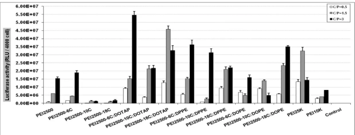
The transfection activity of formed lipopolyplexes was investigated in Neuro2A cells. Modification of lPEI by lipophilic chains caused the reduction in gene delivery ability of vectors except for PEI-6C at C/P= 3. Addition of lipids to the structure of lipopolyplexes increased the transfection activity. As Figure 2 shows the highest transfection efficiency is for PEI-6C: DOTAP formulation. The biological activity of this vector is significantly higher than lPEI (P< 0.05). Compared to the other lipids, the effect of DOPE in improvement of
Figure 1. Ethidium bromide test for liposomal formulations at different C/P ratios.
Liposomal formulations Mean size (nm) Zeta potential (mV)
PEI-6C 178.9±12.2 12.7±0.4
PEI-6C:DOPE 333.0±15.6 13.3±0.8
PEI-6C:DOTAP 166.7±11.1 16.4±2.9
PEI-6C:DPPE 273.6±17.3 5.5±1.9
PEI-10C 141.2±11.2 8.5±0.6
PEI-10C:DOPE 170.3±15.7 8.9±1.2
PEI-10C:DOTAP 95.6±14.1 16.3±2.6
PEI-10C:DPPE 336.8±19.3 3.2±0.6
PEI-18C 333.3±15.6 9.2±0.6
PEI-18C:DOPE 130.8±15.4 14.6±2.5
PEI-18C:DOTAP 95.0±5.9 17.5±4.1
PEI-18C:DPPE 236.5±12.7 14.2±2.6
Figure 2. Transfection efficiency of liposomal formulations at different C/P ratios, (Mean±SD, n= 3).
Figure 3. Cytotoxicity of liposomal formulations at different C/P ratios (Mean±SD, n= 3).
gene expression was less, except for PEI-18C: DOPE at C/P=3. In the most cases, transfection efficiency was improved by increasing at C/P ratio. The transfection activity of some vectors such as PEI2500-6C: DOTAP (at C/P= 3) and PEI2500-18C:DOTAP (at C/P= 1.5) was higher than that of PEI 25 kDa. In comparison with PEI 10 kDa, most of prepared vectors had better gene expression.
As it is presented in Figure 3, no remarkable cytotoxicity was found in Neuro2A cells. The cytotoxicity of vectors were increased slightly by increasing in C/P ratio. The viability of cell in the presence of vectors was ranged between 85 to 100%. PEI 10 kDa and PEI 25 kDa displayed noticeable toxicity especially at C/P= 3.
Discussion
One of the approaches to improve transfection activity is combination of two independent
mechanisms (increasing the cellular uptake by cationic liposome and improving the trafficking of complexes from endosome to nucleus by PEI) in design of gene delivery system (18). It was shown that polymers combined with liposomes would increase the DNA loading, the cell affinity and transfection efficiency (8). So, in the present study, lipopolyplexes were chosen as non-viral vector to improve gene expression.
these particles ranged from 135 to 480 nm. In the present study, lPEI (2500 Da) was modified by acrylate derivatives with different chain lengths (C6, C10, C18). These derivatives formed nanostructures ranging from 141 to 333 nm in size. Several studies had been shown that hydrophobic modification of PEI improved the cell interaction of complexes (14, 19). However, in our study improvement of gene expression was observed only for lPEI modified with hexyl acrylate. Hydrophobic grafting did not alter the ability of other lipoplymers to translocate DNA into the cell.
To examine the effect of PEI molecular weight on polycationic liposome mediated gene transfer, it was observed that PEI with a molecular weight of 1800 Da was as effective as that of 600 Da, but was far less effective than PEI of 25 kDa (11). We showed that lPEI with low molecular weight can improve the transfection activity.
In the present study, three lipids including DOPE, DPPE and DOTAP were used to evaluate the effect of lipid type on transfection activity of nanocarriers. In most cases, transfection efficiency of vectors composed of lipid and lipopolymer was higher than that of lipopolymer. Also the results of transfection studies suggested that the selection of proper lipid is an important factor and it can influence expression of pDNA remarkably. In the study of Yamazaki et al, the highest transfection activity was achieved by addition of DOPE to the formulation in comparison with the egg PC and DPPC (11), but as it was presented in Figure 2, among different lipids, DOTAP had the best impact of gene expression. It demonstrates that the positive charge of DOTAP is more determinant factor than the fusogenic properties of DOPE and DPPE in this case.. Also, it was reported that combination of PEIs (25 kDa) and DOTAP-cholesterol liposomes caused 10-fold increase in gene delivery to cells and high molecular PEI was more effective in gene delivery than low molecular PEI (800 Da) (13).
In the study of Chen et al, addition of DOPE to the structure of vector increased the transfection activity remarkably (8). They suggested that membrane destabilization
property of DOPE has led to the improvement of gene expression. Although, transfection efficiency of lipopolyplexes containing DOPE was higher than that of lipopolymer but DOTAP and DPPE had better impact compared to DOPE. As described previously, lipoplexes containing DOPE showed a perfect hexagonal phase, while lipoplexes composed of DPPE formed a mixed lamellar–hexagonal phase (20, 21). It indicates that the structure of lipids is important to achieve a high level of transfection.
Smaller lipopolyplexes was made when DOTAP was added to the structure. As endocytosis is the main pathways of internalization for both polyplexes and lipoplexes (6) and this internalization process is related to the complex size (22), vector dimensions have maximum effect on the transfection efficiency (14).
Displacement or binding exclusion of ethidium bromide to DNA is one method that is employed to measure the interactions of cationic vectors and DNA. In the present study, the results of ethidium bromide test suggest that these systems should interact with plasmid in a similar manner. No significant differences in DNA binding properties of vectors were observed especially in the C/P ratios higher than 1. Although it was reported that the synergism of ternary complexes of cationic liposomes, PEI and DNA did not related to DNA condensation and complex size (10) but other factors such as morphology and structure of complex, size, net charge and colloidal stability, influence the biological activity of lipoplexes (22).
It was reported by several authors that PEIs with high molecular weight caused marked cytotoxicity (23, 24). In the present study, no significant cytotoxicity was found among the prepared vectors even with the increasing lipid concentration. Although PEI 10 kDa and PEI 25 kDa were toxic at the higher C/P ratio (P> 0.05).
Conclusion
polymer and molecular weight of PEIs should be considered.
Acknowledgment
This study was performed in Pharmaceutical Research Centre, Mashhad University of Medical Sciences (MUMS), Mashhad, Iran.
The results described in this paper were part of a Pharm D student thesis. Authors declare that there is no conflict of interests in this study. Authors would like to thank the Vice Chancellor for Research of MUMS for financial support.
References
1. El-Aneed A. An overview of current delivery systems in cancer gene therapy. J Control Release 2004; 94:1-14. 2. Garcia L, Bunuales M, Duzgunes N, Tros de Ilarduya C. Serum-resistance lipopolyplexes for gene delivery to
liver tumour cells. Eur J Pharm Biopharm 2007; 67:58-66.
3. Matsumoto M, Kishkawa R, Kurosaki T, Nakagawa H, Ichikawa N, Hamamoto T, et al. Hybrid vector including polyethylenimine and cationic lipid, DOTMA, for gene delivery. Int J Pharm 2008; 363:58-65.
4. Gao X, Kim K, Liu D. Nonviral gene delivery:What we know and what is next. AAPS J 2007; 9:E92-104. 5. Luo D, Saltzman WM. Synthetic DNA delivery systems. Nat Biotechnol 2000; 18:33-37.
6. Elouahabi A, Ruysschaert J. Formation and intracellular trafficking of lipoplexes and polyplexes. Mol Ther 2005; 11:336-347.
7. Khalil IA, Kogure K, Akita H, Harashima H. Uptake pathways and subsequent intracellular trafficking in nonviral gene delivery. Pharmacol Rev 2006; 58:32-45.
8. Chen J, Wang H, Gao J, Chen H, Liang W. Liposomes modified with polycation used for gene delivery: Preparation, characterization and transfection in vitro. Int J Pharm 2007; 343:255-261.
9. McNeil SE, Perrie Y. Gene delivery using cationic liposomes. Expert Opin Ther Pat 2005; 16:1371-1382. 10. Hanzlikova M, Soinine P, Lampela P, Mannisto PT, Raasmaja A. The role of PEI structure and size in the
PEI/liposome-mediated synergism of gene transfection. Plasmid 2009; 61:15-21.
11. Yamazaki Y, Nango M, Matsuura M, Hasegawa Y, Hasegawa Y, Oku N. Polycation liposomes, a novel nonviral gene transfer system, constructed from cetylated polyethylenimine. Gene Ther 2000; 7:1148-1155. 12. Merdan T, Kopecek J, Kissel T. Prospects for cationic polymers in gene and oligonucleotide therapy against
cancer. Adv Drug Deliv Rev 2002; 54:715-758.
13. Lee C, Ni Y, Chen C, Chou C, Chang F. Synergistic effect of polyethylenimine and cationic liposmes in nucleic acid delivery to human cancer cells. Biochim Biophys Acta 2003; 1611:55-62.
14. Masotti A, Moretti F, Mancini F, Russo G, Di Lauro N, Checchia P, et al. Physicochemical and biological study of selected hydrophobic polyethylenimine-based polycationic liposomes and their complexes with DNA. Bioorg Med Chem 2007; 15:1504-1515.
15. Ko YT, Kale A, Hartner WC, Papahadjopoulos-Sternberg B, Torchilin VP. Self-assembling micelle-like nanoparticles based on phospholid-polyethyleneimine conjugates for systemic gene delivery. J Control Release 2009; 133:132-138.
16. Malaekeh-Nikouei B, Malaekeh-Nikouei M, Kazemi Oskuee R, Ramezani M. Preparation, characterization and transfection efficiency of nanoliposomes modified with oligoamines as gene carrier. Nanomedicine:NBM 2009; 5:457-462.
17. Zintchenko A, Philipp A, Dehshahri A, Wagner E. Simple modifications of branched PEI lead to highly efficient siRNA carriers with low toxicity. Bioconjug Chem 2008; 19:1448-1455.
18. Lampela P, Elomaa M, Ruponen M, Urtti A, Mannisto PT, Raasmaja A. Different synergistic roles of small polyethylenimine and Dosper in gene delivery. J Control Release 2003; 88:173-178.
19. Incani V, Tunis E, Clements BA, Olson C, Kuchavski C, Lavasanifar A, et al. Palmetic acid substitution on cantianic polymers for effective delivery of plasmid DNA to bone marrow stromal cells. Biomed Mater Res 2006; 81:493-504.
20. Zuhorn IS, Bakowsky U, Polushkin E, Visser WH, Marc CA, Stuart J, et al. Nonbilayer phase of lipoplex– membrane mixture determines endosomal escape of genetic cargo and transfection efficiency. Mol Ther 2005, 11:801-810.
21. Ramezani M, Khoshhamdam M, Dehshahri A, Malaekeh-Nikouei B. The influence of size, lipid composition and bilayer Tm on the transfection efficiency of nanolipoplexes. Colloids Surf B 2009; 72:1-5.
22. Simões S, Filipe A, Faneca H, Mano M, Penacho N, Düzgünes N, et al. Cationic liposomes for gene delivery. Expert Opin Drug Deliv 2005; 2:237-254.
23. Godbey WT, Wu KK, Mikos AG. Polyethyleneimine and its role in gene delivery. J Control Release 1999; 60:149-160.