Control the
Drosophila
Cellular Immune Response
Rami Makki1,2¤a., Marie Meister3¤b., Delphine Pennetier1,2
, Jean-Michel Ubeda3¤c, Anne Braun3¤d, Virginie Daburon1,2, Joanna Krzemien´1,2¤e, Henri-Marc Bourbon1,2, Rui Zhou4, Alain Vincent1,2*, Miche`le Crozatier1,2*
1Universite´ Toulouse 3, Toulouse, France,2Centre de Biologie du De´veloppement UMR5547 CNRS Toulouse, France,3Institut de Biologie Mole´culaire et Cellulaire, UPR9022 CNRS, Strasbourg, France,4Department of Genetics at Harvard Medical School, Boston, Massachusetts, United States of America
Abstract
The posterior signalling centre (PSC), a small group of specialised cells, controls hemocyte (blood cell) homeostasis in the Drosophilalarval hematopoietic organ, the lymph gland. This role of the PSC is very reminiscent of the ‘‘niche,’’ the micro-environment of hematopoietic stem cells in vertebrates. We have recently shown that the PSC acts in a non–cell-autonomous manner to maintain janus tyrosine kinase/signal transducers and activators of transcription (JAK/STAT) signalling in hematopoietic progenitors (prohemocytes), thereby preserving the multipotent character necessary for their differentiation into lamellocytes, a cryptic and dedicated immune cell type required to fight specific immune threats such as wasp parasitism. In this report, on the basis of a knock out generated by homologous recombination, we show that a short type I cytokine-related receptor CG14225/Latran is required for switching off JAK/STAT signalling in prohemocytes. This is a prerequisite to massive differentiation of lamellocytes upon wasp parasitisation. In vivo and cell culture assays indicate that Latran forms heteromers with Domeless, theDrosophilatype I cytokine signalling receptor related to mammalian GP130, and antagonises Domeless activity in a dose-dependent manner. Our analysis further shows that a primary immune response to wasp parasitism is a strong decrease in cytokine mRNA levels in the lymph gland, followed by an increase in the latran/domelessratio. We propose that this sequence of events culminates in the complete inhibition of residual JAK/STAT signalling by Latran. JAK/STAT activity has been associated with several human diseases including leukaemia while knock-out studies in mice point to a central role of this pathway in hematopoiesis and regulation of immune functions. The specific function ofDrosophilaLatran is, to our knowledge, the first in vivo example of a role for a nonsignalling receptor in controlling a dedicated immune response, and thus raises the question of whether short, nonsignalling receptors also control specific aspects of vertebrate cellular immunity.
Citation:Makki R, Meister M, Pennetier D, Ubeda J-M, Braun A, et al. (2010) A Short Receptor Downregulates JAK/STAT Signalling to Control theDrosophila
Cellular Immune Response. PLoS Biol 8(8): e1000441. doi:10.1371/journal.pbio.1000441
Academic Editor:David S. Schneider, Stanford University, United States of America
ReceivedSeptember 29, 2009;AcceptedJune 17, 2010;PublishedAugust 3, 2010
Copyright:ß2010 Makki et al. This is an open-access article distributed under the terms of the Creative Commons Attribution License, which permits unrestricted use, distribution, and reproduction in any medium, provided the original author and source are credited.
Funding:This work was supported by CNRS, Ministere de la Recherche (ACI and ANR programs), and Association de la Recherche contre le Cancer. RM and JK were supported by fellowships from Fondation de la Recherche Medicale and ARC, respectively. RZ is supported by the Leukemia and Lymphoma Society. The funders had no role in study design, data collection and analysis, decision to publish, or preparation of the manuscript.
Competing Interests:The authors have declared that no competing interests exist.
Abbreviations:CBM, cytokine binding motif; Col, Collier; CZ, cortical zone; Dome, Domeless; GFP, green fluorescent protein; JAK/STAT, janus tyrosine kinase/ signal transducers and activators of transcription; Lat, Latran; LDHR, Lat-Dome homology region; LG, lymph gland; MZ, medullary zone; proPo, prophenoloxidase; PSC, posterior signalling centre; RT PCR, reverse transcriptase PCR; SD, standard deviation; Upd, Unpaired; wt, wild type
* E-mail: [email protected] (AV); [email protected] (MC)
¤a Current address: MRC National Institute for Medical Research, London, United Kingdom ¤b Current address: Museum of Zoology, Strasbourg, France
¤c Current address: Laboratoire de Parasitologie et Mycologie, Universite´ Montpellier 1, Montpellier, France ¤d Current address: Charles River Laboratories, Shrewsbury, Massachusetts, United States of America ¤e Current address: Department of Zoology, University of Cambridge, Cambridge, United Kingdom
.These authors contributed equally to this work.
Introduction
The innate immune response–the synthesis of antimicrobial peptides and mobilisation of dedicated immune cells–confers a broad protection against pathogens to all multicellular organisms.
Drosophilahas become a model for studying the role of hematopoietic (blood) cells and the evolution of cellular immunity (reviews by [1,2]). Similar to vertebrates,Drosophilahematopoiesis occurs in two waves during development [3]. A first population of hemocytes is specified in the embryo and gives rise to plasmatocytes involved in
larvae, the LG is composed of several lobes with the anterior-most lobes organised into a medullary zone (MZ) containing the hematopoietic progenitors, a cortical zone (CZ) containing differentiating hemocytes, and a so-called posterior signalling centre
(PSC) (Figure 1F) [7]. These three zones can be identified by the expression of different markers. Differentiating crystal cells and plasmatocytes in the CZ express prophenoloxidase (proPO) and the P1 Nimrod receptor [8], respectively. Hematopoietic progenitors can be distinguished by their expression ofdomeless (dome), which encodes theDrosophilareceptor of the janus tyrosine kinase/signal transducers and activators of transcription (JAK/STAT) pathway andtep4, which encodes a thioester protein [7,9]. PSC cells express the transcription factors Antennapedia (Antp) and Collier/Knot (Col), theDrosophilaEarly B-cell Factor (EBF) ortholog [10], and the morphogen Hedgehog (Hh) [7,11–13]. The PSC controls the balance between multipotent prohemocytes present in the MZ and differentiating hemocytes [9,12]. It acts in a non–cell-autonomous manner, perhaps via Hh signalling, to maintain JAK/STAT signalling in prohemocytes, thus preserving the multipotent character necessary for these cells to adopt a lamellocyte fate upon parasitisation. This key role of the PSC revealed that, inDrosophila, larval hemocyte homeostasis is dependent upon interactions between hematopoietic progenitors and their micro-environment, a role reminiscent of the vertebrate hematopoietic niche [9,12,14]. JAKs and STATs mediate intracellular signalling in response to secreted type I cytokines [15]. In mammals, large families of cytokines and single-pass transmembrane receptors, named type I cytokine receptors, which signal as either homodimers or heteromers, have been identified (for review [16–19]). JAK kinases are anchored to the intracellular part of signalling receptors. Binding of the cytokine induces conformational changes in the latter that bring two JAKs in close proximity. This allows JAK trans-phosphorylation and phosphorylation of the receptor, thereby creating a docking site for STAT transcription factors. Author Summary
A specific microenvironment termed the ‘‘niche’’ supports long term maintenance of hematopoietic stem cells in vertebrates. A small group of specialised cells called the posterior signalling center (PSC) controls hemocyte (blood cell) homeostasis in the Drosophila larval hematopoietic tissue and thus fulfills a similar function to the vertebrate niche. The PSC acts at a distance to maintain JAK/STAT signalling in hematopoietic progenitors (prohemocytes), thereby ensuring their multipotent character. We report here that a short cytokine receptor encoded byCG14225/ latran is required to extinguish JAK/STAT signalling in prohemocytes and thereby ensures their mass differenti-ation into lamellocytes, an immune cell type required to fight specific threats such as wasp parasitism. Domeless, a related receptor in Drosophila, was previously the only known cytokine receptor that signals through the JAK/ STAT pathway. We show that Latran lacks the intracellular domains required for signal transduction and acts instead by antagonizing the function of Domeless in a dose-dependent manner. The role of Drosophila Latran in the repression of JAK/STAT signalling under specific immune conditions raises the question of whether short, non-signalling receptors that antagonize full-length receptors could also control specific aspects of vertebrate immunity.
Figure 1. Latran: homology with JAK/STAT receptors and expression in the LG.(A) Schematic of theD. melanogasterdome-lat genomic region. The ORFs and untranslated 59and 39regions are indicated by black and grey boxes, respectively. Positions of the PG125/domeGal4 P-element insertion [67] and the lat genomic fragment that was deleted to generate a null allele are indicated by an arrowhead and a red bar, respectively. (B) Schematic alignment of the Lat, Dome, and human GP130 proteins. The green box corresponds to the CBM and the blue box to a conserved region between Lat and Dome LDHR; the percentage of sequence identity is given. Fibronectin III (Fn III) motifs are indicated in red, the signal peptide in yellow. TM indicates the transmembrane domain. The intracytoplasmic regions are in grey, the position of the STAT binding site in orange. (C–E). Overlapping expression of lat (ISH red) (C), dome.GFP (GFP, light green) and Col (Ab, bright green, arrows) (D), and triple overlap (E) in LGs of a third instar larva. Nuclei are highlighted in blue (TOPRO-3); anterior is to the left. Scale bar, 80mm. (F) Schematic of an anterior lobe of the LG.
STATs become in turn phosphorylated, leading to their dimerisation and translocation into the nucleus where they function as transcriptional regulators. Recent finding inDrosophila
also point to a noncanonical mode of JAK/STAT signalling, which could directly control heterochromatin stability (for review [20]). Altered JAK/STAT activity has been associated with several human diseases including leukaemia, myocardial hypertrophy, and asthma, while knock-out studies in mice point to a central role in hematopoiesis and regulation of immune functions [21,22]. In contrast to mammals, only one receptor, Domeless (Dome), one JAK (Hopscotch, Hop), one STAT (Stat92E or Marelle) and three cytokines, Unpaired (Upd), Upd2, and Upd3 have been functionally characterised inDrosophila(for review [23]). Sequenc-ing of theD. melanogastergenome revealed, however, the existence of a dome-cognate gene (CG14225, renamed here latran [lat]) (Figure 1) [24,25].
We report here thatlatacts as a negative regulator of the JAK/ STAT pathway during larval hematopoiesis. lat is required for turning off JAK/STAT signalling in hematopoietic progenitors following wasp parasitisation, thereby allowing the massive differentiation of lamellocytes. In vivo and in vitro assays indicate that Latran (Lat) forms heteromers with Dome and antagonises Dome function in a dose-dependent manner. Our studies thus revealed a novel mode of regulation of JAK/STAT signalling, based on differential and tissue-specific expression of signalling and antagonist cognate receptors. The tight tissue-specific regulation of JAK/STAT signalling by latran is crucial for
Drosophila to be able to mount a dedicated cellular immune defense. A negative regulation of JAK/STAT signalling by a nonsignalling receptor chain has, so far, only been reported in primary and cultured mammalian cells, for short versions of class I cytokine receptors [26,27]. However, the in vivo function of these short membrane receptors and how their expression is regulated and linked to tissue homeostasis remain to be established. The specific role of Drosophila lat in controlling a dedicated cellular immune response raises the possibility that nonsignalling receptors could control specific aspects of vertebrate immunity, prefiguring a new field of investigations on this pathway.
Results
CG14225/latranEncodes a JAK/STAT Receptor-Like Protein
Vertebrate class I (one) cytokines bind to receptors composed of various single-pass transmembrane protein chains that form homo- and heteromeric complexes. Dome is the only class I cytokine receptor that has, so far, been characterised inDrosophila
[28–31]. Existence of aD. melanogastergene,CG14225/lat, coding for a protein structurally related to Dome was noticed several years ago [24]. dome and lat are adjacent to each other on the X chromosome and transcribed in the same orientation, suggesting that they originated from a gene duplication event (Figure 1A). The key role of JAK/STAT signalling in regulating larval hemocyte homeostasis [9,32] led us to ask whether lat was involved in this regulation. We first experimentally defined the 59 end oflattranscripts by RACE-PCR, using total RNA from LGs. We positioned thelatmethionine initiation codon and established that 153 bp separate the 39 end of dome mRNAs from the lat
transcription start site (Figures 1 and S1A). Dome and Lat show strong similarity in their extracellular domains, which include, from N- to C-terminal, a signal peptide, a cytokine binding motif (CBM) related to that of vertebrate receptors, and an approxi-mately 200 amino acid region not found in vertebrate receptors (Figures 1B and S2). We designate this region, whose function
remains unknown, as LDHR for Lat-Dome Homology Region. Similar to the human class I cytokine receptor GP130, Dome contains three tandemly arranged fibronectin type III (Fn III) motifs. None Fn III motif are found in Lat. The intracellular region of Lat is shorter than that of Dome and shows no consensus STAT binding site (motif YXXQ, [25]) suggesting thatlatencodes a nonsignalling form of cytokine receptor. A search fordome/lat -related genes in available genomes of other Drosophila species indicated that lat and dome are arranged in tandem, with an intergenic region varying from 150 bp (D. melanogaster) to 2.5 kb (D. virilis) (Figure S3). A higher degree of sequence conservation between orthologous compared to paralogous genes was observed (Figure S3), suggesting thatlatsequences diverge at a much higher rate than those ofdome.
latIs Specifically Expressed in Hematopoietic Progenitors in theDrosophilaLG
In situ hybridisations show that unlikedome,latis not expressed in embryos, a result confirmed by reverse transcriptase PCR (RT-PCR) (unpublished data). Thus, despite genomic proximity, the control oflattranscription is different from that ofdome. In larvae,
lattranscription was detected only in pericardial and LG cells. In situ hybridisation onto LGs expressing a membrane targeted green fluorescent protein (GFP) either in the MZ (dome-Gal4/UAS-mcd8 GFP (dome.GFP)) or the PSC (pcol85/UAS-(dome-Gal4/UAS-mcd8GFP (pcol.GFP)) indicated that lat is only expressed in the MZ (Figure 1C–1F). Coexpression ofdomeandlatin MZ cells, where JAK/STAT signalling is critically required to maintain a pool of hematopoietic progenitors [9], raised the question of the role oflat
in controlling the activation of the JAK/STAT pathway in prohemocytes.
latMutants Are Fully Viable but Unable to Mount a Cellular Immune Response against Wasp Parasitisation
To determine if lat plays a role in larval hematopoiesis, we generated a lat null allele by homologous recombination [33]. Several independent recombination events were obtained and homozygous mutant lines were established (Figure S1). Homozy-gous and trans-heterozyHomozy-gous combinations of these lines produced fertile adults with no obvious morphological defects, indicating that lat is not essential for either viability or germ-line development. In particular, no phenotypic defect was observed in the eye, where the JAK/SAT pathway plays an important role in growth and patterning [34,35]. We then looked at the morphology of the LG inlatmutant larvae, using specific markers for the MZ (tep4), the PSC (col), or for differentiated hemocytes: crystal cells (doxA3) and plasmatocytes (P1). No obvious difference could be found between wild type (wt) and lat mutant larvae, suggesting thatlatis neither required for the ontogeny of the LG, nor for the differentiation of plasmatocytes and crystal cells (Figure S4). The third type of Drosophila hemocytes, the lamellocytes (identified by the integrin a chain [a-PS4 marker]), massively differentiate at the expense of the pool of hematopoietic progenitors upon wasp parasitisation; they start to differentiate in the LG before being released into the hemolymph. In wt larvae, the number of circulating lamellocytes reaches its maximum 48 h after wasp egg-laying [5]. In sharp contrast to wt, virtually no circulating lamellocytes are found in the hemolymph of parasitised
latmutant larvae (Figure 2A, 2B), either 48 or 72 h after wasp egg-laying. Several days later, adult wasps hatch from parasitisedlat
therefore conclude thatlatis required for the massive differenti-ation of lamellocytes in response to wasp parasitisdifferenti-ation.
Lamellocyte production upon parasitisation involves downreg-ulation of JAK/STAT signalling in the MZ, thereby licensing hematopoietic progenitors to differentiate [9]. JAK/STAT activity in the LG can be monitored by the expression of a reporter transgene,dome-MESO-lacZ(dome-MESO), where LacZ is under the control of an intronic dome regulatory element [9,36,37]. Under normal conditions, LacZ expression is observed in the MZ of lat mutant as in wt larvae, indicating that the JAK/STAT pathway is active and that lat is not required for this activity (Figures 2 and S4). 30 h postinfestation a strong reduction of dome-MESO expression is observed in wt LGs (Figure 2D, 2E) correlating with lamellocyte differentiation (Figure 2E, insert) and premature LG dispersal [5]. In sharp contrast, dome-MESO remains expressed inlatmutant LGs and these, unlike wt LG, do not prematurely disperse (Figure 2F, 2G), correlating with the absence of circulating lamellocytes in the hemolymph. This shows thatlatis required for the downregulation of JAK/STAT activity in hematopoietic progenitors following parasitisation. The obser-vation of few differentiated lamellocytes in lat mutant larvae (Figure 2G and insert) indicates, however, thatlatis not required for the lamellocyte differentiation programper se. In cells were the JAK/STAT pathway is activated, Stat has a predominantly nuclear localisation. In order to follow the activity of the pathway after parasitism, we analysed the subcellular localisation of a fluorescent Stat protein, Stat-GFP [38], expressed in the LG ( srp-Gal4;STAT92E-GFP). In noninfectious conditions, Stat-GFP is mainly found in the nuclei, in either wt (Figure 2H) or lat
(unpublished data) mutant larvae, consistent with active signalling. 4–6 h after wasp egg-laying, Stat-GFP is found both in the cytoplasm and the nucleus in wt LG, whereas it remains predominantly localised in the nucleus in lat mutant LG (Figure 2I, 2J). These data show a decreased activity of JAK/ STAT signalling in wt LG, already 4–6 h after wasp parasitisation, whereas no change can be detected inlatmutant LG.
Lat and PSC Activity in the LG: Robustness of Hemocyte Homeostasis
The PSC (niche) is critically required to maintain a balance between JAK/STAT-positive progenitors and JAK/STAT-nega-tive differentiating hemocytes in third instar LG [9]. The function oflatin the MZ raised the question of the relative contribution of positive and negative regulation by the PSC andlat, respectively, in the maintenance of this balance. Therefore, we examined the proportion of prohemocytes (expressing dome-MESO) and differentiating hemocytes in LGs double mutant for lat and col
(Figure S5). Whereas incolmutant LGs, which lack a PSC, the MZ disappears and all prohemocytes differentiate [9], we observed a less severe phenotype inlat;coldouble mutants, namely the loss of an organised MZ with remaining prohemocytes intermingled with differentiated hemocytes (Figure S5A). Intermingling of prohemo-cytes and differentiated hemoprohemo-cytes was also observed in lat;col
double mutants following wasp parasitisation with, in this case, some lamellocytes among differentiated hemocytes (Figure S5B). The persistence of prohemocytes in thelat;coldouble mutant LG underlines the crucial role of lat in the complete switch from progenitor to differentiated state that is observed either in col
mutant larvae or following parasitisation.
Lat Is a Negative Regulator of JAK/STAT Signalling The structural similarity between Lat and Dome together with
latfunction suggested thatlatencodes a novel negative regulator of the JAK/STAT pathway. To test this hypothesis, we overex-Figure 2. lat mutant LGs do not properly respond to wasp
parasitisation.(A–C) DAPI staining of circulating hemocytes from (A) wt, (B)latmutant, and (C)latmutant larvae expressinglatin the LG (srp.lat). 48 h after parasitisation, lamellocytes are found in large numbers in the hemolymph of wt larvae (A, insert) but lacking inlat mutants. This lack-of-lamellocyte phenotype is rescued by expressing latin the LG (C, arrows). (D–I) Prohemocytes, marked by dome-MESO expression (green) are not maintained in wt LGs following wasp parasitisation (D and E); this is paralleled by massive lamellocyte differentiation (integrinachain [a-PS4], red) (E, insert). Inlatmutant larvae, dome-MESO remains expressed after parasitisation (F, G); few lamellocytes differentiate (G, insert). (H–J) Stat-GFP (green) subcellular localisation in wt (H and I) andlatmutant LG (J), either without (H) or 4– 6 h after parasitism (I and J).srp-Gal4 was used to drive UAS-Stat-GFP in the LG (srp.Stat-GFP). Stat-GFP localisation is nuclear in nonparasitised wt and parasitisedlatLG, whereas it is both cytoplasmic and nuclear in parasitised wt LG. The scattered distribution of GFP-labelled cells is due to nonuniform activation of thesrp-Gal4 driver in the LG [11]. Nuclei (TOPRO-3) are in blue. Scale bars, 50mm (A); 80mm (D).
pressed lat in the MZ and followed JAK/STAT activity using dome-MESO expression. lat overexpression led to a complete inhibition of JAK/STAT signalling in the MZ while both crystal cells and plasmatocytes were still able to differentiate (Figure 3A– 3D). To further investigate the possible mechanism behind this inhibition, we turned to reporter assay developed in cultured
DrosophilaSchneider (S2-NP) cells [29]. S2-NP cells display a basal level of endogenous JAK/STAT activity, as shown by transfection of a STAT reporter gene (10XStat92E-Luciferase reporter). A much stronger activity is observed upon coexpression of either of the cytokines Upd, Upd2 [29,30,36], or Upd3 (Figure S6). To assess for lat function, we transfected S2-NP cells with 10XStat92E-luciferase, Actin promoter-driven Renilla luciferase, Upd expression vectors (Act-renilla and Act-upd), together with Actin promoter-driven Dome (Act-dome) and/or Lat (Act-lat) expression vectors at various relative concentrations. Since high level of forced Dome expression could act as a dominant-negative
[39], we transfected low levels of Act-dome, which modestly increased JAK/STAT signalling (Figure 3E). In contrast, trans-fection of similar amounts of Act-lat severely decreased signalling (.4-fold), confirming that latacts as a negative regulator of the pathway (Figure 3E) without affecting the level of Dome expression (unpublished data). Lat function is independent of the added cytokine (Figure S6). Intermediate levels of JAK/STAT inhibition were observed for different relative amounts of Act-dome and Act-lat indicating that the ratio between Lat and Dome is critical (Figure 3E). These data both confirmed that lat is a negative regulator of JAK/STAT signalling and suggested that the ratio between Dome and Lat is a key factor in controlling JAK/ STAT activity.
Binding of Upd to Dome induces endocytosis of receptor-ligand complexes and their trafficking through the endosomal compart-ments, a trafficking required to activate JAK/STAT signalling [40]. We looked at the intracellular localisation of a tagged Dome (Dome-V5) in cells transfected with both Upd and Act-Dome-V5, where the pathway is active. Dome(-V5) was localised in cytoplasmic vesicles as previously described (Figure 3F) [40]. This localisation was unchanged in cells cotransfected with a tagged Lat (Act-HA-Lat), which results in inactivation of the pathway (Figure 3F, 3G). Both Dome-V5 and HA-Lat were localised in mostly overlapping intracytoplasmic vesicles, indicating that negative regulation of Dome activity by Lat is not linked to a defect in Dome internalisation.
Lat and Dome Can Form Heterodimers In Vivo
Long and short forms of vertebrate class 1 cytokine receptors can form heterodimers/multimers [17,41]. While Dome was previously shown to form homodimers [42], we tested the possibility that Lat could form heteromers with Dome, by using coimmunoprecipita-tion (co-IP) assays. S2-NP cells were transfected with equivalent amounts of plasmids encoding tagged Dome(-V5) and Lat(-HA). Cell lysates were prepared 72 h post-transfection and subjected to IP with either anti-V5 or anti-HA antibodies, followed by Western blot analysis with one or the other antibody. Lat and Dome co-IP in both directions indicated that they form heteromers in cell culture (Figure 4A). We then tested whether Lat and Dome form heteromers in vivo, using thebblue-bblaub-galactosidase comple-mentation technique developed to detect protein-protein interac-tions in vivo [43–45] and already applied to show that Dome forms homodimers [42]. Briefly, this technique uses twob-Gal mutant forms (DaandDv) that are separately inactive but can complement each other if brought into proximity by fusing them to proteins that physically interact. Like for Dome [42],DaandDv b-galactosidases were fused to the Lat C terminus. We used theda-Gal4 driver to coexpress different combinations of Lat and Dome fusion proteins in embryos (Figure 4B–4E), because it leads to strong ubiquitous embryonic expression of the proteins (Figure 4C and unpublished data) [42]. Contrasting with this ubiquitous expression, X-gal staining was only detected in the salivary glands and hindgut when DomeDaand DomeDvwere coexpressed as previously reported (Figure 4B) [42]. A similar staining pattern was observed upon coexpression of LatDa/LatDv LatDa/DomeDv or DomeDa/ LatDv(Figure 4D, 4E, and not shown), while no staining could be detected when only one fusion protein was expressed. These results indicate that Lat is able to form homodimers and heterodimers with Dome in vivo. We then testedb-gal comple-mentation in the MZ using thedome-Gal4 driver becauseda-Gal4 is not expressed in the LG (unpublished data). Staining was observed upon coexpression of LatDaand DomeDv(Figure 4F) or DomeDa and LatDv(unpublished data), while no staining could be observed upon expression of either fusion protein alone. These complemen-Figure 3. Lat acts as a dominant-negative JAK/STAT receptor.
(A–D) dome-Gal4 driven expression oflatin the MZ switches off JAK/ STAT signalling, as monitored by dome-MESO expression (LacZ, green). proPO (red, A and B) and P1 stainings (red, C and D) indicate differentiating crystal cells and plasmatocytes, respectively. (E) Dro-sophilaS2-NP cells were transfected with 106STAT92E-luciferase, Act-Renilla, and Act-upd, and various amounts of Act-dome and Act-lat plasmids (indicated in nanograms). Luciferase assays were performed 4 d later and the reporter activity normalised as the ratio of firefly luciferase/Renilla. The results are from three independent experiments. Vertical bars correspond to SD. (F–G) Immunodetection of Dome-V5 (green) and HA-Lat (red) inDrosophilaS2-NP cells, in conditions where the JAK/STAT pathway is either on (F) or off (G) (MM and Figure 3E). HA-Lat and Dome-V5 colocalise in trafficking cytoplasmic vesicles in both conditions. (G) Shows a phase-contrast view of a transfected cell. Scale bars, 80mm (A); 8mm (G).
tation assays show that Dome and Lat form heterodimers in the LG. Unfortunately we could not determine whether Dome or Lat can also form homodimers in the LG sincedome-Gal4 driven expression of two copies of either Dome or Lat leads to early larval lethality. We observed, however, that dome-Gal4-driven expression of one copy of either Lat, LatDa or LatDvresulted in an ‘‘outstretched wing’’ phenotype in adult flies, a phenotype previously described for
updmutant flies, hence the nameoutstretched (os/upd)given to these mutants [46,47]. This observation indicates that both the native and Lat fusion proteins are able to downregulate the JAK/STAT pathway. eyeless-Gal4 driven expression of latin eye discs led to smaller eyes, another phenotype reminiscent ofupdmutants (Figure S7) [39], confirming thatlatis able to downregulate JAK/STAT signalling in tissues other than the LG when ectopically expressed. Taken together, cell culture and in vivo data indicate that Lat forms inactive heteromers with Dome and is a novel negative regulator of the JAK/STAT pathway.
Wasp Infestation Results in an Increased Lat/Dome Ratio Both cell culture and in vivo data pointed to the Lat/Dome ratio as a key component in the regulation of JAK/STAT signalling. To determine whether this ratio is modified upon wasp parasitisation, we compared the levels of accumulation oflatand
domemRNAs in LGs relative to internal controls (rp49andrpL17, ribosomal protein mRNAs). Quantitative RT-PCR (qRT-PCR) measurements were performed on total LG RNA from control larvae and larvae 4 h after wasp egg-laying in order to mainly detect primary changes that occur in response to parasitism [48]. We detected an about 2-fold increase inlattranscripts and 2-fold decrease indometranscripts, which results in a significant drop in the dome/lat ratio (Figure 5A). Since JAK/STAT signalling remains on after infestation inlatmutant larvae, we repeated the analysis on RNAs fromlatmutant LGs. In this case, no decrease in the level ofdometranscripts was observed upon wasp infestation, indicating that this decrease depends uponlatactivity (Figure 5B). In order to strengthen this conclusion, we tested whether decreasing JAK/STAT signalling in the LG by expressing dsRNA-hop(srp-Gal4.dsRNA-hop) could rescue thelatmutant phenotype. We indeed observed that lowering the level of Hop activity restored the ability oflatmutant LG to massively produce lamellocytes following wasp parasitism (Figure 5C), confirming thatlatacts upstream of hopin the signalling cascade. Together, these data lead us to propose that the shift in the relative levels of
dome and lat expression observed following wasp egg-laying operates as a switch leading to a complete extinction of JAK/ STAT signalling, thus allowing prohemocytes to differentiate.
upd3Is Expressed and Required for JAK/STAT Signalling Activity in the MZ
Three different ligands, Upd, Upd2, and Upd3, are known to activate JAK/STAT signalling inDrosophila [47,49]. In order to determine which of them are expressed in the LG, we first performed RT-PCR experiments, starting from LG mRNA. We detectedupd3and very low amounts ofupd2but noupdtranscripts (Figures 5, 6, and S8). Sinceupd2mutants have no hematopoietic phenotype (unpublished data),upd2was not further considered in this study. We then focused onupd3expression and function. Since only genome annotation data were available, we determined the 59 end ofupd3transcript by RACE-PCR and repositioned the ATG initiation codon (Figure S8A). In situ hybridisation of upd3
transcripts in Dome.GFP and pcol.GFP LGs indicated that
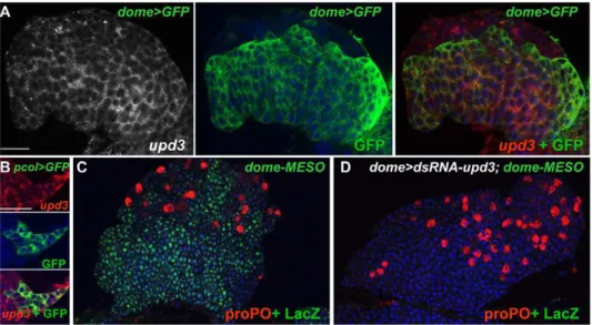
upd3is expressed in the MZ, the PSC, and in few scattered cells of the CZ (Figure 6A, 6B). While aupd3loss of function mutant is not available, studies performed in vivo and in cell culture have established that upd3dsRNA expression can efficiently suppress
upd3activity [50]. We looked at the consequence ofupd3dsRNA expression (UAS-upd3 dsRNA/dome-Gal4), which drastically reduces
upd3 mRNA level in the LG (Figure 5D), on dome-MESO expression. No dome-MESO expression could be detected, showing thatupd3expression in the MZ is required to maintain JAK/STAT signalling active (Figure 6C, 6D). Whenupd3dsRNA expression was targeted to the PSC (pcol-Gal4), dome-MESO expression was unperturbed (unpublished data). We then deter-mined whetherupd3levels are modified upon wasp parasitisation. The drastic decrease of upd3 transcripts observed 4 h after infestation (Figure 5A) shows that upd3 downregulation is an immediate response to wasp parasitisation.
While JAK/STAT signalling is dependent upon the binding of Upd to Dome,domeis itself a target of the JAK/STAT pathway in the embryonic mesoderm [36], a regulatory loop reproduced by the dome-MESO enhancer in the LG [9,36]. To directly test whether the decreased amount ofdometranscripts in the LG that Figure 4. Lat forms heterodimers with Dome in the LG. (A)
Immunoprecipitation of the Lat/Dome protein complex. Lysates from either control S2-NP cells or cells transfected with pAc-DomeV5 and pAc-HALat were immunoprecipitated with either anti-V5 or anti-HA antibodies as indicated above each lane and subjected to Western blot analysis. Both conditions indicate the formation of Dome/Lat protein complexes. (B, D–E) Formation of Dome homodimers, Lat homodimers, and Lat/Dome heterodimers in (B) da-Gal4xUAS-DomeDa /UAS-Do-meDv, (D) da-Gal4; UAS-LatDa/UAS-LatDv, and (E) da-Gal4; UAS-LatDa/UAS-DomeDv embryos, respectively, as visualised by X-Gal staining (blue). In stage 13 embryos, strong staining is observed in the salivary glands (black arrow) and the hindgut (black arrowhead). (C) da-Gal4; UAS-LatDaubiquitous expression of LatDadetected by LacZ immunostaining. (F) Formation of Dome/Lat heterodimers in the MZ of the LG. Scale bars, 40mm (F).
follows wasp parasitisation could result from the drop of upd3
activity, we measured the relative amounts of dome and lat
transcripts uponupd3 dsRNAexpression in the MZ. Whereas thelat
level was not affected, a 2-fold decrease was observed for dome
transcripts (Figure 5D). We conclude that the decrease in dome
transcripts is a secondary response consecutive to decreased levels of upd3 mRNA. Unlike dome, however, upd3 downregulation is independent of lat function (Figure 5B). Thus, we propose the following model: wasp parasitism results in a drastic decrease in
upd3 levels, which in turn leads to a downregulation of JAK/ Figure 5. Downregulation ofupd3anddometranscripts in response to wasp infestation.Quantitative analysis ofdome,lat, andupd3 relative to rp49 transcripts is given in either (A) wt or (B) latmutant LGs. A significant decrease of dome (p= 561025) and increase of lat (p= 1.5610216) transcripts is observed 4 h after wasp egg-laying. The level ofupd3transcripts drops to almost undetectable. This decrease ofupd3 mRNA is also observed inlatmutant LGs (B). (C)aPS4 (lamellocytes, black) immunostaining of wt,latmutant, andlat,srp.dsRNA-hop LGs30 h following wasp infestation. Massive lamellocyte differentiation is observed inlat;srp.dsRNA-hopmutant LGs similar to wt. (D)dome-Gal4.induced degradation ofupd3transcripts in the LG leads to a significant decrease ofdome(p= 161028) transcripts whereas the level oflat(p= 0.2) is not affected.rp49andrpL17mRNAs were used as internal controls (unpublished data). Three independent experiments were performed, vertical bars correspond to SD.
doi:10.1371/journal.pbio.1000441.g005
Figure 6.upd3is expressed and required to maintain JAK/STAT signalling in prohemocytes.(A)upd3expression (white and red, left and right panels, respectively) overlaps that of dome.GFP (green, middle and right panels) in LGs of third instar larvae. (B)upd3(red, top panel) is also expressed in the PSC (pcol.GFP, green, middle panel), overlay at the bottom. (C and D) dome-Gal4 driven expression ofupd3dsRNA leads to loss of dome-MESO expression (LacZ, green) in the MZ. proPO staining (red) indicates differentiating crystal cells. Nuclei (TOPRO-3) are in blue. Scale bars, 40mm (A); 60mm (B).
STAT signalling and a decrease of dome transcription. This, in turn, results in an increased lat/dome ratio, which subsequently leads to the complete shut down of the JAK/STAT pathway. The complete and efficient inhibition of JAK/STAT signalling in the LG thus requireslatfunction (Figure 7).
Discussion
The evolutionarily conserved JAK/STAT signalling pathway was discovered from studies on the role of interferon in the control of immune responses. Vertebrate genomes encode multiple forms of all major JAK/STAT pathway components, including multiple receptor subunits. As opposed to this, in Drosophila, only one functional receptor, Dome, had so far been characterised. However, sequence similarity betweendomeand the neighbouring geneCG14225/latsuggested a gene duplication event and raised the question oflatfunction.
Switching off JAK/STAT Signalling and Orienting
Prohemocytes towards a Lamellocyte Fate: Two Facets of theDrosophilaImmune Response to Wasp Parasitisation In order to neutralise parasitoid wasp eggs, theDrosophilalarval hematopoietic organ must rapidly release large amounts of lamellocytes in the hemolymph. In normal developmental conditions, the PSC maintains JAK/STAT signalling in the hematopoietic progenitor zone (MZ) thereby preserving their prohemocyte character. Upon wasp infestation, however, JAK/ STAT signalling is switched off, leading to a concerted differentiation of prohemocytes into lamellocytes [9]. Incolmutant LGs, which are devoid of PSC cells, no lamellocytes differentiate after wasp infestation as a consequence of the premature loss of multipotent prohemocytes; conversely, in lat mutant larvae, prohemocytes are maintained and very few lamellocytes differen-tiate. Prohemocytes persist inlat;coldouble mutant LGs, suggesting that a basal level of JAK/STAT signalling subsists in this mutant context, leading to a stochastic rather than global differentiation of prohemocytes. The comparison of col, lat, and lat;col mutant phenotypes, therefore, allows to conclude that latfunctions as a switch. In normal developmental conditions, PSC activity overrides lat function in the MZ. Upon wasp infestation, PSC activity is short-circuited andlatplays a decisive role in completely silencing the JAK/STAT pathway in all prohemocytes. Hemocyte
homeostasis in the LG thus relies on both extrinsic signals from the niche and intrinsic JAK/STAT activity in progenitor cells (Figure 7). Inlatmutants, some lamellocytes differentiate following wasp parasitisation indicating thatlatis not strictly required for the lamellocyte differentiation programme per se. Thus, switching off JAK/STAT signalling and orienting prohemocytes towards a lamellocyte fate are two distinct responses to wasp parasitisation (Figure 7).
Differential Regulation oflatanddomeExpression Warrants Inactivation of the JAK/STAT Pathway upon Wasp Infestation
In situ hybridisation and LG-targeted RNA interference experiments show thatupd3is expressed and required in the MZ to maintain JAK/STAT activity in prohemocytes, therefore acting in an autocrine and/or paracrine manner, as previously reported for Upd in embryos [47,51,52]. The drastic decrease of upd3
expression induced by wasp egg-laying is accompanied by a significant decrease indometranscripts, showing thatdomeis both a component and a target of JAK/STAT signalling in the MZ (Figure 5), as previously documented in the embryonic mesoderm [36].latanddomemRNA levels are not, however, coregulated in response to parasitisation even though the two genes lie very close to each other on the chromosome, a tandem organisation conserved in other Drosophila species. The uncoupling between
dome and lat expression results in an increased lat/dome ratio following wasp infestation, which is determinant for the ability of Lat to antagonise Dome activity. Comparative analyses of RNAs from wt andlatmutant LGs show that the primary component of the JAK/STAT pathway that is affected by wasp infestation is the level ofupd3transcripts. Although we do not know yet howupd3is downregulated, it is tempting to speculate that it could be at a transcriptional level, similar to the importance of post-transcriptional regulation for cytokine levels in vertebrates (for review [53]). In summary, our results show that a primary immune response to wasp egg-laying is a strong decrease inupd3mRNA levels in the LG, which induces a downregulation of the JAK/ STAT pathway, followed by a decrease ofdomeand increase oflat
levels. This results in an increasedlat/domeratio that further and completely turns off the JAK/STAT pathway. Since in the absence oflatthe decrease inupd3level does not completely switch off the JAK/STAT pathway. We conclude thatLatacts as a switch
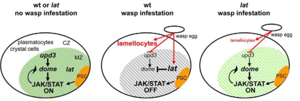
Figure 7. Model forlatfunction inDrosophilalarval hematopoiesis.During normal development (left panel), PSC cells (orange) act, in a non– cell-autonomous manner (arrow) to maintain JAK/STAT signalling activity and preserve a pool of multipotent prohemocytes in the MZ (green shades). The PSC signal overrideslatfunction in the MZ (grey shades). In response to parasitisation (middle panel), there is a decrease ofupd3anddomeand increase oflattranscripts, which ultimately lead to an increasedlat/domeratio. The PSC signal is short-circuited. As a result, JAK/STAT signalling is switched off, thus licensing prohemocytes to differentiate into lamellocytes. Lat activity is strictly required in the LG for this switch. In the absence of lat(right panel), residualupd3levels maintain JAK/STAT activity, therefore preserving a pool of prohemocytes (grey shades). Upon wasp parasitisation some differentiating hemocytes become lamellocytes, however, indicating thatlatis not required for this differentiation program per se. Arrows indicate activation, vertical bars repression.
that is required for the total arrest of JAK/STAT signalling in hematopoietic progenitors in response to wasp parasitisation, a prerequisite to massive differentiation of lamellocytes and efficient immune response (Figure 7).
dome andlat, a Pair of Duplicated Genes with Antagonistic Functions
Dome is related to the human GP130 and cognate GP130-like (GPL) signalling receptors, which form heteromeric complexes with short, nonsignalling receptors such as IL-6R or Oncostatin M receptor (OSM-R) to mediate signalling (Figure S9) [26,54,55].lat
encodes a short-type receptor that could either act as IL-6R and confer signalling specificity to Dome or as a dominant-negative receptor similar to what has been described ex vivo for short receptors such as GPL and IL13Ra2 [27]. Cell-culture and in vivo assays show that Lat antagonises Dome activity in a dose-dependent manner and forms heteromers with Dome thereby acting as a dominant-negative receptor. Altogether, these data suggest that, following parasitism, which leads to decreased cytokine levels, Lat blocks Dome activity in the LG through the formation of inactive heteromers.
While our analysis indicates thatlatis specifically required in the larval hematopoietic organ for massive lamellocyte production in response to an immune challenge, phenotypes induced by ectopic
latexpression show that it can antagonise JAK/STAT activity in other tissues. Together, the phenotypic and protein interaction data suggest that LG specific lat expression has been selected during evolution to fulfil specific immune functions.
Cytokine signalling pathways are subject to extensive positive and negative feedback regulations, which are crucial to generate appropriate physiological responses [22]. Two genome-wide RNA interference (RNAi) screens for JAK/STAT signalling components were conducted in Drosophilacultured cells. While they identified large sets of putative positive and negative regulators, they failed to detectlat, either because its expression level in cell culture is too low to be functional or because thelatdsRNA constructs used in these screens were not efficient enough [29,30,56].col/kn was identified in one of these RNAi screens, however, as a positive regulator acting downstream of Hop [30], suggesting another possible level of regulation of JAK/STAT signalling in the LG. Initial evidence for the involvement of JAK/STAT signalling in Drosophila cellular immunity came from the observation that a dominant gain-of-function mutation of the JAK kinase (hopTum) provokes the apparition of lamellocytes and melanotic masses in the absence of wasp infection. This finding led to the conclusion that upregulation of JAK/STAT signalling triggers lamellocyte differentiation, which is in apparent contradiction with our present data [32]. Whether constitutive JAK/STAT signalling in differentiating hemocytes could instruct them to become lamellocytes remains an interesting possibility. Of note, a STAT target,chimno, was recently shown to be expressed at higher levels in differentiating CZ cells as compared to undifferentiated MZ cells [57]. Recent studies further suggest a dual role of Wg signalling in the maintenance of prohemocytes and PSC cells [58]. A tight control of ROS levels in the MZ is also required to maintain a pool of prohemocytes [59]. How these different signalling pathways are interconnected with JAK/STAT signalling in order to maintain hemocyte homeostasis in the LG are important questions to be addressed in the future.
A Conserved Role for Dominant-Negative Short Receptors in the JAK/STAT Pathway?
The type I cytokine receptor family has considerably expanded in vertebrates [60]. This expansion results both from an increased
number of receptor genes and from the generation of various protein isoforms that can act as either receptors or coreceptors [61]. Soluble versions of short receptors isolated from diverse body fluids have also been identified, which behave as antagonists by competing with membrane-associated receptors for ligand binding [62,63]. These soluble receptors are generated by either limited proteolysis or translation from differently spliced mRNAs. Finally, studies on IL13Ra2 [27] or GPL [64] suggested that short receptors anchored to the membrane could also behave as dominant negative receptors. However, the exact function of these receptors and how their expression is regulated and linked in vivo to tissue homeostasis, remains unknown. Our studies in
Drosophilaindicate that Lat acts as a dominant-negative receptor rather than a coreceptor, extending in vivo the few observations made in mammalian cell cultures [65,66]. Tissue-specific regula-tion of JAK/STAT signalling in response to environmental cues is crucial for the ability ofDrosophila to mount a cellular immune defense. Our results bring to light a new mode of fine tuning of the JAK/STAT pathway, that is, differential expression of signalling and antagonist cognate receptors. Whether and when regulated expression of long and short receptor isoforms is employed in controlling specific aspects of immunity in vertebrates certainly deserves further investigation.
Materials and Methods
DrosophilaStrains
The following strains were used: pcol85-Gal4;UASmcd8GFP (pcol.GFP) [9]; PG125-dome-Gal4 (dome-Gal4) [67] andsrp-Gal4 [11].ey-Gal4 andda-Gal4 were obtained from the Bloomington
Drosophila Stock Center. The dome-MESO, UAS-dome, UAS-DomeDa, and UAS-DomeDv strains are from [42]; the UAS-upd3dsRNA from [50]; and the P[70FLP][70I-SceI)/TM3 and P[ry+; FLP)10 (Chromosome II) from F. Karch.whitestrains were used as wt. Mid-second instar larvae subjected for 30–60 min to egg-laying byLeptopilina boulardi(G464) were allowed to develop at the appropriate temperature and analysed 4, 24, or 48 h later.
Generation of alatNul Mutant by Site-Directed Recombination
The procedure was adapted from the Ends out Knock Out method [33]. A lat KO ‘‘donor’’ transgene was constructed in pW25 [68] by inserting 4 kb of 59and of 39flanking sequences of thelatgene separated by the mini-whitegene and used to transform
whitemutant flies (Figure S1B). Two different inserts on the second chromosome were selected for the recombination-targeting protocol. Several independent lat KO lines were obtained and verified for deletion oflatand insertion of mini-whiteby PCR and Southern blot analyses (Figure S1C). Thelat18A
line was chosen for all the experiments.
Constructs
The mapping oflatandupd3transcript 59ends was performed by 59RACE PCR (Marathon cDNA amplification kit, Clontech, and BD Smart RACE kit, BD Biosciences), using either polyA+
Act-Lat, Act-HAAct-Lat, and Act-DomeV5 plasmids were constructed and used for cell culture experiments.
RNA Probes
A526-bplatgenomic fragment amplified using primers 6 and 8 (Figure S1A) was cloned in the Invitrogen pCRBluntII-TOPO vector. Two differentupd3probes of 836 bp (primers 1 and 2) and 2,057 bp (primers 5 and 6) were designed for in situ hybridisation (Figure S8).
In Situ Hybridisation, Antibody Staining, and Western Blotting
Dissections, in situ hybridisation, and immunostaining proce-dures were as described in [9,70]. The following antibodies were used: rabbit anti-GFP (Torrey) 1/500; mouse anti-b-galactosidase (Promega) 1/800; rabbit anti-proPO 1/200; mouse anti-Col 1/50; rabbit anti-aPS4 [9] 1/200; mouse anti-V5 (Invitrogen) 1/5,000; mouse and rabbit anti-HA (Covance and Santa Cruz, respectively) 1/1,000. Mounting in Vectashield medium (Vector Laboratories) preceded analysis by confocal microscopy (Leica SP2).
X-Gal Staining
X-Gal staining was as described in [42].
RNA Amplification and Quantitative RT-PCR
Dissected LGs were collected in trizol and total RNA was extracted using trizol reagent (Invitrogen) according to the manufacturer. Superscript Reverse Transcriptase II (Invitrogen) and oligo dT primers were used for reverse transcription. Real-time quantitative PCR was performed on a MyiQ single color real time PCR detection system (Biorad). CT values were collected and analysis was performed according to the 2DDCTmethod [71] using
rp49 and rpL17A to normalize estimates of relative expression. Primers used: 1 and 3 and 5 and 7 fordomeand lat, respectively (Figure S1), 3 and 4 forupd3(Figure S8). No significant differences were detected in the level of control RNAs in wt,lat and dome.
upd3dsRNA experiments (Figure 5). Primers sequences for rp49,
rpL17A,upd, andupd2are available on request. All qRT-PCR data are representative of three independent experiments and presented as means 6 standard deviation (SD). Statistical analyses were performed using Student’sttest.
Cell Culture Experiments
Various amounts of Act-Lat, 0.2 ng of Act-Dome, and 1 ng of either Act-Upd, Upd2, or Upd3 were used to transfect S2-NP cells [29]. Luciferase assays were performed 4 d later, and the reporter activity was normalised as the ratio of firefly luciferase/Renilla. The results are from three independent experiments. For immunostaining, S2-NP cells were transfected with 1 ng of upd and 0.2 ng of Dome-V5 with or without 1 ng of Act-HALat (Figure 3E and 3F).
Immunoprecipitation of HALat/DomeV5 Complex
Drosophila S2-NP cells [29] were maintained in Schneider medium +10% FCS + penicillin-streptomycin (Sigma 1/100) at 25uC without supplemental CO2. Cells (36106 per well) were seeded and cultured in six-well plates. 24 h later, transfections using Effectene (Quiagen) were performed. Each well was transfected with 20 ng of plasmid encoding either HA-Lat, Dome-V5, or both, and completed with plasmid DNA encoding the empty vector (pHA vector) to a final amount of 400 ng of DNA. 48 h later, cells from each well were washed in PBS and lysed in 150ml of ice-cold buffer containing 50 mmol Tris
(pH 7.4), 150 mmol NaCl, 1 mmol EDTA, 1% NP40, and antiprotease cocktail (Roche) for 20 min. 140ml of the crude lysate was used for IP. Protein G sepharose beads (Sigma) were first incubated with 1mg of anti-HA or anti-V5 antibodies for 1 h
at 4uC and then with the cleared supernatant for 2 h at 4uC. Beads were then boiled in denaturing sample buffer and the released proteins loaded on a gel with 3ml of the crude lysate (1/50 of the
total preparation) used as a control lane. The separated proteins were analysed by Western blotting with either mouse anti-V5 or mouse anti-HA antibodies.
Supporting Information
Figure S1 TheD. melanogaster dome/lat/zwgenomic region.(A) Nucleotide sequence of theD. melanogaster dome/lat/zw
genomic region between thedomeand zw transcription starts, as extracted from Flybase. Vertical arrows in the margin indicate the direction of transcription. ORFs are in bold capital letters, untranslated 59 and 39 sequences are in bold italic lower case, introns and intergenic regions are in lower case. Transcription starts are indicated by an arrowhead with+1, translation initiation codons (ATG) are underlined, and stop codons are circled. Primers used for PCR and RT-PCR experiments are underlined and numbered. Note that the position of thelatATG differs from that found in FlyBase. The genomic region deleted by homologous recombination inlatmutant is labelled in yellow. The dashed line indicates the DNA fragment used to detect thelat sequence on Southern blots (see below). (B) Schematic of the donor DNA fragment used to generate a lat KO. Top line, lat genomic structure, (see Figure 1A); bottom, lat KO transgene, with the positions of primers, as indicated in (A). (C) Southern blot analysis of genomic DNA from three independent lat KO strains (18A,
18C,and 21D) and controls. Position of thelatprobe is indicated (Figure S1A, dashed line). In contrast to control flies, no DNA fragments corresponding to lat were detected in lat mutants, whereas two separate fragments were detected forwhite, confirm-ing the insertion of the mini-white gene.
Found at: doi:10.1371/journal.pbio.1000441.s001 (0.43 MB DOC)
Figure S2 Sequence alignment of the D. melanogaster
Dome and Lat proteins. ClustalW alignment of the Dome and Lat amino-acid sequences (http://npsapbil.ibcp.fr/cgibin/ npsa_automat.pl?page = /NPSA/npsa_clustalw.html). The CBM (green letters, the signature of the motif is in bold), LDHR (blue), fibronectin repeats (red), transmembrane domain (bold), and STAT binding site (purple) are indicated. Stars and points indicate identical and similar amino acids, respectively. Black arrowheads indicate the position of introns.
Found at: doi:10.1371/journal.pbio.1000441.s002 (0.71 MB DOC)
Figure S3 An evolutionary dendogram of Dome and Lat.
Search fordome/latrelated genes was based on blast analyses using either the CBM region or the entire protein sequences. Complete amino-acid sequences encoded by each gene were compared with ClustalW. The dendogram was drawn, on the basis of the CBM sequence using the Phylip-Neighbor program (http://toolkit. tuebingen.mpg.de/sections/classification). Species abbreviations: Dmel (D. melanogaster), Dyak (D. yakuba), Dana (D. ananassae), Dmoja (D. mojavensis), Dpse (D. pseudoobscura), Dvir (D. virilis). Scale bar represents the number of substitution/site.
Found at: doi:10.1371/journal.pbio.1000441.s003 (1.07 MB TIF)
cells under nonimmune conditions. The MZ and PSC develop inlatmutant LGs (B, D, F, H, J) as in wild type (A, C, E, G, I), as visualised bytep4(A, B), LacZ (dome-MESO, C, D) and
col (E, F), respectively. Differentiating plasmatocytes (P1, H) and crystal cells (doxA3, J) are found in the CZ (G, I).
Found at: doi:10.1371/journal.pbio.1000441.s004 (2.51 MB TIF)
Figure S5 Stochastic differentiation of hemocytes in
lat;coldouble mutant LG.(A, B) Intermingling of prohemo-cytes (green) and differentiating hemoprohemo-cytes, here crystal cells expressing proPO (red), is observed inlat;coldouble mutant LGs; (I) some lamellocytes differentiate following wasp egg-laying (integrina chain [a-PS4], red arrow). Nuclei (TOPRO-3) are in blue.
Found at: doi:10.1371/journal.pbio.1000441.s005 (1.45 MB TIF)
Figure S6 latnegatively regulates the JAK/STAT path-way.DrosophilaS2-NP cells were transfected with 106
STAT92E-luciferase, Act-Renilla and 1 ng of either Act-upd (upd), Act-upd2
(upd2, top panel), orAct upd3 (upd3, bottom panel) together with 0.2 ng of either Act-Dome or Act-Lat. Luciferase assays were performed 4 d later, and the reporter activity was normalised as the ratio of Firefly luciferase/Renilla. The results are from three independent experiments. Vertical bars correspond to SD. Found at: doi:10.1371/journal.pbio.1000441.s006 (0.26 MB TIF)
Figure S7 latantagonises the JAK/STAT pathway in the eye.eyeless(ey)-Gal4driven expression oflatin the eye disc leads to significant reduction of the eye. Flies were raised at 29uC. Found at: doi:10.1371/journal.pbio.1000441.s007 (0.78 MB TIF)
Figure S8 Mapping the upd3 transcription start and initiation codon.(A) Nucleotide sequence of theD. melanogaster upd3genomic region as extracted from Flybase. ORFs are in bold capital letters, untranslated 59and 39sequences in bold italic lower case, introns and intergenic regions in lower case. Since only genome annotation data were available forupd3, we verified the 59 end ofupd3by RACE-PCR, starting from total RNA isolated from larval LGs. The transcription start is indicated by an arrowhead
with+1, the translation initiation codon (ATG) underlined, and the stop codon circled. Primers used are underlined and numbered. (B) RT-PCR analysis ofupd,upd2, andupd3expression in LGs. Left, PCR amplification on control genomic DNA; right, PCR amplification from RNA of dissected LG. Only upd3
expression is detected at significant levels.
Found at: doi:10.1371/journal.pbio.1000441.s008 (2.16 MB DOC)
Figure S9 Schematic of type I cytokine receptors from
Drosophilaand Vertebrates.The green box corresponds to the CBM and the blue box to the LDHR; Fibronectin III (FnIII) motifs are indicated in red; the signal peptide in yellow. TM indicates the transmembrane domain. The intracytoplasmic regions are in grey, with the position of the STAT and JAK binding sites in orange and black, respectively. Ig-like domains are highlighted by circles.
Found at: doi:10.1371/journal.pbio.1000441.s009 (0.21 MB TIF)
Acknowledgments
We thank H. Agaisse, K. Basler, S. Carreno, H. Gascan, J. Hombria-Castelli-Gair, F. Karch, and R. Maeda for plasmids and fly strains and the Bloomington Stock Center for fly stocks; I. Ando, H.M. Mu¨ller for P1 and proPO antibodies; S. Bernat-Fabre, H. Foussard, Y. Carrier, V. Gobert, and N. Vanzo for technical advice and discussions; and M. Boube, J. Oyallon, S. Plaza, and L. Waltzer for critical reading of the manuscript. We thank N. Perrimon for hospitality and help with the cell culture assays. We are grateful to B. Ronsin and A. Le Ru for assistance with confocal microscopy (Plateforme Imagerie Cellulaire RIO, CBD/IFR109, Toulouse).
Author Contributions
The author(s) have made the following declarations about their contributions: Conceived and designed the experiments: RM MM DP AV MC. Performed the experiments: RM MM DP VD MC. Analyzed the data: RM MM DP AV MC. Contributed reagents/materials/analysis tools: RM JMU AB JK HMB RZ MC. Wrote the paper: AV MC.
References
1. Lemaitre B, Hoffmann J (2007) The host defense of Drosophila melanogaster. Annu Rev Immunol 25: 697–743.
2. Crozatier M, Meister M (2007) Drosophila haematopoiesis. Cell Microbiol 9: 1117–1126.
3. Evans CJ, Banerjee U (2003) Transcriptional regulation of hematopoiesis in Drosophila. Blood Cells Mol Dis 30: 223–228.
4. Bataille L, Auge B, Ferjoux G, Haenlin M, Waltzer L (2005) Resolving embryonic blood cell fate choice in Drosophila: interplay of GCM and RUNX factors. Development 132: 4635–4644.
5. Lanot R, Zachary D, Holder F, Meister M (2001) Postembryonic hematopoiesis in Drosophila. Dev Biol 230: 243–257.
6. Rizki RM, Rizki TM (1984) Selective destruction of a host blood cell type by a parasitoid wasp. Proc Natl Acad Sci U S A 81: 6154–6158.
7. Jung SH, Evans CJ, Uemura C, Banerjee U (2005) The Drosophila lymph gland as a developmental model of hematopoiesis. Development 132: 2521–2533. 8. Kurucz E, Markus R, Zsamboki J, Folkl-Medzihradszky K, Darula Z, et al.
(2007) Nimrod, a putative phagocytosis receptor with EGF repeats in Drosophila plasmatocytes. Curr Biol 17: 649–654.
9. Krzemien J, Dubois L, Makki R, Meister M, Vincent A, Crozatier M (2007) Control of blood cell homeostasis in Drosophila larvae by the posterior signalling centre. Nature 446: 325–328.
10. Dubois L, Vincent A (2001) The COE—Collier/Olf1/EBF—transcription factors: structural conservation and diversity of developmental functions. Mech Dev 108: 3–12.
11. Crozatier M, Ubeda JM, Vincent A, Meister M (2004) Cellular immune response to parasitization in Drosophila requires the EBF orthologue collier. PLoS Biol 2: E196. doi:10.1371/journal.pbio.0020196.
12. Mandal L, Martinez-Agosto JA, Evans CJ, Hartenstein V, Banerjee U (2007) A haematopoietic niche defined by Antennapedia expression uses Hedgehog for the maintenance of blood cell precursors in Drosophila. Nature 446: 320–324. 13. Lebestky T, Jung SH, Banerjee U (2003) A Serrate-expressing signaling center
controls Drosophila hematopoiesis. Genes Dev 17: 348–353.
14. Crozatier M, Krzemien J, Vincent A (2007) The hematopoietic niche: a Drosophila model, at last. Cell Cycle 6: 1443–1444.
15. Darnell JE, Jr. (1997) STATs and gene regulation. Science 277: 1630–1635. 16. Kristensen DM, Kalisz M, Nielsen JH (2005) Cytokine signalling in embryonic
stem cells. Apmis 113: 756–772.
17. Heinrich PC, Behrmann I, Haan S, Hermanns HM, Muller-Newen G, et al. (2003) Principles of interleukin (IL)-6-type cytokine signalling and its regulation. Biochem J 374: 1–20.
18. Taga T, Kishimoto T (1997) Gp130 and the interleukin-6 family of cytokines. Annu Rev Immunol 15: 797–819.
19. O’Shea JJ, Gadina M, Schreiber RD (2002) Cytokine signaling in 2002: new surprises in the Jak/Stat pathway. Cell 109 Suppl: S121–131.
20. Li WX (2008) Canonical and non-canonical JAK-STAT signaling. Trends Cell Biol 18: 545–551.
21. O’Shea JJ, Murray PJ (2008) Cytokine signaling modules in inflammatory responses. Immunity 28: 477–487.
22. Shuai K, Liu B (2003) Regulation of JAK-STAT signalling in the immune system. Nat Rev Immunol 3: 900–911.
23. Arbouzova NI, Zeidler MP (2006) JAK/STAT signalling in Drosophila: insights into conserved regulatory and cellular functions. Development 133: 2605– 2616.
24. Hombria JC, Brown S (2002) The fertile field of Drosophila Jak/STAT signalling. Curr Biol 12: R569–575.
25. Hou SX, Zheng Z, Chen X, Perrimon N (2002) The Jak/STAT pathway in model organisms: emerging roles in cell movement. Dev Cell 3: 765–778. 26. Diveu C, Lak-Hal AH, Froger J, Ravon E, Grimaud L, et al. (2004)
Predominant expression of the long isoform of GP130-like (GPL) receptor is required for interleukin-31 signaling. Eur Cytokine Netw 15: 291–302. 27. Rahaman SO, Sharma P, Harbor PC, Aman MJ, Vogelbaum MA, et al. (2002)
28. Brown S, Hu N, Hombria JC (2001) Identification of the first invertebrate interleukin JAK/STAT receptor, the Drosophila gene domeless. Curr Biol 11: 1700–1705.
29. Baeg GH, Zhou R, Perrimon N (2005) Genome-wide RNAi analysis of JAK/ STAT signaling components in Drosophila. Genes Dev 19: 1861–1870. 30. Muller P, Kuttenkeuler D, Gesellchen V, Zeidler MP, Boutros M (2005)
Identification of JAK/STAT signalling components by genome-wide RNA interference. Nature 436: 871–875.
31. Chen HW, Chen X, Oh SW, Marinissen MJ, Gutkind JS, et al. (2002) mom identifies a receptor for the Drosophila JAK/STAT signal transduction pathway and encodes a protein distantly related to the mammalian cytokine receptor family. Genes Dev 16: 388–398.
32. Gregory L, Came PJ, Brown S (2008) Stem cell regulation by JAK/STAT signaling in Drosophila. Semin Cell Dev Biol 19: 407–413.
33. Gong WJ, Golic KG (2003) Ends-out, or replacement, gene targeting in Drosophila. Proc Natl Acad Sci U S A 100: 2556–2561.
34. Tsai YC, Sun YH (2004) Long-range effect of upd, a ligand for Jak/STAT pathway, on cell cycle in Drosophila eye development. Genesis 39: 141–153. 35. Ekas LA, Baeg GH, Flaherty MS, Ayala-Camargo A, Bach EA (2006) JAK/
STAT signaling promotes regional specification by negatively regulating wingless expression in Drosophila. Development 133: 4721–4729.
36. Hombria JC, Brown S, Hader S, Zeidler MP (2005) Characterisation of Upd2, a Drosophila JAK/STAT pathway ligand. Dev Biol 288: 420–433.
37. Rivas ML, Cobreros L, Zeidler MP, Hombria JC (2008) Plasticity of Drosophila Stat DNA binding shows an evolutionary basis for Stat transcription factor preferences. EMBO Rep 9: 1114–1120.
38. Karsten P, Plischke I, Perrimon N, Zeidler MP (2006) Mutational analysis reveals separable DNA binding and trans-activation of Drosophila STAT92E. Cell Signal 18: 819–829.
39. Bach EA, Vincent S, Zeidler MP, Perrimon N (2003) A sensitized genetic screen to identify novel regulators and components of the Drosophila janus kinase/ signal transducer and activator of transcription pathway. Genetics 165: 1149–1166.
40. Devergne O, Ghiglione C, Noselli S (2007) The endocytic control of JAK/ STAT signalling in Drosophila. J Cell Sci 120: 3457–3464.
41. Stahl N, Boulton TG, Farruggella T, Ip NY, Davis S, et al. (1994) Association and activation of Jak-Tyk kinases by CNTF-LIF-OSM-IL-6 beta receptor components. Science 263: 92–95.
42. Brown S, Hu N, Hombria JC (2003) Novel level of signalling control in the JAK/STAT pathway revealed by in situ visualisation of protein-protein interaction during Drosophila development. Development 130: 3077–3084. 43. Rossi F, Charlton CA, Blau HM (1997) Monitoring protein-protein interactions
in intact eukaryotic cells by beta-galactosidase complementation. Proc Natl Acad Sci U S A 94: 8405–8410.
44. Mohler WA, Blau HM (1996) Gene expression and cell fusion analyzed by lacZ complementation in mammalian cells. Proc Natl Acad Sci U S A 93: 12423–12427.
45. Blakely BT, Rossi FM, Tillotson B, Palmer M, Estelles A, et al. (2000) Epidermal growth factor receptor dimerization monitored in live cells. Nat Biotechnol 18: 218–222.
46. Wieschaus W, Nusslein-Volhard C, Jurgens G (1984) Mutations affecting the pattern of the larval cuticle in Drosophila melanogaster III. Zygotic loci on the X chromosome and fourth chromosome. Roux’s Arch DevBiol 193: 296–307. 47. Harrison DA, McCoon PE, Binari R, Gilman M, Perrimon N (1998) Drosophila
unpaired encodes a secreted protein that activates the JAK signaling pathway. Genes Dev 12: 3252–3263.
48. Wertheim B, Kraaijeveld AR, Schuster E, Blanc E, Hopkins M, et al. (2005) Genome-wide gene expression in response to parasitoid attack in Drosophila. Genome Biol 6: R94.
49. Gilbert MM, Weaver BK, Gergen JP, Reich NC (2005) A novel functional activator of the Drosophila JAK/STAT pathway, unpaired2, is revealed by an in vivo reporter of pathway activation. Mech Dev 122: 939–948.
50. Agaisse H, Petersen UM, Boutros M, Mathey-Prevot B, Perrimon N (2003) Signaling role of hemocytes in Drosophila JAK/STAT-dependent response to septic injury. Dev Cell 5: 441–450.
51. Gergen JP, Wieschaus E (1986) Dosage requirements for runt in the segmentation of Drosophila embryos. Cell 45: 289–299.
52. Sefton L, Timmer JR, Zhang Y, Beranger F, Cline TW (2000) An extracellular activator of the Drosophila JAK/STAT pathway is a sex-determination signal element. Nature 405: 970–973.
53. Anderson P (2008) Post-transcriptional control of cytokine production. Nat Immunol 9: 353–359.
54. Diveu C, Lelievre E, Perret D, Lak-Hal AH, Froger J, et al. (2003) GPL, a novel cytokine receptor related to GP130 and leukemia inhibitory factor receptor. J Biol Chem 278: 49850–49859.
55. Ernst M, Jenkins BJ (2004) Acquiring signalling specificity from the cytokine receptor gp130. Trends Genet 20: 23–32.
56. Muller P, Boutros M, Zeidler MP (2008) Identification of JAK/STAT pathway regulators—insights from RNAi screens. Semin Cell Dev Biol 19: 360–369. 57. Flaherty MC, Salis P, Evans CJ, Ekas LA, Marouf A, et al. (2010) chinmo is a
functional effector of the JAK/STAT pathway that regulates eye development, tumor formation, and stem cell self-renewal in Drosophila. Develpmental Cell 18: 556–568.
58. Sinenko SA, Mandal L, Martinez-Agosto JA, Banerjee U (2009) Dual role of wingless signaling in stem-like hematopoietic precursor maintenance in Drosophila. Dev Cell 16: 756–763.
59. Owusu-Ansah E, Banerjee U (2009) Reactive oxygen species prime Drosophila haematopoietic progenitors for differentiation. Nature 461: 537–541. 60. Boulay JL, O’Shea JJ, Paul WE (2003) Molecular phylogeny within type I
cytokines and their cognate receptors. Immunity 19: 159–163.
61. Skiniotis G, Boulanger MJ, Garcia KC, Walz T (2005) Signaling conformations of the tall cytokine receptor gp130 when in complex with IL-6 and IL-6 receptor. Nat Struct Mol Biol 12: 545–551.
62. Haque SJ, Sharma P (2006) Interleukins and STAT signaling. Vitam Horm 74: 165–206.
63. Mantovani A, Garlanda C, Locati M, Rodriguez TV, Feo SG, et al. (2007) Regulatory pathways in inflammation. Autoimmun Rev 7: 8–11.
64. Diveu C, Venereau E, Froger J, Ravon E, Grimaud L, et al. (2006) Molecular and functional characterization of a soluble form of oncostatin M/interleukin-31 shared receptor. J Biol Chem 281: 36673–36682.
65. Perrot-Applanat M, Gualillo O, Pezet A, Vincent V, Edery M, et al. (1997) Dominant negative and cooperative effects of mutant forms of prolactin receptor. Mol Endocrinol 11: 1020–1032.
66. Ross RJ, Esposito N, Shen XY, Von Laue S, Chew SL, et al. (1997) A short isoform of the human growth hormone receptor functions as a dominant negative inhibitor of the full-length receptor and generates large amounts of binding protein. Mol Endocrinol 11: 265–273.
67. Bourbon HM, Gonzy-Treboul G, Peronnet F, Alin MF, Ardourel C, et al. (2002) A P-insertion screen identifying novel X-linked essential genes in Drosophila. Mech Dev 110: 71–83.
68. Gong WJ, Golic KG (2004) Genomic deletions of the Drosophila melanogaster Hsp70 genes. Genetics 168: 1467–1476.
69. Bischof J, Maeda R, Hediger M, Karch F, Basler K (2007) Highly efficient.transgenesis in Drosophila with germline-specific expression of fC31 integrase. Proc Natl Acad Sci U S A 104: 3312–3317.
70. Dubois L, Enriquez J, Daburon V, Crozet F, Lebreton G, et al. (2007) collier transcription in a single Drosophila muscle lineage: the combinatorial control of muscle identity. Development 134: 4347–4355.