Molecular Epidemiology of Imported Cases
of Leishmaniasis in Australia from 2008 to
2014
Tamalee Roberts1,2*, Joel Barratt2,3, Indy Sandaradura1, Rogan Lee4, John Harkness1, Deborah Marriott1, John Ellis2, Damien Stark1
1Department of Microbiology, SydPath, St. Vincent’s Hospital, Victoria St, Darlinghurst, N.S.W, Australia,2
School of Medical and Molecular Sciences, University of Technology, Sydney, Ultimo, N.S.W, Australia,3i3 Institute, University of Technology, Sydney, Ultimo, N.S.W, Australia,4Centre for Infectious Diseases and Microbiology Laboratory Services, ICPMR, Westmead Hospital, Westmead, N.S.W, Australia
Abstract
Leishmaniasis is a vector borne disease caused by protozoa of the genusLeishmania. Human leishmaniasis is not endemic in Australia though imported cases are regularly en-countered. This study aimed to provide an update on the molecular epidemiology of im-ported leishmaniasis in Australia. Of a total of 206 biopsies and bone marrow specimens submitted to St Vincent’s Hospital Sydney for leishmaniasis diagnosis by PCR, 55 were found to be positive forLeishmaniaDNA. All PCR products were subjected to restriction fragment length polymorphism analysis for identification of the causative species. Five Leishmaniaspecies/species complexes were identified withLeishmania tropicabeing the most common (30/55). Travel or prior residence in aLeishmaniaendemic region was the most common route of acquisition with ~47% of patients having lived in or travelled to Af-ghanistan. Cutaneous leishmaniasis was the most common manifestation (94%) with only 3 cases of visceral leishmaniasis and no cases of mucocutaneous leishmaniasis encoun-tered. This report indicates that imported leishmaniasis is becoming increasingly common in Australia due to an increase in global travel and immigration. As such, Australian clini-cians must be made aware of this trend and consider leishmaniasis in patients with suspi-cious symptoms and a history of travel in endemic areas. This study also discusses the recent identification of a uniqueLeishmaniaspecies found in native kangaroos and a poten-tial vector host which could create the opportunity for the establishment of a local transmis-sion cycle within humans.
Introduction
Leishmaniasis is a vector-borne disease caused by protozoa of the genusLeishmania. The dis-ease is transmitted via sand flies of the genusPhlebotomusin the old world (Europe, Asia and Africa) andLutzomyiain the new world (the Americas) [1]. There are over 14 species of
OPEN ACCESS
Citation:Roberts T, Barratt J, Sandaradura I, Lee R, Harkness J, Marriott D, et al. (2015) Molecular Epidemiology of Imported Cases of Leishmaniasis in Australia from 2008 to 2014. PLoS ONE 10(3): e0119212. doi:10.1371/journal.pone.0119212
Academic Editor:Sima Rafati, Pasteur Institute of Iran, IRAN, ISLAMIC REPUBLIC OF
Received:December 11, 2014
Accepted:January 28, 2015
Published:March 3, 2015
Copyright:© 2015 Roberts et al. This is an open access article distributed under the terms of the
Creative Commons Attribution License, which permits unrestricted use, distribution, and reproduction in any medium, provided the original author and source are credited.
Data Availability Statement:All relevant data are within the paper and its Supporting Information files.
Funding:The authors received no specific funding for this work.
Leishmaniawhich may cause up to three different clinical syndromes [2,3]: cutaneous leish-maniasis (CL) manifests as ulcerated skin lesions; mucocutaneous leishleish-maniasis (MCL) affects the mucous membranes of the nose, mouth and throat and can lead to partial or total destruc-tion of the associated membranes; and visceral leishmaniasis (VL) is a systemic, potentially le-thal disease caused by parasites of theLeishmania donovaniicomplex [4,5]. There are an estimated 1.2 million cases of CL and 400,000 cases of VL reported annually worldwide [6], while MCL is a much rarer illness. In Australia leishmaniasis is an imported disease with no lo-cally acquired human infections described to date. Imported cases of leishmaniasis are becom-ing increasbecom-ingly common in non-endemic regions such as Australia, North America and Northern Europe, due to increased international travel, immigration and deployment of de-fence personnel to endemic areas [5,7,8]. There are several clinically importantLeishmaniasp. and each are morphologically indistinguishable. Whilst a patient’s travel history may facilitate determination of the causative species, different species often occupy overlapping geographic ranges [5]. As the response to treatment in clinical cases of leishmaniasis may be species specif-ic [9], accurate speciation is important to determine optimal treatment and precise prognosis.
The life cycle ofLeishmaniasp. is a two-stage cycle involving a vertebrate host and an insect vector. During this cycle, the parasite exists in two different morphological states: as amasti-gotes inside phagocytes of their vertebrate hosts, or as flagellated promastiamasti-gotes within the gut of their insect vector, usually a phlebotamine sand fly [10]. Within its endemic range, leish-maniasis is a common zoonoses and infects a variety of animals including feral dogs, marsupi-als, rodents and domestic animals [3,11]. Until recently Australia was thought to be free of leishmaniasis though with the confirmation of CL in native Australian macropods, Antarctica is now the only continent thought to beLeishmaniafree [12,13]. The discovery of a native spe-cies ofLeishmaniain Australia raises two important queries; (1) whether this native Leishman-iasp. has the capacity to cause human disease under certain circumstances, and (2) whether the proposed native Australian insect vector has the capacity to transmit other, clinically im-portantLeishmaniasp.
Over the last decade the number of cases of imported leishmaniasis has doubled in the Netherlands and tripled in the UK [14].There are few recent reports describing imported cases of leishmaniasis in Australia [15–20] and as a consequence, current data on the molecular epi-demiology of Australian imported leishmaniasis is lacking. Furthermore, with the increase in global travel, immigration to Australia, and the despatch of Australian military personnel to endemic regions, it is important that the current status of leishmaniasis in Australia is regularly monitored. Therefore, the aim of this study was to provide a current, large-scale report on the molecular epidemiology of imported leishmaniasis in Australia.
Materials and Methods
A total of 206 punch biopsies or bone marrow aspirates from patients with suspected leishman-iasis infection were submitted to the Department of Microbiology, St. Vincent’s Hospital, Syd-ney, Australia between July 2008 and March 2014. DNA was extracted from all tissue samples using a Qiagen Tissue extraction kit (Qiagen, Hilden, Germany) and the Qiagen Biorobot EZ1. The DNA extracts were used as template for a conventional PCR assay targeting the ITS1 re-gion ofLeishmaniasp., which has been previously described [21]. A portion of all PCR prod-ucts was subjected to agarose gel electrophoresis on a 2% agarose gel (Life Technologies). Gels were visualised under UV light to confirm the presence of a PCR product from 300–350bp in size, which is indicative of aLeishmaniapositive sample. Confirmation of the causative
restriction enzymeHaeIII (as per [7]). Digestion was carried out for 60 minutes using the con-ditions recommended by the supplier (Sigma-Aldrich). Restriction fragments were then sub-jected to agarose gel electrophoresis on a 4% gel (Life Technologies) and viewed under UV light for visualisation of the restriction patterns. Determination of the causativeLeishmania
species was performed by comparing the resulting restriction patterns to those previously pub-lished [7]. All PCR-RFLPs were accompanied by a positive control consisting ofLeishmania
DNA extracted from a clinical isolate ofLeishmania tropicawhich had been previously isolated into culture in the Department of Microbiology lab at St Vincent’s Hospital, Sydney.
Ethics Statement
This retrospective study was approved by the institutional ethics review committee at
St. Vincent’s Hospital, Sydney (HREC reference: LNR /14/SVH/374, SSA reference: LNRSSA/ 14/SVH/378) and all patient details were de-identified.
Results
Of the 206 samples submitted to the Department of Microbiology between July 2008 and March 2014 for investigation of leishmaniasis,Leishmaniainfection was confirmed in 55 pa-tients by PCR. The vast majority of PCR confirmed cases were male (n = 41) with only 14 con-firmed female cases. CL was the most common clinical manifestation (n = 52, 94%) with VL in three patients (5%). Travel toLeishmaniaendemic regions was the source of infection in most cases. Patients consisted of 5 defence force personnel who had toured in an endemic country as part of their duties, 28 people who had travelled overseas for holiday or to visit family in an en-demic area and 21 patients who had immigrated to Australia from an enen-demic country. For two patients who had a history of travel or who had immigrated to Australia, the exact region in which they had lived or travelled was not specified. A history of travel or residence in Af-ghanistan was noted in the majority of cases (n = 24) while 13 patients reported travel or resi-dence in other Middle Eastern countries. There were nine patients that had reportedly travelled in Central or South America and four patients who had travelled to countries in the Mediterra-nean, two of whom acquired their infection from Southern Spain. Five species ofLeishmania/
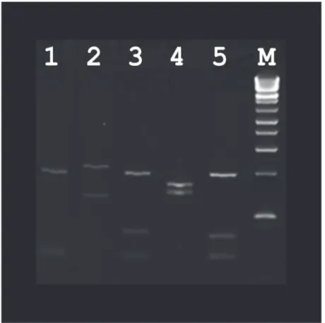
Leishmaniacomplex organisms were identified; 30L.tropica, 7L.donovaniicomplex, 5L. bra-ziliensiscomplex, 6L.majorand 2L.mexicana. For five specimens, speciation could not be de-termined due to the presence of a very weak positive PCR product resulting in a restriction pattern that was difficult to interpret. Most patients acquired leishmaniasis in the Old World (n = 43). For patients with CL, ulcerative lesions on the legs (n = 14) and arms (n = 12) were the most common manifestations. One patient who was affected by VL acquired their infection by transplacental transmission. The patient’s mother was a Sudanese refugee who had been liv-ing in Australia for two years prior to becomliv-ing pregnant. This patient died from infection at 2 years old. No cases of MCL were identified in this study. A summary of these results can be found inTable 1.Fig. 1shows the digestion of amplified ITS1 regions with the restriction endo-nucleaseHaeIII of different species ofLeishmaniafrom isolates from this study on a 4% agarose gel.
Discussion
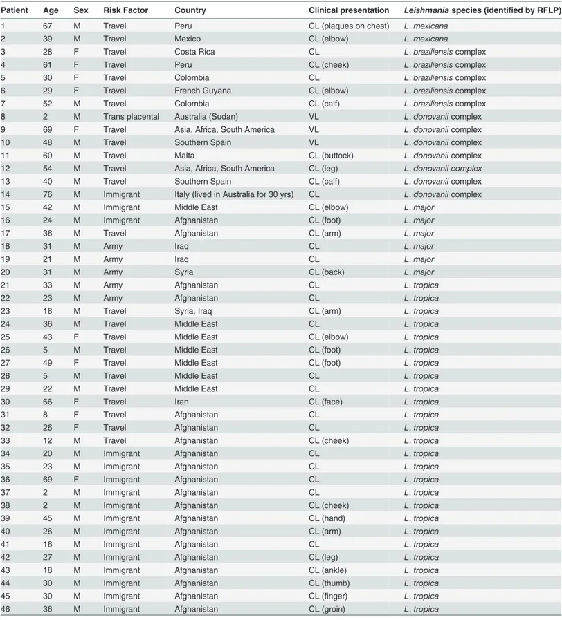
Table 1. Patient details, associated risk factor, clinical presentation (site if known) and species identified.
Patient Age Sex Risk Factor Country Clinical presentation Leishmaniaspecies (identified by RFLP)
1 67 M Travel Peru CL (plaques on chest) L.mexicana
2 39 M Travel Mexico CL (elbow) L.mexicana
3 28 F Travel Costa Rica CL L.braziliensiscomplex
4 61 F Travel Peru CL (cheek) L.braziliensiscomplex
5 30 F Travel Colombia CL L.braziliensiscomplex
6 29 F Travel French Guyana CL (elbow) L.braziliensiscomplex
7 52 M Travel Colombia CL (calf) L.braziliensiscomplex
8 2 M Trans placental Australia (Sudan) VL L.donovaniicomplex
9 69 F Travel Asia, Africa, South America VL L.donovaniicomplex
10 48 M Travel Southern Spain VL L.donovaniicomplex
11 60 M Travel Malta CL (buttock) L.donovaniicomplex
12 54 M Travel Asia, Africa, South America CL (leg) L.donovanii complex
13 40 M Travel Southern Spain CL (calf) L.donovaniicomplex
14 76 M Immigrant Italy (lived in Australia for 30 yrs) CL L.donovaniicomplex
15 42 M Immigrant Middle East CL (elbow) L.major
16 24 M Immigrant Afghanistan CL (foot) L.major
17 36 M Travel Afghanistan CL (arm) L.major
18 31 M Army Iraq CL L.major
19 21 M Army Iraq CL L.major
20 31 M Army Syria CL (back) L.major
21 33 M Army Afghanistan CL L.tropica
22 23 M Army Afghanistan CL L.tropica
23 18 M Travel Syria, Iraq CL (arm) L.tropica
24 36 M Travel Middle East CL L.tropica
25 43 F Travel Middle East CL (elbow) L.tropica
26 5 M Travel Middle East CL (foot) L.tropica
27 49 F Travel Middle East CL (foot) L.tropica
28 5 M Travel Middle East CL L.tropica
29 22 M Travel Middle East CL L.tropica
30 66 F Travel Iran CL (face) L.tropica
31 8 F Travel Afghanistan CL L.tropica
32 26 F Travel Afghanistan CL L.tropica
33 12 M Travel Afghanistan CL (cheek) L.tropica
34 20 M Immigrant Afghanistan CL L.tropica
35 23 M Immigrant Afghanistan CL L.tropica
36 69 F Immigrant Afghanistan CL L.tropica
37 2 M Immigrant Afghanistan CL L.tropica
38 2 M Immigrant Afghanistan CL (cheek) L.tropica
39 45 M Immigrant Afghanistan CL (hand) L.tropica
40 26 M Immigrant Afghanistan CL (arm) L.tropica
41 16 M Immigrant Afghanistan CL L.tropica
42 27 M Immigrant Afghanistan CL (leg) L.tropica
43 18 M Immigrant Afghanistan CL (ankle) L.tropica
44 30 M Immigrant Afghanistan CL (thumb) L.tropica
45 30 M Immigrant Afghanistan CL (finger) L.tropica
46 36 M Immigrant Afghanistan CL (groin) L.tropica
with CL following entry into Australia [20]. The second largest Australian study to date de-scribed 20 cases of imported leishmaniasis diagnosed over a three year period [16]. The present study provides the most recent update and reports a further 55 cases of leishmaniasis diagnosed over a six year period. This study also provides information on the causative species based on the results of a PCR-RFLP technique previously described [7,21].Leishmania tropicawas the
Table 1. (Continued)
Patient Age Sex Risk Factor Country Clinical presentation Leishmaniaspecies (identified by RFLP)
47 16 M Immigrant Afghanistan CL (ear) L.tropica
48 36 F Immigrant Afghanistan CL (arm) L.tropica
49 18 M Immigrant Afghanistan CL (arm) L.tropica
50 1 M Immigrant Afghanistan CL (ankle) L.tropica
51 28 M Travel Pakistan CL (nose) No ID*
52 36 F Travel Panama CL No ID*
53 26 F Travel Peru CL (arm) No ID*
54 34 M Travel - CL (calf) No ID*
55 26 M Immigrant - CL No ID*
*Due to a very weak PCR positive result which made restriction patterns difficult to interpret
doi:10.1371/journal.pone.0119212.t001
Fig 1. Digestion of amplified ITS1 regions with the restriction endonucleaseHaeIII of different species ofLeishmaniafrom isolates from this study on a 4% agarose gel.Lane 1:L.tropica, lane 2:L.major, lane 3:L.donovanicomplex, lane 4:L.braziliensiscomplex, lane 5:L.mexicana. A 100bp ladder was used as the molecular size marker (M).
most common species identified in this study (55%) which is higher than the previous Austra-lian study which reportedL.tropicain only 35% of cases [16]. The slight increase in imported
L.tropicainfections in comparison to the previous study is probably attributable to the recent increase in immigration from Afghanistan to Australia and the deployment of defence person-nel to Afghanistan. In the previous Australian study [16] members of theL.braziliensis com-plex were the second most common cause of leishmaniasis, in contrast to the present study in whichL.donovaniicomplex cases were the second most common (n = 7). The proportion of importedL.majorandL.mexicanacases reported in this study is similar to those reported in the previous Australian study [16].
In the present study, CL was the most common clinical syndrome described (n = 52). Ulcers on the arms and legs were the most common manifestation in CL patients. Consequently, cuta-neous lesions on the limbs of travellers returning from endemicLeishmaniaregions should im-mediately alert clinicians to the possibility ofLeishmaniainfection. Unsurprisingly, no cases of MCL were observed in this study, which is in agreement with previous reports of imported leishmaniasis. Whilst MCL is the most disfiguring of the threeLeishmaniasyndromes, it is also the least common [3]. MCL usually occurs as a result of infections with New WorldLeishmania
species (usuallyLeishmania braziliensiscomplex) and generally occurs concurrently with or following a cutaneous infection, albeit rarely [3].
Leishmania tropicawas the most common species identified in the study cohort which coin-cides with the patients' travel history where more than half of the patients had a history of trav-el to or immigration from Afghanistan or the Middle East. A report from the Netherlands describedL.majoras the most common species identified in a cohort consisting predominant-ly of Dutch soldiers who had been deployed to Afghanistan [5]. In our study it was observed that three of the five defence personnel were also infected withL.major. In addition, in the Dutch studyL.tropicawas only identified in civilians that had a history of travel or residence in Afghanistan [5]. Most of the cohort examined in this study were infected withL.tropicaand it should be noted that the vast majority were civilian immigrants from Afghanistan.L.major
is commonly found in rural areas whereasL.tropicais predominantly found in urban areas. This could explain the difference in species found in people who had travelled from the same country as defence personnel are more likely to be deployed to rural areas and therefore more likely to be infected withL.major.
In total we were able to identify 25 other series [5,7,16,22–41] reporting three or more cases of imported leishmaniasis which also included demographic and travel history together with speciation of isolates, summarised inS1 Table(S1 Published series of travel related leishmania-sis). Similar to our study most imported cases tend to occur in men, presumably due to risk taking behaviour, and present as the cutaneous form of the disease. In contrast, New World species were much less common in our study and the Old World species were mostly acquired in the Middle East rather than Southern Europe. In a study from the UK, the majority of Old World leishmaniasis cases were reportedly acquired whilst travelling in Southern Europe [41]. There were only three patients in the present study who acquired their infection after travel to a Southern European country (Malta and Spain) and a fourth patient from this study is as-sumed to have acquired their infection in Southern Europe as they had immigrated to Australia from Italy 30 years prior. These differences are likely explained by the differences in travel pat-terns of Australian residents and the large number of immigrants in the study. Deployment of Australian troops to the Middle East during this period likely contributed as well.
global travel, immigration and deployment of defence personnel toLeishmaniaendemic areas there is an increased need for clinicians in non-endemic areas to be more aware of leishmania-sis and to consider it in patients displaying clinical manifestations resembling those of CL, VL or MCL. Furthermore, clinicians should also be aware that in rare cases, paediatric VL can occur in patients with no history of travel. In these unusual cases, the familial history should also be considered given the potential for transplacental transmission ofLeishmaniato occur as observed once in this study.
Accurate speciation in clinical cases of leishmaniasis is not only important from an epidemi-ological perspective, but can be important for predicting the clinical outcome and selecting an appropriate treatment regimen. New World leishmaniasis caused by species within theL. bra-ziliensiscomplex (L.braziliensis,L.guyanensis,L.panamensisandL.peruviana) are more likely to lead to secondary MCL than other New World species. As MCL can occur up to two years after cutaneous lesions resolve [42], knowledge of the causative species can facilitate diagnosis should lesions appear on the mucous membranes at a later time. Furthermore,L.braziliensis
complex organisms are resistant to miltefosine while other species are not [14], so miltefosine is unlikely to be effective in such cases.
PCR is currently the tool of choice for diagnosis of leishmaniasis. Traditionally microscopy, histopathology and culture were used though these techniques do not differentiate between
Leishmaniaspecies. Prior to the advent of PCR, isoenzyme analysis was the gold standard for speciation ofLeishmaniasp. though this technique is comparatively laborious and requires prior cultivation of parasitesin vitro[7]. Several PCR-based tools have been developed which are capable of differentiating between certain species and/or complexes ofLeishmania, though each of these has its advantages and limitations [7,43–46]. One study showed that Kinetoplast DNA (kDNA) had the highest sensitivity for the detection ofLeishmaniasp. over ITS1 PCR and splice leader mini-exon PCR, however this technique does not allow for speciation [47]. RT-PCRs have been developed but most have the limitation of only differentiating to complex level and not having the ability to speciate within that complex. The advantage of this though is that RT-PCR is much faster than conventional PCR, which has to be followed by either RFLP or sequencing for any kind of speciation, if only the complex level is desired. Generally PCR is highly sensitive and when coupled with RFLP analysis, the assay employed in this study, can differentiate between mostLeishmaniaspecies. This PCR-RFLP does have its limitations how-ever, as it cannot differentiate between species within theL.donovanicomplex (L.donovani
andL.infantum/L.chagasi) and those within theL.braziliensiscomplex. Sequencing of PCR products to differentiate between species is also complicated by the heterogenous nature of
LeishmaniaITS1 sequences. The ribosomal RNA (rRNA) genes exist in eukaryotic genomes as tandem repeats with many copies [48,49]. In some protozoa (e.g.Toxoplasma gondiiand rela-tives), the ITS1 region is identical for each copy [50,51], which is conducive to sequencing. In contrast, the ITS1 region in individualLeishmaniaisolates varies greatly between copies [52]. As a result, PCR products derived from the ITS1 ofLeishmaniado not produce a clean se-quencing read. Regardless, because the rRNA genes exist in multiple copies in the genome, there is still the benefit of increased sensitivity for PCRs targeting these genes compared to those targeting single copy genes.
(Lasiohelea)[10]. While there is little evidence to suggest that this macropod-infecting Leish-maniasp. can infect humans, the possibility that the Australian midge vector could transmit pathogenicLeishmaniasp. is a cause for concern. In a recent study, midges of the genus Culi-coides(family Ceratopogonidae) were found to support the replication ofLeishmania infantum
andLeishmania majorin their midgut for at least 3 days following an experimental infection [54,55]. Considering that the proposed AustralianLeishmaniavector is also a member of the family Ceratopogonidae, it is not unreasonable to suggest that this native midge may also be ca-pable of transiently supporting the growth of clinically importantLeishmaniasp.. With cases of imported human leishmaniasis becoming a regular occurrence in Australia, it is possible that the native midge vector will come into contact with human infectingLeishmaniaspecies, providing an opportunity for the establishment of a local transmission cycle. While the support for such an event is limited, it is not unprecedented and certainly warrants continued investigation.
Conclusion
This represents the largest study to examine the molecular epidemiology of imported leish-maniasis in Australia. Of 206 patients suspected of harbouring aLeishmaniainfection over a six-year period, 55 were confirmed by PCR. The majority of infected patients had travelled to or immigrated from Afghanistan andL.tropicawas the most common species identified. These results indicate that imported cases of leishmaniasis are an ongoing occurrence in Aus-tralia and highlights the need for AusAus-tralian clinicians to consider leishmaniasis when assessing patients with a history of travel to or residence in endemic regions presenting with Leishman-ia-associated clinical manifestations.
Supporting Information
S1 Table. Published series of travel related leishmaniasis
(DOCX)
Author Contributions
Conceived and designed the experiments: TR DS JH DM RL JE. Performed the experiments: TR DS. Analyzed the data: TR DS JB IS JH JE DM RL. Contributed reagents/materials/analysis tools: TR DS JB. Wrote the paper: TR JB IS DS.
References
1. Dostalova A, Volf P (2012) Leishmania development in sand flies: parasite-vector interactions over-view. Parasit Vectors 5: 276. doi:10.1186/1756-3305-5-276PMID:23206339
2. Herwaldt BL (1999) Leishmaniasis. Lancet 354: 1191–1199. PMID:10513726
3. Barratt JL, Harkness J, Marriott D, Ellis JT, Stark D (2010) Importance of nonenteric protozoan infec-tions in immunocompromised people. Clin Microbiol Rev 23: 795–836. doi:10.1128/CMR.00001-10
PMID:20930074
4. Alvar J, Yactayo S, Bern C (2006) Leishmaniasis and poverty. Trends Parasitol 22: 552–557. PMID:
17023215
5. Bart A, van Thiel PP, de Vries HJ, Hodiamont CJ, Van Gool T (2013) Imported leishmaniasis in the Netherlands from 2005 to 2012: epidemiology, diagnostic techniques and sequence-based species typing from 195 patients. Euro Surveill 18: 20544. PMID:23929178
8. Khairnar K, Lynde C, Ralevski F, Keystone J, McCarthy A, et al. (2013) Molecular diagnosis of leish-maniasis at the subgenus and species level. Parasitol Res.
9. Bailey MS, Lockwood DN (2007) Cutaneous leishmaniasis. Clin Dermatol 25: 203–211. PMID:
17350500
10. Dougall AM, Alexander B, Holt DC, Harris T, Sultan AH, et al. (2011) Evidence incriminating midges (Diptera: Ceratopogonidae) as potential vectors of Leishmania in Australia. Int J Parasitol 41: 571–
579. doi:10.1016/j.ijpara.2010.12.008PMID:21251914
11. Diniz MM, Galvis Ovallos F, de Castro Gomes CM, de Oliveira Lavitschka C, Bianchi Galati EA (2014) Host-biting rate and susceptibility of some suspected vectors to Leishmania braziliensis. Parasit Vec-tors 7: 139. doi:10.1186/1756-3305-7-139PMID:24684943
12. Rose K (2004) Cutaneous leishmaniasis in red kangaroos. Aust Vet J 82: 440. PMID:15354854 13. Rose K, Curtis J, Baldwin T, Mathis A, Kumar B, et al. (2004) Cutaneous leishmaniasis in red
kanga-roos: isolation and characterisation of the causative organisms. Int J Parasitol 34: 655–664. PMID:
15111087
14. Schwartz E, Hatz C, Blum J (2006) New world cutaneous leishmaniasis in travellers. Lancet Infect Dis 6: 342–349. PMID:16728320
15. Stark D, Pett S, Marriott D, Harkness J (2006) Post-kala-azar dermal leishmaniasis due to Leishmania infantum in a human immunodeficiency virus type 1-infected patient. J Clin Microbiol 44: 1178–1180. PMID:16517925
16. Stark D, van Hal S, Lee R, Marriott D, Harkness J (2008) Leishmaniasis, an emerging imported infec-tion: report of 20 cases from Australia. J Travel Med 15: 351–354. doi:10.1111/j.1708-8305.2008. 00223.xPMID:19006509
17. Konecny P, Stark DJ (2007) An Australian case of New World cutaneous leishmaniasis. Med J Aust 186: 315–317. PMID:17371215
18. Ju O, Grove DI, Jaksic WJ, Dart GW (2004) Visceral leishmaniasis: a trip to the Greek Islands is not al-ways idyllic. Med J Aust 181: 446–447. PMID:15487964
19. Maguire GP, Bastian I, Arianayagam S, Bryceson A, Currie BJ (1998) New World cutaneous leishmani-asis imported into Australia. Pathology 30: 73–76. PMID:9534213
20. Storer E, Wayte J (2005) Cutaneous leishmaniasis in Afghani refugees. Australas J Dermatol 46: 80–
83. PMID:15842398
21. el Tai NO, Osman OF, el Fari M, Presber W, Schonian G (2000) Genetic heterogeneity of ribosomal in-ternal transcribed spacer in clinical samples of Leishmania donovani spotted on filter paper as revealed by single-strand conformation polymorphisms and sequencing. Trans R Soc Trop Med Hyg 94: 575–
579. PMID:11132393
22. Antinori S, Gianelli E, Calattini S, Longhi E, Gramiccia M, et al. (2005) Cutaneous leishmaniasis: an in-creasing threat for travellers. Clin Microbiol Infect 11: 343–346. PMID:15819858
23. Bailey MS, Caddy AJ, McKinnon KA, Fogg LF, Roscoe M, et al. (2012) Outbreak of zoonotic cutaneous leishmaniasis with local dissemination in Balkh, Afghanistan. J R Army Med Corps 158: 225–228. PMID:23472571
24. Blonski KM, Blodorn-Schlicht N, Falk TM, Faye RS, Clausen OP (2012) Increased detection of cutane-ous leishmaniasis in Norway by use of polymerase chain reaction. APMIS 120: 591–596. doi:10.1111/ j.1600-0463.2012.02875.xPMID:22716214
25. Center AFHS (2007) Leishmaniasis in relation to service in Iraq/Afghanistan, U.S. Armed Forces, 2001–2006. Medical Surveillance Monthly Report (MSMR) 14: 2–5.
26. Centers for Disease C (1992) Viscerotropic leishmaniasis in persons returning from Operation Desert Storm—1990–1991. MMWR Morb Mortal Wkly Rep 41: 131–134. PMID:1738359
27. Harms G, Scherbaum H, Reiter-Owona I, Stich A, Richter J (2011) Treatment of imported New World cutaneous leishmaniasis in Germany. Int J Dermatol 50: 1336–1342. doi:10.1111/j.1365-4632.2011. 04987.xPMID:22004484
28. Harms G, Schonian G, Feldmeier H (2003) Leishmaniasis in Germany. Emerg Infect Dis 9: 872–875. PMID:12890332
29. Hepburn NC, Tidman MJ, Hunter JA (1993) Cutaneous leishmaniasis in British troops from Belize. Br J Dermatol 128: 63–68. PMID:8381299
30. Lachaud L, Dedet JP, Marty P, Faraut F, Buffet P, et al. (2013) Surveillance of leishmaniases in France, 1999 to 2012. Euro Surveill 18: 20534. PMID:23929121
32. Morizot G, Kendjo E, Mouri O, Thellier M, Perignon A, et al. (2013) Travelers with cutaneous leishmani-asis cured without systemic therapy. Clin Infect Dis 57: 370–380. doi:10.1093/cid/cit269PMID:
23633111
33. Poeppl W, Oeser C, Grabmeier-Pfistershammer K, Walochnik J, Burgmann H (2013) Clinical findings and management of imported cutaneous leishmaniasis: report of 14 cases from Austria. Travel Med In-fect Dis 11: 90–94. doi:10.1016/j.tmaid.2013.03.002PMID:23522841
34. Scarisbrick JJ, Chiodini PL, Watson J, Moody A, Armstrong M, et al. (2006) Clinical features and diag-nosis of 42 travellers with cutaneous leishmaniasis. Travel Med Infect Dis 4: 14–21. PMID:16887720 35. Scope A, Trau H, Anders G, Barzilai A, Confino Y, et al. (2003) Experience with New World cutaneous
leishmaniasis in travelers. J Am Acad Dermatol 49: 672–678. PMID:14512915
36. Seaton RA, Morrison J, Man I, Watson J, Nathwani D (1999) Out-patient parenteral antimicrobial thera-py—a viable option for the management of cutaneous leishmaniasis. QJM 92: 659–667. PMID:
10542306
37. Solomon M, Baum S, Barzilai A, Scope A, Trau H, et al. (2007) Liposomal amphotericin B in compari-son to sodium stibogluconate for cutaneous infection due to Leishmania braziliensis. J Am Acad Der-matol 56: 612–616. PMID:17276541
38. van Thiel PP, Zeegelaar JE, van Gool T, Faber WR, Kager PA (2011) Cutaneous leishmaniasis in three Dutch military cohorts following jungle training in Belize. Travel Med Infect Dis 9: 153–160. doi:10. 1016/j.tmaid.2011.03.001PMID:21450527
39. Weina PJ, Neafie RC, Wortmann G, Polhemus M, Aronson NE (2004) Old world leishmaniasis: an emerging infection among deployed US military and civilian workers. Clin Infect Dis 39: 1674–1680. PMID:15578370
40. Zeegelaar JE, Steketee WH, van Thiel PP, Wetsteyn JC, Kager PA, et al. (2005) Changing pattern of imported cutaneous leishmaniasis in the Netherlands. Clin Exp Dermatol 30: 1–5. PMID:15663490 41. Wall EC, Watson J, Armstrong M, Chiodini PL, Lockwood DN (2012) Epidemiology of imported
cutane-ous leishmaniasis at the Hospital for Tropical Diseases, London, United Kingdom: use of polymerase chain reaction to identify the species. Am J Trop Med Hyg 86: 115–118. doi: 10.4269/ajtmh.2012.10-0558PMID:22232460
42. Lawn SD, Whetham J, Chiodini PL, Kanagalingam J, Watson J, et al. (2004) New world mucosal and cutaneous leishmaniasis: an emerging health problem among British travellers. QJM 97: 781–788. PMID:15569809
43. Kumar A, Boggula VR, Misra P, Sundar S, Shasany AK, et al. (2010) Amplified fragment length poly-morphism (AFLP) analysis is useful for distinguishing Leishmania species of visceral and cutaneous forms. Acta Trop 113: 202–206. doi:10.1016/j.actatropica.2009.10.006PMID:19854144
44. Haouas N, Garrab S, Gorcii M, Khorchani H, Chargui N, et al. (2010) Development of a polymerase chain reaction-restriction fragment length polymorphism assay for Leishmania major/Leishmania kill-icki/Leishmania infantum discrimination from clinical samples, application in a Tunisian focus. Diagn Microbiol Infect Dis 68: 152–158. doi:10.1016/j.diagmicrobio.2010.06.011PMID:20846587 45. Weirather JL, Jeronimo SM, Gautam S, Sundar S, Kang M, et al. (2011) Serial quantitative PCR assay
for detection, species discrimination, and quantification of Leishmania spp. in human samples. J Clin Microbiol 49: 3892–3904. doi:10.1128/JCM.r00764-11PMID:22042830
46. Pita-Pereira D, Lins R, Oliveira MP, Lima RB, Pereira BA, et al. (2012) SYBR Green-based real-time PCR targeting kinetoplast DNA can be used to discriminate between the main etiologic agents of Brazil-ian cutaneous and visceral leishmaniases. Parasit Vectors 5: 15. doi:10.1186/1756-3305-5-15PMID:
22240199
47. Bensoussan E, Nasereddin A, Jonas F, Schnur LF, Jaffe CL (2006) Comparison of PCR assays for di-agnosis of cutaneous leishmaniasis. J Clin Microbiol 44: 1435–1439. PMID:16597873
48. Boothroyd JC, Wang A, Campbell DA, Wang CC (1987) An unusually compact ribosomal DNA repeat in the protozoan Giardia lamblia. Nucleic Acids Res 15: 4065–4084. PMID:3588284
49. Guay JM, Huot A, Gagnon S, Tremblay A, Levesque RC (1992) Physical and genetic mapping of cloned ribosomal DNA from Toxoplasma gondii: primary and secondary structure of the 5S gene. Gene 114: 165–171. PMID:1601300
50. Barratt J, Al Qassab S, Reichel MP, Ellis JT (2008) The development and evaluation of a nested PCR assay for detection of Neospora caninum and Hammondia heydorni in feral mouse tissues. Mol Cell Probes 22: 228–233. doi:10.1016/j.mcp.2008.03.001PMID:18420378
52. Ghatee MA, Sharifi I, Kuhls K, Kanannejad Z, Fasihi Harandi M, et al. (2014) Heterogeneity of the inter-nal transcribed spacer region in Leishmania tropica isolates from southern Iran. Exp Parasitol 144: 44–
51. doi:10.1016/j.exppara.2014.06.003PMID:24932536
53. Dougall A, Shilton C, Low Choy J, Alexander B, Walton S (2009) New reports of Australian cutaneous leishmaniasis in Northern Australian macropods. Epidemiol Infect 137: 1516–1520. doi:10.1017/ S0950268809002313PMID:19288959
54. Seblova V, Sadlova J, Carpenter S, Volf P (2012) Development of Leishmania parasites in Culicoides nubeculosus (Diptera: Ceratopogonidae) and implications for screening vector competence. J Med Entomol 49: 967–970. PMID:23025175