Localization and Can Be Used to Express Heterologous
Proteins on the Mycobacterial Surface
Valentina Dona`1¤a, Marcello Ventura1, Michela Sali2, Alessandro Cascioferro1¤b, Roberta Provvedi3, Giorgio Palu`1, Giovanni Delogu2, Riccardo Manganelli1*
1Department of Molecular Medicine, University of Padua, Padua, Italy,2Institute of Microbiology, Universita` Cattolica del Sacro Cuore, Rome, Italy,3Department of Biology, University of Padua, Padua, Italy
Abstract
PPE represent a peculiar family of mycobacterial proteins characterized by a 180 aminoacids conserved N-terminal domain. Several PPE genes are co-transcribed with a gene encoding for a protein belonging to another family of mycobacterial specific proteins named PE. Only one PE-PPE couple has been extensively characterized so far (PE25-PPE41) and it was shown that these two proteins form a heterodimer and that this interaction is essential for PPE41 stability and translocation through the mycobacterial cell wall. In this study we characterize the PE11-PPE17 couple. In contrast with what was found for PE25-PPE41, we show that PPE17 is not secreted but surface exposed. Moreover, we demonstrate that the presence of PE11 is not necessary for PPE17 stability or for its localization on the mycobacterial surface. Finally, we show that the PPE domain of PPE17 targets the mycobacterial cell wall and that this domain can be used as a fusion partner to expose heterologous proteins on the mycobacterial surface.
Citation:Dona` V, Ventura M, Sali M, Cascioferro A, Provvedi R, et al. (2013) The PPE Domain of PPE17 Is Responsible for Its Surface Localization and Can Be Used to Express Heterologous Proteins on the Mycobacterial Surface. PLoS ONE 8(3): e57517. doi:10.1371/journal.pone.0057517
Editor:Pere-Joan Cardona, Fundacio´ Institut d’Investigacio´ en Cie`ncies de la Salut Germans Trias i Pujol. Universitat Auto`noma de Barcelona. CIBERES, Spain
ReceivedAugust 31, 2012;AcceptedJanuary 23, 2013;PublishedMarch 1, 2013
Copyright:ß2013 Dona` et al. This is an open-access article distributed under the terms of the Creative Commons Attribution License, which permits unrestricted use, distribution, and reproduction in any medium, provided the original author and source are credited.
Funding:This work was supported by the European Community’s Seventh Framework Programme (FP7/2007–2013) under grant agreement nu201762 and by Ministero dell’Universita` e della Ricerca Scientifica, Progetti di Interesse Nazionale 2008 (code number 2008Y8RZTF). The funders had no role in study design, data collection and analysis, decision to publish, or preparation of the manuscript.
Competing Interests:The authors have declared that no competing interests exist.
* E-mail: [email protected]
¤a Current address: Tuberculosis Research Section, Laboratory of Clinical Infectious Disease, National Institute of Allergy and Infectious Diseases, National Institutes of Health, Bethesda, Maryland, United States of America
¤b Current address: Integrated Mycobacterial Pathogenomics Unit, Institut Pasteur, Paris, France
Introduction
One of the most intriguing features of the genomes of slow growing mycobacteria is the presence of a high number of genes encoding for members of two peculiar protein families named PE and PPE. The members of these families are characterized from highly conserved N-terminal domains of about 110 or 180 aminoacids, respectively, typically containing the motif PE or PPE at the beginning of their amino acidic sequence, after which they are named [1]. Some members of these protein families are represented by a sole PE or PPE domain, while most of them present a second larger C-terminal extension which can be unique or belong to one of several subclasses [2]. Although some PE and PPE have been shown to be involved in the modulation of the immune response and/or to be essential for virulence [3,4,5,6], their precise function has been elucidated or proposed only for a few of them [7,8,9].
All the PE and PPE proteins characterized to date were shown to be surface exposed or secreted [10,11]. Interestingly, almost all PE nor PPE do not present canonical secretion signals and recently it has been demonstrated that both the N- and the C-terminus of the PE domain contain the information necessary to drive the translocation of this family of proteins [12,13,14]. In a recent study we showed that the catalytic domain of the secreted
lipase LipY is fused to a PE domain inMycobacterium tuberculosis, to a PPE domain inMycobacterium marinumand to a canonical signal peptide inMycobacterium gilvum, suggesting that these domains are interchangeable modules used by mycobacteria to target proteins to the bacterial surface [15]. Secretion of several PE and PPE is dependent on the type VII secretion system ESX-5, although the molecular mechanism of the transport process is still unknown [10,16].We also reported that the PE domain of one of the better characterized PE proteins, PE_PGRS33, was able to drive the surface localization of MPT64 when it was fused at its C-terminus [12,13]. The expression of this chimeric protein on the surface of a recombinant Mycobacterium bovis BCG increased its protection againstM. tuberculosisinfection, opening the possibility to exploit the function of these proteins to develop an improved vaccine against tuberculosis [17].
was recently shown to interact with Toll-like receptor-2 resulting in downstream activation of nuclear factor-kb and HIV-1 LTR trans-activation [21]. Here we demonstrate that PPE17 is stable and surface exposed, even when expressed in the absence of the cognate PE. We also demonstrate that the PPE domain of PPE17 contains the information necessary for secretion and anchorage to the cell wall and that it can be used as a fusion partner to express antigens on the mycobacterial surface.
Materials and Methods
Ethics Statement
All procedures involving the use of animal were approved by the Ethical Committee of the Catholic University of the Sacred Heart, Rome.
Bacterial Strains, Media and Growth Conditions
M. smegmatis mc2155 [22] and M. bovis BCG (Pasteur)were grown at 37uC in Middlebrook 7H9 (liquid medium) or 7H10 (solid medium; Difco), supplemented with 0.05% v/v Tween 80 (Sigma-Aldrich) and 0.2% v/v glycerol (Sigma-Aldrich). M. bovis
BCG cultures were also added with ADN (2% glucose, 5% BSA, 0,85% NaCl). Strains processed for proteinase K degradation assay, and cell subfractioning were grown in Sauton (Difco). For cloning procedures E. coli strain HB101 was grown in Luria Bertani medium (LB) [23]. Hygromycin (Roche) was used at a final concentration of 100mg ml21(solid media) or 50mg ml21(liquid
media) for M. smegmatis and at a final concentration of 200mg ml21forE. coli.
DNA Manipulation
All genes expressed in this work were amplified from theM. tuberculosisH37Rv chromosomal DNA with Pfu DNA polymerase (Stratagene). For the amplification of the sequence encoding
HA-tagged PPE17 or its domain PPE domain (PPEd) with and without co-expression of PE11, an upper primer was designed containing anXbaI restriction site immediately before the start codon of the structural gene of PPE17 or PE11, respectively (RP93, RP91) (Table S1). The lower primer for the amplification of the whole gene encoding PPE17 was designed to remove the stop codon of the gene which was fused to the HA coding sequence and a stop codon followed by anXbaI restriction site(RP561) (Table S1). The same strategy was used to design the lower primer for the amplification of the sequence encoding the PPEd (RP560) (Table S1). All fragments were cloned into pMV10-25 [12], in order to place the transcription of the HA-tagged proteins under the control of the strong mycobacterial promoter Phsp60obtaining the following replicative plasmids: pVD28 (PE11-PPE17-HA), pVD27 (PE11-PPEd-HA), pVD31 (PPE17-HA), and pAL27 (PPEd-HA) (Table S2). The correct orientation of the inserted fragments was verified by PCR.
In order to develop a surface expression system based on the PPEd, we constructed the mycobacterial expression vector pAL26, where the sequence encoding this domain was placed under the control of Phsp60 and upstream of an in frame polylinker to facilitate cloning. At this purpose, the 567 bp sequence encoding the PPEd plus the polylinker was amplified using the primers RP233 and RP234. The upper primer was designed to contain an
XbaI site immediately before the start codon of the PPE coding sequence, while the lower primer was designed to have a polylinker containing BamHI, PacI and NcoI restriction sites before a stop codon and aKpnI site. This fragment was transcriptionally fused to the Phsp60present in the shuttle vector pMV10-25 [24] digested by NheI andKpnI (Tables S1 and S2).
To construct the translational fusion between PPEd and the mycobacterial antigen Mpt64, a fragment encoding Mpt64, deprived of its first 23 aminoacids (aa) (D-MPT64) tagged with the HA epitope was excised from pSTE2 [12,13] and cloned into
Figure 1. Maps of the PE11-PPE17 encoding region and of their derivatives used for this study.A) Map of theM. tuberculosisgenomic region encoding PE11 and PPE17; B) Maps of the plasmids used to express PE11, PPE17 and MPT64 derivatives. The gene encoding PE11 is shown in white, the gene encoding PPE17 is shown in grey, the gene encoding the leaderless MPT64 is shown in stripes, while the 9 amino acid HA epitope is shown in black. The molecular weight of the PPE-derived proteins encoded by the relative genes is shown.
pAL26. Briefly, vectors pSTE2 and pAL26 were digested by
BamHI andNcoI and the respective fragments were separated by agarose gels, purified and ligated to obtain pAL29 (Tables S1 and S2).
Electroporation
Electroporation of mycobacteria was performed as previously described [25]. Briefly, mid-exponential cultures were extensively washed in 10% glycerol and concentrated approximately 40-fold. One hundredml of concentrated cells were mixed with 1mg of DNA and transferred to 0.2 cm gap cuvettes (Eppendorf). Samples were electroporated using an Electroporator Gene Pulser Trans-fection Apparatus (Biorad; capacitance 25mF; voltage12.5 kV cm21; resistance 200V). After the pulse, the cells were diluted in
900 ml of liquid medium, incubated for 3 h (M. smegmatis), or 24 h (M. bovisBCG) and then plated on selective solid medium.
Protein Samples Preparation
Protein samples were prepared as previously described [13]. Briefly, mid-exponential cultures were separated from culture supernatants by centrifugation, and secreted proteins were pre-cipitated from culture supernatants with 10% TCA (w/v). Cells were washed with PBS and thereafter subjected to proteinase K degradation, Genapol extraction or subcellular fractionation as described below. Proteins samples were boiled and separated by SDS-PAGE as described below.
Proteinase K Degradation Assay
Proteinase K degradation assay was performed as previously described [12]. Briefly, bacteria were grown for 14 h starting from an OD600 of 0.1 in 20 ml of medium. Cells were washed once in TBS buffer (Tris HCl pH 7.5, NaCl 150 mM, KCl 3 mM) and resuspended in 1 ml of the same buffer. Each sample was divided in two identical aliquots. One aliquot was treated with proteinase K (Sigma-Aldrich) up to a concentration of100 mg ml21
, whereas the other was left untreated and incubated for 30 min at 4uC. The reaction was stopped by the addition of 1X complete EDTA-free protease inhibitor (Roche). Subsequently, samples were washed once in TBS and resuspended in TBS plus loading buffer 5X (sucrose 50% w/ v, SDS 10% w/v, 0.3 M Tris HCl pH 6.8, bromophenol blue 0.05%w/v, b-mercaptoethanol 5% v/v) or subjected to sub-cellular fractionation as described below. Finally, samples were boiled for10 min to allow bacterial lysis and loaded on a polyacrylamide gel in equal amounts. Treated and untreated samples were processed in parallel using the same procedure to allow their comparison. Each experiment was performed at least twice with different biological samples.
Subcellular Fractionation
Subcellular fractionation was performed as previously described [12,13] with some modifications after proteinase K treatment, bacterial pellets were resuspended in PBS 1X/phenyl methane sulphonyl fluoride (Sigma-Aldrich, PMSF) and subjected to sonication to lyse the cells. The lysates were centrifuged at 10006g at 4uC to precipitate cellular debris and unlysed cells. Supernatants were transferred to fresh tubes and sedimented at 27.0006g for 30 min at 4uC to recover cell wall-associated proteins. Once again, the supernatant was precipitated at 100.0006g for 2 h to separate cytoplasmic membrane from
cytosolic fraction. Cytosolic proteins were subsequently concen-trated on Amicon centrifugal filters (cutoff 3 kDa) to a final volume of 1 ml. Pellets were washed once after each step of centrifugation
in PBS/PMSF 1 mM and finally resuspended in 1 ml of PBS plus Loading buffer 5X. Finally, samples were boiled for 5 min before being separated on polyacrylamide gels and subjected to Western blotting as described below.
SDS-PAGE and Immunoblot
SDS-PAGE was performed according to standard protocols. Briefly, proteins were separated on 10%, polyacrylamide gels [23], and subsequently transferred to polyvinylidene fluoride mem-branes (PVDF; Bio-Rad) by Western blotting. Proteins were visualized by immunoblotting using monoclonal antibodies di-rected against the HA epitope (Anti-HA.11; Covance, dilution 1:2000), or GFP (Chemicon; dilution 1:2500). Secondary goat anti-mouse (Santa Cruz Biotechnology; dilution 1:2000) horserad-ish peroxidase conjugates were used to detect proteins. The West Dura Signal Kit (Pierce) was used to develop the chemilumines-cent signal. Image acquisitions and quantifications were performed using a Versadoc Imaging System (Bio-Rad) and Quantity One4.2.3 software (Bio-Rad).
Enzyme-linked Immunosorbent Assay with Whole Cells ofM. smegmatisorM. bovisBCG
The assay was performed as previously described [26] with some modifications: cells were grown to an OD600of about 0.8, harvested by centrifugation at 30006g for 10 minutes at room
temperature, washed twice in TBST buffer (50 mM Tris-HCl pH 8.0, 150 mM NaCl, 1 mM MgCl2 and 0.05% Tween 80). Pellets were finally resuspended in 1 ml of TBST and divided into two identical aliquots. One aliquot was treated with proteinase K (Sigma-Aldrich) 100mg ml21, whereas the other
was left untreated and incubated for 30 min at 4uC. The reaction was stopped adding 1X complete EDTA-free protease inhibitor (Roche). Samples were then centrifuged at 4uC for 5 minutes at 30006g and washed with 500ml of TBST. Finally pellets were resuspended in 100ml of NaHCO350 mM, pH 9,6 to yield a cell concentration of about 16109cells ml21. One
hundredml of the so obtained cell suspensions were transferred to each well of a 96-wellsmicrotitre plate (NUNC-Immuno Maxi Sorp Surface, Nalge Nunc International). After 24 h of in-cubation at 4uC, the microplate was centrifuged and the supernatant discarded. Samples were then blocked with 200ml of 3% powdered skim milk in TBST for 90 minutes at 37uC (M. smegmatis) or at room temperature (M. bovis BCG), and subsequently washed once with 200ml of TBST. The primary antibody (a monoclonal anti -HA.11, Covance, or a polyclonal anti-MPT64 mouse anti-serum) was diluted in 1% powdered skim milk in TBST (1:10000 dilution for anti-HA and 1:6400 for anti-MPT64) and 100ml added to each well. After an incubation of 1 h at 37uC (M. smegmatis) or at room temperature (M. bovisBCG), the wells were washed three times with 200ml of TBST. The secondary antibody (alkaline phosphatase conjugate goat anti-mouse, Santa Cruz Biotechnology) was diluted 1:5000 in TBST containing 1% powdered skim milk and 100ml were added to each well. Samples were incubated for 1 h at 37uC (M. smegmatis) or at room temperature (M. bovis
Evaluation of the Protective Activity of Recombinant BCG Groups of C57Bl/6 mice were immunized subcutaneously with 56106CFU of BCG PPE-DMpt64, BCG PPE or, as a control, BCG Pasteur on day 0. Ten-weeks following the immunization, vaccinated and control mice were infected aerosolizing about 100 CFUs ofM. tuberculosisErdman using a Middlebrook chamber (Glas-Col) as described previously [27]. The vaccinated and control mice were sacrificed 28 days after challenge and bacterial colonization of lung and spleen tissues assessed as described earlier [28]. Briefly, to assess the bacterial growthin vivo, five mice per group were sacrificed, and the lungs and spleens were removed aseptically and homogenized separately in 5 ml of 0.04% Tween 80-PBS using a Seward Stomacher 80 blender (Tekmar). The homogenates were diluted serially in the Tween-PBS solution, and 50-ml aliquots were plated on Middlebrook 7H11 agar (Difco) containing 2-thiophenecarboxylic acid hydrazide (2mg/ml). The number of CFUs in the infected organs was determined after 14 to 21 days of incubation at 37uC in sealed plastic bags.
Results
Construction of Mycobacterial Strains Expressing HA-labelled PPE17
The gene encoding PPE17, as several other PPE genes, is co-transcribed with a gene encoding a PE domain (PE11). To determine PPE17 localization, and to assess whether co-expression with PE11 plays a role in PPE17 stability or localization, we constructed a series of replicative mycobacterial expression vectors, in which the gene encoding PPE17 or just its N-terminal PPE domain (PPEd) fused to the HA epitope coding sequence (to facilitate detection), were expressed either in the presence or in the absence of the gene encoding PE11 (Fig. 1B). Each construct was placed under the transcriptional control of the strong mycobac-terial promoter Phsp60. The resulting plasmids were then in-troduced by electroporation in M. bovis BCG and M. smegmatis
mc2155. Protein extracts of the resulting strains were analysed by Western blot to assess the expression of the recombinant proteins. We found that PPE17 was not secreted into the culture supernatant of M.bovis BCG (Fig. S1) or extracted by the non-ionic detergent Genapol inM. smegmatis(Fig. S2).
Surface Localization of PPE17 does not Depend on the Presence of PE11
The four M. bovis BCG strains containing the first four constructs shown in Figure 1 were grown in liquid media, and divided into two equal aliquots. One aliquot of each strain was subjected to proteinase K degradation. The resulting samples were then used to perform an ELISA assay on whole cells. The ELISA was developed as described previously [20] with an anti-HA primary antibody and a secondary antibody linked to a horseradish peroxidase. After the addition of the substrate we were expecting to detect absorbance at 405 nm for recombinant proteins exposed on the mycobacterial surface in the untreated samples, which should not be detectable in the respective samples previously treated with proteinase K. We could detect a signal above background for PPE17 regardless of PE11 co-expression (Fig. 2). Moreover, in both cases, PPE17 was sensitive to proteinase K degradation, further confirming its surface exposure. However, we could not detect any signal deriving from PPEd, suggesting that this moiety may be imbedded inside the cell wall structure, as we already reported for the PE domain of PE_PGRS33[12].
To further corroborate these results we repeated the proteinase K degradation assay onM. bovisBCG strains expressing PPE17. After the treatment all samples were lysed by sonication and
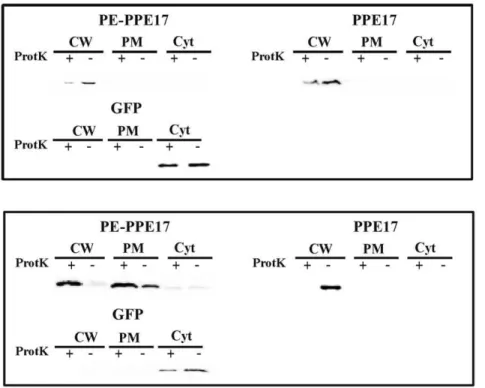
subjected to subcellular fractionation. Finally, protein samples were separated on an SDS page gel and analysed by Western blot with a monoclonal antibody against the HA epitope. The intracellular protein GFP was used as a control. As shown in Figure 3A, PPE17 mainly localized in the cell wall fraction regardless of PE11 co-expression. In all samples it was sensitive to proteinase K degradation confirming the results shown in the ELISA assay. Since inM. bovisBCG the chromosomal copy of the gene encoding PE11 might complement the absence of this gene in the expression plasmid, we repeated this experiment in Mycobac-terium smegmatis, which does not encodes close homologs of PE11 or PPE17: as shown in Figure 3B, also in this species PPE17 mainly localized in the cell wall fraction regardless of PE11 co-expression and was sensitive to proteinase K degradation.
Construction and Characterization of Mycobacterial Strains Expressing Heterologous Proteins on their Surface
In order to develop a mycobacterial expression system for surface localization of chimeric proteins based on the PPEd, we constructed an expression vector in which the sequence encoding this domain was placed downstream of Phsp60and upstream of an in-frame polylinker to facilitate cloning (pAL26) (Fig. 4).
The sequence encoding MPT64 (a protective antigen absent in several M. bovis BCG strains) [17,27] deprived of its signal sequence (DMPT64) was cloned in frame with the sequence encoding the PPE domain and the HA epitope. The resulting vector was electroporated inM. smegmatisandM. bovisBCG. The resulting strains were subjected to proteinase K degradation assay followed by whole-cell ELISA. As shown in Figure 5, the fusion protein was clearly detectable in bothM. smegmatisand M. bovis
BCG, whereas strains expressing DMPT64 showed a signal comparable to the negative control, represented by strains not expressing MPT64, demonstrating that PPEd was driving the localization of the chimeric protein on the mycobacterial surface. Moreover, in both mycobacterial species, the fusion protein was sensitive to the proteolytic activity of the proteinase K, confirming that PPEd-DMpt64 is surface exposed. To further corroborate Figure 2. Whole-cell ELISA ofM. bovisBCG strains expressing different proteins.Cell cultures ofM. bovisBCG were treated (grey) or not treated (white) with proteinase K to allow degradation of surface exposed proteins. Control represents wtM. bovis BCG. Proteins were detected using monoclonal antibodies against HA.
these data theses trains were also subjected to proteinase K degradation assay followed by subcellular fractionation. As shown in Figure 6 in both organisms the chimeric protein was mainly found in the cell wall fraction and was sensitive to degradation.
Evaluation of the Protective Activity Induced by rBCG Expressing PPEd-DMPT64 in the Mouse Model of Tuberculosis
In order to evaluate if the expression of surface exposed
PPEd-DMPT64 on can improve the protection against M. tuberculosis
infection of the vaccineM. bovisBCG strain, female C57Bl/6 mice were immunized s.c. withi)the recombinant BCG expressing the PPEd-DMPT64 chimeric protein; ii) the recombinant BCG expressing PPEd and iii) the parental BCG strain following standard procedures. Ten weeks after vaccination, mice were infected by aerosol with a low dose ofM. tuberculosisErdman. Four weeks later the mice were sacrificed and the bacterial loads in the lung and spleen tissues were assessed by CFU counting. As shown in Figure 7, all mice previously immunized with BCG showed a significant reduction in lung CFUs compared to non-immunized mice, but no differences were observed between mice immunized with recombinant or parental BCG. Similar results were observed in the spleen. Taken together these results indicate that expression
of the candidate antigen MPT64 on the surface using the PPEd-based delivery system does not provide enhanced anti-TB immunity. In a previous study we showed that a recombinant
M. bovisBCG strain expressing the surface exposed fusion protein between the PE domain of PE_PGRS33 and DMPT64 does improve protection againstM. tuberculosis infection compared to the parental strain. A potential explanation for this difference is that a higher amount of MPT64 is expressed on the surface of the
M. bovis BCG strain expressing PE-DMPT64 than in that expressing PPE-DMPT64. In order to test this hypothesis, we performed a whole-cell ELISA onM. bovisBCG strains expressing the two different chimeras. As shown in Figure 8, the signal of the strain expressing PE-DMPT64 was higher than that obtained from the strain expressing PPEd-DMpt64, suggesting that the exposure of the PE-based chimera was more efficient than that of the PPE-based chimera.
Discussion
PPE proteins are divided into 5 subfamilies (I–V) depending on their evolutionary lineage [2]. The single member of subfamily I, 4 out of 10 members of subfamily II, 3 out of 6 members of subfamily III, and 9 out of 26 members of subfamily IV are encoded by genes located immediately downstream of a gene Figure 3. Subcellular fractionation analysis ofM. bovisBCG andM. smegmatisexpressing PPE17.Western blot analysis of different cellular fractions ofM. bovis(A) orM. smegmatis(B) strains expressing PPE17 in presence or absence of PE11. CW: cell wall; PM plasma membrane; Cyt: cytoplasm;+treated with proteinase K; -: not treated with proteinase K. Proteins were detected using monoclonal antibodies against HA or GFP. doi:10.1371/journal.pone.0057517.g003
encoding a PE protein. With two exceptions (Rv2769c: PE27, 275 aa and Rv3018a: PE27a, 28 aa) the PE genes associated with PPE genes encode for proteins of about 120 aa containing only the PE domain [2], which is likely able to interact with PPE domains. The PE and PPE domains of the well characterized PE25-PPE41 couple have been previously shown to form a heterodimer essential for the stability and/or the secretion of the cognate PPE [19]. Moreover, the interaction between several PE and PPE domains not encoded by adjacent genes has also been predicted [29].
In a previous study we determined that the PE domain of PE_PGRS33is responsible for its surface localization [12,13] and that it can be used as a fusion partner to expose heterologous antigens on theM. bovisBCG surface leading to an increase of its protective activity againstM. tuberculosisinfection [17].
The aim of this work was to assess the role of the PPE domain of PPE17 and of its cognate PE protein (PE11) in PPE17 cellular localization. PPE17 is a 346 aminoacids protein containing a 180 aa PPE domain followed by a large domain exhibiting the GxxSVPxxW motif of unknown function which typically char-acterizes the members of PPE subfamily III [2]. The genes encoding PE11 and PPE17 are strongly induced inM. tuberculosis
following surface stress and PPE17 was recently shown to interact with Toll-like receptor-2 resulting in downstream activation of nuclear factor-kband HIV-1 LTR trans-activation [21]. Finally, it is worth knowing that in the genome of M. bovis(both wt and BCG) the PPE17-encoding gene contains a mismatch resulting in the production of a truncated protein of 543 aminoacids.
We constructed four plasmids expressing full-length HA-labeled PPE17, or just its HA-labeled PPE domain, with or without PE11 co-expression, which were introduced in M. bovis BCG (Fig. 1). The surface exposure of PPE17 and PPEd with or without PE11 co-expression was tested in M. bovis BCG through whole-cell ELISA performed on cultures previously subjected to the pro-teinase K degradation assay (Fig. 2). We could easily detect the presence of PPE17 on the bacterial surface regardless of PE11 co-expression, strongly suggesting that PE11 is not essential for PPE17 transport across the cell wall.
The subcellular localization of these recombinant proteins inM. bovisBCG was further characterized by a proteinase K degrada-tion assay followed by subcellular fracdegrada-tionadegrada-tion, which confirmed the results obtained in the ELISA assay. PPE17 was principally found in the cell wall fraction, and was degraded by proteinase K further confirming its surface localization (Fig. 3). Surprisingly, the presence of PE11 was not required for stability or for surface
localization of PPE17, in contrast to PPE41 which requires the presence of PE25 [14,18], suggesting that different PE-PPE couples might have different molecular roles in transport and assembly. Even if we cannot exclude that in M. bovis BCG the chromosomal copy of the gene encoding PE11 might complement the absence of this gene in the expression plasmid, this is unlikely since PPE17 was surface exposed also when expressed in M. smegmatiswhich does not encode a PE11 close homolog (Fig. 3B). Since overexpression from a strong promoter might affect cellular localization as well, we performed preliminary experi-ments with strains expressing PE11 and PPE17, whose expression was placed under the control of the weak promoter PRv1818c: subcellular fractionation experiments gave results perfectly over-lapping those obtained with strains expressing PE11 and PPE17 under the control of the strong promoter Phsp60, ruling out the possibility of artifacts due to overexpression. However, the level of expression in these strains was too low to allow protein detection in whole-cell ELISA experiments (data not shown).
In conclusion, we demonstrated that PPE17 is surface exposed regardless of the presence of PE11. We further hypothesized, that its PPE domain may contain the signal sufficient for surface Figure 5. Whole-cell ELISA ofM. smegmatis andM. bovisBCG
expressing PPEd-DMPT64.Cell cultures of A)M. smegmatisor B)M. bovisBCG were treated (grey) or not treated (white) with proteinase K to allow degradation of surface exposed proteins. Control represents wt M. smegmatis or M. bovis BCG. Proteins were detected using monoclonal antibodies against HA.
doi:10.1371/journal.pone.0057517.g005
Figure 6. Subcellular fractionation analysis ofM. smegmatisand
M. bovisBCG expressing PPEd-DMPT64.Western blot analysis of different cellular fractions of A) M. smegmatis and B)M. bovis BCG strains expressing PPEd-DMPT64. CW: cell wall; PM plasma membrane; Cyt: cytoplasm;+treated with proteinase K; -: not treated with pro-teinase K. Proteins were detected using monoclonal antibodies against HA or GFP.
doi:10.1371/journal.pone.0057517.g006
Figure 7. Protection after challenge with virulentM. tuberculo-sis.Protective activity induced by BCG expressing PPEd-DMPT64 in two independent experiments. Immunized and control mice were infected 10 weeks post-immunization withM. tuberculosisErdman. Twenty-eight days later mice were sacrificed and lung and spleen bacterial load were determined by CFU counting.
localization. In order to confirm this hypothesis, we constructed a plasmid expressing a chimeric protein, in which PPEd was fused to a leaderless MPT64, a protective antigen ofM. tuberculosisabsent in severalM. bovisBCG strains. This plasmid was then introduced in both M. smegmatisand M. bovis BCG. Whole-cell ELISA and subcellular fractionation experiments performed after proteinase K treatment confirmed that the PPEd-DMPT64 chimera was surface expressed in both species (Fig. 5 and 6). These data confirm that the PPE domain of PPE17, as previously shown for the PPE domain of LipY in M. marinum [15], and for the PE domains of PE11, PE_PGRS33 and LipY in M. tuberculosis[12] contains the information necessary for directing the protein to the cell envelope and suggest that not all PPE proteins (not even those coexpressed with a cognate PE protein) require a PE partner for their surface localization.
Secretion of several PE and PPE proteins in the model organism
M. marinumrequire ESX-5 [10]. Moreover, the secretion of several PE and PPE proteins sharing some immunodominant epitopes with PE19 and PPE25, whose structural genes are physically associated to ESX-5, has been recently shown to require a functional ESX-5 secretion system for their translocation across theM. tuberculosisenvelope [4]. PE11 and PPE17 do not contain any of these shared epitopes rendering a prediction of their dependence on ESX-5 impossible at this time. Additional studies, for instance the determination of the subcellular localization of these proteins in mycobacterial mutant strains lacking the ESX-5 secretion system, are required in order to assess the relevance of this secretion system for PE11-PPE17 translocation across the mycobacterial envelope.
In a previous study we showed that surface-expression of
DMPT64 in M. bovis BCG driven by the PE domain of
PE_PGRS33caused an increase of the protective potential of this recombinant vaccine strain against virulentM. tuberculosisinfection in mice in comparison to the parental strain [17]. These results prompted us to perform a protection study using the recombinant
M. bovisBCG strain expressing the PPEd-DMPT64 chimera on its surface for immunization. Surprisingly, in this case we could not detect any increase in protection after challenge with virulentM. tuberculosis (Fig. 7).The fact that the amount of the PE-based chimera exposed on the bacterial surface was higher than that of the PPE-based chimera (Fig. 8), might explain the reason of this difference in the protective efficacy of the two recombinant BCG strains. At present we do not know if the difference in the amount of exposed protein was due to a difference in the efficiency of the PE and PPE domains in exporting proteins or simply to difference in the stability of the two chimeras. Alternatively, the sole PE domain of PE_PGRS33was shown previously to be able to elicit predominantly cell-mediated immunity and subsequent protection against challenge when expressed in a DNA vaccine [25], and might thus have adjuvant properties also when used as a fusion partner, whereas the immunogenic properties of the PPE domain of PPE17 are still unknown.
Thus, although the expression of antigens on theM. bovisBCG surface appears to be a promising strategy to increase the protective potential of this vaccine strain, further studies are indeed required in order to draw final conclusions on the efficacy and versatility of this approach. Further understanding of the mechanisms of transport and cell wall anchorage of PE and PPE proteins, as well as their differential immunogenic properties, will be absolutely necessary to finally reveal the role of these peculiar proteins inM. tuberculosisphysiology and virulence, and for their biotechnological exploitation.
Supporting Information
Figure S1 Western blot of recombinantM. bovis BCG surnatants.1–3:M. bovisBCG expressing the secreted protein MPT64-HA; 4–6:M. bovisBCG expressing PE-PPE17-HA. 1 and 4: surnatant; 2 and 5: surnatant 1:5; 3 and 6: proteins extracted from the pellet. Molecular weight in kDa are shown on the right. Proteins were detected using monoclonal antibodies against HA. (JPG)
Figure S2 Genapol surnatant from M. smegmatis strains expressing different proteins. PE-PPE17-HA (1), PE_PGRS33-HA (2), delta-MPT64-HA (3). Molecular weight in kDa are shown on the right. Proteins were detected by Western blot using monoclonal antibodies against HA.
(JPG)
Table S1 Primers used in this study. (DOC)
Table S2 Plasmids used in this study. (DOCX)
Acknowledgments
The authors wish to thank Wilber Bitter and Maria H. Daleke for helpful discussion.
Author Contributions
Conceived and designed the experiments: RM GD. Performed the experiments: VD MV MS AC. Analyzed the data: VD MS MV RP GD RM. Contributed reagents/materials/analysis tools: GP. Wrote the paper: RM VD GD RP.
Figure 8. Whole-cell ELISA of M. bovisBCGstrains expressing PE-DMPT64 or PPEd-DMPT64.Controls are represented byM. bovis BCG wt or expressing intracellular DMpt64. Proteins were detected using a polyclonal antibody against MPT64.
References
1. Cole ST, Brosch R, Parkhill J, Garnier T, Churcher C, et al. (1998) Deciphering the biology ofMycobacterium tuberculosisfrom the complete genome sequence. Nature 393: 537–544.
2. Gey van Pittius NC, Sampson SL, Lee H, Kim Y, van Helden PD, et al. (2006) Evolution and expansion of theMycobacterium tuberculosisPE and PPE multigene families and their association with the duplication of the ESAT-6 (esx) gene cluster regions. BMC Evol Biol 6: 95.
3. Dong D, Wang D, Li M, Wang H, Yu J, et al. (2012) PPE38 modulates the innate immune response and is required forMycobacterium marinumvirulence. Infect Immun 80: 43–54.
4. Sayes F, Sun L, Di Luca M, Simeone R, Degaiffier N, et al. (2012) Strong immunogenicity and cross-reactivity ofMycobacterium tuberculosisESX-5 type VII secretion: encoded PE-PPE proteins predicts vaccine potential. Cell Host Microbe 11: 352–363.
5. Basu S, Pathak SK, Banerjee A, Pathak S, Bhattacharyya A, et al. (2007) Execution of macrophage apoptosis by PE_PGRS33 ofMycobacterium tuberculosis
is mediated by Toll-like receptor 2-dependent release of tumor necrosis factor-alpha. J Biol Chem 282: 1039–1050.
6. Iantomasi R, Sali M, Cascioferro A, Palucci I, Zumbo A, et al. (2012) PE_PGRS30 is required for the full virulence ofMycobacterium tuberculosis. Cell Microbiol 14: 356–367.
7. Sultana R, Tanneeru K, Guruprasad L (2011) The PE-PPE domain in mycobacterium reveals a serine alpha/beta hydrolase fold and function: an in-silico analysis. PLoS One 6: e16745.
8. Chaturvedi R, Bansal K, Narayana Y, Kapoor N, Sukumar N, et al. (2010) The multifunctional PE_PGRS11 protein fromMycobacterium tuberculosisplays a role in regulating resistance to oxidative stress. J Biol Chem 285: 30389–30403. 9. Deb C, Daniel J, Sirakova TD, Abomoelak B, Dubey VS, et al. (2006) A novel
lipase belonging to the hormone-sensitive lipase family induced under starvation to utilize stored triacylglycerol inMycobacterium tuberculosis. J Biol Chem 281: 3866–3875.
10. Abdallah AM, Verboom T, Weerdenburg EM, van Pittius NC, Mahasha PW, et al. (2009) PPE and PE_PGRS proteins ofMycobacterium marinumare transported via the type VII secretion system ESX-5. Mol Microbiol 73: 329–340. 11. Sampson SL, Lukey P, Warren RM, van Helden PD, Richardson M, et al.
(2001) Expression, characterization and subcellular localization of the Mycobac-terium tuberculosisPPE gene Rv1917c. Tuberculosis (Edinb) 81: 305–317. 12. Cascioferro A, Delogu G, Colone M, Sali M, Stringaro A, et al. (2007) PE is
a functional domain responsible for protein translocation and localization on mycobacterial cell wall. Mol Microbiol 66: 1536–1547.
13. Cascioferro A, Daleke MH, Ventura M, Dona V, Delogu G, et al. (2011) Functional dissection of the PE domain responsible for translocation of PE_PGRS33 across the mycobacterial cell wall. PLoS One 6: e27713. 14. Daleke MH, Ummels R, Bawono P, Heringa J, Vandenbroucke-Grauls CM, et
al. (2012) General secretion signal for the mycobacterial type VII secretion pathway. Proc Natl Acad Sci U S A.
15. Daleke MH, Cascioferro A, de Punder K, Ummels R, Abdallah AM, et al. (2011) Conserved Pro-Glu (PE) and Pro-Pro-Glu (PPE) protein domains target
LipY lipases of pathogenic mycobacteria to the cell surface via the ESX-5 pathway. J Biol Chem 286: 19024–19034.
16. Bottai D, Di Luca M, Majlessi L, Frigui W, Simeone R, et al. (2012) Disruption of the ESX-5 system of Mycobacterium tuberculosiscauses loss of PPE protein secretion, reduction of cell wall integrity and strong attenuation. Mol Microbiol 83: 1195–1209.
17. Sali M, Di Sante G, Cascioferro A, Zumbo A, Nicolo C, et al. (2010) Surface expression of MPT64 as a fusion with the PE domain of PE_PGRS33 enhances
Mycobacterium bovisBCG protective activity againstMycobacterium tuberculosisin mice. Infect Immun 78: 5202–5213.
18. Abdallah AM, Verboom T, Hannes F, Safi M, Strong M, et al. (2006) A specific secretion system mediates PPE41 transport in pathogenic mycobacteria. Mol Microbiol 62: 667–679.
19. Strong M, Sawaya MR, Wang S, Phillips M, Cascio D, et al. (2006) Toward the structural genomics of complexes: crystal structure of a PE/PPE protein complex fromMycobacterium tuberculosis. Proc Natl Acad Sci U S A 103: 8060–8065. 20. Manganelli R, Voskuil MI, Schoolnik GK, Smith I (2001) TheMycobacterium
tuberculosisECF sigma factor SigE: role in global gene expression and survival in macrophages. Mol Microbiol 41: 423–437.
21. Bhat KH, Chaitanya CK, Parveen N, Varman R, Ghosh S, et al. (2012) Proline-Proline-Glutamic acid (PPE) protein Rv1168c of Mycobacterium tuberculosis
augments transcription from HIV-1 long terminal repeat promoter. J Biol Chem 287: 16930–16946.
22. Snapper SB, Melton RE, Mustafa S, Kieser T, Jacobs WR Jr (1990) Isolation and characterization of efficient plasmid transformation mutants ofMycobacterium smegmatis. Mol Microbiol 4: 1911–1919.
23. Sambrook J, Fritsch EF, Maniatis T (1989) Molecular cloning: a laboratory manual. Cold Spring Harbor, N.Y.: Cold Spring Harbor Laboratory. 24. Delogu G, Pusceddu C, Bua A, Fadda G, Brennan MJ, et al. (2004)
Rv1818c-encoded PE_PGRS protein ofMycobacterium tuberculosisis surface exposed and influences bacterial cell structure. Mol Microbiol 52: 725–733.
25. Maciag A, Dainese E, Rodriguez GM, Milano A, Provvedi R, et al. (2007) Global analysis of theMycobacterium tuberculosisZur (FurB) regulon. J Bacteriol 189: 730–740.
26. Song H, Sandie R, Wang Y, Andrade-Navarro MA, Niederweis M (2008) Identification of outer membrane proteins ofMycobacterium tuberculosis. Tuber-culosis (Edinb).
27. Li Z, Howard A, Kelley C, Delogu G, Collins F, et al. (1999) Immunogenicity of DNA vaccines expressing tuberculosis proteins fused to tissue plasminogen activator signal sequences. Infect Immun 67: 4780–4786.
28. Sali M, Clarizio S, Pusceddu C, Zumbo A, Pecorini G, et al. (2008) Evaluation of the anti-tuberculosis activity generated by different multigene DNA vaccine constructs. Microbes Infect 10: 605–612.