Duplication Quantitative Fluorescent PCR
Xiangdong Kong1., Lin Li2., Lei Sun3*., Kepeng Fu3
, Ju Long3, Xunjin Weng3, Xuehe Ye3, Xinxiong Liu3, Bo Wang4, Shanhuo Yan3, Haiming Ye3, Zuqian Fan3
1Prenatal Diagnosis Center, the First Affiliated Hospital of Zhengzhou University, Henan, China,2Department of Genetic Laboratory, Lin Yi People’s Hospital, Shandong, China,3Laboratory of Medical Genetics, Qinzhou Maternal and Child Health Care Hospital, Guangxi, China,4Genetics Laboratory, Hubei Maternal and Child Health Hospital, Hubei, China
Abstract
The aim of this study was use a simple and rapid procedure, called segmental duplication quantitative fluorescent polymerase chain reaction (SD-QF-PCR), for the prenatal diagnosis of fetal chromosomal aneuploidies. This method is based on the co-amplification of segmental duplications located on two different chromosomes using a single pair of fluorescent primers. The PCR products of different sizes were subsequently analyzed through capillary electrophoresis, and the aneuploidies were determined based on the relative dosage between the two chromosomes. Each primer set, containing five pairs of primers, was designed to simultaneously detect aneuploidies located on chromosomes 21, 18, 13, X and Y in a single reaction. We applied these two primer sets to DNA samples isolated from individuals with trisomy 21 (n = 36); trisomy 18 (n = 6); trisomy 13 (n = 4); 45, X (n = 5); 47, XXX (n = 3); 48, XXYY (n = 2); and unaffected controls (n = 40). We evaluated the performance of this method using the karyotyping results. A correct and unambiguous diagnosis with 100% sensitivity and 100% specificity, was achieved for clinical samples examined. Thus, the present study demonstrates that SD-QF-PCR is a robust, rapid and sensitive method for the diagnosis of common aneuploidies, and these analyses can be performed in less than 4 hours for a single sample, providing a competitive alternative for routine use.
Citation:Kong X, Li L, Sun L, Fu K, Long J, et al. (2014) Rapid Diagnosis of Aneuploidy Using Segmental Duplication Quantitative Fluorescent PCR. PLoS ONE 9(3): e88932. doi:10.1371/journal.pone.0088932
Editor:Osman El-Maarri, University of Bonn, Institut of experimental hematology and transfusion medicine, Germany ReceivedSeptember 1, 2013;AcceptedJanuary 14, 2014;PublishedMarch 13, 2014
Copyright:ß2014 Kong et al. This is an open-access article distributed under the terms of the Creative Commons Attribution License, which permits unrestricted use, distribution, and reproduction in any medium, provided the original author and source are credited.
Funding:The study was supported by Scientific Research and Technological Development of Guangxi Province (1140003A-48), Then Health Project of Guangxi Province (project no. Z2010032) and Scientific Research and Technological Development of Qinzhou City (20100901 and 20136104). The funders had no role in study design, data collection and analysis, decision to publish, or preparation of the manuscript.
Competing Interests:The authors have declared that no competing interests exist. * E-mail: [email protected]
.These authors contributed equally to this work.
Introduction
Chromosomal aneuploidies cause a number of syndromes attributable to chromosomal aberration, and these abnormalities are typically associated with severe mental retardation, multiple dysmorphic features, growth retardation, etc. The frequency of chromosomal aneuploidy is approximately 1 out of 160 live births in the human population [1]. These diseases cannot be cured, and prenatal diagnosis is important to predict these syndromes. The prenatal diagnosis of chromosome aneuploidies is typically performed using G-band cytogenetic analysis within vitrocultures
of nucleated fetal cells retrieved through amniocentesis, chorionic biopsy or fetal blood sampling. This conventional cytogenetic technique detects a wide range of aberrations with high reliability and has become the gold standard for the detection of fetal chromosomal abnormalities since the 1970s [2]. However, conventional cytogenetics is time consuming (up to 2 weeks) and has technical issues, such as culture failure or external contam-ination. This method is also unable to detect submicroscopic duplications and deletions.
Quantitative methods, measuring the contributions from two different chromosomes, have been used for the rapid detection of trisomy 21 and other chromosome abnormalities. These methods
segmental duplications [7,8]. These tests maintain the original ratio between the target and control chromosomes and can clearly differentiate between samples from patients with common aneuploidies and those from unaffected controls. However, these two tests require several separate PCR reactions for each sample, reducing sample throughput and increasing the probability of handling errors.
Quantitative fluorescence polymerase chain reaction (QF-PCR) is highly accurate and robust, processing 96 samples at one time in an automated system, and the results are available in less than 48 hours [9,10]. However, a major drawback to this technique is that the informative polymorphisms observed in one human popula-tion might not be applicable in another [11–13]. Multiplex ligation-dependent probe amplification (MLPA) has been validat-ed for the diagnosis of changes in genomic copy numbers, and this technique has recently been applied for the analysis of chromo-somal aneuploidy [14]. However, MLPA requires an overnight step for hybridization to human genomic DNA, making this technique time consuming and difficult to develop [15,16].
To address these drawbacks, we developed two novel segmental duplications for each target chromosome (chromosomes 21, 18, 13, X and Y) to detect aneuploidies. Segmental duplications are two similar sequences with different fragment lengths, located on two different chromosomes. These sequences maintain the original ratio between the two different chromosomes when co-amplified using a single pair of fluorescent primers. The PCR products of different sizes are subsequently analyzed through capillary electrophoresis, and the aneuploidies are determined based on the relative dosage between the two chromosomes in a single reaction. In the present study, we applied this method, called segmental duplication quantitative fluorescent polymerase chain reaction (SD-QF-PCR), to analyze 96 DNA samples. Here, we
show that this method is a reliable, simple and high-throughput alternative for the diagnosis of targeted aneuploidies.
Materials and Methods
Ethics Statement
This study was conducted according to the principles expressed in the Declaration of Helsinki. The protocol for this study was approved through the Research Ethics Committee of Qinzhou Maternal and Child Health Hospital. Written informed consent was obtained from each participant for the collection of samples and subsequent analyses.
Samples
A total of 96 samples were collected in this study, of which 36 samples were obtained from patients with trisomy 21 (15 peripheral blood samples; 15 uncultured amniotic fluid samples and 6 cord blood samples); 2 samples were obtained from unbalanced translocation cases, including 46,XX,der(14;21)(q10; q10),+21; 4 cord blood samples were obtained from patients with trisomy 13; 6 samples from patients with trisomy 18 (3 uncultured amniotic fluid samples and 3 cord blood samples); 5 peripheral blood samples were obtained from 45, X patients; 3 peripheral blood samples were obtained from 47, XXX patients; and 2 peripheral blood samples were obtained from 48, XXYY patients. The samples obtained from 40 unaffected individuals (10 peripheral blood samples, 15 uncultured amniotic fluid samples and 15 cord blood samples) were used as unaffected controls. The karyotypes for all samples were previously validated through full karyotyping analysis, and these data were withheld from the technician who performed the SD-QF-PCR assays.
Table 1.Information for primers and amplicons.
Segmental duplications Amplicon location Amplicon size (bp) Primer sequence (59R39)
Primer set one 21/11 chr21:38471113-38471259 147 F:GTGCCATTGACACAGGAGGAC
chr11:66961762-66961889 128 R:FAM-CTTTACCCCCAGCTGTCCC
Primer set two 21/6 chr21:33921446-33921704 259 F:FAM-CAGGACCTGACCCTGGCT
chr6:156574766-156575012 247 R:CGGGAGACTGCCATTGATGA
Primer set one 18/10 chr18:1362392-1362584 193 F:AGAGGACACACCAAACTAGATCA
chr10:89514395-89514555 161 R:FAM-AGATGAAATTCGGCCTGTTCA
Primer set two 18/1 chr18:25655313-25655497 185 F:AGAGCTTCAGTTTCTCTGGTCC
chr1:60749715-60749904 190 R:FAM-TCTAGATCATTGCCCATTGCCC
Primer set one X/Y chrX:11314897-11315111 215 F:TTGAGGCCAACCATCAGAGC
chrY:6737876-6738096 221 R:FAM-AGCTACCACCTCATCCTGGG
Primer set two X/Y chrX:90738412-90738622 211 F:TCTTTTCTACCACTGAAACAGAGT
chrY:4592828-4593027 200 R:FAM-AGGCGAGTAGCTGTCTATGA
Primer set one 13/11 chr13:90824978-90825215 238 F:AGGAATTCATCTTTCAAGGTCAAAA
chr11:13751134-13751377 244 R:FAM-TGTTTTGAAGCAGGAGGATTTCT
Primer set two 13/9 chr13:19635510-19635629 120 F:CTTGGGAGAGGCCAAGAAAGA
chr9:96393435-96393536 102 R:FAM-TGAGGCTGGGTCGAGGG
Primer set one 3/X chr3:25797801-25798115 315 F:FAM-GGTTTTGCCTAGGTCCAGTG
chrX:77386140-77386460 321 R:CCTGGTAATACAGCTCAGTGTCA
Primer set two 18/X chr18:30092561-30092811 251 F:GCACCAGTGAAGATGATGGC
chrX:79816393-79816660 268 R:FAM-TTGACCTGCCATACGAAGCA
doi:10.1371/journal.pone.0088932.t001
DNA Extraction
DNA was extracted from uncultured amniotic fluid samples and blood samples using the QIAamp DNA Mini Kit (Qiagen) and the QIAamp DNA blood mini kit (Qiagen) according to the manufacturer’s instructions. Approximately 5–20 ng of DNA was diluted to 25–50mL and used in a SD-QF-PCR multiplex-based reaction. The concentration of the extracted genomic DNA was spectrophotometrically determined at an absorbance of 260 nm (Quawell). A total of 96 DNA samples, stored at 2
80uC, were used in the QF-PCR analysis.
Segmental Duplications and Primers
The segmental duplication sequences were obtained from two public databases: the Segmental Duplication Database (http:// humanparalogy.gs.washington.edu/) and NCBI (http://www. ncbi.org/). Two independent primer sets (two segmental duplica-tions per chromosome) were designed to simultaneously detect aneuploidies. The sequences of the PCR primers, available from the NCBI database, are presented in Table 1. One primer was unlabeled, and the other primer was labeled with FAM (6-carboxyfluorescein). All PCR primers and probes were constructed and purified through HPLC at GenScript.
PCR
The PCR amplification was performed using the Gene AmpH
PCR System 9700 (Applied Biosystems) in a total reaction volume of 25mL containing 16Reaction Master Mix (Tiangen Biotech),
10 ng genomic DNA and 0.5mmol/L of each forward and reverse primer. The reaction mixture was preheated at 95uC for 3
at 60uC, and 30 seconds at 72uC and a final extension step at 72uC for 10 minutes.
Electrophoretic Analysis
Approximately 1mL of the PCR product was mixed with 24mL of formamide and 1mL of the GeneScan 500 Rox size standard (Applied Biosystems). The mixture was denatured at 95uC for 3 minutes and placed on ice to prevent re-annealing until further analysis. The electrophoretic analysis was performed using a POP4 gel (Applied Biosystems) on the ABI 31306l Genetic
Analyzer (Applied Biosystems). The PCR products were separated and visualized using GeneScan Analysis software (Applied Biosystems). GeneMapperHID Software v3.2 (Applied Biosystems) was used for the data analysis. The relative probe signal ratios were calculated based on the peak area of the segmental duplication (length). The expected value for a euploid sample is 1, and the expected value for a trisomic sample is 1.5, reflecting the additional target chromosome (Figure 1C).
Results and Discussion
Segmental Duplication Selection and Primer Design
Segmental duplications are nearly identical DNA segments widespread throughout the human genome [17,18]. The infor-mation for segmental duplications was acquired from the Segmental Duplication Database (http://humanparalogy.gs. washington.edu/). We selected sequences with two hits from the whole genome: one hit must be located on the target chromosome (e.g., primer set one), and the other hit, with different sizes, must be located on a chromosome other than the target chromosome
Figure 1. Ideogram of the segmental duplicates.A) The segmental duplications of primer set one located on human chromosomes (Chr). B) A single pair of primers was used to simultaneously amplify a 128-bp sequence on chr 11 and a 147-bp sequence on chr 21 from segmental duplications. C) Normal samples with a 1:1 pattern ratio of chromosome dosages. D) Trisomy 21 samples with a 2:3 pattern ratio of chromosome dosages.
Figure 2. Electrophoretograms of primer set one. The aneuploidies of chromosomes 21, 18 and 13 could be detected using segmental duplications A, B and D, respectively; common sex chromosome aneuploidies could be identified through combinatorial analyses using segmental duplications C and E; F represents the karyotype of each individual.
doi:10.1371/journal.pone.0088932.g002
Table 2.Assay patterns for common sex chromosome abnormalities.
Karyotype Chr X:Chr Y Autosome:Chr X
45,X 1:0 2:1
46,XX 2:0 2:2
46,XY 1:1 2:1
48,XXYY 2:2 2:2
47,XXX 3:0 2:3
doi:10.1371/journal.pone.0088932.t002
blasted this sequence against the human genome in NCBI (http:// www.ncbi.nlm.nih.gov/tools/primer-blast) to obtain a suitable pair of primers, matching both sequences (Figure 1B), which can maintain the original ratio between the target and control chromosomes during amplification (Figure 1C).
We originally designed more than ten pairs of primers for capillary electrophoresis, and these primers were pre-screened under a number of PCR conditions. The following criteria were used to select the primers: (a) no obvious nonspecific primer extension products when the segmental duplications were ampli-fied; (b) segmental duplications were amplified with equal
(c) a clear, non-overlapping difference between normal and abnormal samples after electrophoresis; and (d) minimal deviation from the mean. After PCR and capillary electrophoresis, we generated two primer sets (2 primers for each type of aneuploidy) with five pairs of primers each. Each primer met the criteria described above, and both primer sets could simultaneously detect the aneuploidies of chromosomes 21, 18, 13, X and Y in a single reaction (Table 1).
While we did not observe SNPs or CNVs within the target regions of the designed primers, these sequences might appear in some samples due to individual differences. Sequence variants in
Figure 3. Electrophoretograms of primer set two. The aneuploidies of chromosomes 13, 18 and 21 could be detected using segmental duplications A, B and D, respectively; common sex chromosome aneuploidies could be identified through combinatorial analyses using segmental duplications C and E; F represents the karyotype of each individual.
results. To resolve this problem, we analyzed two segmental duplications per chromosome to provide further accuracy in the
analysis of a given aneuploidy. To reduce the mutual interference of the primers (which might result in imbalanced amplification and
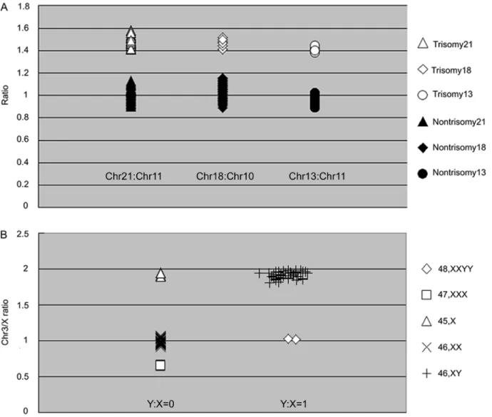
Figure 4. The individual distributions observed in the assays.A) Individual relative ratios of chromosomes 21,18 and 13 in normal and trisomic samples. B) Distribution of the X vs. Y and Chr 3 vs. X ratio; the x-axis represents the X/Y ratio, and the y-axis represents the Chr3/X ratio. doi:10.1371/journal.pone.0088932.g004
Table 3.Specificity and sensitivity of the autosomal chromosomes for primer set one.
Primer set Primer set one
Segmental duplication Chr 21 Chr 18 Chr 13
Mean control 1.01 1.03 0.95
SD control 0.06 0.06 0.05
95% CI control 0.96–1.07 0.96–1.09 0.91–0.98
Mean trisomic 1.49 1.47 1.40
SD trisomic 0.05 0.04 0.03
95% CI trisomic 1.44–1.54 1.43–1.51 1.38–1.43
Total samples 96 96 96
Sensitivity 1 1 1
Specificity 1 1 1
doi:10.1371/journal.pone.0088932.t003
a failed experiment), we designed two independent primer sets to simultaneously detect aneuploidies. Thus, this assay design reduces the probability of detecting sequence variants in the two target regions at the same time. Furthermore, in clinical practice, differences between the two assays might reflect structural abnormalities. In such cases, full karyotyping, FISH, or array comparative genomic hybridization should be applied for diag-nostic confirmation. The major drawback of this approach, similar to many other PCR-based assays, is that it is possible to miss some cases of structural abnormalities, low-level mosaicism or maternal cell contamination. However, this drawback does not hinder the wide application of this technology.
Detection of the Common Chromosome Aneuploidies
To reduce the probability of a variant in the primer region, we selected two target regions per chromosome and designed two primer sets to simultaneously detect aneuploidies. For primer set one, a series of qualified segmental duplications, located on chromosomes 21 and 11, chromosomes 18 and 10 and chromo-somes 13 and 11, were selected to detect the aneuploidies of chromosomes 21, 18 and 13, respectively (Figures 2A, 2B and 2D). To detect abnormalities on the sex chromosomes, we designed two pairs of primers: segmental duplications located on chromosomes X and Y (Figure 2C) were designed to quantify the ratio between the X and Y chromosomes, and segmental duplications located on chromosomes 3 and X were designed to quantify the ratio between chromosomes 3 and X (Figure 2E). In theory, the common abnormalities of sex chromosomes can be identified using these two primers pairs (Table 2).
For primer set two, the segmental duplications located on chromosomes 13 and 9, chromosomes 18 and 1 and chromosomes 21 and 6 were selected to detect the aneuploidies of chromosomes 13, 18 and 21, respectively (Figures 3A, 3B and 3D). Segmental duplications located on chromosomes X and 18 and chromosomes X and Y were designed to detect abnormalities on the sex chromosomes (Figures 3C and 3E).
We examined 96 DNA samples in each of the two assays and obtained a correct and unambiguous diagnosis in all cases. In total, 56 aneuploidies were detected: 36 cases of trisomy 21; 4 cases of trisomy 13; 6 cases of trisomy 18; 5 cases of trisomy 45, X; 3 cases of trisomy 47, XXX; and 2 cases of trisomy 48, XXYY. A total of 40 unaffected DNA controls were also screened in each assay. The diagnostic sensitivity and specificity of all samples was 100%, and the results correlated with their respective karyotypes. We attribute this high reliably to the use of segmental duplications as detection targets. A single pair of primers can perfectly match both loci to co-amplify the segmental duplications located on two different chromosomes, while maintaining almost identical amplification efficiencies between target chromosomes and control chromo-somes. Therefore, segmental duplications can be used to reliably detect chromosome abnormalities. For primer set one, the means and standard deviations for the segmental duplications on the autosomal chromosomes are shown in Table 3, and all 96 samples were categorized into different groups according to their mean values (Figure 4).
A total of 36 samples with trisomy 21 were observed, including two unbalanced translocations (46,XX,der(14; 21)(q10;q10),+21) correctly diagnosed as trisomy 21 using SD-QF-PCR. For the determination of trisomy 21, two pairs of segmental duplications (Chr21/Chr11 and Chr21/Chr9) were selected for the identifica-tion of addiidentifica-tional copies of chromosome 21 (Figure 2A and
Figure 3D). The segmental duplications were located on the long arm of chromosome 21 [19]. Thus, these results demonstrate that this approach is not only an acceptable method for detecting an extra target chromosome, but also for detecting the relative dosage of the chromosomes in which the segmental duplications are located, i.e., chromosome deletion or duplication.
Methodology Comparison
Fluorescent in situ hybridization (FISH) is a routine method used for the rapid detection of prenatal aneuploidies in interphase amniocytes. This method avoids the need to culture cells and reduces the time required for diagnosis. However, this technique is relatively labor intensive and requires technical expertise, requirements that prevent the efficient processing of a large number of samples in clinical diagnostic settings [20,21]. These drawbacks have encouraged the development of more rapid and efficient methods for the detection of fetal chromosomal aneuploidies.
QF-PCR and MLPA are the most extensively validated DNA-based techniques described for the rapid detection of fetal chromosomal aneuploidies [9–12]. However, for QF-PCR, based on the amplification of polymorphic microsatellite repeats, the differences in the number of informative polymorphisms might limit the universality of this assay. Segmental duplications are not dependent on multiple informative polymorphism markers, applicable to all human populations. Thus, this method could be considered as an alternative or complement strategy to QF-PCR. MLPA, which requires an overnight step for hybridization, requires approximately 16 hours to detect a single sample. SD-QF-PCR exhibits equal application efficiency and saves more than 12 hours turnaround time, making SD-QF-PCR faster and easier. Compared with conventional karyotype analyses, SD-QF-PCR reduces the turnaround time from 2–4 weeks to less than 4 hours for a single aneuploidy test [2]. Using the current protocol, a single operator can manage at least 96 samples a day and report the results in less than 24 hours, which should meet the needs of most diagnostic laboratories.
Conclusions
In the present study, we designed two novel sets of segmental duplications and described a simple and rapid SD-QF-PCR method as an alternative approach to simultaneously detect the aneuploidies of chromosomes 21, 18, 13, X and Y in a single reaction. This method can process 96 samples at a time in an automated system for each primer set, and the results are available the same day. The retrospective study of 96 DNA samples was entirely consistent with the previous results of conventional cytogenetics. Thus, this procedure represents a competitive alternative diagnostic tool for use in prenatal screening.
Acknowledgments
The authors would like to thank Yangqing Lu at Guangxi University for assistance with the English.
Author Contributions
References
1. Driscoll DA, Gross S (2009) Clinical practice. Prenatal screening for aneuploidy. N Engl J Med 360: 2556–2562.
2. Caspersson T, Zech L, Johansson C, Modest EJ (1970) Identification of human chromosomes by DNA-binding fluorescent agents. Chromosoma 30: 215–227. 3. Von Eggeling F, Freytag M, Fahsold R, Horsthemke B, Claussen U (1993) Rapid detection of trisomy 21 by quantitative PCR. Hum Genet 91: 567–570. 4. Lee HH, Chang JG, Lin SP, Chao HT, Yang ML, et al. (1997) Rapid detection of trisomy 21 by homologous gene quantitative PCR (HGQ-PCR). Hum Genet 99: 364–367.
5. Zimmermann B, Holzgreve W, Wenzel F, Hahn S (2002) Novel real-time quantitative PCR test for trisomy 21. Clin Chem 48: 362–363.
6. Helmy SM, Ismail S, Bassiouni R, Gaber KR (2009) Sensitivity of DCSR3/ GAPDH ratio using quantitative real-time PCR in the rapid prenatal diagnosis for down syndrome. Fetal Diagn Ther 25: 220–223.
7. Deutsch S, Choudhury U, Merla G, Howald C, Sylvan A, et al. (2004) Detection of aneuploidies by paralogous sequence quantification. J Med Genet 41: 908– 915.
8. Guo Q, Xiao L, Zhou Y (2012) Rapid diagnosis of aneuploidy by high-resolution melting analysis of segmental duplications. Clin Chem 58: 1019–1025. 9. Mansfield ES (1993) Diagnosis of Down syndrome and other aneuploidies using
quantitative polymerase chain reaction and small tandem repeat polymorphisms. Hum Mol Genet 2: 43–50.
10. Mann K, Donaghue C, Fox SP, Docherty Z, Ogilvie CM (2004) Strategies for the rapid prenatal diagnosis of chromosome aneuploidy. Eur J Hum Genet 12: 907–915.
11. Atef SH, Hafez S, Helmy S, Helmy N (2011) QF-PCR as a Rapid Technique for Routine Prenatal Diagnosis of Fetal Aneuploidies. Pediatr Res 70: 412–412. 12. Slater HR, Bruno DL, Ren H, Pertile M, Schouten JP, et al. (2003) Rapid, high
throughput prenatal detection of aneuploidy using a novel quantitative method (MLPA). J Med Genet 40: 907–912.
13. Dudarewicz L, Holzgreve W, Jeziorowska A, Jakubowski L, Zimmermann B (2005) Molecular methods for rapid detection of aneuploidy. J Appl Genet 46: 207–215.
14. Schouten JP, McElgunn CJ, Waaijer R, Zwijnenburg D, Diepvens F, et al. (2002) Relative quantification of 40 nucleic acid sequences by multiplex ligation-dependent probe amplification. Nucleic acids res 30: e57.
15. Boormans EM, Birnie E, Oepkes D, Galjaard RJ, Schuring-Blom GH, et al. (2010) Comparison of multiplex ligation-dependent probe amplification and karyotyping in prenatal diagnosis. Obstet Gynecol 115: 297–303.
16. Willis AS, Veyver I, Eng CM (2012) Multiplex ligation-dependent probe amplification (MLPA) and prenatal diagnosis. Prenat Diagn 32: 315–320. 17. She X, Jiang Z, Clark RA, Liu G, Cheng Z, et al. (2004) Shotgun sequence
assembly and recent segmental duplications within the human genome. Nature 431: 927–930.
18. Bailey JA, Yavor AM, Massa HF, Trask BJ, Eichler EE (2001) Segmental duplications: organization and impact within the current human genome project assembly. Genome Res 11: 1005–1017.
19. Baldus CD, Liyanarachchi S, Mro´zek K, Auer H, Tanner SM, et al. (2004) Acute myeloid leukemia with complex karyotypes and abnormal chromosome 21: Amplification discloses overexpression of APP, ETS2, and ERG genes. PNAS 101: 3915–3920.
20. Caine A, Maltby AE, Parkin CA, Waters JJ, Crolla JA, et al. (2005) Prenatal detection of Down’s syndrome by rapid aneuploidy testing for chromosomes 13, 18, and 21 by FISH or PCR without a full karyotype: a cytogenetic risk assessment. Lancet 366: 123–128.
21. Ho SS, Chua C, Gole L, Biswas A, Koay E, et al. (2012) Same-day prenatal diagnosis of common chromosomal aneuploidies using microfluidics-fluores-cence in situ hybridization. Prenat Diagn 32: 321–328.